Targeting IDH1/2 mutant cancers with combinations of ATR and PARP inhibitors
←
→
Page content transcription
If your browser does not render page correctly, please read the page content below
Published online 17 May 2021                                                                                 NAR Cancer, 2021, Vol. 3, No. 2 1
                                                                                                                  doi: 10.1093/narcan/zcab018

Targeting IDH1/2 mutant cancers with combinations
of ATR and PARP inhibitors
Amrita Sule 1 , Jinny Van Doorn1 , Ranjini K. Sundaram1 , Sachita Ganesa1 ,
Juan C. Vasquez2 and Ranjit S. Bindra 1,*
1
    Department of Therapeutic Radiology, Yale University School of Medicine, New Haven, CT 06511, USA and
2
    Department of Pediatrics, Yale School of Medicine, New Haven, CT 06511, USA

                                                                                                                                                              Downloaded from https://academic.oup.com/narcancer/article/3/2/zcab018/6276970 by guest on 13 December 2021
Received January 14, 2021; Revised April 17, 2021; Editorial Decision April 26, 2021; Accepted May 04, 2021

ABSTRACT                                                                        GRAPHICAL ABSTRACT
Mutations in the isocitrate dehydrogenase-1 and -2
(IDH1/2) genes were first identified in glioma and
acute myeloid leukemia (AML), and subsequently
found in multiple other tumor types. These neomor-
phic mutations convert the normal product of en-
zyme, ␣-ketoglutarate (␣KG), to the oncometabo-
lite 2-hydroxyglutarate (2HG). Our group recently
demonstrated that 2HG suppresses the high-fidelity
homologous recombination (HR) DNA repair path-
way, resulting in a state referred to as ‘BRCAness’,
which confers exquisite sensitivity to poly(ADP-
ribose) polymerase (PARP) inhibitors. In this study,
we sought to elucidate sensitivity of IDH1/2-mutant
cells to DNA damage response (DDR) inhibitors and,
whether combination therapies could enhance de-
scribed synthetic lethal interactions. Here, we re-
port that ATR (ataxia telangiectasia and Rad3-related
protein kinase) inhibitors are active against IDH1/2-
mutant cells, and that this activity is further potenti-                        INTRODUCTION
ated in combination with PARP inhibitors. We demon-
                                                                                Isocitrate dehydrogenase-1 and -2 (IDH1/2) are enzymes
strate this interaction across multiple cell line mod-                          which convert the Kreb’s cycle intermediate isocitrate to ␣-
els with engineered and endogenous IDH1/2 mu-                                   ketoglutarate (␣KG). IDH1/2 mutations occur frequently
tations, with robust anti-tumor activity in vitro and                           in subsets of human malignancies derived from a wide range
in vivo. Mechanistically, we found ATR and PARP                                 of tissues (1). IDH1 mutations have been recurrently re-
inhibitor treatment induces premature mitotic en-                               ported in low-grade glioma, glioblastoma multiforme (1),
try, which is significantly elevated in the setting of                          acute myeloid leukemia (AML) (2), cholangiocarcinoma
IDH1/2-mutations. These data highlight the poten-                               (3), chondrosarcoma, and also melanoma (4,5). Mutations
tial efficacy of targeting HR defects in IDH1/2-mutant                          in the IDH2 gene also have been reported in gliomas and
cancers and support the development of this combi-                              AML (1,6). They are heterozygous missense mutations
nation in future clinical trials.                                               which primarily occur at the R132 residue in IDH1, and the
                                                                                R140 or R172 residues in IDH2 genes (1). These mutations
                                                                                in IDH1/2 are neomorphic, in that they convert the normal
                                                                                product of the WT enzyme, ␣KG, into 2-hydroxyglutarate
                                                                                (2HG). 2HG is an oncometabolite which is thought to drive
                                                                                transformation and tumor progression by altering a diverse
                                                                                range of cellular processes, including metabolism and epi-
                                                                                genetic changes (7–9).

* To   whom correspondence should be addressed. Tel: +1 203 200 3672; Fax: +1 203 200 3673; Email: ranjit.bindra@yale.edu


C The Author(s) 2021. Published by Oxford University Press on behalf of NAR Cancer.
This is an Open Access article distributed under the terms of the Creative Commons Attribution License (http://creativecommons.org/licenses/by/4.0/), which
permits unrestricted reuse, distribution, and reproduction in any medium, provided the original work is properly cited.
2 NAR Cancer, 2021, Vol. 3, No. 2
Previous work from our lab and others have demon- Coy’s 5A with 10% FBS (Gibco). SW1353 cells were cul-
strated that IDH1/2-mutant cancers are defective in ho- tured in RPMI 1640 with 10% FBS (Gibco).
mologous recombination (HR), which confer sensitivity
to poly (ADP-ribose) polymerase (PARP) inhibitors (10– Antibodies and reagents
13). Mechanistically, we have shown that 2HG produc-
tion results in aberrant hypermethylation of histone 3 ly- Anti-Histone H3 phospho-Ser-10 (ab14955, Abcam),
sine 9 (H3K9) at DNA break sites, masking a H3K9 anti-GAPDH (60004-1-Ig, Proteintech), anti-Caspase-
trimethylation signal required for recruitment of homology- 3 (9662, Cell Signaling Technology), anti-Cleaved-
dependent repair factors. This results in suppression of HR Caspase-3(9664, Cell Signaling Technology), anti-PARP1
and subsequent PARP inhibitor sensitivity, which is com- (VMA00016, BioRad), anti-Cleaved -PARP (5625, Cell
monly referred to as a BRCAness phenotype (10,14). We Signaling Technology), anti-Rabbit-IgG, HRP conju-
also have extended these findings to other tumor-associated gate (SA00001-2, Proteintech), anti-Mouse-IgG, HRP
Downloaded from https://academic.oup.com/narcancer/article/3/2/zcab018/6276970 by guest on 13 December 2021
oncometabolites, such as succinate and fumarate (15). conjugate (SA00001-1, Proteintech), anti-Cyclin A
PARP is involved in base excision repair (BER) and is im- (SC-271682, Santa Cruz Biotechnology), anti-RPA70
portant for repairing single-strand breaks (SSBs) (16). Cells (ab12320,Abcam), ␥ -H2AX(05–636, Millipore Sigma),
with HR defects are thought to accumulate SSBs result- Alexa Fluor 647 (A21236, ThermoFisher Scientific),
ing from replication fork stalling or collapse when treated Alexa Flour (A32744, ThermoFisher Scientific), Click-
with PARP inhibitors, which leads to lethal double strand iT™ EdU (C10419,ThermoFisher Scientific), RNase/PI
breaks (DSBs) (17,18). PARP inhibitors have been tested buffer (550825, BD Biosciences), Vectashield DAPI
extensively in HR-deficient cancer models, such as those (H-1200-10, Vector Laboratories) were used. (2R)-Octyl-
with BRCA1 and BRCA2 mutations (19), as single agents ␣-hydroxyglutarate (2-HG) was purchased from Cayman
or in combination with a range of DNA damaging agents chemical. Drugs were purchased from Selleckchem.
and DNA repair inhibitors (20,21). ATR (ataxia telangiec- AZD6738 and Olaparib were provided by AstraZeneca.
tasia and Rad3-related protein kinase) is a key cell cycle
regulator, which halts the cell cycle in an event of DNA Short-term cell viability assays
damage and initiates DNA damage response (DDR). ATR
maintains genomic stability by responding to replication Cells were plated in 96-well plates at a concentration of
stress and DNA damage in S-phase by activating the S- 2000 cells per well and allowed to adhere at room tempera-
phase checkpoint, thereby allowing DNA repair and pre- ture for 60 min before returning them to the incubator. Af-
venting premature mitotic entry (22). ATR inhibition al- ter 24 h, the media was changed and replaced with respec-
lows damaged cells to proceed past the S-phase checkpoint, tive drugs in triplicates at indicated concentrations. Five
promoting the induction of DSBs and premature mitotic days after drug treatment, the cells were fixed with 3.7%
entry; ultimately leading to apoptosis (23,24). Independent paraformaldehyde and stained with Hoechst (1 g/ml). The
of replication stress ATR also regulates the G2/M check plates were then imaged on a Cytation 3 automated im-
point and hence abrogation of ATR could potentially lead ager (BioTek), and cells were counted using CellProfiler
to pre-mitotic entry (25–27). Several preclinical studies have (http://cellprofiler.org/).
demonstrated the efficacy of PARP inhibitors in combina-
tion with ATR inhibitors in many BRCA-deficient, as well Clonogenic survival assays
as ATM-deficient, cancer models (25–27). Cells were counted and diluted in media containing vari-
In this study, we sought to further elucidate the spectrum ous concentrations of respective drugs. They were immedi-
of DDR inhibitor sensitivity in IDH1/2-mutant cells, and ately transferred into six-well plates in triplicate at three-
whether combination therapies could further enhance this fold dilutions ranging from 9000 to 37 cells per well. After
synthetic lethal interaction. Here, we report that ATR in- 12–14 days, plates were washed with PBS and stained with
hibitors are active against IDH1/2-mutant cells and that crystal violet. Colonies were counted by hand. Counts were
this activity can be further potentiated by combination with normalized to plating efficiency of corresponding treatment
PARP inhibitors both in vitro and in vivo. condition, unless otherwise noted.
MATERIALS AND METHODS U2OS DR-GFP assay
Cell culture U2OS DR-GFP assay was carried out as previously de-
scribed (28). To test the effect of 2HG, U2OS cells express-
The U87 IDH1 R132H/+ (ATCC® HTB-14IG™), HT1080 ing the reporter were cultured with or without 300 M
IDH1 R132C (ATCC® CCL-121™) and SW1353 IDH2 2HG. Drugs were added 24 h prior to ligand addition. Lig-
R172S (ATCC® HTB-94) cell lines was purchased from ands were washed off after 24 h and media was replenished
ATCC. HCT116 IDH1 R132H/+ was purchased from with respective drugs and 2HG. Cells were analyzed on a
(Horizon Discovery HD 104-013). LN229 cells with doxy- BD FACS Calibur flow cytometer after 72 h.
cyclin inducible IDH1 R132H/+ have been described pre-
viously (10). LN229, HT1080 and SW1353 cells were cul-
Flow cytometry
tured in DMEM with 10% FBS (Gibco). U87 cells were
cultured in DMEM-F12 with 10% FBS (Gibco). HCT116 For propidium iodide (PI) staining, cells were seeded in 60
(parental and IDH1 R132H/+) cells were cultured in Mc- mm dishes. 24–48 h after plating, cells were treated with
NAR Cancer, 2021, Vol. 3, No. 2 3
drugs. Twenty four hours post drug treatment, cells were In vivo Olaparib and AZD6738 efficacy studies
harvested by trypsinization and fixed with 70% ethanol.
Female athymic nu/nu mice (Hsd:Athymic Nude-Foxn1nu,
Cells were then stained in RNase/PI buffer (BD Bio-
Envigo) were used for in vivo xenograft studies. One million
sciences). For EdU staining, cells were treated with 10
HCT116 IDH1 R132H/+ or HT1080 IDH1 R132C/+ cells
M EdU 1.5 h prior to harvesting and processed ac-
were injected subcutaneously into the flank at a concentra-
cording to manufacture’s protocol (Thermo Fisher:Click
tion of 1 × 10⁶ cells/100 l of PBS. Prior to drug treatment,
iT™ EdU Cell Proliferation Kit). For p-H3 staining, cells
mice were randomized and placed into groups of 5–8 ani-
were trypsinized 24 h post drug treatment and processed
mals such that the average starting tumor volume of each
as previously described by Forment et.al. (29). Cells were
group was approximately equal (100 mm3 ). Olaparib and
treated with an extraction buffer, 0.2% Triton (PBS) for
AZD6738 were administered daily up to 21 or 28 days. Each
10 min, washed with PBS (1% BSA) followed by fixa-
dose of olaparib was delivered via oral gavage at 25 or 50
tion with 70% ethanol. Cells were incubated in primary
Downloaded from https://academic.oup.com/narcancer/article/3/2/zcab018/6276970 by guest on 13 December 2021
mg/kg. Each dose of AZD6738 was delivered via oral gav-
and secondary antibody solutions for 1 h and 30 min, re-
age at 25 or 50 mg/kg. Olaparib and AZD6738 were sol-
spectively, at room temperature. Cells were run on a BD
ubilized in DMSO and diluted with 10% (w/v) 2-hydroxy-
FACS Calibur flow cytometer and analyzed with FlowJo
propyl-b-cyclodextrin (Sigma) to obtain the desired concen-
software.
tration. Tumors were measured by calipers and volume was
calculated using the equation for ellipsoid volume: volume
NAD quantification = /6 × (length) × (width)2 . IACUC protocols at the Yale
School of Medicine were followed throughout the study.
NAD level analyses were performed using a NAD/NADH
Quantification kit (Sigma), as per manufacturer’s specifica- Statistical analysis
tions.
Data are presented as mean ± SD or SEM. Comparisons
were made using Student’s t test (Clonogenic survival as-
Immunofluorescence say). Two-way analysis of variance (ANOVA) was used to
test for significant differences between groups (xenograft
25,000 cells were seeded on glass chamber slides. Slides were
studies). All tests were two-sided. Statistical analyses were
treated with drugs for 24 h. Cells were fixed with 3.7%
carried out using GraphPad Prism. P values are indicated
paraformaldehyde. Cells were then washed with PBS and
either directly on figures or using *P < 0.05, **P < 0.01,
permeabilized with 0.2% Triton–PBS. Cells were incubated
***P < 0.001 and ****P < 0.0001.
with primary antibody (anti-␥ -H2AX, anti-pH3 or anti-
cyclin A) overnight at 4◦ C and secondary antibody (Alexa
Flour 594 or Alexa Flour488) at room temperature for 1 h. RESULTS
For RPA70 immunostaining, cells were pre-extracted for 1 IDH1 mutation confers sensitivity to ATR inhibitors
min in 0.1 M PIPES pH 6.9, 1 mM EGTA, 4 M glycerol and
0.2% Triton X-100. Cells were washed once for 2 min in 0.1 Previous studies from our group and others have established
M PIPES pH 6.9, 1 mM EGTA, and 4 M glycerol in H2O, mutant IDH1/2-induced HR defects and PARP inhibitor
and then fixed for 20 min with 3.7% formaldehyde, 50 mM sensitivity (10–13), although the spectrum of sensitivity to
PIPES pH 6.9, 5 mM EGTA, and 1 mM MgCl2 . Cells were other DDR inhibitors has yet to be fully elucidated. ATR
blocked in casein + 5% goat serum before adding primary inhibitors have been shown to be synthetic lethal with DNA
antibody (anti-RPA70) at 1:500 overnight at 4◦ C. The next repair defects caused by ATM, TP53 or Rad51 loss in can-
day, cells were washed with PBS before secondary antibody cer cells (31–34), which prompted us to test whether they
(Alexa-flour-488) incubation at 1:500 for 2 h at room tem- would be active against IDH1/2-mutant cells. We tested
perature. Cells were analyzed on a Keyence BZ-X800. Foci four different ATR inhibitors; AZD6738, ATRIN-175, VE-
were analyzed with the Focinator v2-21 software as previ- 822 and BAY-1895344 in U87, HCT116 and LN229 IDH1
ously described (30). R132H/+ isogenic cell line models using short-term viabil-
ity assays in vitro. We observed evidence of ATR inhibitor
sensitivity which was greatest in IDH1-mutant versus -WT
Protein detection by western blot cells (Figure 1A–C), with WT/mutant IC50 ratios rang-
ing from 1.28 to 6.78 (Figure 1D). We confirmed this dif-
Whole cell lysates were prepared using RIPA buffer (Cell ferential sensitivity between IDH1-mutant and -WT cells
Signaling Technology) with 1× protease and phosphatase in a clonogenic survival assay, which revealed a >10-fold
inhibitor (78442, ThermoFisher Scientfic). Proteins were sensitivity of IDH1-mutant cells to AZD6738 at a dose of
separated by SDS-PAGE and transferred to a PVDF mem- 250 nM (Figure 1E). These data confirm that like PARP
brane for analysis. Blots were blocked in 5% BSA (Gold inhibition, ATR inhibition is synthetic lethal with mutant
Biotechnology) in 1× TBST (American Bio) and probed IDH1/2-associated HR defects.
overnight with respective primary antibodies. Blots were
washed with 1× TBST and incubated with HRP conjugated
ATR and PARP inhibition are synthetically lethal in IDH1/2
secondary antibodies for 1 h at room temperature. Blots
mutant cancers
were exposed using Clarity Western ECL substrate (Bio-
Rad), and imaged on a ChemiDoc (BioRad) imaging sys- A number of studies have shown that ATR and PARP in-
tem. hibitor combinations are more cytotoxic than PARP in-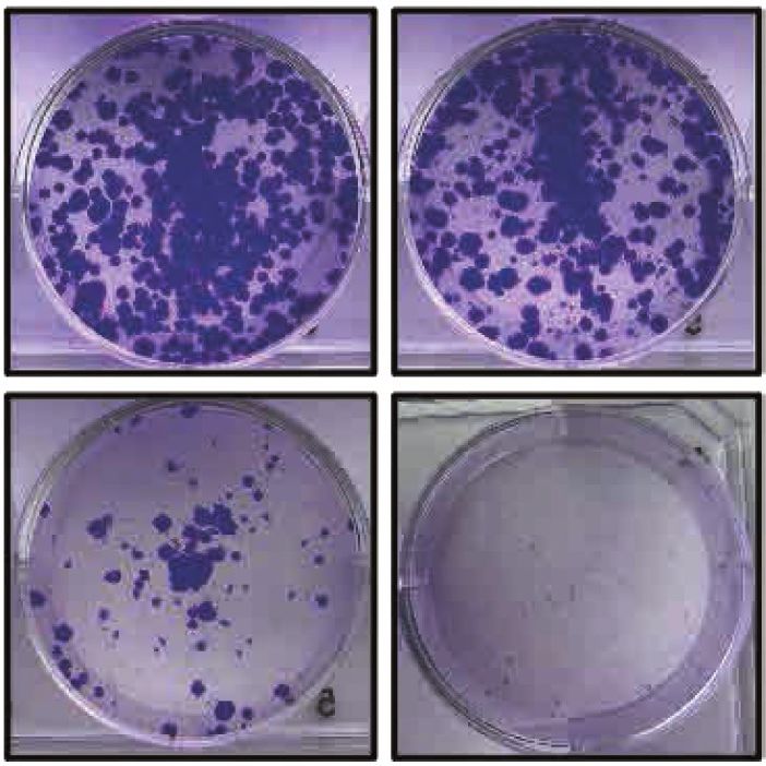
4 NAR Cancer, 2021, Vol. 3, No. 2

                                                                                                                                                          Downloaded from https://academic.oup.com/narcancer/article/3/2/zcab018/6276970 by guest on 13 December 2021
Figure 1. IDH1 mutation confers sensitivity to ATR inhibitors. (A–C) Five-day short-term cell viability assays of four separate ATR inhibitors AZD6738,
ATRIN175, VE-822 and BAY-1895344. U87 (WT and IDH1 R132H/+), HCT116 (WT and IDH1 R132H/+) and LN229 (WT and doxycycline inducible
IDH1 R132H/+) cells were used for this assay. (D) IC50 ratios of screened WT and the IDH1 mutant cells indicate that the IDH1 mutant cells are more
sensitive to all the four ATR inhibitors tested. (E) HCT116 WT and R132H/+ cells were treated with AZD6738 for 14 days (n = 6). Error bars represent
means ± SEM, *P < 0.05.

hibitor monotherapy in BRCA1/2-deficient cells (26,35).                       genic survival assays (Figure 2A), which was also observed
Given our previous findings of oncometabolite-induced                         in the U87 WT and IDH1-mutant cell line pair (Figure 2B).
PARP inhibitor sensitivity, and our new findings above                        We validated the ATRi and PARPi combination efficacy
which demonstrate robust ATR inhibitor sensitivity, we                        with another ATR inhibitor, BAY1895344, in combination
hypothesized that this combination would also be effec-                       with olaparib in HCT116 isogenic cell line (Supplementary
tive against IDH1/2-mutant cells. As shown in Figure 2A,                      Figure S2A).
combination therapy of a PARPi, olaparib and an ATRi,                            We also sought to extend these findings to cell line mod-
AZD6738 in HCT116 WT and IDH1-mutant cells was sig-                           els with endogenous IDH1/2 mutations. To this end, we
nificantly more cytotoxic in IDH1-mutant cells in clono-                      selected several IDH1/2-WT and -mutant cholangiocarci-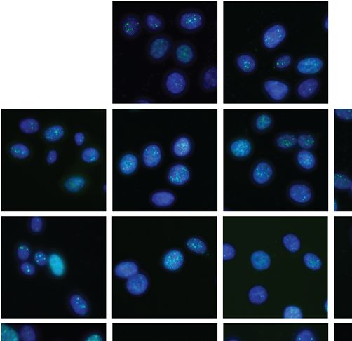
NAR Cancer, 2021, Vol. 3, No. 2 5
Downloaded from https://academic.oup.com/narcancer/article/3/2/zcab018/6276970 by guest on 13 December 2021
Figure 2. ATR inhibitor increases PARP inhibitor mediated sensitivity in IDH1 mutant cells. (A) Quantification and representative images of clonogenic
survival assays of HCT116 WT and IDH1-mutant (R132H/+) cells were treated with AZD6738 alone (solid lines) and in combination with 1 M olaparib
(dashed lines) for 14 days (n = 6). (B) Quantification representative images of clonogenic survival assays of U87 WT and IDH1-mutant (R132H/+) cells
were treated with AZD6738 alone (solid lines) and in combination with 2 M olaparib (dashed lines) for 14 days (n = 4). (C) Quantification of clonogenic
survival assay of SW1353 (IDH2 R172S/+) cells treated with AZD6738 alone (blue line) and in combination with 1 M olaparib (red line) for 14 days.
(D) Quantification of clonogenic survival assay of RBE (IDH1 R132S/+) cells treated with AZD6738 alone (blue line) and in combination with 2 M
Olaparib (red line) for 14 days. Error bars represent means ± SEM. ***P < 0.001, **P < 0.01, *P < 0.05.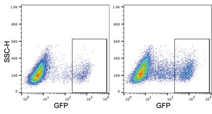
6 NAR Cancer, 2021, Vol. 3, No. 2
noma, fibrosarcoma and chondrosarcoma cell lines, and We further looked at effect of olaparib, AZD6738 and
we first tested monotherapy PARP and ATR inhibitor sen- the combination treatment on homologous recombination
sitivity in order to identify the appropriate dose ranges (HR) repair efficacy. We used a ligand-dependent I-SceI sys-
for the combination drug studies (Supplementary Figure tem combined with the U2OS DR-GFP assay to measure
S1A and B). We then selected the chondrosarcoma cell line HR (40) (Figure 3C). As these cells do not have IDH1/2
SW1353, which harbors an IDH2 mutation (R172S/+), as mutations, we added 2HG directly to the cells. As previously
well as the cholangiocarcinoma cell line RBE, which har- reported (10), we observed that 2HG alone suppresses HR.
bors an IDH1 mutation (R132S/+) for combination ATR Cells not treated with 2HG have 9.5% GFP+ cell popula-
and PARP inhibitor studies. Both cell lines exhibited in- tion which goes down to around 6.5% when treated with
creased sensitivity to the olaparib and AZD6738 combina- 2HG. We observed that in cells treated with 2HG; olaparib
tion relative to AZD6738 monotherapy in clonogenic sur- (6.7%), AZD6738 (4.1%) as well as the combination treat-
vival assays (Figure 2C, D, Supplementary Figure S2B, C ment (3.9%) significantly reduced the number of GFP+ cells
Downloaded from https://academic.oup.com/narcancer/article/3/2/zcab018/6276970 by guest on 13 December 2021
and S3E). Of note, olaparib monotherapy was not as effec- thereby significantly suppressing HR (Figure 3C and D).
tive in short-term cell viability assays with RBE cells in com- AZD6738 treatment also caused a suppression of HR in
parison to the other cell lines tested (Supplementary Fig- cells not treated with 2HG. These data, coupled with find-
ure S1A). However, clonogenic survival assays in combina- ings in Figure 3B that ␥ -H2AX levels are highest in the
tion with AZD6738 revealed a thousand-fold sensitization PARP/ATR inhibitor-treated IDH1-mutant cells, suggest
in RBE cells to olaparib, indicating the exquisite potenti- that sensitization occurs via a combination of decreased HR
ation associated with the combination therapy compare to and unrepaired DSBs.
single agent treatment (Figure 2D). As reported previously
(36,37), we also saw some cytotoxicity of the combination in
ATR and PARP inhibitor treatment cause premature mitotic
the WT cells, however the cytotoxicity in the IDH1-mutant
entry
cells was more heightened.
We also tested the efficacy of two other PARP inhibitors, Next, we sought to further probe the mechanistic basis for
niraparib and talazoparib, by themselves and in combi- enhanced tumor cell kill induced by AZD6738 and olaparib
nation with AZD6738 in HCT116 WT and IDH1-mutant combination treatment in IDH1/2-mutant cells. We first ex-
cell line (Supplementary Figure S3A–E). Additionally, we amined the effect of AZD6738 and Olaparib on cell cy-
also assessed the NAD levels in the isogenic cell line cle distribution in our IDH1-mutant and -WT isogenic cell
pairs to validate if PARP inhibitor sensitivity was affected line pairs. We found that treatment of HCT116 WT with
by the NAD. We did not observe any significant differ- 500 nM AZD6738 and 1 M Olaparib, either alone or in
ence in the NAD levels of HCT116 and U87 (WT and combination, did not induce significant changes in cell cy-
IDH1-mutant) cells (Supplementary Figure S3F). Taken to- cle profiles after 24 h (Supplementary Figure S4A and B).
gether, our data suggests that ATR and PARP inhibitor However, both olaparib and AZD6738 induced increases in
combinations are highly effective in IDH1/2-mutant cells the G2/M phase fractions of HCT116- IDH1-mutant cells,
in vitro. which was further augmented with the combination treat-
ment (Supplementary Figure S4A and B). We therefore as-
sessed the mitotic cell population by immunofluorescence
ATR and PARP inhibitor combination increase DSBs and
(IF) staining for p-Histone 3 (p-H3) at Ser 10, which identi-
suppress HR
fies cells in late G2 and mitosis. These experiments revealed
ATR protects replicating DNA and in an advent of DNA a significant increase in p-H3 positive cells after treatment
damage, delays cell cycle progression via replication fork with olaparib and AZD6738 in HCT116 IDH1-mutant ver-
stabilization (38). Increased replication stress in cancers sus -WT cells (Figure 4A), which was also observed in the
makes them sensitive to ATR inhibitors (39). To assess if U87 isogenic pair (Figure 4B). We also observed a small, but
IDH1/2-mutant cells are sensitive to ATR inhibitors due significant, increase in p-H3 levels after olaparib treatment
to replication stress, we looked at RPA70 foci formation alone in HCT116 IDH1-mutant versus -WT cells. Repre-
in U87 WT and IDH-mutant cells treated with olaparib, sentative images IF images for these experiments are shown
AZD6738 or both (Figure 3A). We did not observe any sig- in Supplementary Figure S5. We then validated this result
nificant differences between the WT and IDH1-mutant cells using multiparametric flow cytometry to measure p-H3 lev-
in response to olaparib or the combination treatment after els and cell cycle phase, which again revealed a significant
4 hours of treatment (Figure 3A). After 24 h of treatment increase in p-H3 levels after treatment with olaparib and
the RPA70 foci increased overall in all treatment groups AZD6738 in HCT116 IDH1-mutant versus -WT cells (Fig-
however, we did not observe any significant difference be- ure 4C).
tween WT and IDH1-mutant cells. ATR inhibitor exerts We went on to profile DNA synthesis using EdU prolif-
sensitivity possibly by a mechanism independent of repli- eration assays after treatment with olaparib and AZD6738,
cation stress. We then assessed presence DNA damage by either alone or in combination, in order to assess whether
looking at ␥ -H2AX foci in HCT116 WT and IDH1-mutant there also were mutant IDH1-dependent differences in
cells post olaparib, AZD6738 and combination treatment. replication. We saw that olaparib and AZD6738 alone re-
After 24 h, IDH1-mutant cells had significantly increased duced the number of cells replicating in S-phase in IDH1-
proportion of cells with ␥ -H2AX foci relative to WT cells mutant cells relative to untreated control, which was fur-
suggesting increased levels of unrepaired DNA damage af- ther reduced with the combination (Figure 4D). Although
ter drug treatment (Figure 3B). not significant, the number of cells in S-phase were less in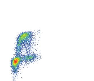
NAR Cancer, 2021, Vol. 3, No. 2 7
Downloaded from https://academic.oup.com/narcancer/article/3/2/zcab018/6276970 by guest on 13 December 2021
Figure 3. ATR and PARP inhibition increases unrepaired damage and suppresses HR. (A) U87 WT and IDH1-R132H/+ cells were treated with olaparib
(10 M), AZD6738 (5 nM) or both for 4 and 24 h. Cell were fixed and stained for RPA70 and counterstained with DAPI. Cells with more than 20 RPA70
foci were counted in seven distinct fields. In total 250–350 cells were analyzed. Representative images of cells stained with RPA (green) and counterstained
with DAPI (Blue) are shown. The images shown were acquired using a 40× objective lens. The scale bar is 10 m. (B) HCT116 WT and IDH1-R132H/+
cells were treated with olaparib (1 M), AZD6738 (500 nM) or both for 24 h. Cell were fixed and stained for ␥ -H2AX and counterstained with DAPI.
Cells with more than 10 ␥ -H2AX foci were counted in eight distinct fields. In total 800–1000 cells were analyzed. Representative images of cells stained
with ␥ -H2AX (green) and counterstained with DAPI (Blue) are shown. The images shown were acquired using a 40× objective lens. The scale bar is 10
m. (C) Quantitation of ligand inducible U2OS-DR-GFP assay where cells were cultured with or without 300 M 2HG for 3 days and with and without
olaparib (1 M), AZD6738 (500 nM) or both for 24 h prior to ligand induction (n = 3). (D) Quantitation of control U2OS-DR-GFP assay without ligand
induction. Dotplots show GFP population with and without ligand addition. Error bars represent means ± SEM. ***P < 0.001, **P < 0.01, *P < 0.05.
8 NAR Cancer, 2021, Vol. 3, No. 2
Downloaded from https://academic.oup.com/narcancer/article/3/2/zcab018/6276970 by guest on 13 December 2021
Figure 4. ATR and PARP inhibition cause IDH1/2 mutants to prematurely enter mitosis. (A and B) HCT116 and U87 (WT and IDH1-R132H/+) cells
were treated with olaparib (1M), AZD6738 (500 nM) or both for 24 h. Cell were fixed and stained for p-H3 (Ser10) and counterstained with DAPI.
Number of p-H3 positive cells were analyzed in 5 distinct fields (n = 2). (C) Representative p-H3-Alexa-647 plots of HCT116 WT and R132H/+ cells that
were treated with olaparib (1 M), AZD6738 (500 nM) or both for 24 h. DNA content (propidium iodide) and pH3-Ser10/Alexa-488 were assessed by
flow cytometry (n = 3). Quantification of result shown on the right panel. (D) Representative EdU-Alexa-647 plots of HCT116 WT and R132H/+ cells
were treated with olaparib (1 M), AZD6738 (500 nM) or both for 24 h DNA content (propidium iodide) and EdU -Ser10/Alexa-488 were assessed by
flow cytometry (n = 3). (E) HCT116 (WT and IDH1-R132H/+) cells were treated with olaparib (1 M), AZD6738 (500 nM) or both for 24 h. Cell were
fixed and stained for cyclin-A and counterstained with DAPI. Number of cylin-A positive cells were analyzed in five distinct fields (n = 3). Quantification
of result shown on the right panel. Error bars represent means ± SEM. ***P < 0.001, **P < 0.01, *P < 0.05.
NAR Cancer, 2021, Vol. 3, No. 2 9
Downloaded from https://academic.oup.com/narcancer/article/3/2/zcab018/6276970 by guest on 13 December 2021
Figure 5. ATRi and PARPi combination caused increased apoptosis in IDH1-mutant cells. HCT116 (WT and IDH1-R132H/+) cells were treated with
olaparib (1 M), AZD6738 (500 nM) or both for 24 h (left panel) or 48 h (right panel). Lysates were harvested after 24 and 48 h. Cleaved caspase-3 and
cleaved PARP was evaluated by western blot analysis.
the IDH1-mutant cells relative to IDH-WT cells. We then tion treatment caused a significant delay in tumor growth
validated this result by IF staining for cyclin A which iden- relative to Olaparib or AZD6738 alone in the HCT116
tifies cells primarily in S phase and early G2-phase (41,42). IDH1-mutant flank model without any significant change
We observed that olaparib monotherapy increased cyclin A in body weight (Figure 6B, Supplementary Figure S7A and
positive cells in the IDH1 WT but not in the IDH1-mutant B). Similarly, in mice bearing the fibrosarcoma HT1080 tu-
cells. In line with our previous EdU staining experiment mors, the combination of olaparib and AZD6738 signifi-
we saw that Olaparib and AZD6738 combination signifi- cantly delayed tumor growth relative to single agent ther-
cantly reduced the number of replicating cells in the IDH1- apy but did not result in a significant drop in body weight
mutant cell line (Figure 4E). Representative images IF im- (Figure 6C and Supplementary Figure S6C). We saw similar
ages for these experiments are shown in Supplementary Fig- levels of tumor suppression in another independent study
ure S6. We also saw significantly differences in p-H3 and with HCT116 IDH1-mutant xenograft model, with smaller
Cyclin A population in the WT in single vs combination sample size (n = 5), where the combination therapy at lower
treatment. dose of AZD6738 (25 mg/kg) and olaparib (25 mg/kg) was
Finally, we assessed the expression of apoptotic mark- also effective in causing a significant tumor growth delay
ers, cleaved-PARP and cleaved-caspase-3 in response to (Figure 6D and Supplementary Figure S8).
the drug treatments. HCT116 WT and IDH1 mutant cells
treated with olaparib, AZD6738 or AZD6738 in combina-
tion with olaparib for 24 and 48 h. Cell lysates were an- DISCUSSION
alyzed by western blotting. The combination of olaparib This study identifies a novel synthetic lethal interaction be-
and AZD6738 did not cause any increase in the apoptotic tween ATR inhibitors and IDH1/2 mutations, which can
proteins at 24 h (Figure 5, left panel). However, post 48 be significantly enhanced by combination with PARP in-
h of treatment there was significantly high expression of hibitors. We have seen the significant effects of olaparib
cleaved-PARP and cleaved caspase-3 in the IDH1 mutant and AZD6738 treatment not only in engineered, isogenic
cells treated with AZD6738 and olaparib combination rel- IDH1/2-mutant cell lines, but also in patient-derived cell
ative to single drug treatments and the WT cells. (Figure lines with endogenous IDH1/2 mutations. In each of these
5, right panel). Taken together, these data suggest that ola- models, the combination of AZD6738 and olaparib proved
parib and AZD6738 specifically induce a defect in mitotic to be significantly cytotoxic than either drug alone. We
entry, most pronounced in IDH1-mutant cells, which drives have corroborated our in vitro cytotoxic studies with in
them to apoptosis. vivo xenograft models, where we observed a significant de-
lay in tumor growth, using both the engineered HCT116
IDH1-mutant xenograft model, as well as the fibrosar-
ATRi and PARPi is active against IDH1-mutant tumors in
coma HT1080 xenograft model with an endogenous IDH1
vivo
R132C mutation. While the inhibition of ATR and PARP
Finally, we tested the efficacy of the AZD6738 and causes moderate cytotoxicity in the WT cells, they exhibit
olaparib in two independent flank animal models. We much exacerbated cytotoxic effects in IDH1/2-mutant cells.
subcutaneously implanted HCT116 IDH1-mutant (IDH1 These preclinical studies show that combined AZD6738
R132H/+) and HT1080 (IDH1 R132C/+) cells in the hind and olaparib treatment may have more long-lasting effects
flank of athymic nude mice and treated them with vehi- relative to monotherapy.
cle, AZD6738, olaparib, or AZD6738 plus olaparib (Figure Previous studies have established enhanced efficacy with
6A). Mice (n = 8) were treated daily with vehicle, olaparib the combination of AZD6738 and olaparib in BRCA- mu-
(50 mg/kg), AZD6738 (50 mg/kg), or olaparib (50 mg/kg) tant (25,43,44) and ATM-deficient (27) cancer models. One
plus AZD6738 (50 mg/kg), post tumor implantation (∼100 advantage to this combination approach is that ATR in-
mm3 ) for up to 44 and 40 days respectively. The combina- hibitors may prevent the emergence of PARP inhibitor re-
10 NAR Cancer, 2021, Vol. 3, No. 2

                                                                                                                                                       Downloaded from https://academic.oup.com/narcancer/article/3/2/zcab018/6276970 by guest on 13 December 2021

Figure 6. Combination of ATRi and PRAPi was effective in IDH1-mutant mouse xenograft model. (A) Athymic nude mice received subcutaneous injection
of HCT116 IDH1 R132H/+ or HT1080 IDH1 R132C/+ cells. Twelve days after injection, the hind flank tumors were measured and equally distributed
to four-arm treatment groups (B) Left panel: Mice carrying flank tumors of HCT116 R132H/+ cells were treated with no treatment (n = 8), Olaparib
alone (50 mg/kg) (n = 8), AZD6738 (50 mg/kg) (n = 8), or Olaparib (50 mg/kg) and AZD6738 (50 mg/kg) (n = 8). Mice were treated daily for 28 days.
Mean tumor volume per group with SEM is plotted on y-axis. Right panel: mean body weight with SEM of mice during HCT116 IDH1 R132H/+ flank
tumor experiment. (C) Left panel: mice carrying flank tumors of HT1080 `cells were treated with no treatment (n = 7), Olaparib alone (50 mg/kg) (n =
7), AZD6738 (50 mg/kg) (n = 7), or Olaparib (50 mg/kg) and AZD6738 (50 mg/kg) (n = 8). Mice were treated daily for 28 days. Right panel: mean body
weight with SEM of mice during HT1080 flank tumor experiment. (D) Left panel: mice carrying flank tumors of HCT116 R132H/+ cells were treated with
no treatment (n = 5), Olaparib alone (25 mg/kg) (n = 5), AZD6738 (25 mg/kg) (n = 5), or Olaparib (25 mg/kg) and AZD6738 (25 mg/kg) (n = 5). Mice
were treated daily for 21 days. Mean tumor volume per group with SEM is plotted on y-axis. Right panel: mean body weight with SEM of mice during
HCT116 R12H/+ flank tumor experiment. Error bars represent means ± SEM. P values were calculated using two-way ANOVA.
NAR Cancer, 2021, Vol. 3, No. 2 11
sistance (45) when HR-defective cells are treated with the DATA AVAILIBILITY
latter class of drugs as a monotherapy (43). In this study
The raw .fcs files have been submitted to FlowRepository
we have reported that IDH1/2 mutant cells are sensitive to
under the following repository IDs: FR-FCM-Z3DN (EdU
ATR inhibitors. We have shown that ATR inhibitors ex-
staining), FR-FCM-Z3DM (PI staining), FR-FCM-Z3DX
ert sensitivity in IDH1/2 mutant cells via suppressing HR,
(p-H3) and FR-FCM-Z3MT (U2OS-DRGFP assay).
resulting in increased unrepaired DNA. Mechanistically,
ATR inhibition has been shown to potentiate the effect of
PARP inhibition via several mechanisms in BRCA-mutant SUPPLEMENTARY DATA
cells, including the disruption of replication fork protection
(44). Regarding the mechanistic basis for the enhanced ef- Supplementary Data are available at NAR Cancer Online.
ficacy of this combination IDH1/2-mutant cells, the data
presented here suggest that premature mitotic entry could
Downloaded from https://academic.oup.com/narcancer/article/3/2/zcab018/6276970 by guest on 13 December 2021
ACKNOWLEDGEMENTS
play a role, especially given the important role of ATR in
regulating the S-G2 transition (22). Taken together we think We thank Yale Flow Cytometry for their assistance for
that presence of high genomic instability in HR deficient their assistance with FACS service. The core is supported
IDH1/2 mutant cells (10) and accumulation of olaparib me- in part by an NCI Cancer Center Support Grant #NIH
diated DNA breaks, ATR inhibition can lead to abrogation P30 CA016359, the BD symphony was funded by shared
of S-phase arrest and cause apoptosis by premature mitotic instrument grant #NIH S1- ODO26996. We also thank As-
entry. traZeneca to provide us with drugs.
Achieving maximum efficacy and minimum toxicity are
paramount aspects of clinical drug development. Olaparib
has been extensively investigated in a multitude of trials FUNDING
and maximum tolerated doses have been identified (46). National Institutes of Health [R01 CA215453-02 to R.S.B.];
AZD6738 is a highly selective orally bioavailable inhibitor America Association of Cancer Research [18-40-12-SULE
of ATR kinase (47) and is currently being investigated in to A.S.]; Pilot Funding from the National Brain Tumor So-
early-phase clinical trials. A dose escalation ongoing phase ciety [Grant 2021].
I PATRIOT study (NCT02223923) is looking at safety of Conflict of interest statement. Ranjit Bindra: Cybrexa,
AZD6738 as a monotherapy and palliative radiotherapy Athena; founder, consultant, equity, sponsored research.
with solid tumors (48). In another phase I study by Yap
et al. (49) where AZD6738 toxicity was assessed in com-
bination with other therapies, the AZD6738 and olaparib REFERENCES
arm was found to be well tolerated (50). The AZD6738 1. Yan,H., Parsons,D.W., Jin,G., McLendon,R., Rasheed,B.A.,
and olaparib combination is currently being tested in phase Yuan,W., Kos,I., Batinic-Haberle,I., Jones,S., Riggins,G.J. et al.
II trials, which includes the CAPRI study for recurrent (2009) IDH1 and IDH2 mutations in gliomas. N. Engl. J. Med., 360,
ovarian cancers (NCT03462342), as well as OLAPCO 765–773.
study in patients with HR defects beyond those associ- 2. Mardis,E.R., Ding,L., Dooling,D.J., Larson,D.E., McLellan,M.D.,
Chen,K., Koboldt,D.C., Fulton,R.S., Delehaunty,K.D.,
ated with BRCA1/2 mutations (NCT02576444). Bindra McGrath,S.D. et al. (2009) Recurring mutations found by sequencing
and colleagues recently reported the interim results of the an acute myeloid leukemia genome. N. Engl. J. Med., 361, 1058–1066.
OLAPCO trial (NCT02576444), which tested the safety and 3. Jiao,Y., Pawlik,T.M., Anders,R.A., Selaru,F.M., Streppel,M.M.,
efficacy of monotherapy olaparib against IDH1/2-mutant Lucas,D.J., Niknafs,N., Guthrie,V.B., Maitra,A., Argani,P. et al.
(2013) Exome sequencing identifies frequent inactivating mutations in
tumors. Efficacy was seen in four out of six IDH1/2-mutant BAP1, ARID1A and PBRM1 in intrahepatic cholangiocarcinomas.
mesenchymal sarcomas, but no clinical benefit was seen in Nat. Genet., 45, 1470–1473.
four patients with IDH1/2-mutant cholangiocarcinomas. 4. Krauthammer,M., Kong,Y., Ha,B.H., Evans,P., Bacchiocchi,A.,
In addition, while the antitumor effects were robust, tumor McCusker,J.P., Cheng,E., Davis,M.J., Goh,G., Choi,M. et al. (2012)
recurrences were seen in all responding patients within 14 Exome sequencing identifies recurrent somatic RAC1 mutations in
melanoma. Nat. Genet., 44, 1006–1014.
months. While this a preliminary readout of promising effi- 5. Amary,M.F., Bacsi,K., Maggiani,F., Damato,S., Halai,D., Berisha,F.,
cacy in a small number of patients, these data suggest that Pollock,R., O’Donnell,P., Grigoriadis,A., Diss,T. et al. (2011) IDH1
monotherapy PARP inhibitor treatment may be efficacious and IDH2 mutations are frequent events in central chondrosarcoma
in some tumor types, but not in others, suggesting that com- and central and periosteal chondromas but not in other mesenchymal
tumours. J. Pathol., 224, 334–343.
bination strategies are needed (51). Additional efforts to test 6. Clark,O., Yen,K. and Mellinghoff,I.K. (2016) Molecular pathways:
the combination of olaparib and AZD6738 is underway in isocitrate dehydrogenase mutations in cancer. Clin. Cancer Res., 22,
phase II trial in IDH1- and IDH2-mutant cholangiocarci- 1837–1842.
noma and solid tumors, led by LoRusso and colleagues at 7. Dang,L., White,D.W., Gross,S., Bennett,B.D., Bittinger,M.A.,
our institution (NCT03878095). In this pre-clinical study, Driggers,E.M., Fantin,V.R., Jang,H.G., Jin,S., Keenan,M.C. et al.
(2010) Cancer-associated IDH1 mutations produce
we demonstrate that combined treatment with AZD6738 2-hydroxyglutarate. Nature, 465, 966.
and olaparib is well-tolerated by mice and is not associated 8. Turcan,S., Makarov,V., Taranda,J., Wang,Y., Fabius,A.W.M., Wu,W.,
with significant weight loss. Moreover, we demonstrate that Zheng,Y., El-Amine,N., Haddock,S., Nanjangud,G. et al. (2018)
combination treatment remains effective even at decreased Mutant-IDH1-dependent chromatin state reprogramming,
reversibility, and persistence. Nat. Genet., 50, 62–72.
dosing ranges, which could have important implications for 9. Waitkus,M.S. and Yan,H. (2020) Targeting isocitrate dehydrogenase
the development of clinical trials and strategies to minimize mutations in cancer: emerging evidence and diverging strategies. Clin.
toxicities. Cancer Res., 27, 383–388.12 NAR Cancer, 2021, Vol. 3, No. 2
10. Parker,L.S., Christopher,D.C., Nathaniel,D.R., Susan,E.S., 29. Forment,J.V. and Jackson,S.P. (2015) A flow cytometry-based method
Karin,R.P., Hanwen,B., Yanfeng,L., Ranjini,K.S., Denise,C.H., to simplify the analysis and quantification of protein association to
Nathan,R.F. et al. (2017) 2-Hydroxyglutarate produced by chromatin in mammalian cells. Nat. Protoc., 10, 1297–1307.
neomorphic IDH mutations suppresses homologous recombination 30. Oeck,S., Malewicz,N.M., Hurst,S., Al-Refae,K., Krysztofiak,A. and
and induces PARP inhibitor sensitivity. Science Transl. Med, 9, 15. Jendrossek,V. (2017) The Focinator v2-0 - graphical interface, four
11. Wang,Y., Wild,A.T., Turcan,S., Wu,W.H., Sigel,C., Klimstra,D.S., channels, colocalization analysis and cell phase identification. Radiat.
Ma,X., Gong,Y., Holland,E.C., Huse,J.T. et al. (2020) Targeting Res., 188, 114–120.
therapeutic vulnerabilities with PARP inhibition and radiation in 31. Kwok,M., Davies,N., Agathanggelou,A., Smith,E., Petermann,E.,
IDH-mutant gliomas and cholangiocarcinomas. Sci. Adv., 6, Yates,E., Brown,J., Lau,A. and Stankovic,T. (2015) Synthetic lethality
eaaz3221. in chronic lymphocytic leukaemia with DNA damage response
12. Lu,Y., Kwintkiewicz,J., Liu,Y., Tech,K., Frady,L.N., Su,Y.T., defects by targeting the ATR pathway. Lancet, 385, S58.
Bautista,W., Moon,S.I., MacDonald,J., Ewend,M.G. et al. (2017) 32. Vendetti,F.P., Lau,A., Schamus,S., Conrads,T.P., O’Connor,M.J. and
Chemosensitivity of IDH1-mutated gliomas due to an impairment in Bakkenist,C.J. (2015) The orally active and bioavailable ATR kinase
PARP1-mediated DNA repair. Cancer Res., 77, 1709–1718. inhibitor AZD6738 potentiates the anti-tumor effects of cisplatin to
Downloaded from https://academic.oup.com/narcancer/article/3/2/zcab018/6276970 by guest on 13 December 2021
13. Molenaar,R.J., Radivoyevitch,T., Nagata,Y., Khurshed,M., resolve ATM-deficient non-small cell lung cancer in vivo. Oncotarget,
Przychodzen,B., Makishima,H., Xu,M., Bleeker,F.E., Wilmink,J.W., 6, 44289–44305.
Carraway,H.E. et al. (2018) IDH1/2 mutations sensitize acute 33. Krajewska,M., Fehrmann,R.S.N., Schoonen,P.M., Labib,S., de
myeloid leukemia to PARP inhibition and this is reversed by Vries,E.G.E., Franke,L. and van Vugt,M.A.T.M. (2015) ATR
IDH1/2-mutant inhibitors. Clin. Cancer Res., 24, 1705–1715. inhibition preferentially targets homologous recombination-deficient
14. Sulkowski,P.L., Oeck,S., Dow,J., Economos,N.G., Mirfakhraie,L., tumor cells. Oncogene, 34, 3474–3481.
Liu,Y., Noronha,K., Bao,X., Li,J., Shuch,B.M. et al. (2020) 34. Reaper,P.M., Griffiths,M.R., Long,J.M., Charrier,J.D.,
Oncometabolites suppress DNA repair by disrupting local chromatin Maccormick,S., Charlton,P.A., Golec,J.M. and Pollard,J.R. (2011)
signalling. Nature, 582, 586–591. Selective killing of ATM- or p53-deficient cancer cells through
15. Sulkowski,P.L., Sundaram,R.K., Oeck,S., Corso,C.D., Liu,Y., inhibition of ATR. Nat. Chem. Biol., 7, 428–430.
Noorbakhsh,S., Niger,M., Boeke,M., Ueno,D., Kalathil,A.N. et al. 35. Kim,H., George,E., Ragland,R.L., Rafail,S., Zhang,R., Krepler,C.,
(2018) Krebs-cycle-deficient hereditary cancer syndromes are defined Morgan,M., Herlyn,M., Brown,E.J. and Simpkins,F. (2016) Targeting
by defects in homologous-recombination DNA repair. Nat. Genet., the ATR/CHK1 axis with PARP inhibition results in tumor
50, 1086–1092. regression in BRCA mutant ovarian cancer models. Clin. Cancer
16. Benjamin,R.C. and Gill,D.M. (1980) ADP-ribosylation in Res.23 30973108
mammalian cell ghosts. Dependence of poly(ADP-ribose) synthesis 36. Peasland,A., Wang,L.Z., Rowling,E., Kyle,S., Chen,T., Hopkins,A.,
on strand breakage in DNA. J. Biol. Chem., 255, 10493–10501. Cliby,W.A., Sarkaria,J., Beale,G., Edmondson,R.J. et al. (2011)
17. Bryant,H.E., Schultz,N., Thomas,H.D., Parker,K.M., Flower,D., Identification and evaluation of a potent novel ATR inhibitor,
Lopez,E., Kyle,S., Meuth,M., Curtin,N.J. and Helleday,T. (2005) NU6027, in breast and ovarian cancer cell lines. Br. J. Cancer, 105,
Specific killing of BRCA2-deficient tumours with inhibitors of 372–381.
poly(ADP-ribose) polymerase. Nature, 434, 913–917. 37. Mohni,K.N., Thompson,P.S., Luzwick,J.W., Glick,G.G.,
18. Farmer,H., McCabe,N., Lord,C.J., Tutt,A.N., Johnson,D.A., Pendleton,C.S., Lehmann,B.D., Pietenpol,J.A. and Cortez,D. (2015)
Richardson,T.B., Santarosa,M., Dillon,K.J., Hickson,I., Knights,C. A synthetic lethal screen identifies DNA repair pathways that
et al. (2005) Targeting the DNA repair defect in BRCA mutant cells sensitize cancer cells to combined ATR inhibition and cisplatin
as a therapeutic strategy. Nature, 434, 917–921. treatments. PLoS One, 10, e0125482.
19. O’Connor,M.J. (2015) Targeting the DNA damage response in 38. Dueva,R. and Iliakis,G. (2020) Replication protein A: a
cancer. Mol. Cell, 60, 547–560. multifunctional protein with roles in DNA replication, repair and
20. Lord,C.J. and Ashworth,A. (2017) PARP inhibitors: synthetic beyond. NAR Cancer, 2, zcaa022.
lethality in the clinic. Science, 355, 1152–1158. 39. Macheret,M. and Halazonetis,T.D. (2015) DNA replication stress as
21. Mateo,J., Lord,C.J., Serra,V., Tutt,A., Balmana,J., a hallmark of cancer. Annu Rev Pathol, 10, 425–448.
Castroviejo-Bermejo,M., Cruz,C., Oaknin,A., Kaye,S.B. and de 40. Bindra,R.S., Goglia,A.G., Jasin,M. and Powell,S.N. (2013)
Bono,J.S. (2019) A decade of clinical development of PARP Development of an assay to measure mutagenic non-homologous
inhibitors in perspective. Ann. Oncol., 30, 1437–1447. end-joining repair activity in mammalian cells. Nucleic Acids Res., 41,
22. Lecona,E. and Fernandez-Capetillo,O. (2018) Targeting ATR in e115.
cancer. Nat. Rev. Cancer, 18, 586–595. 41. Pines,J. and Hunter,T. (1989) Isolation of a human cyclin cDNA:
23. Cimprich,K.A. and Cortez,D. (2008) ATR: an essential regulator of evidence for cyclin mRNA and protein regulation in the cell cycle and
genome integrity. Nat. Rev. Mol. Cell Biol., 9, 616–627. for interaction with p34cdc2. Cell, 58, 833–846.
24. Forment,J.V. and O’Connor,M.J. (2018) Targeting the replication 42. Sherr,C.J. (1996) Cancer Cell Cycles. Science, 274, 1672.
stress response in cancer. Pharmacol. Ther., 188, 155–167. 43. Kim,H., Xu,H., George,E., Hallberg,D., Kumar,S., Jagannathan,V.,
25. Kim,H., George,E., Ragland,R.L., Rafail,S., Zhang,R., Krepler,C., Medvedev,S., Kinose,Y., Devins,K., Verma,P. et al. (2020) Combining
Morgan,M.A., Herlyn,M., Brown,E.J. and Simpkins,F. (2017) PARP with ATR inhibition overcomes PARP inhibitor and platinum
Targeting the ATR/CHK1 axis with PARP inhibition results in resistance in ovarian cancer models. Nat. Commun., 11, 3726.
tumor regression in BRCA-mutant ovarian cancer models. Clin. 44. Yazinski,S.A., Comaills,V., Buisson,R., Genois,M.-M.,
Cancer Res., 23, 3097–3108. Nguyen,H.D., Ho,C.K., Kwan,T.T., Morris,R., Lauffer,S.,
26. Schoonen,P.M., Kok,Y.P., Wierenga,E., Bakker,B., Foijer,F., Nussenzweig,A. et al. (2017) ATR inhibition disrupts rewired
Spierings,D.C.J. and Vugt,M.A.T.M. (2019) Premature mitotic entry homologous recombination and fork protection pathways in PARP
induced by ATR inhibition potentiates olaparib inhibition-mediated inhibitor-resistant BRCA-deficient cancer cells. Gene Dev, 31,
genomic instability, inflammatory signaling, and cytotoxicity in 318–332.
BRCA2-deficient cancer cells. Mol Oncol, 13, 2422–2440. 45. Murai,J., Feng,Y., Yu,G.K., Ru,Y., Tang,S.-W., Shen,Y. and
27. Lloyd,R.L., Wijnhoven,P.W.G., Ramos-Montoya,A., Wilson,Z., Pommier,Y. (2014) Resistance to PARP inhibitors by SLFN11
Illuzzi,G., Falenta,K., Jones,G.N., James,N., Chabbert,C.D., Stott,J. inactivation can be overcome by ATR inhibition. Oncotarget, 5,
et al. (2020) Combined PARP and ATR inhibition potentiates 76534–76550.
genome instability and cell death in ATM-deficient cancer cells. 46. Guo,X.X., Wu,H.L., Shi,H.Y., Su,L. and Zhang,X. (2018) The
Oncogene, 39, 4869–4883. efficacy and safety of olaparib in the treatment of cancers: a
28. Goglia,A.G., Delsite,R., Luz,A.N., Shahbazian,D., Salem,A.F., meta-analysis of randomized controlled trials. Cancer Manag Res, 10,
Sundaram,R.K., Chiaravalli,J., Hendrikx,P.J., Wilshire,J.A., Jasin,M. 2553–2562.
et al. (2015) Identification of novel radiosensitizers in a 47. Foote,K.M., Nissink,J.W.M., McGuire,T., Turner,P., Guichard,S.,
high-throughput, cell-based screen for DSB repair inhibitors. Mol. Yates,J.W.T., Lau,A., Blades,K., Heathcote,D., Odedra,R. et al.
Cancer Ther., 14, 326–342. (2018) Discovery and characterization of AZD6738, a potent
inhibitor of ataxia telangiectasia mutated and Rad3 related (ATR)NAR Cancer, 2021, Vol. 3, No. 2 13
kinase with application as an anticancer agent. J. Med. Chem., 61, carboplatin, olaparib or durvalumab in patients (pts) with advanced
9889–9907. cancers. Eur. J. Cancer, 69, S2.
48. Dillon,M.T., Boylan,Z., Smith,D., Guevara,J., Mohammed,K., 50. Krebs,M.G., Lopez,J., El-Khoueiry,A., Bang,Y.J., Postel-Vinay,S.,
Peckitt,C., Saunders,M., Banerji,U., Clack,G., Smith,S.A. et al. Abida,W., Carter,L., Xu,W., Im,S.A., Pierce,A. et al. (2018) Phase I
(2018) PATRIOT: A phase I study to assess the tolerability, safety study of AZD6738, an inhibitor of ataxia telangiectasia Rad3-related
and biological effects of a specific ataxia telangiectasia and (ATR), in combination with olaparib or durvalumab in patients (pts)
Rad3-related (ATR) inhibitor (AZD6738) as a single agent and in with advanced solid cancers. Cancer Res., 78,
combination with palliative radiation therapy in patients with solid doi:10.1158/1538-7445.AM2018-CT026.
tumours. Clin. Transl. Radiat. Oncol., 12, 16–20. 51. Eder,J.P., Doroshow,D.B., Do,K.T., Keedy,V.L., Sklar,J.S., Glazer,P.,
49. Yap,T.A., Krebs,M.G., Postel-Vinay,S., Bang,Y.J., El-Khoueiry,A., Bindra,R. and Shapiro,G.I. (2021) Clinical efficacy of Olaparib in
Abida,W., Harrington,K., Sundar,R., Carter,L., IDH1/IDH2-mutant mesenchymal sarcomas. JCO Precis. Oncol., 5,
Castanon-Alvarez,E. et al. (2016) 1LBA - phase I modular study of 466–472.
AZD6738, a novel oral, potent and selective ataxia telangiectasia
Rad3-related (ATR) inhibitor in combination (combo) with
Downloaded from https://academic.oup.com/narcancer/article/3/2/zcab018/6276970 by guest on 13 December 2021You can also read