JBC Papers in Press. Published on March 31, 2020 as Manuscript RA119.011537 latest ...
←
→
Page content transcription
If your browser does not render page correctly, please read the page content below
JBC Papers in Press. Published on March 31, 2020 as Manuscript RA119.011537
      The latest version is at https://www.jbc.org/cgi/doi/10.1074/jbc.RA119.011537

 A flexible network of Vimentin intermediate filaments promotes the migration of amoeboid cancer cells
                                    through confined environments

  Sandrine B. Lavenus*, Sara M. Tudor*, Maria F. Ullo, Karl W. Vosatka, and Jeremy S. Logue

From the Department of Regenerative & Cancer Cell Biology, Albany Medical College, 47 New Scotland
Ave, Albany, NY 12208, USA

                Running title: Inhibiting fast amoeboid migration with Vimentin bundles

*Equal contribution

To whom correspondence should be addressed: Jeremy S. Logue: Department of Regenerative & Cancer
Cell Biology, Albany Medical College, 47 New Scotland Ave, Bldg. Medical Sciences (MS), Rm.
MS420, Albany, NY 12208, USA; E-mail: loguej@mail.amc.edu

Keywords: Cancer, metastasis, epithelial-to-mesenchymal transition, cell migration, amoeboid, bleb,

                                                                                                               Downloaded from http://www.jbc.org/ by guest on October 25, 2020
cytoskeleton, Vimentin, Simvastatin, bundling
_____________________________________________________________________________________

Abstract                                                 filament network promotes LBBM of amoeboid
Tumor cells can spread to distant sites through          cancer cells in confined environments and that
their ability to switch between mesenchymal and          Vimentin bundling perturbs cell mechanical
amoeboid (bleb-based) migration. Because of this         properties and thereby inhibits the invasive
difference, inhibitors of metastasis must account        properties of cancer cells.
for each migration mode. However, the role of            ________________________________________
Vimentin in amoeboid migration has not been              Introduction
determined. Since amoeboid, Leader Bleb-Based            Cell migration is required for embryonic
Migration (LBBM) occurs in confined spaces and           development, immune surveillance, and wound
Vimentin is known to strongly influence cell             healing in healthy individuals. However, the
mechanical properties, we hypothesized that a            uncontrolled migration of tumor cells to distant
flexible Vimentin network is required for fast           sites is a hallmark of metastasis and is associated
amoeboid migration.       To this end, here we           with poor prognosis. In recent years, it has been
determined the precise role of the Vimentin              demonstrated that cells can adopt multiple modes
intermediate filament system in regulating the           of migration, including mesenchymal, collective,
migration of amoeboid human cancer cells.                lobopodial, osmotic engine, and amoeboid (1,2).
Vimentin is a classic marker of epithelial-to-           This is important because blocking metastasis will
mesenchymal transition and is therefore an ideal         require that each mode of migration be targeted.
target for a metastasis inhibitor.       Using a         To this aim, here we determine the role of a well-
previously developed PDMS slab-based approach            established regulator of mesenchymal migration,
to confine cells, RNAi-based Vimentin silencing,         the Vimentin Intermediate Filament (VIF)
Vimentin       over-expression,   pharmacological        cytoskeleton, in regulating the amoeboid migration
treatments, and measurements of cell stiffness, we       of cancer cells.
found that RNAi-mediated depletion of Vimentin                    The switch from a predominantly Keratin
increases LBBM by ~50% compared with control             to Vimentin expression pattern is a classic marker
cells and that Vimentin over-expression and              of Epithelial-to-mesenchymal Transition (EMT).
Simvastatin-induced Vimentin bundling inhibit            Accordingly, Vimentin is known to increase the
fast amoeboid migration and proliferation.               size and strength of focal adhesions, template
Importantly, these effects were independent of           microtubules, and during lobopodial migration is a
changes in actomyosin contractility. Our results         critical component of the nuclear piston
indicate that a flexible Vimentin intermediate           mechanism (3-6). In contrast, the role of Vimentin

                                                     1
Inhibiting fast amoeboid migration with Vimentin bundles

in amoeboid (blebbing) cells has not been                 specificity, as opposed to other molecules (e.g.,
determined. Using in vivo and in vitro approaches,        Withaferin A) that effect other components of the
it has been shown that highly contractile                 cytoskeleton (19,20).      Here, by combining
(metastatic) cancer cells will switch from a              Simvastatin with our recently described approach
mesenchymal to “fast amoeboid” mode of                    for the confinement of cells, we describe the
migration in response to physically confining             precise role of a flexible (unbundled) Vimentin
environments, such as those found in micro-               network in amoeboid human cancer cells (21).
lymphatics/capillaries and perivascular spaces (7-
11).      Additionally, certain drug treatments                   Our data show that the concentration of
including, Matrix Metalloprotease (MMP) and               Vimentin and its bundling are potent regulators of
tyrosine kinase inhibitors (e.g., Dasatinib), will        mesenchymal       and      amoeboid      migration,
induce a switch to bleb-based migration (12-14).          mechanics, and the survival of human cancer cells
Fast amoeboid migration relies on the formation of        in confinement. Collectively, this work sheds new
what we termed a leader bleb (7). In confined             light on the potential of Vimentin as a therapeutic
environments, leader blebs are typically very large       target.
and stable blebs containing a rapid cortical
actomyosin flow (7-10). Whereas mesenchymal               Results
                                                          Because a high level of Vimentin expression is

                                                                                                                 Downloaded from http://www.jbc.org/ by guest on October 25, 2020
cells utilize integrin-Extracellular Matrix (ECM)
interactions for migration, fast amoeboid or              correlated with hematogenous metastasis within a
Leader Bleb-Based Migration (LBBM) only                   wide array of melanoma samples, we set out to
requires friction between the cortical actomyosin         determine the localization of Vimentin in
flow and the extracellular environment (10). This         melanoma A375-M2 cells (22). Moreover, this
property likely promotes the invasive properties of       highly metastatic sub-line has been observed by
cancer cells in vivo.                                     intravital imaging to undergo amoeboid migration
                                                          in tumors (23).        Using a combination of
         Because metastasis requires that cells           immunofluorescence and FusionRed tagged
migrate within the confines of tissues, we                Vimentin (Vimentin-FusionRed), we determined
hypothesized that confined cancer cell migration          the localization of Vimentin in A375-M2 cells
(i.e., LBBM) requires a flexible intermediate             using high-resolution imaging. In cells adhered to
filament network. In support of this theory,              fibronectin coated glass, an isotropic network of
epithelial cells treated with TGF-β, which                Vimentin was concentrated near the cell center
promotes a switch from a predominantly Keratin            (Fig. 1A, left).      Similarly, in non-adherent
to Vimentin expression pattern, will undergo fast         (blebbing) cells on uncoated glass, Vimentin
amoeboid migration (8). Unlike Keratin, which             surrounded the nucleus and was excluded from
stiffens by bundling in response to force (i.e.,          blebs (Fig. 1A, middle). In order to evaluate the
strain stiffens), Vimentin remains unbundled and          localization of Vimentin in cells with leader blebs,
flexible (15).        Moreover, photobleaching            we promoted the conversion of A375-M2 cells to
experiments have shown that Vimentin undergoes            this morphology by confinement using our
subunit exchange an order of magnitude faster             Polydimethylsiloxane       (PDMS)        slab-based
than Keratin and are therefore, considered to be          approach (21). This involves placing cells under a
more dynamic (16). Recently, a statin used for            Bovine Serum Albumin (BSA; 1%) coated slab of
lowering blood cholesterol, Simvastatin, was              PDMS, which is held at a defined height above
identified in a screen for Vimentin binding               cover glass by ~3 µm beads. This confinement
molecules (17). In contrast to other statins, such        height was previously shown to be optimal for
as Pravastatin, Simvastatin was found to directly         stimulating the transition to fast amoeboid
bind Vimentin filaments and induce bundling. In           migration (8). Using this approach, we found that
cell-based assays, Simvastatin was shown to block         Vimentin was kept entirely within the cell body as
the proliferation of adrenal carcinoma cells,             opposed to leader blebs (Fig. 1A-B, right & Movie
possibly because Vimentin bundling inhibits its           S1). This is significant because the cell body
degradation required for cell division (18).              resists the motile force generated by the cortical
Importantly, Simvastatin binds Vimentin with high         actomyosin flow in leader blebs, thus Vimentin

                                                      2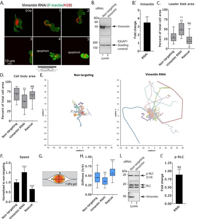
Inhibiting fast amoeboid migration with Vimentin bundles

may play an important role in regulating LBBM               (h/d), which is a function of the opposing force, is
speed (8).                                                  used to define the “cell stiffness.” After Vimentin
                                                            RNAi, cells were found to be ~25% softer than
         To directly test the notion that Vimentin          control (Fig. 2H). In contrast, over-expressing an
concentration in the cell body limits LBBM speed,           RNAi resistant form of Vimentin-FusionRed (i.e.,
we depleted A375-M2 cells of Vimentin using a               rescue) led to an increase in cell stiffness (Fig.
Locked Nucleic Acid (LNA), which offer                      2H). Because cortical actomyosin is also expected
enhanced specificity and stability over traditional         to regulate stiffness, we confirmed that the level of
small interfering RNAs (siRNAs) (24). Because               active (phosphorylated) Regulatory Light Chain
of the long half-life of Vimentin, cells were               (p-RLC) is not affected by Vimentin RNAi (Fig.
incubated with LNAs for 5 days to achieve a                 2I). Thus, we propose Vimentin limits cell
~75% reduction in protein levels (Fig. 2B).                 migration in confinement through increasing cell
Moreover, because these cells predominantly                 stiffness.
express Vimentin, they are an ideal (simplified)
model for defining the role of intermediate                          Because depleting A375-M2 cells of
filaments in LBBM (Fig. 1C). Using our PDMS                 Vimentin had a striking effect on confined
slab-based approach, LBBM was quantitatively                migration, we set out to quantitatively evaluate the

                                                                                                                    Downloaded from http://www.jbc.org/ by guest on October 25, 2020
evaluated for LNA treated cells by live imaging             effect of Vimentin Over-Expression (OE) on
over 5 hr. Strikingly, in cells depleted of                 LBBM. To this end, we transiently transfected
Vimentin, the speed of LBBM was increased over              cells with Vimentin-FusionRed and performed live
control by ~50%, whereas over-expressing an                 imaging (Fig. 3A). Although leader bleb and cell
RNAi resistant form of Vimentin-FusionRed (i.e.,            body area were unaffected, we found a ~35%
rescue) had the opposite effect (Fig. 2E-F &                decrease in LBBM speed after Vimentin OE when
Movie S2).        Directionality over time was              compared to cells expressing EGFP alone (Fig.
unchanged in Vimentin RNAi cells (Fig. S1B).                3B-D). Directionality over time was unchanged in
Quantitation proved that leader bleb area, which is         Vimentin OE cells (Fig S2D). The survival of
defined as the single largest bleb within a given           confined cells was only slightly affected by
frame, is close to double the size of control (Fig.         Vimentin      OE      (Fig.     S1E).           Using
2C). Interestingly, quantitation of cell body area          immunofluorescence, transfected cells were found
found that in Vimentin RNAi cells, the cell body            to have a ~2-fold increase in Vimentin (Fig. S1F).
area was decreased by over 25% (Fig. 2D). We                Consistent with this result, cell stiffness was found
speculate that this result is consistent with the           to be increased by ~50% after Vimentin OE (Fig.
location of Vimentin in these cells, which may              3E). Supported by our finding that Vimentin is
limit the degree that cortical actomyosin is able to        entirely localized to the cell body, we speculate
contract the cell body. Consequently, more                  that increasing the level of Vimentin decreases
cytoplasm from the cell body can enter leader               LBBM speed through stiffening the cell body.
blebs, increasing their size. Strikingly, the nucleus
in Vimentin RNAi cells was observed to undergo                       Next, we set out to determine if
large shape changes, which may reflect an increase          modulating the architecture of Vimentin can
in the degree of force transmitted to the nucleus           impact LBBM. Recently, a potent and selective
from cortical actomyosin (Fig. 2A). A more than             inducer of Vimentin bundling was identified by an
25% increase in the number of Vimentin RNAi                 image-based screen (17). In this screen, the
cells undergoing apoptosis is consistent with               cholesterol lowering statin, Simvastatin, was
reports of nuclear rupture and DNA damage in                shown to directly bind Vimentin and likely
confined cells (Fig. S2C) (25,26). To test the              through limiting electrostatic repulsion between
hypothesis that Vimentin regulates the stiffness of         filaments, is able to induce filament bundling (17).
A375-M2 cells, we used an approach described by             This result is significant because in large cohort
the Piel Lab (Institut Curie) that involves                 studies, patients taking statins for cholesterol have
compressing cells between two Polyacrylamide                decreased cancer associated morbidity (27).
(PA; 1 kPa) gels (Fig. 2G & S1A) (8). Using this            Therefore, we treated A375-M2 cells with
approach, cell height divided by the diameter               Simvastatin (10 µM) and performed live imaging

                                                        3
Inhibiting fast amoeboid migration with Vimentin bundles
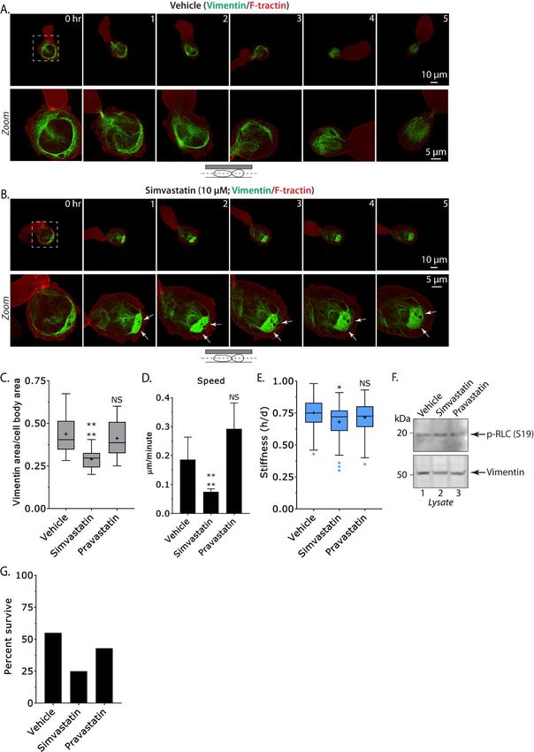
of Vimentin-FusionRed. Compared to Vehicle In order to evaluate if Simvastatin has a
treated (DMSO), the Vimentin network was general effect on cancer cell motility, we subjected
observed to progressively collapse in cells treated drug treated A375-M2 and lung cancer A549 cells,
with Simvastatin (Fig. 4A-B & Movies 3-4). To which also express Keratin (Fig. 1B), to
quantitatively evaluate this affect we measured the transmigration assays. Using filters with 8 µm
total area of the Vimentin network in Vehicle, pores, we found that Simvastatin decreases
Simvastatin, and Pravastatin treated cells. The transmigration for A375-M2 (~80%) and to a
related statin, Pravastatin, is used here for lesser extent A549 cells (~15%), whereas
comparison with Simvastatin. Using this Pravastatin did not have a significant effect (Fig.
approach, the average area of the Vimentin 4A-B). Interestingly, when using filters with
network was decreased by ~35% in Simvastatin larger pores (12 µm) we did not observe a
treated cells, whereas treating cells with statistically significant difference in the
Pravastatin did not have a significant effect (Fig. transmigration of A375-M2 cells after Simvastatin
4C). Next, we quantified the speed of LBBM for treatment (Fig. 5A, right). Because we observed a
Vehicle, Simvastatin, and Pravastatin treated cells. ~50% increase in the number of apoptotic cells
This analysis showed that cells treated with after Simvastatin treatment (Fig. 4G), we next
Simvastatin were ~60% slower than those treated determined if Simvastatin had a general effect on
Downloaded from http://www.jbc.org/ by guest on October 25, 2020
with Vehicle and Pravastatin (Fig. 4D). To cell proliferation. To accomplish this, we counted
evaluate the effect of Simvastatin on cell stiffness, cells over 5 consecutive days in order to generate
we again used the gel sandwich approach. For growth curves for A375-M2 and A549 cells.
cells treated with Simvastatin, we observed a small Strikingly, we found that Simvastatin but not
decrease (~10%) in cell stiffness, whereas treating Pravastatin treatment inhibited the proliferation of
cells with Pravastatin had no effect when both cell types (Fig. 5C-D). However, this effect
compared to Vehicle (Fig. 4E). Because the was significantly more pronounced for A375-M2
bundling of Vimentin causes the network to cells, which express high levels of Vimentin (Fig.
collapse into a small area of the cytoplasm, this 1B). Consistent with Vimentin increasing the size
result might be expected since the gel sandwich and strength of focal adhesions, A375-M2 and
assay measures the stiffness of the entire cell. A549 cells were frequently less spread one day
Although we are unable to directly measure their after Simvastatin treatment (Fig. 5E) (3). Again,
stiffness, based on previous studies of bundled this effect was much more pronounced for A375-
intermediate filaments we predict a local (large) M2 cells. In contrast, cells treated with
increase in mechanical properties (15). In Pravastatin were not significantly different from
agreement with this idea, leader blebs appear to be Vehicle (Fig. 5E). Similarly, we found
resisted by the cell body after Simvastatin Simvastatin but not Pravastatin inhibits the
treatment (Fig. 4B & Movie 4). Because the transmigration and proliferation of WM983-B and
actomyosin cytoskeleton is also expected to effect MDA-MB-231 cells, which both express high
cell stiffness, we also measured the level of active levels of Vimentin (Fig. S2). Altogether, these
myosin (p-RLC) in drug treated cells. By Western results suggest that confined migration and the
blotting, we confirmed that treatment with proliferation of cancer cells is inhibited by
Vehicle, Simvastatin, and Pravastatin did not Vimentin bundling.
affect the level of p-RLC (Fig. 4F). Moreover, the
concentration of Vimentin was unchanged in these Discussion
cells (Fig. 4F). Therefore, by inducing Vimentin Here, we describe for the first time the
bundling with Simvastatin, migration in confined contribution of the Vimentin intermediate filament
environments is inhibited. As tissue culture cells network to LBBM. In contrast to mesenchymal
obtain cholesterol from serum, statin treatment is migration, we demonstrate that depleting cells of
not expected to alter the level of intracellular Vimentin increases the speed of LBBM. A result
cholesterol, which is a critical component of the that may be explained by our observation that
plasma membrane (28). Therefore, our data Vimentin is entirely localized to the cell body,
supports a model whereby local stiffening of the which is thought to resist the motile force
cell body by Vimentin bundling inhibits LBBM. produced by leader blebs. In agreement with this
4
Inhibiting fast amoeboid migration with Vimentin bundles

concept, leader blebs that have spontaneously               pores. However, this effect appears to be more
separated from the cell body are extremely fast             pronounced in cells that express high levels of
(8). Our measurements found leader bleb area to             Vimentin. Additionally, the proliferation of cells
be increased after Vimentin RNAi, whereas the               expressing high levels of Vimentin was
cell body area was reduced. These results suggest           dramatically inhibited by Simvastatin. This result
that Vimentin limits the compressibility of the cell        may be due to the loss of adhesion we observed
body by cortical actomyosin, therefore more                 after Simvastatin but not Pravastatin treatment.
cytoplasm can flow into leader blebs to increase            Alternatively, these results may be due to defects
their size. Consistent with previous reports, our           in the microtubule network, as microtubules and
measurements of stiffness found that Vimentin               Vimentin reciprocally template their growth (4,5).
protects the cell against compression (29).                 Moreover, Vimentin expression has been reported
Vimentin may be particularly important for                  to reinforce EMT through the regulation of
protecting the nucleus from compressive force, as           intracellular signaling (37,38). Thus, future work
several adaptor proteins have been reported to              will need to resolve these alternative explanations.
connect it to the nuclear envelope (30-32).                          Our results are significant because in large
Accordingly, Vimentin has been recently reported            cohort studies, patients taking statins for
to protect the nucleus from rupture and DNA                 cholesterol have decreased cancer associated

                                                                                                                    Downloaded from http://www.jbc.org/ by guest on October 25, 2020
damage in confinement (33,34). In line with these           morbidity (27). However, statins are selectively
results, we find the nucleus in Vimentin RNAi               localized to the liver, which is the major site of
cells to undergo dramatic shape changes during              cholesterol production; therefore, this effect is
LBBM. Moreover, cells depleted of Vimentin                  more likely to be due to a decrease in blood
were found to more frequently undergo apoptosis.            cholesterol. Accordingly, rapidly growing cancer
In mesenchymal cells, the density of the Vimentin           cells require high uptake of extracellular
network has been reported to inversely correlate            cholesterol (39,40). Therefore, patients are not
with the actin retrograde flow rate in lamellipodia         likely to benefit much from the effect of
(35,36).     Similarly, we speculate that the               Simvastatin on Vimentin. Together with the work
localization of Vimentin in the cell body may be            of others, our studies support the derivatization of
important to not restrict the cortical actomyosin           Simvastatin for generating an entirely Vimentin
flow in leader blebs.        Therefore, our work            selective molecule.        Notably, mice lacking
identifies Vimentin to be a fundamental regulator           Vimentin exhibit few defects, therefore the
of LBBM.                                                    systemic administration of a Vimentin selective
         Our results suggest that Vimentin                  molecule may be well tolerated (41-43). Thus, the
increases the stiffness of the cell body to                 perturbation of Vimentin function in cancer cells
negatively regulate LBBM. To test this model, we            represents an attractive strategy for the prevention
used the cholesterol lowering statin, Simvastatin,          of metastasis.
which was identified after a screen of 1,120
biochemically active molecules to specifically              SUPPLEMENTAL INFORMATION
bind and induce the bundling of Vimentin
filaments (17). Indeed, using high-resolution live          Supplemental information includes 2 figures and 4
imaging of melanoma cells, we observed the                  movies and can be found with this article online.
Vimentin network to progressively collapse after
Simvastatin treatment and not with another statin,          EXPERIMENTAL PROCEDURES
Pravastatin. Because tissue culture cells obtain
cholesterol from serum, this effect is not likely due       Cell culture
to the lowering of cholesterol. In line with our            A375-M2 (CRL-3223), A549 (CCL-185), and
model, Vimentin bundles led to the cell body                MDA-MB-231 cells were obtained from the
being cemented in place, inhibiting LBBM. Using             American Type Culture Collection (ATCC;
transmigration assays, which involves the                   Manassas, VA). WM983-B cells were purchased
migration of cells through fibronectin coated               from Rockland Immunochemicals (Pottstown,
filters, we determined that Vimentin bundling               PA). All cells were maintained for up to 30
could inhibit migration through 8 but not 12 µm             passages in DMEM supplemented with 10% FBS

                                                        5
Inhibiting fast amoeboid migration with Vimentin bundles
(cat no. 12106C; Sigma Aldrich, St. Louis, MO), Non-targeting (cat no. 4390844) and Vimentin (cat
GlutaMAX (Thermo Fisher Scientific, Carlsbad, no. 4390824; s14798) LNAs were purchased from
CA), antibiotic-antimycotic (Thermo Fisher Thermo Fisher Scientific. 50 nM (final
Scientific) and 20 mM Hepes pH 7.4. Cells were concentration) LNA was transfected into cells
plated on 6-well glass bottom plates (Cellvis, using RNAiMAX (Thermo Fisher Scientific)
Mountain View, CA) either directly or after diluted in OptiMEM (Thermo Fisher Scientific).
coating with 10 µg/ml human plasma fibronectin
(cat no. FC010; Millipore, Billerica, MA), as Microscopy
noted in the figure legend. Live high-resolution imaging was performed using
a General Electric (Boston, MA) DeltaVision Elite
Pharmacological treatments imaging system mounted on an Olympus (Japan)
Simvastatin (cat no.1965) and Pravastatin (cat no. IX71 stand with a computerized stage,
2318) were purchased from Tocris Bioscience environment chamber (heat, CO2, and humidifier),
(Bristol, UK). DMSO (Sigma Aldrich) was used ultrafast solid-state illumination with
to make 1000X stock solutions for a working excitation/emission filter sets for DAPI, CFP,
concentration of 10 µM. Prior to imaging under GFP, YFP, and Cy5, critical illumination,
confinement, plates with PDMS slabs were Olympus PlanApo N 60X/1.42 NA DIC (oil)
Downloaded from http://www.jbc.org/ by guest on October 25, 2020
incubated overnight in media with DMSO, objective, Photometrics (Tucson, AZ) CoolSNAP
Simvastatin, or Pravastatin. The following day, HQ2 camera, proprietary constrained iterative
this media was replaced with fresh complete deconvolution, and vibration isolation table.
media containing DMSO, Simvastatin, or
Pravastatin. Confinement
This protocol has been described in detail
Plasmids elsewhere (21). Briefly, PDMS (Dow Corning
Vimentin-FusionRed and H2B-FusionRed were 184 SYLGARD) was purchased from Krayden
purchased from Evrogen (Russia). mEmerald- (Westminster, CO). 2 mL was cured overnight at
Vimentin-7 and mEmerald-F-tractin-N13 were 37 °C in each well of a 6-well glass bottom plate
gifts from Michael Davidson (Florida State (Cellvis). Using a biopsy punch (cat no. 504535;
University). F-tractin-FusionRed has been World Precision Instruments, Sarasota, FL), an 8
previously described (7). 1 µg of plasmid was mm hole was cut and 3 mL of serum free media
transfected using a Nucleofector 2b device (Kit V; containing 1% BSA was added to each well and
Lonza, Basel, Switzerland). incubated overnight at 37 °C. After removing the
serum free media containing 1% BSA, 200 µL of
Mutagenesis complete media containing trypsinized cells
The QuikChange II XL Site-Directed Mutagenesis (250,000 to 1 million) and 2 µL of beads (3.11
Kit (Agilent Technologies; Santa Clara, CA) was µm; Bangs Laboratories, Fishers, IN) were then
used according to the manufacture’s protocol for pipetted into the round opening. The vacuum
generating an RNAi resistant form of Vimentin- created by briefly lifting one side of the hole with
FusionRed. The following primers were used for a 1 mL pipette tip was used to move cells and
PCR: beads underneath the PDMS. Finally, 3 mL of
complete media was added to each well and cells
F: CTGGCACGTCTTGATCTTGAACGCAAAG were recovered for ~60 min before imaging.
R: CTTTGCGTTCAAGATCAAGACGTGCCAG
Leader bleb, cell body, and Vimentin area
This yields a single (silent; C->T) mutation measurements
centrally located within the LNA target sequence For leader bleb, cell body, and Vimentin areas,
(GTCTTGACCTTGAACGCAA). Clones were freshly confined cells were traced from high-
verified by sequencing using a commercially resolution images with the free-hand circle tool in
available resource (Thermo Fisher Scientific). Fiji (https://fiji.sc/). From every other frame, the
percent of cell body area for leader blebs, percent
LNAs of total for cell body areas, and percent of cell
6Inhibiting fast amoeboid migration with Vimentin bundles
body area for Vimentin was calculated in gels, whereas the cell diameter was measured
Microsoft Excel (Redmond, WA). Frame-by- using a far-red plasma membrane dye (cat no.
frame measurements were then used to generate an C10046; ThermoFisher). Stiffness was defined as
average for each cell. All statistical analyses were the height (h) divided by the diameter (d). If drugs
performed in GraphPad Prism (La Jolla, CA). were used, gels were first incubated with drug in
media for 30 min before an experiment.
Cell migration
To perform cell speed and directionality analyses, Transmigration
we used a Microsoft Excel plugin, DiPer, Prior to transmigration assays, polycarbonate
developed by Gorelik and colleagues and the Fiji filters with 8 or 12 µm pores (Corning; Corning,
plugin, MTrackJ, developed by Erik Meijering for NY) were coated with 10 µg/mL fibronectin
manual tracking (44,45). For minimizing (Millipore) by air drying for 1 hr. After permitting
positional error, cells were tracked every other ~100,000 cells in serum free media to attach (1
frame. Brightfield imaging was used to confirm hr), DMSO, Simvastatin, or Pravastatin were
that beads were not obstructing the path of a cell. added to each well. Bottom chambers contained
All statistical analyses were performed in 20% FBS in media to attract cells. After 24 hr,
GraphPad Prism. cells from the bottom of the filter were trypsinized
Downloaded from http://www.jbc.org/ by guest on October 25, 2020
and counted using an automated cell counter
Cell stiffness measurements (TC20; Bio-Rad, Hercules, CA). Transmigration
The gel sandwich assay described in detail was then calculated as the ratio of cells on the
elsewhere was used with minor modifications (8) . bottom of the filter vs. the total. All statistical
Briefly, 6-well glass bottom plates (Cellvis) and analyses were performed in GraphPad Prism.
18 mm coverslips were activated using 3-
aminopropyltrimethoxysilane (Sigma Aldrich) for Growth curves
5 min and then for 30 min with 0.5% On day zero, ~125,000 cells were plated in 6-well
glutaraldehyde (Electron Microscopy Sciences, tissue culture plates in complete media with
Hatfield, PA) in PBS. 1 kPa Polyacrylamide (PA) DMSO, Simvastatin, or Pravastatin. For 5
gels were made using 2 µL of blue fluorescent consecutive days, cells were trypsinized and
beads (200 nm; ThermoFisher), 18.8 µL of 40% counted using an automated cell counter (TC20;
acrylamide solution (cat no. 161-0140; Bio-Rad, Bio-Rad). Each day, wells were supplemented
Hercules, CA), and 12.5 µL of bis-acrylamide (cat with fresh media and drug till their day to be
no. 161-0142; Bio-Rad) in 250 µL of PBS. counted. All plots were generated using GraphPad
Finally, 2.5 µL of Ammonium Persulfate (APS; Prism.
10% in water) and 0.5 µL of
Tetramethylethylenediamine (TMED) was added Immunofluorescence
before spreading 9 µL drops onto treated glass After washing with Hepes Buffered Saline (HBS),
under coverslips. After polymerizing for 40 min, cells in 6-well glass bottom plates (Cellvis) were
the coverslip was lifted in PBS, extensively rinsed fixed on ice with methanol containing 1%
and incubated overnight in PBS. Before each Paraformaldehyde (PFA; Electron Microscopy
experiment, the gel attached to the coverslip was Sciences) for 20 min. Blocking, permeabilization,
placed on a 14 mm diameter, 2 cm high PDMS antibody incubations, and washing were done in
column for applying a slight pressure to the HBS with 1% BSA, 1% fish gelatin, 0.1% Triton
coverslip with its own weight. Then, both gels X-100, and 5 mM EDTA. A 1:100 dilution of
were incubated for 30 min in medium before Vimentin antibody (cat no. 5741; Cell Signaling
seeding cells in plates. After the bottom gels in Technology, Danvers, MA) was incubated with
plates was placed on the microscope stage, the cells overnight at 4°C. After extensive washing, a
PDMS column with the top gel was placed on top 1:400 dilution of Alexa Fluor 488 conjugated anti-
of the cells seeded on the bottom gels, confining rabbit secondary antibody (cat no. A-21206;
cells between the two gels (Figure 2G). After 1 hr Thermo Fisher Scientific) was then incubated with
of adaptation, the height of cells was measured cells for 2 hr at room temperature. Cells were
with beads by measuring the distance between again extensively washed and then imaged in
7Inhibiting fast amoeboid migration with Vimentin bundles
HBS. For calculating the level of Vimentin- no. 3671; Cell Signaling Technology), or RLC (cat
FusionRed OE, we used a custom macro in Fiji no. 8505; Cell Signaling Technology) were
(https://fiji.sc/) for automatically quantifying incubated with membranes overnight at 4 °C.
Vimentin in transfected vs. untransfected cells. Bands were then resolved with Horse Radish
Transfected cells were identified using FusionRed. Peroxidase (HRP) conjugated secondary
antibodies and a C-Digit imager (LI-COR
Western blotting Biosciences, Lincoln, NE).
Whole-cell lysates were prepared by scraping cells
into ice cold RIPA buffer (50 mM Hepes pH Statistics
7.4,150 mM NaCl, 5 mM EDTA, 0.1% SDS, 0.5% All sample sizes were empirically determined
deoxycholate, and 1% Triton X-100) containing based on saturation. As noted in each figure
protease and phosphatase inhibitors (Roche, legend, statistical significance was determined by
Switzerland). Before loading onto 4–12% either a two-tailed (unpaired) Student’s t-test, F
NuPAGE Bis-Tris gradient gels (Thermo Fisher test, or ordinary one-way ANOVA followed by a
Scientific), lysates were cleared by centrifugation. post-hoc multiple comparisons test. Normality
Following SDS-PAGE, proteins in gels were was determined by a D’Agostino & Pearson test in
transferred to nitrocellulose membranes and GraphPad Prism. * - p ≤ 0.05, ** - p ≤ 0.01, and
Downloaded from http://www.jbc.org/ by guest on October 25, 2020
subsequently immobilized by air drying overnight. *** - p ≤ 0.001
After blocking in Tris-Buffered Saline containing
0.1% Tween 20 (TBS-T) and 1% BSA, primary Data availability
antibodies against Vimentin, (cat no. 5741; Cell The data that support the findings of this study are
Signaling Technology), pan-Keratin (cat no. 4545; available from the corresponding author, J.S.L.,
Cell Signaling Technology), IQGAP1 (cat no. upon reasonable request.
20648; Cell Signaling Technology), p-RLC (cat
8Inhibiting fast amoeboid migration with Vimentin bundles
ACKNOWLEDGEMENTS
We would like to thank Justin Kusiel for helping initiate the Simvastatin studies and all members of the
Logue Lab for insightful discussions and critical reading of this manuscript. We would also like to thank
the administrative staff within the Department of Regenerative and Cancer Cell Biology at the Albany
Medical College. This work was supported by start-up funds provided by the Albany Medical College.
COMPETING FINANCIAL INTERESTS
The authors declare no competing financial interests.
AUTHOR CONTRIBUTIONS
J.S.L. conceived and designed the study. S.B.L. and S.M.T. contributed equally to all experiments.
M.F.U. performed the transmigration, growth curves, and Western blot assays for WM983-B and MDA-
MB-231 cells. K.W.V. contributed to the quantitative image analysis of rescue (RNAi + Vimentin-
FusionRed) cells. J.S.L. wrote the manuscript with comments from all lab members.
Downloaded from http://www.jbc.org/ by guest on October 25, 2020
9Inhibiting fast amoeboid migration with Vimentin bundles
REFERENCES
1. Yamada, K. M., and Sixt, M. (2019) Mechanisms of 3D cell migration. Nature Reviews
Molecular Cell Biology 20, 738-752
2. Stroka, K. M., Jiang, H., Chen, S.-H., Tong, Z., Wirtz, D., Sun, S. X., and Konstantopoulos, K.
(2014) Water permeation drives tumor cell migration in confined microenvironments. Cell 157,
611-623
3. Mendez, M. G., Kojima, S.-I., and Goldman, R. D. (2010) Vimentin induces changes in cell
shape, motility, and adhesion during the epithelial to mesenchymal transition. The FASEB
Journal 24, 1838-1851
4. Gan, Z., Ding, L., Burckhardt, C. J., Lowery, J., Zaritsky, A., Sitterley, K., Mota, A., Costigliola,
N., Starker, C. G., and Voytas, D. F. (2016) Vimentin intermediate filaments template
microtubule networks to enhance persistence in cell polarity and directed migration. Cell systems
3, 252-263. e258
5. Leduc, C., and Etienne-Manneville, S. (2017) Regulation of microtubule-associated motors drives
intermediate filament network polarization. J Cell Biol 216, 1689-1703
6. Petrie, R. J., Koo, H., and Yamada, K. M. (2014) Generation of compartmentalized pressure by a
Downloaded from http://www.jbc.org/ by guest on October 25, 2020
nuclear piston governs cell motility in a 3D matrix. Science 345, 1062-1065
7. Logue, J. S., Cartagena-Rivera, A. X., Baird, M. A., Davidson, M. W., Chadwick, R. S., and
Waterman, C. M. (2015) Erk regulation of actin capping and bundling by Eps8 promotes cortex
tension and leader bleb-based migration. Elife 4
8. Liu, Y. J., Le Berre, M., Lautenschlaeger, F., Maiuri, P., Callan-Jones, A., Heuze, M., Takaki, T.,
Voituriez, R., and Piel, M. (2015) Confinement and low adhesion induce fast amoeboid migration
of slow mesenchymal cells. Cell 160, 659-672
9. Ruprecht, V., Wieser, S., Callan-Jones, A., Smutny, M., Morita, H., Sako, K., Barone, V., Ritsch-
Marte, M., Sixt, M., Voituriez, R., and Heisenberg, C. P. (2015) Cortical contractility triggers a
stochastic switch to fast amoeboid cell motility. Cell 160, 673-685
10. Bergert, M., Erzberger, A., Desai, R. A., Aspalter, I. M., Oates, A. C., Charras, G., Salbreux, G.,
and Paluch, E. K. (2015) Force transmission during adhesion-independent migration. Nat Cell
Biol 17, 524-529
11. Paul, C. D., Mistriotis, P., and Konstantopoulos, K. (2017) Cancer cell motility: lessons from
migration in confined spaces. Nature Reviews Cancer 17, 131
12. Coussens, L. M., Fingleton, B., and Matrisian, L. M. (2002) Matrix metalloproteinase inhibitors
and cancer: trials and tribulations. Science 295, 2387-2392
13. Wolf, K., Mazo, I., Leung, H., Engelke, K., von Andrian, U. H., Deryugina, E. I., Strongin, A. Y.,
Brocker, E. B., and Friedl, P. (2003) Compensation mechanism in tumor cell migration:
mesenchymal-amoeboid transition after blocking of pericellular proteolysis. J Cell Biol 160, 267-
277
14. Logue, J. S., Cartagena-Rivera, A. X., and Chadwick, R. S. (2018) c-Src activity is differentially
required by cancer cell motility modes. Oncogene
15. Ma, L., Xu, J., Coulombe, P. A., and Wirtz, D. (1999) Keratin filament suspensions show unique
micromechanical properties. Journal of Biological Chemistry 274, 19145-19151
16. Liao, G., and Gundersen, G. G. (1998) Kinesin is a candidate for cross-bridging microtubules and
intermediate filaments selective binding of Kinesin to detyrosinated tubulin and vimentin.
Journal of Biological Chemistry 273, 9797-9803
17. Trogden, K. P., Battaglia, R. A., Kabiraj, P., Madden, V. J., Herrmann, H., and Snider, N. T.
(2018) An image-based small-molecule screen identifies vimentin as a pharmacologically
relevant target of simvastatin in cancer cells. The FASEB Journal, fj. 201700663R
18. Chou, Y. H., Ngai, K. L., and Goldman, R. (1991) The regulation of intermediate filament
reorganization in mitosis. p34cdc2 phosphorylates vimentin at a unique N-terminal site. J Biol
Chem 266, 7325-7328
10Inhibiting fast amoeboid migration with Vimentin bundles
19. Berghe, W. V., Sabbe, L., Kaileh, M., Haegeman, G., and Heyninck, K. (2012) Molecular insight
in the multifunctional activities of Withaferin A. Biochemical pharmacology 84, 1282-1291
20. Grin, B., Mahammad, S., Wedig, T., Cleland, M. M., Tsai, L., Herrmann, H., and Goldman, R. D.
(2012) Withaferin a alters intermediate filament organization, cell shape and behavior. PloS one
7, e39065
21. Logue, J., Chadwick, R., and Waterman, C. (2018) A simple method for precisely controlling the
confinement of cells in culture.
22. Li, M., Zhang, B., Sun, B., Wang, X., Ban, X., Sun, T., Liu, Z., and Zhao, X. (2010) A novel
function for vimentin: the potential biomarker for predicting melanoma hematogenous metastasis.
Journal of Experimental & Clinical Cancer Research 29, 109
23. Tozluoglu, M., Tournier, A. L., Jenkins, R. P., Hooper, S., Bates, P. A., and Sahai, E. (2013)
Matrix geometry determines optimal cancer cell migration strategy and modulates response to
interventions. Nat Cell Biol 15, 751-762
24. Elmén, J., Thonberg, H., Ljungberg, K., Frieden, M., Westergaard, M., Xu, Y., Wahren, B.,
Liang, Z., Ørum, H., and Koch, T. (2005) Locked nucleic acid (LNA) mediated improvements in
siRNA stability and functionality. Nucleic acids research 33, 439-447
25. Raab, M., Gentili, M., de Belly, H., Thiam, H.-R., Vargas, P., Jimenez, A. J., Lautenschlaeger, F.,
Downloaded from http://www.jbc.org/ by guest on October 25, 2020
Voituriez, R., Lennon-Duménil, A.-M., and Manel, N. (2016) ESCRT III repairs nuclear
envelope ruptures during cell migration to limit DNA damage and cell death. Science 352, 359-
362
26. Denais, C. M., Gilbert, R. M., Isermann, P., McGregor, A. L., te Lindert, M., Weigelin, B.,
Davidson, P. M., Friedl, P., Wolf, K., and Lammerding, J. (2016) Nuclear envelope rupture and
repair during cancer cell migration. Science 352, 353-358
27. Nielsen, S. F., Nordestgaard, B. G., and Bojesen, S. E. (2012) Statin Use and Reduced Cancer-
Related Mortality. New England Journal of Medicine 367, 1792-1802
28. DeGrella, R. F., and Simoni, R. D. (1982) Intracellular transport of cholesterol to the plasma
membrane. J Biol Chem 257, 14256-14262
29. Mendez, M. G., Restle, D., and Janmey, P. A. (2014) Vimentin enhances cell elastic behavior and
protects against compressive stress. Biophys J 107, 314-323
30. Wilhelmsen, K., Litjens, S. H., Kuikman, I., Tshimbalanga, N., Janssen, H., van den Bout, I.,
Raymond, K., and Sonnenberg, A. (2005) Nesprin-3, a novel outer nuclear membrane protein,
associates with the cytoskeletal linker protein plectin. The Journal of cell biology 171, 799-810
31. Makise, M., Nakamura, H., and Kuniyasu, A. (2018) The role of vimentin in the tumor marker
Nup88-dependent multinucleated phenotype. BMC Cancer 18, 519
32. Georgatos, S. D., and Blobel, G. (1987) Lamin B constitutes an intermediate filament attachment
site at the nuclear envelope. J Cell Biol 105, 117-125
33. Stankevicins, L. D. C., Urbanska, M., Flormann, D. A., Terriac, E., Mostajeran, Z., Gad, A. K.
B., Cheng, F., Eriksson, J. E., and Lautenschläger, F. (2019) Vimentin provides the mechanical
resilience required for amoeboid migration and protection of the nucleus. bioRxiv
34. Patteson, A. E., Vahabikashi, A., Pogoda, K., Adam, S. A., Goldman, A., Goldman, R., and
Janmey, P. (2019) Vimentin protects the structural integrity of the nucleus and suppresses nuclear
damage caused by large deformations. bioRxiv
35. Costigliola, N., Ding, L., Burckhardt, C. J., Han, S. J., Gutierrez, E., Mota, A., Groisman, A.,
Mitchison, T. J., and Danuser, G. (2017) Vimentin fibers orient traction stress. Proc Natl Acad
Sci U S A 114, 5195-5200
36. Jiu, Y., Lehtimaki, J., Tojkander, S., Cheng, F., Jaalinoja, H., Liu, X., Varjosalo, M., Eriksson, J.
E., and Lappalainen, P. (2015) Bidirectional Interplay between Vimentin Intermediate Filaments
and Contractile Actin Stress Fibers. Cell Rep 11, 1511-1518
37. Vuoriluoto, K., Haugen, H., Kiviluoto, S., Mpindi, J., Nevo, J., Gjerdrum, C., Tiron, C., Lorens,
J., and Ivaska, J. (2011) Vimentin regulates EMT induction by Slug and oncogenic H-Ras and
migration by governing Axl expression in breast cancer. Oncogene 30, 1436-1448
11Inhibiting fast amoeboid migration with Vimentin bundles
38. Phua, D. C., Humbert, P. O., and Hunziker, W. (2009) Vimentin regulates scribble activity by
protecting it from proteasomal degradation. Molecular biology of the cell 20, 2841-2855
39. Fiorenza, A. M., Branchi, A., and Sommariva, D. (2000) Serum lipoprotein profile in patients
with cancer. A comparison with non-cancer subjects. International Journal of Clinical and
Laboratory Research 30, 141-145
40. Benn, M., Tybjærg-Hansen, A., Stender, S., Frikke-Schmidt, R., and Nordestgaard, B. G. (2011)
Low-Density Lipoprotein Cholesterol and the Risk of Cancer: A Mendelian Randomization
Study. JNCI: Journal of the National Cancer Institute 103, 508-519
41. Galou, M., Colucci-Guyon, E., Ensergueix, D., Ridet, J. L., Gimenez y Ribotta, M., Privat, A.,
Babinet, C., and Dupouey, P. (1996) Disrupted glial fibrillary acidic protein network in astrocytes
from vimentin knockout mice. J Cell Biol 133, 853-863
42. Henrion, D., Terzi, F., Matrougui, K., Duriez, M., Boulanger, C. M., Colucci-Guyon, E., Babinet,
C., Briand, P., Friedlander, G., Poitevin, P., and Levy, B. I. (1997) Impaired flow-induced
dilation in mesenteric resistance arteries from mice lacking vimentin. J Clin Invest 100, 2909-
2914
43. Eckes, B., Colucci-Guyon, E., Smola, H., Nodder, S., Babinet, C., Krieg, T., and Martin, P.
(2000) Impaired wound healing in embryonic and adult mice lacking vimentin. J Cell Sci 113 ( Pt
Downloaded from http://www.jbc.org/ by guest on October 25, 2020
13), 2455-2462
44. Gorelik, R., and Gautreau, A. (2014) Quantitative and unbiased analysis of directional persistence
in cell migration. Nat Protoc 9, 1931-1943
45. Meijering, E., Dzyubachyk, O., and Smal, I. (2012) Methods for cell and particle tracking.
Methods Enzymol 504, 183-200
12Inhibiting fast amoeboid migration with Vimentin bundles
Downloaded from http://www.jbc.org/ by guest on October 25, 2020
Figure 1. Vimentin localizes to the cell body of leader bleb forming cells. A. Localization in adhered
to fibronectin (left), uncoated glass (blebbing; middle), and confined under PDMS (forming a leader bleb;
right) of either endogenous (A; adhered) or transiently expressed Vimentin (A’; Vimentin-FusionRed).
B. Percent of confined cells with Vimentin being localized in either the cell body or leader bleb. C.
Lysates from A375-M2 and A549 cells probed for endogenous Vimentin and pan-Keratin. Densitometry
(C’; n=3) was used to determine the ratio of Vimentin in A375-M2 vs. A549 cells. Statistical significance
was determined by a one-sample (hypothetical value=1) Student’s t-test. Error is SEM. All data are
representative of at least three independent experiments. * - p ≤ 0.05, ** - p ≤ 0.01, *** - p ≤ 0.001, and
**** - p ≤ 0.0001
13Inhibiting fast amoeboid migration with Vimentin bundles
Downloaded from http://www.jbc.org/ by guest on October 25, 2020
Figure 2. RNAi of Vimentin promotes rapid leader bleb-based migration. A. Montage of an A375-
M2 cell under PDMS depleted of Vimentin by RNAi expressing F-tractin-mEmerald and H2B-
FusionRed. B. Western blot confirming the depletion of Vimentin by RNAi in A375-M2 cells.
Densitometry (B’; n=3) was used to determine the fold change in Vimentin after RNAi. Error is SEM.
C-D. Quantitative evaluation of leader bleb (C) and cell body (D) area in control (non-targeting),
Vimentin RNAi, and rescue (RNAi + Vimentin-FusionRed) cells. Statistical significance was determined
by an ordinary one-way ANOVA followed by a post-hoc multiple comparisons test. E. Migration tracks
for non-targeting (left; n=40) and Vimentin RNAi (right; n=43) cells under PDMS. F. Amalgamated
instantaneous speeds for non-targeting, Vimentin RNAi, and rescue (RNAi + Vimentin-FusionRed) cells.
14Inhibiting fast amoeboid migration with Vimentin bundles
Data were normalized around the average for non-targeting collected at the time of Vimentin RNAi or
rescue. Statistical significance was determined by an F test. Error is SEM. G. Cartoon of the gel
sandwich approach for measuring cell stiffness. H. Cell stiffness for non-targeting (n=77), Vimentin
RNAi (n=30), and rescue (RNAi + Vimentin-FusionRed; n=23) cells. Statistical significance was
determined by an ordinary one-way ANOVA followed by a post-hoc multiple comparisons test. I.
Western blots of endogenous phosphorylated Regulatory Light Chain (p-RLC; S19) and RLC in non-
targeting and Vimentin RNAi cells. Densitometry (I’; n=3) was used to determine the fold change in p-
RLC after RNAi. Statistical significance was determined by a one-sample (hypothetical value=1)
Student’s t-test. Error is SEM. Tukey box plots in which “+” and line denote the mean and median,
respectively. All data are representative of at least three independent experiments. * - p ≤ 0.05, ** - p ≤
0.01, *** - p ≤ 0.001, and **** - p ≤ 0.0001
Downloaded from http://www.jbc.org/ by guest on October 25, 2020
15Inhibiting fast amoeboid migration with Vimentin bundles
Downloaded from http://www.jbc.org/ by guest on October 25, 2020
Figure 3. Over-expressing Vimentin in confined cells. A. Montage of an A375-M2 cell over-
expressing Vimentin and the marker of Filamentous actin (F-actin), F-tractin, under PDMS. B-C.
Quantitative evaluation of leader bleb (B) and cell body (C) area for cells over-expressing EGFP alone
and Vimentin-FusionRed. Statistical significance was determined by two-tailed (unpaired) Student’s t-
tests. D. Instantaneous speeds for cells over-expressing EGFP alone (n=41) and Vimentin-FusionRed
(n=41). Statistical significance was determined by an F test. Error is SEM. E. Cell stiffness for EGFP
alone (n=34) and Vimentin-FusionRed (n=31) over-expressing cells. Statistical significance was
determined by a two-tailed (unpaired) Student’s t-test. Tukey box plots in which “+” and line denote the
mean and median, respectively. All data are representative of at least three independent experiments. * -
p ≤ 0.05, ** - p ≤ 0.01, *** - p ≤ 0.001, and **** - p ≤ 0.0001
16Downloaded from http://www.jbc.org/ by guest on October 25, 2020
Inhibiting fast amoeboid migration with Vimentin bundles
17Inhibiting fast amoeboid migration with Vimentin bundles
Figure 4. Vimentin bundling inhibits leader bleb-based migration. A-B. Montage of an A375-M2
cell treated with Vehicle (A; DMSO) or Simvastatin (B; 10 µM) expressing F-tractin-mEmerald and
Vimentin-FusionRed under PDMS. Arrows point to areas of collapsed Vimentin in the cell body. C.
Quantitative evaluation of Vimentin network collapse for Vehicle (n=39), Simvastatin (n=26), and
Pravastatin (n=27) treated cells. Statistical significance was determined by an ordinary one-way ANOVA
followed by a post-hoc multiple comparisons test. D. Quantitative evaluation of instantaneous speeds for
Vehicle (n=28), Simvastatin (n=19), and Pravastatin (n=20) treated cells. Statistical significance was
determined by F tests. Error is SEM. E. Cell stiffness measurements for Vehicle (n=77), Simvastatin
(n=63), and Pravastatin (n=62). Statistical significance was determined by a Kruskal-Wallis test followed
by a post-hoc multiple comparisons. F. Western blots of endogenous p-RLC, RLC, and Vimentin in drug
treated cells. G. Percent of live (drug treated) cells after 5 hr under PDMS. Tukey box plots in which
“+” and line denote the mean and median, respectively. All data are representative of at least three
independent experiments. * - p ≤ 0.05, ** - p ≤ 0.01, *** - p ≤ 0.001, and **** - p ≤ 0.0001
Downloaded from http://www.jbc.org/ by guest on October 25, 2020
18Inhibiting fast amoeboid migration with Vimentin bundles
Downloaded from http://www.jbc.org/ by guest on October 25, 2020
Figure 5. Simvastatin impairs the migration and proliferation of cells expressing high levels of
Vimentin. A-B. Quantitative evaluation of transmigration for drug treated A375-M2 (A) and A549 (B)
cells through fibronectin coated (10 µg/mL) polycarbonate filters with 8 or 12 µm pores. Statistical
significance was determined by a two-tailed (unpaired) Student’s t-test. Error is SEM. C-D. Growth
curves for drug treated A375-M2 (C) and A549 (D) cells on tissue culture plastic. Error is SEM. E.
Brightfield images of A375-M2 (top) and A549 (bottom) cells one day after treatment with Vehicle,
19Inhibiting fast amoeboid migration with Vimentin bundles
Simvastatin, or Pravastatin. All experiments were performed at least three times. * - p ≤ 0.05, ** - p ≤
0.01, *** - p ≤ 0.001, and **** - p ≤ 0.0001
Downloaded from http://www.jbc.org/ by guest on October 25, 2020
20A flexible network of Vimentin intermediate filaments promotes the migration of
amoeboid cancer cells through confined environments
Sandrine B Lavenus, Sara M Tudor, Maria F Ullo, Karl W Vosatka and Jeremy S Logue
J. Biol. Chem. published online March 31, 2020
Access the most updated version of this article at doi: 10.1074/jbc.RA119.011537
Alerts:
• When this article is cited
• When a correction for this article is posted
Click here to choose from all of JBC's e-mail alerts
Downloaded from http://www.jbc.org/ by guest on October 25, 2020You can also read