A CROSS-REACTIVE HUMAN IGA MONOCLONAL ANTIBODY BLOCKS SARS-COV-2 SPIKE-ACE2 INTERACTION - NATURE
←
→
Page content transcription
If your browser does not render page correctly, please read the page content below
ARTICLE
                  https://doi.org/10.1038/s41467-020-18058-8                OPEN

                  A cross-reactive human IgA monoclonal antibody
                  blocks SARS-CoV-2 spike-ACE2 interaction
                  Monir Ejemel1,5, Qi Li1,5, Shurong Hou 2,5, Zachary A. Schiller 1,5, Julia A. Tree3, Aaron Wallace1,
                  Alla Amcheslavsky1, Nese Kurt Yilmaz2, Karen R. Buttigieg3, Michael J. Elmore 3, Kerry Godwin3,
                  Naomi Coombes3, Jacqueline R. Toomey1, Ryan Schneider1, Anudeep S. Ramchetty1, Brianna J. Close                                             4,

                  Da-Yuan Chen4, Hasahn L. Conway 4, Mohsan Saeed4, Chandrashekar Ganesa1, Miles W. Carroll3,
                  Lisa A. Cavacini 1 ✉, Mark S. Klempner1 ✉, Celia A. Schiffer 2 ✉ & Yang Wang 1 ✉
1234567890():,;

                  COVID-19 caused by SARS-CoV-2 has become a global pandemic requiring the development
                  of interventions for the prevention or treatment to curtail mortality and morbidity. No vaccine
                  to boost mucosal immunity, or as a therapeutic, has yet been developed to SARS-CoV-2. In
                  this study, we discover and characterize a cross-reactive human IgA monoclonal antibody,
                  MAb362. MAb362 binds to both SARS-CoV and SARS-CoV-2 spike proteins and competi-
                  tively blocks ACE2 receptor binding, by overlapping the ACE2 structural binding epitope.
                  Furthermore, MAb362 IgA neutralizes both pseudotyped SARS-CoV and SARS-CoV-2 in 293
                  cells expressing ACE2. When converted to secretory IgA, MAb326 also neutralizes authentic
                  SARS-CoV-2 virus while the IgG isotype shows no neutralization. Our results suggest that
                  SARS-CoV-2 specific IgA antibodies, such as MAb362, may provide effective immunity
                  against SARS-CoV-2 by inducing mucosal immunity within the respiratory system, a
                  potentially critical feature of an effective vaccine.

                  1 MassBiologics of the University of Massachusetts Medical School, Boston, MA, USA. 2 Biochemistry and Molecular Pharmacology, University of

                  Massachusetts Medical School, Boston, MA, USA. 3 National Infection Service, Public Health England, Porton Down, Salisbury, Wiltshire, UK. 4 National
                  Emerging Infectious Diseases Laboratories, Boston University, Boston, MA, USA. 5These authors contributed equally: Monir Ejemel, Qi Li, Shurong Hou,
                  Zachary A. Schiller. ✉email: Lisa.Cavacini@umassmed.edu; Mark.Klempner@umassmed.edu; Celia.Schiffer@umassmed.edu; Yang.Wang@umassmed.edu

                  NATURE COMMUNICATIONS | (2020)11:4198 | https://doi.org/10.1038/s41467-020-18058-8 | www.nature.com/naturecommunications                            1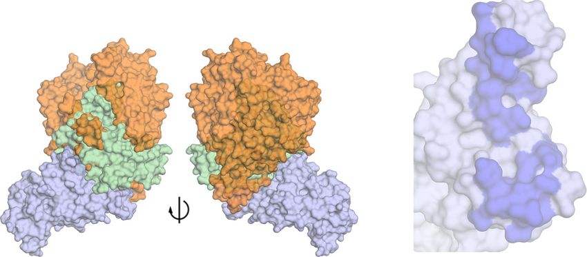
ARTICLE                                                                 NATURE COMMUNICATIONS | https://doi.org/10.1038/s41467-020-18058-8

I
    n December 2019, a novel coronavirus (SARS-CoV-2) was                    and adjuvants for 6–8 weeks. Hybridomas were generated fol-
    identified as the cause of an outbreak of acute respiratory               lowing a standard fusion protocol9. A panel of over 36 hybridomas
    infections. The coronavirus disease 2019 (COVID-19) ranges               were isolated based on various neutralization activities against
from mild to severe acute respiratory infection, with a fatality rate        SARS-CoV with lead antibodies showing protective potency in
estimated to range from 2 to 3%1–4. Within 3 months of the first              mice and hamster models9,10. To explore the possibility that some
report cases, COVID-19 rapidly disseminated through the human                of the SARS-CoV-specific hybridoma may have cross-reactivity
population and had become a global pandemic by March 2020.                   against SARS-CoV-2, these hybridomas were recovered and
Phylogenic analysis has classified SARS-CoV-2 within the sar-                 screened by ELISA against the SARS-CoV-2 spike protein.
becoviruses subgenus, the β lineage that also contains SARS-CoV,             MAb362 was identified with cross-binding activity against both
sharing ~79.6% sequence identity4.                                           the RBD and S1 subunit of the SARS-CoV and SARS-CoV-2 spike
   Interventions for the prevention or treatment of COVID-19 are             proteins (Supplementary Table 1).
crucial for the ongoing outbreak. Pre- or post-exposure immu-                   While both IgG and IgA are expressed at the mucosa, IgA is more
notherapies with neutralizing antibodies, would be of great use by           effective on a molar basis and thus the natural choice for mucosal
providing immediate mucosal immunity against SARS-CoV-2.                     passive immunization as we recently demonstrated in other mucosal
Although concerns, as occurred with SARS-CoV5,6, that vaccines               infectious disease20,21. To further characterize the functionality of
may cause disease enhancement still need to be addressed. The                MAb362, variable sequences of MAb362 were cloned into
feasibility of human monoclonal antibodies (MAbs) as immu-                   expression vectors as either IgG or monomeric IgA isotypes. Both
noprophylaxis or therapy against coronaviruses including SARS-               MAb362 IgG and IgA were assessed in ELISA-binding assays
CoV7–10 and MERS-CoV11 has been demonstrated. These anti-                    against the RBD of the S1 subunit for SARS-CoV (S270–510) and
coronavirus MAbs primarily target the viral spike (S) glycopro-              SARS-CoV-2 (S319–541) (Fig. 1a, b). MAb362 IgA showed better
tein, a type I transmembrane glycoprotein that produces recog-               binding activities, compared with its IgG counterpart against SARS-
nizable crown-like spike structures on the virus surface. The                CoV-2 S319–541 (Fig. 1b). Assessment of the binding kinetics was
receptor-binding domain (RBD) of the S protein facilitates viral             consistent with the ELISA-binding trends. The binding affinity of
entry into human cells through human angiotensin-converting                  IgA with RBD of SARS-CoV-2 is significantly higher (0.3 nM) than
enzyme 2 (ACE2) receptor binding leveraging a similar                        that of IgG (13 nM) due to a much slower dissociation rate as
mechanism as SARS-CoV12–14.                                                  an IgA (Koff = 1.13 × 10⁻³ ± 1.06 × 10⁻⁴) compared with an IgG
   Most current anti-SARS-CoV MAbs neutralize virus by bind-                 (Koff = 7.75 × 10⁻⁵ ± 5.46 × 10⁻⁵) (Fig. 1e, f). Of note, MAb362 IgA
ing to epitopes on the spike protein RBD of SARS-CoV15. We and               and IgG showed similar binding affinity with SARS-CoV S270–510
others have demonstrated that neutralizing MAbs that block                   (Fig. 1c, d).
RBD-ACE2 binding could confer potent protection against                         To confirm binding results, the full ectodomain of spike was
SARS-CoV as both prophylaxis and treatment in various animal                 expressed including residues 1−1208 of SARS-CoV-2 with
models7,9,10. Several anti-SARS-CoV MAbs have demonstrated                   stabilizing proline mutations and a C-terminal T4 fibritin
cross-neutralizing activities against the S protein of SARS-CoV-             trimerization motif as described recently22 (Supplementary Fig. 1).
216,17.                                                                      MAb362 IgA still showed better binding activities with the
   Antibody-dependent enhancement of viral infections are one                stabilized trimer form as compared with its IgG isotype in ELISA
of the major hurdles in the development of effective vaccines.               (Fig. 1b) and affinity assays. The binding affinity of MAb362 IgA
This enhancement is likely facilitated by the Fc domain of IgG but           with the ectodomain of SARS-CoV-2 is 0.17 nM as compared
not for its isotype variant IgA18. The avidity of mucosal IgA, in            with the 27 nM of IgG (Fig. 1g, h).
comparison with IgG, owing to the multimeric structure,
enhances the antibody binding with antigens. In addition, the
diverse, high level of glycosylation of IgA antibodies, further              Structural modeling of MAb362 binding to RBD. To correlate
protects the mucosal surface with non-specific interference. In               the epitope binding with functionality, MAb362 IgG and IgA
animal models, high titers of mucosal IgA in the lung is correlated          were tested in a receptor-blocking assay with Vero E6 cells. The
with reduced pathology upon viral challenge with SARS-CoV19.                 result suggested that both MAb362 IgG and IgA block SARS-
How precisely which isotype may protect the mucosa from SARS-                CoV-2 RBD binding to receptors in a concentration-dependent
CoV-2 infection remains an open question.                                    manner starting at ~30 nM (Fig. 2a, Supplementary Fig. 2a).
   In the current study, we describe the discovery of a cross-               Mutational scanning with a combination of alanine (to introduce
neutralizing human IgA monoclonal antibody, MAb362 IgA. This                 a loss of interaction), tryptophan (to introduce a steric challenge),
IgA antibody binds to SARS-CoV-2 RBD with high affinity                       and lysine to introduce charge mutations were performed to
competing at the ACE2 binding interface by blocking interactions             better delineate the binding surface (Fig. 2b). The results showed
with the receptor. MAb362 IgA neutralizes both pseudotyped                   that that key residues (Y449A, Y453A, F456A, A475W, Y489A,
SARS-CoV and SARS-CoV-2 in 293 cells expressing ACE2. The                    and Q493W) were critical for the complex and presumably,
secretory IgA form of MAb326 also neutralizes authentic SARS-                alterations in the packing caused marked loss of binding affinity
CoV-2 virus. Our results demonstrate that the IgA isotype may                (Fig. 2b and Supplementary Fig. 2b). Among the mutant we
play a critical role in SARS-CoV-2 neutralization.                           tested, A475W and Y489A also disrupted ACE2 binding (Sup-
                                                                             plementary Fig. 3). Interestingly, introduction of lysine mutations
                                                                             had little effect on binding, and some even showed enhanced
Results                                                                      binding, presumably owing to an overall more favorable charged
Selection of MAb binding to RBD of SARS-CoV-2 in ELISA.                      interaction with the MAb362.
We have previously developed and characterized a panel of                       To better define the antibody-binding epitope, known co-
human MAbs that targets the RBD of the SARS-CoV S glyco-                     crystal and cryo-electron microscopy complexes from SARS-CoV
protein, isolated from transgenic mice expressing human immu-                and MERS spike protein in complex with neutralizing antibodies
noglobulin genes9,10. These transgenic mice contains human                   were evaluated for their potential to competitively block ACE2
immunoglobulin genes and inactivated mouse heavy chain and                   binding, based on the structural interface of ACE2-SARS-CoV-2-
kappa light chain genes (Bristol-Myers Squibb). Transgenic mice              RBD (PDB ID-6VW1)23. The 80R-SARS-CoV-RBD complex
were immunized weekly with 10 mg of SARS-CoV spike protein                   (PDB ID-2GHW)24, a crystal structure of SARS-CoV-RBD in

2                NATURE COMMUNICATIONS | (2020)11:4198 | https://doi.org/10.1038/s41467-020-18058-8 | www.nature.com/naturecommunications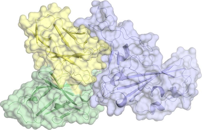
NATURE COMMUNICATIONS | https://doi.org/10.1038/s41467-020-18058-8 ARTICLE
a SARS-CoV
S1 and RBD
c MAb362 IgG
KD 1.3 ± 0.59 nM
e MAb362 IgG
KD 13 ± 4.2 nM
g MAb362 IgG
KD 27.0 ± 5.8 nM
1.5
0.6 0.6 0.8
1000 nM 1000 nM 1000 nM
IgG on SARS-CoV-2 RBD
IgG on SARS-CoV RBD
IgG on SARS-CoV-2
500 nM
Ectodomain Trimer
500 nM 500 nM
1.0 0.6
OD450 nm
0.4 250 nM 0.4 250 nM 250 nM
125 nM 125 nM 125 nM
0.4
62.5 nM 62.5 nM 62.5 nM
0.5 0.2 0.2
0.2
0.0 0.0 0.0 0.0
0.0001 0.001 0.01 0.1 1 10 100 0 100 200 300 0 100 200 300 0 200 400 600
MAb concentration (nM) Time (s) Time (s) Time (s)
MAb362 IgG, S1 MAb362 IgA, S1 Irrelevant IgG, S1
MAb362 IgG, RBD MAb362 IgA, RBD Irrelevant IgG, RBD
b SARS-CoV-2
S1, RBD, and Ectodomain Trimer
d MAb362 IgA
KD 1.4 ± 0.27 nM
f MAb362 IgA
KD 0.3 ± 0.1 nM
h MAb362 IgA
KD 0.17 ± 0.03 nM
2.0
0.25 0.6 0.6
1000 nM 1000 nM 1000 nM
IgA on SARS-CoV-2 RBD
IgA on SARS-CoV RBD
1.5
IgA on SARS-CoV-2
0.20 500 nM 500 nM
Ectodomain Trimer
500 nM
OD450 nm
250 nM 0.4 250 nM 0.4 250 nM
0.15
1.0 125 nM 125 nM 125 nM
0.10 62.5 nM 62.5 nM 62.5 nM
0.2 0.2
0.5
0.05
0.0 0.00 0.0 0.0
0.0001 0.001 0.01 0.1 1 10 100 0 100 200 300 0 100 200 300 0 200 400 600
MAb concentration (nM) Time (s) Time (s) Time (s)
MAb362 IgG, S1 MAb362 IgA, S1 Irrelevant IgG, S1
MAb362 IgG, RBD MAb362 IgA, RBD Irrelevant IgG, RBD
MAb362 IgG, Trimer MAb362 IgA, Trimer Irrelevant IgG, Trimer
Fig. 1 Binding of MAb362 IgG and IgA to spikes of SARS-CoV and SARS-CoV-2. MAb362 IgG and IgA bind to purified SARS-CoV S1 (S1–590) and RBD
(S270–510) truncations a and SARS-CoV-2 S1 (S1–604), RBD (S319–541), and ectodomain trimer b. IgGs are red lines, IgAs are blue lines, and irrelevant IgGs are
black. Affinity measurements of MAb362 IgG c, e, g and IgA d, f, h against the RBD truncations of S glycoprotein of SARS-CoV and SARS-CoV-2 c–f, as
well as ectodomain trimer of SARS-CoV-2 g, h were conducted using bio-layer interferometry and demonstrate nano and sub-nanomolar affinities. Data
are plotted as the mean ± s.d. from n = 3 independent experiments a, b. Source data are provided as a Source Data file.
complex with a neutralizing antibody, 80 R, was found most and SARS-CoV-2. This finding was consistent with the strong
closely to have these characteristics. When the sequence was activity of MAb362 of compromising RBD–receptor interaction.
evaluated, we ascertained that the two antibodies, MAb362 and As with the binding of ACE2, the predicted MAb362-binding
80 R, had frameworks with 90% amino-acid sequence identity epitope can only be exposed if the RBD was in the open or up
(Supplementary Fig. 4). Thus, the crystal structure 2GHW conformation in the trimer (Fig. 3d). In the closed conformation,
provided a suitable scaffold to generate a homology model of this epitope would not be accessible to MAb362 without major
MAb362. Protein–protein docking was performed using the steric clashes. However, unlike CR3022 for instance, MAb362
Schrodinger suite with tethers based on the mutational analysis. could access the ACE2-binding epitope(s) if one or more of the
The complex that satisfied the energetics and mutational data was trimers is in this open conformation, potentially accounting for
then further interrogated with a 300 ns fully solvated molecular the added neutralizing activity.
dynamics simulation in which the complex-structure remained
stable after equilibration. The final frame of the simulation is the
current model of the structure of the MAb362:SARS-CoV-2-RBD MAb362 IgA neutralizes SARS-CoV and SARS-CoV-2. To
complex (Fig. 2c). evaluate the neutralization potency of cross-reactive MAb362, a
The interface of the complex is predicted to form an extensive pseudovirus assay using lentiviral pseudovirions on 293T cells
interface (Fig. 2d and Supplementary Fig. 3) with the CDRs of expressing ACE2 receptor29 was performed. Both MAb362 IgG
both the heavy and light chains forming interactions with SARS- and IgA showed potent neutralization activity against SARS-CoV
CoV-2-RBD. Interestingly, the mutational analysis in combina- (Fig. 4a). MAb362 IgG weakly neutralized SARS-CoV-2 pseudo-
tion with this model indicates that the light chain’s contribution virus despite its activities to block receptor binding. Interestingly,
to this complex may be more significant than the heavy chain isotype switch to MAb362 IgA resulted in significantly enhanced
(Supplementary Fig. 3c). Complementing the receptor-blocking neutralization potency with an IC50 value of 1.26 µg ml−1, com-
assay and mutational analysis, our structural analysis further pared with its IgG subclass variant (IC50 = 58.67 µg ml−1)
confirms that the MAb362 epitope is directly competing for the (Fig. 4b). Monomeric MAb362 IgA was also co-expressed with J
ACE2 binding epitope on SARS-CoV-2 spike protein. chain to produce dimeric IgA (dIgA) and secretory component to
produce secretory IgA (sIgA) as described in Supplementary
MAb362 structural epitope. The model of the structure of the Fig. 630. Both dIgA and sIgA were significantly more effective at
MAb362:SARS-CoV-2-RBD complex permitted the superposition neutralizing SARS-CoV-2 pseudovirus with an IC50 of 30 ng ml−1
of the ACE2:SARS-CoV-2-RBD (PDB: 6VWI23) (Fig. 3a). and 10 ng ml−1, respectively (Fig. 4b). Of note, all MAb362 IgG
MAb362 is predicted to overlaps with the ACE2 epitope on the and IgA isotype variants showed comparable neutralization
RBD. This interface of MAb362 (Fig. 2d) is very similar with the activity against SARS-CoV (Fig. 4a). Further, the most potent
ACE2 interface projected onto the SARS-CoV-2-RBD (Fig. 3b). form MAb362 sIgA was tested in authentic virus neutralization
However, this predicted epitope of MAb362 is different from the assay against SARS-CoV-2. MAb362 sIgA neutralized SARS-CoV-
other recently reported MAb complexes to the SARS-CoV-2-RBD 2 virus with an IC50 value of 9.54 µg ml−1 (Fig. 4c). MAb362 IgG
(Fig. 3c and Supplementary Fig. 5), including: CR302217 (PDB: failed to neutralize live virus at the highest tested concentration.
6W41); S30916 (PDB: 6WPT); REGN10933 and REGN1098725; This is consistent with our prior study showing isotype switch to
(PDB: 6XDG); P2B-2F626 (PDB: 7BWJ); CB627 (PDB: 7C01) and IgA lead to improved antibody neutralization of HIV infection31.
B3828 (PDB: 7BZ5). MAb362 is predicted to block ACE2-binding Our data extend this observation to coronavirus, suggesting that
interface through a unique epitope conserved between SARS-CoV IgA may play an important role in SARS-CoV-2 neutralization.
NATURE COMMUNICATIONS | (2020)11:4198 | https://doi.org/10.1038/s41467-020-18058-8 | www.nature.com/naturecommunications 3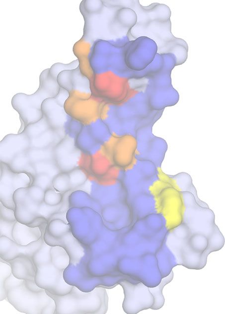
ARTICLE NATURE COMMUNICATIONS | https://doi.org/10.1038/s41467-020-18058-8
a b
ELISA EC50 Fold change
Receptor blocking activity (µg mL–1) to WT
SARS-CoV-2 S1 Wild type 0.64 1.00
150 Y449A 2.03 3.18
MAb362 IgG
Y453A 64.31 100.69
MAb362 IgA
Binding relative to no antibody
125 L455K 0.27 0.42
Irrelevant IgA
A456A 4.00 6.26
100 A475W 6.59 10.32
control (%)
F486A 0.48 0.75
75
N487K 0.63 0.99
50 Y489A 13.04 20.42
Q493W 4.54 7.11
25 Q496K 0.21 0.33
Q498A 0.21 0.33
0
T500A 0.91 1.42
1 10 100 1000 10,000
N501A 0.34 0.52
MAb Concentration (nM)
G502K 0.18 0.28
Y505A 0.42 0.65
c d
A475
VL
Y489
F456
Q493
Y453
Y449
VH
SARS-CoV-2 RBD SARS-CoV-2 RBD (MAb362)
MAb362
Fig. 2 Mutationally guided molecular modeling of MAb362 binding to RBD. a SARS-CoV-2 S1 was pre-incubated with MAb362 IgG (red circles) and IgA
(blue squares) ranging from ~2 to 2000 nM. Both MAb362 isotypes demonstrated concentration-dependent inhibition of SARS-CoV-2 RBD binding to
Vero E6 cells at concentrations >30 nM. Data are plotted as the mean ± s.d. from n = 3 independent experiments. b Mutational scanning was performed to
better delineate the binding surface. Key residues were mutated and expressed as recombinant proteins. Identified critical residues (orange) were
experimental confirmed by shifts in EC50 values for MAb362 binding in ELISA relative to wild-type RBD (blue). EC50 values calculated from n = 3
independent experiments. c Surface representation of the predicted molecular model of MAb362 SARS-CoV-2 RBD complex; the light chain of MAb362
(light yellow), the heavy chain (green), and the SARS-CoV-2 RBD (violet). d The predicted binding interface on SARS-CoV-2 RBD with MAb362. The
residues identified by mutagenesis from b are labeled and colored according to influence degree; red represents strongest defects, orange for medium
defects and yellow for subtle defects. Source data are provided as a Source Data file a, b.
Discussion the shorter hinge in IgG. Our results suggest that compared with
This study reports a unique cross-reactive epitope within the core IgG, SARS-CoV-2-specific IgA antibody may play an important
receptor-binding interface of the S protein of both SARS-CoV independent role in providing protective mucosal immunity. A
and SARS-CoV-2. MAb362 IgA neutralizes the virus by com- similar finding has been observed for IgA antibodies to other
peting with S protein binding to ACE2 receptors. Interestingly, viruses such as influenza and HIV. When monoclonal antibodies
our results show that despite the same blocking of spike inter- are expressed as IgG or IgA1 isotypes with identical variable
action with ACE2, MAb362 IgG weakly neutralizes SARS-CoV-2, regions, antibody binding (affinity, breadth) as well as neu-
whereas IgA as monomer, dimer, or secretory antibody has sig- tralization are enhanced as IgA1 molecules34,35. Though serum
nificantly enhanced neutralization potency. Structural studies half-life of monomeric IgA is relatively short, at the mucosal
demonstrated that IgA1 has a lengthy hinge region with a 13-a.a. epithelial interface, IgA is typically present as secretory antibody.
insertion and a relaxed “T” like structure as compared with the Polymeric IgA (predominantly as a dimer) is produced by local
more rigid “Y” like structure in IgG32,33. Thus, the increase plasma cells and binds to the polymeric Ig receptor at the baso-
flexibility of IgA1 would likely afford a greater reach toward its lateral surface, is transported through the epithelial cell for release
epitopes on the target and decrease steric hindrance. MAb362 IgA at the apical side as secretory IgA, which is the polymeric IgA
binds when the spike protein (trimer) is in open form. The longer with addition of the secretory component contributed by the
IgA1 hinge may allow two Fabs to reach two RBDs of the trimer polymeric Ig receptor. The secretory component protects the IgA
at the same time without clashes, which may not be achieved by from harsh conditions prolonging half-life. In addition, the
4 NATURE COMMUNICATIONS | (2020)11:4198 | https://doi.org/10.1038/s41467-020-18058-8 | www.nature.com/naturecommunications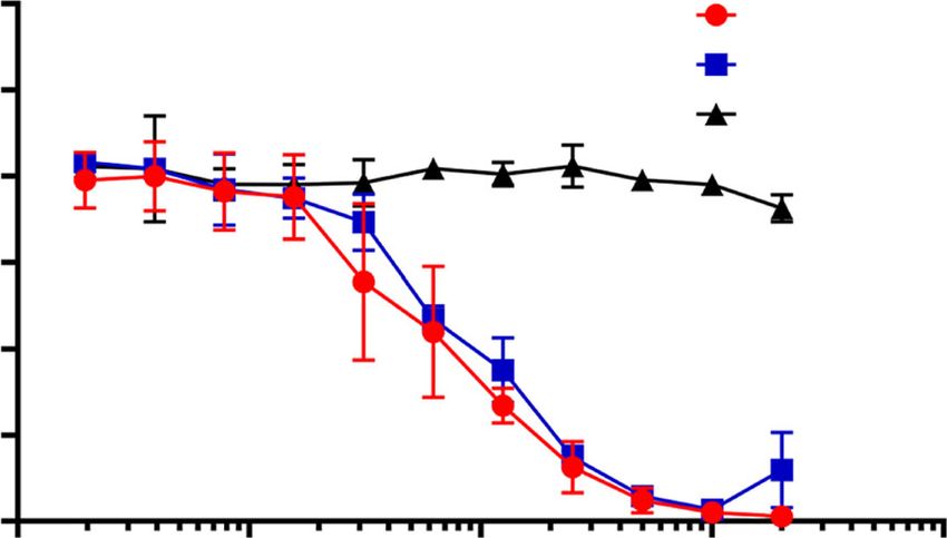
NATURE COMMUNICATIONS | https://doi.org/10.1038/s41467-020-18058-8 ARTICLE
a SARS-CoV-2 receptor
b
(ACE2)
MAb362
180°
SARS-CoV-2 spike protein RBD SARS-CoV-2 RBD (ACE2)
c d
MAb362
REGN10987
REGN10933 B38 MAb362
S309
CB6 SARS-CoV-2
CR3022 RBD
P2B-2F6 180°
SARS-CoV-2 RBD
SARS-CoV-2
spike protein
(open state)
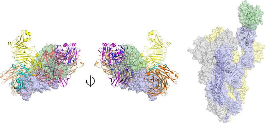
Fig. 3 Predicted MAb362 structural epitope. a Superposition of the space filling molecular model of MAb362 (green) complex on the crystal structure
of the complex of ACE2 (orange) -SARS-CoV-2 RBD (violet) (6VW123) two views are rotated 180°. b The binding interface on SARS-CoV-2 RBD with
ACE2 calculated from the co-crystal structure of the complex. The binding interface shown as darker shade is defined as having vdW contacts great than
−0.5 kcal mol−1. c Positioning of MAb362 on SARS-CoV-2 RBD (violet) relative to the binding of other currently published SARS-CoV-2 RBD-neutralizing
antibodies: CR3022 (PDB: 6W4146; orange); S309 (PDB: 6WPT16; cyan); REGN10933 and REGN10987 (PDB: 6XDG25; magenta and yellow); P2B-2F6
(PDB: 7BWJ26; salmon); CB6 (PDB: 7C0127; wheat) and B38 (PDB: 7BZ528; blue). MAb362 recognized a unique epitope overlapping with the binding
interface of ACE2. d Predicted MAb362 molecular model on the spike trimer in open conformation with one RBD domain exposed 6VYB45.
carbohydrate moieties of sIgA molecules can bind to adhesion Methods
molecules expressed by many pathogens and interfere initial S glycoprotein expression and purification. The amino-acid sequence of the
SARS-CoV S glycoprotein (Urbani strain, National Center for Biotechnology
binding of virus to the target cells as the first line of defense. Thus, Information [strain no. AAP13441]) and SARS-CoV-2 S glycoprotein sequence
mucosal passive immunization of secretory IgA directly to the (GeneBank: MN908947) were used to design a codon-optimized version for
infection site could additionally be an effective approach of sys- mammalian cell expression of the gene encoding the ectodomain of the S glyco-
temic delivery of other IgG treatment to achieve immediate proteins a.a. 1–1190 (S1–1190) for SARS-CoV and a.a. 1–1255 (S1–1255) for SARS-
protection. To date, innovative approaches are being explored for CoV-222. The synthetic gene was cloned into pcDNA 3.1 Myc/His in-frame with c-
Myc and 6-histidine epitope tags that enabled detection and purification. Trun-
sIgA production in mammalian and especially plant expression cated soluble S glycoproteins were generated by polymerase chain reaction (PCR)
systems for cost-effective production including ongoing work in amplification of the desired fragments from the vectors encoding S1255 and S1273.
our laboratories36,37. The SARS-CoV-2 RBD constructs carrying point mutation were generated by
Other recent structure studies have characterized antibodies following the standard protocol from QuikChange II XL Kit (Agilent). The cloned
genes were sequenced to confirm that no errors had accumulated during the PCR
targeting the RBD domain distal from the receptor-binding process. All constructs were transfected into Expi293 cells using ExpiFectamine 293
core interface of SARS-CoV-2 but lack the characteristics of Transfection Kit (Thermo Fisher).
how MAb362 interacts the ACE2-binding epitope. These neu- The plasmid of stabilized trimer of ectodomain of SARS-CoV-2, NIAID
tralizing IgGs, 47D11 and 309, neutralize SARS-CoV-2 with VRC7471, and its expression and purification protocol was kindly provided by
Dr. Kizzmekia S. Corbett, PhD, at Vaccine Research Center of National Institute of
high potency, but do not block receptor binding to ACE16,38. Allergy and Infectious Diseases as part of large-scale production contract awarded to
Potentially, ACE2 may not be the sole receptor for SARS-CoV- MassBiologics of UMMS (U24AI126683)22 In this construct, a gene encoding
2, similar to SARS-CoV39, or these antibodies may prevent a residues 1−1208 of SARS-CoV-2 S glycoprotein sequence (GenBank: MN908947)
conformational change necessary for viral entry. Further study was modified by adding two proline substitutions at residues 986 and 987, a “GSAS”
substitution at residues 682–685, a C-terminal T4 fibritin trimerization motif, an
of the interaction between MAb362, and other receptor HRV3C protease cleavage site, a TwinStrepTag and an 8x HisTag. The construct was
blocking and neutralizing antibodies against SARS-CoV-2 will cloned into the mammalian expression vector pCDNA 3.1. The construct was then
provide insight into the design of vaccine and prophylactic/ transfected into Expi293 cells using ExpiFectamine 293 Transfection Kit (Thermo
therapeutic antibodies against future emerging infections Fisher). Protein was purified from using StrepTactin resin (IBA) followed by size-
caused by this viral family. exclusion chromatography using a Superose 6 10/300 column (GE Healthcare).
NATURE COMMUNICATIONS | (2020)11:4198 | https://doi.org/10.1038/s41467-020-18058-8 | www.nature.com/naturecommunications 5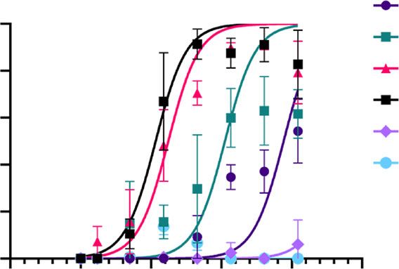
ARTICLE NATURE COMMUNICATIONS | https://doi.org/10.1038/s41467-020-18058-8
a Neutralization of SARS-CoV b Neutralization of SARS-CoV-2
MAb362 IgG MAb362 IgG
100 100
MAb362 IgA MAb362 IgA
Neutralization (%) MAb362 dIgA MAb362 dIgA
Neutralization (%)
80 80
MAb362 sIgA MAb362 sIgA
60 60
Irrelevant IgG Irrelevant IgG
40 Irrelevant sIgA 40 Irrelevant sIgA
20 20
0 0
-2 -1 0 1 2 3 -6 -4 -2 0 2 4
Antibody Concentration Antibody Concentration
Log10 (µg mL–1) Log10 (µg mL–1)
MAb362 MAb362 MAb362 MAb362 MAb362 MAb362 MAb362 MAb362
Antibody Antibody
IgG IgA dIgA sIgA IgG IgA dIgA sIgA
IC50 IC50
1.56 0.84 0.63 0.94 58.67 1.26 0.03 0.01
–1
(µg mL ) (µg mL–1)
c 100
Plaque reduction (%)
75
50
25
0
0.01 1.0 100
Antibody concentration
(µg mL–1)
Antibody MAb362 MAb362
IgG sIgA
IC50
–1 >50 9.54 ± 5.88
(µg mL )
Fig. 4 IgA isotype switch enhances MAb362 neutralization of SARS-CoV-2. MAb362 antibody-mediated neutralization of luciferase-encoding
pseudovirions with spike proteins of SARS-CoV a and SARS-CoV-2 b. SARS-CoV and SARS-CoV-2 pseudovirions pre-incubated with serial dilutions of
MAb362 were used to infect 293 cells expressing ACE2 receptor. Pseudoviral transduction was measured by luciferase activities in cell lysates 48 h post
transduction to calculate neutralization (%) relative to non-antibody-treated controls. IC50 values were calculated by nonlinear regression analysis using Prism
version 8.1.1. Isotype switching improved SARS-CoV-2 IC50. Data are plotted as the mean ± s.d. from n = 3 independent experiments a, b. c Dose–response
curve for PRNT with MAb362 at a starting concentration of 50 µg mL−1 titrated 1:2. MAb362 sIgA had a 50% endpoint titer of 9.54 ± 5.88 µg mL−1 calculated
by Spearman–Kärber method, from n = 2 biologically independent experiments. Representative data are plotted with a Probit mid-point analysis curve ± 95%
CI from one experiment with n = 2 technical replicates, using R programming language v3.5.3 and Library ggplot2 v3.3.0 for statistical computing and
graphics47,48. Source data are provided as a Source Data file a–c.
All recombinant proteins were purified by immobilized metal chelate affinity of antibodies (GE life Sciences). Purified antibodies were dialyzed against PBS
chromatography using nickel-nitrilotriacetic acid (Ni-NTA) agarose beads. before being moved onto size-exclusion chromatography on fast performance
Proteins were eluted from the columns using 250 mmol/L imidazole and then liquid chromatography to separate out the desired dimeric or secretory antibodies
dialyzed into phosphate-buffered saline (PBS), pH 7.2 and checked for size and using a HiLoad 26/600 Superdex 200-pg size-exclusion column (GE life Sciences).
purity by sodium dodecyl sulfate polyacrylamide gel electrophoresis (SDS-PAGE). The desired fractions were pooled, concentrated, and quality analyzed by SDS-
The stabilized trimer is also analyzed by high performance liquid chromatography PAGE and HPLC with representative HPLC profile and gel image shown in
(HPLC) (Supplementary Fig. 1). Supplementary Fig. 630.
Generation of MAbs. Previously generated frozen hybridomas of anti-SARS-CoV ELISA. Dilutions of purified MAbs were tested in ELISA for reactivity against
MAbs9 were recovered and scaled up. Hybridoma supernatants were screened for recombinant S protein. In brief, 96-well plates were coated with S proteins followed
reactivity to the SARS-CoV-2 S protein. Positive cell clones were selected for by incubation overnight at 4°C. The plates were blocked with 1% BSA with 0.05%
antibody sequencing. For MAb362, the heavy chain and light chain variable regions Tween 20 in PBS. Hybridoma supernatant or purified antibody diluted in 1× PBS
were amplified from hybridoma cells and cloned into an immunoglobulin G1 (IgG) plus 0.1% Tween 20 and added to the 96-well plates and incubated for 1 h at room
expression vector. Isotype switching was conducted using primers designed to temperature. Plates were stained with horseradish peroxidase-conjugated anti-
amplify the variable heavy chain of the IgG antibody. Products were digested and kappa (Company Southern biotech, #2060-05,1:2000 dilution) for 1 h and devel-
ligated into a pcDNA 3.1 vector containing the heavy constant IgA1 chain. The oped using 3,3′,5,5′-tetramethylbenzidine. Absorbance at an optical density at
vector was transformed in NEB5-α-competent cells, and sequences were verified 450 nm (OD450) was measured on an Emax precision plate reader (Molecular
ahead of transient transfection. IgG and monomeric IgA1 antibodies were trans- Devices) using Softmax Pro v4.3.1 LS.
fected in Expi293 cells. Cell supernatants were harvested 5 days post transfection
for antibody purification by protein A sepharose for IgG and Capto L resin for IgA
(GE life Sciences). For dimeric IgA1 (dIgA), the heavy and light chain vectors were ELISA-based ACE2-binding assay. In all, 250 ng of ACE2 protein was coated on
co-transfected with pcDNA-containing DNA for the connecting J chain. For ELISA plates overnight at 4 °C. After blocking with 1% BSA in PBS with 0.05% Tween
secretory IgA1 (sIgA) expression, a pcDNA-vector containing gene sequence of 20 for 1 h at room temperature, threefold of serial dilutions started from 10 µg ml−1 of
secretory component was added to the transfection reaction in a 1:1 ratio. wild type and point mutations S protein were added into the plates and incubated for
Supernatant was run through a column of Capto L resin to capture the light chain 1 h at room temperature. Then plates were stained with mouse-anti-Myc antibody
6 NATURE COMMUNICATIONS | (2020)11:4198 | https://doi.org/10.1038/s41467-020-18058-8 | www.nature.com/naturecommunications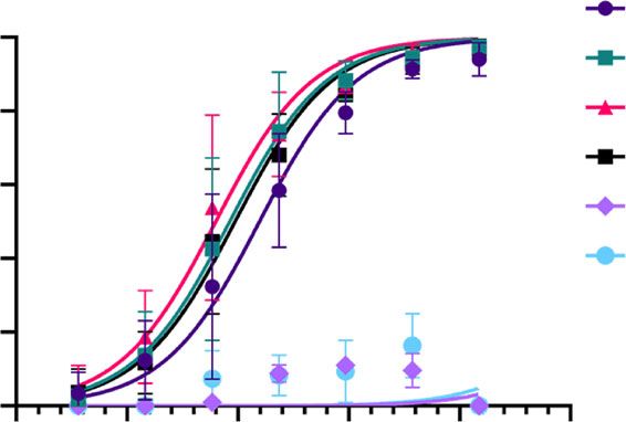
NATURE COMMUNICATIONS | https://doi.org/10.1038/s41467-020-18058-8                                                                                             ARTICLE

(BD Pharmingen #551101), at 2 µg ml−1 for 1 h, followed by horseradish peroxidase-        Mutational scanning to identify MAb362-binding residues. SARS-CoV-2 RBD
conjugated goat anti-mouse (Jackson ImmnuoResearch #115-035-062, 1:2000 dilu-             residues were individually mutated with a combination of alanine (to introduce a
tion) for 1 h and developed using 3,3′,5,5′-tetramethylbenzidine. Absorbance at an        loss of interaction), tryptophan (to introduce a steric challenge), and lysine
optical density at 450 nm (OD450) was measured on an Emax precision plate reader          mutations to introduce charge using QuikChange II XL Kit (Agilent) or BioXp
(Molecular Devices) using Softmax Pro v4.3.1 LS.                                          3200 System (SGI-DNA). The genes were cloned into RBD expression vectors and
                                                                                          RBD proteins were purified as described above. Mutant RBDs were confirmed
                                                                                          intact expression on proteins gels, and the same amount of proteins were coated on
Flow cytometry-based receptor-binding inhibition assay. Vero E6 cells were                the plate for ELISA assays.
harvested with PBS containing 5 mM ethylenediaminetetraacetic acid and ali-                   Dilutions of purified MAbs were tested in ELISA for reactivity against mutant
quoted to 1 × 10⁶ cells per reaction. Cells were pelleted then resuspended in PBS         RBD proteins. In all, 96-well plates were coated with 100 µl of 5 µg of RBD mutants
containing 10% FBS. Before mixing with the cells, Myc-tagged SARS-CoV-2 S1–604            followed by incubation overnight at 4°C. The plates were blocked with 1% BSA
was incubated with the MAb at varying concentrations for 1 h at room tempera-             with 0.05% Tween 20 in PBS. Purified antibody diluted in 1× PBS plus 0.1% Tween
ture, then the S protein was added to the Vero cells to a final concentration of           20 and added to the 96-well plates and incubated for 1 h at room temperature.
10 nM. The cells–S protein mixture was incubated for 1 h at room temperature.             Plates were stained with alkaline phosphatase affiniPure goat anti-Human IgG
After incubation, the cell pellets were washed and then resuspended in PBS with           (Jackson ImmunoResearch #109-055-098, 1:1000 dilution) for 1 h at room
2% FBS and incubated with 10 µg mL−1 of mouse-anti-Myc antibody (BD Phar-                 temperature. Alkaline phosphatase affiniPure goat anti-Mouse IgG (Jackson
mingen #551101, 1:100 dilution) for 1 h at 4 °C. Pellets were washed again then           ImmunoResearch #115-055-003, 1:1000 dilution) was used to detect his tag in a
subsequently incubated with a phycoerythrin-conjugated anti-mouse IgG (Jackson            separate ELISA to verify protein expression and coating. Plates were developed
ImmunoResearch, #115-116-071, 1:20 dilution) for 40 min at 4 °C. Cells were               using p-Nitrophenyl Phosphate (Thermo Fisher Scientific). Absorbance at an
washed twice then subjected to flow cytometric analysis using a MACSquant Flow             optical density at 405 nm (OD405) was measured on an Emax precision plate
Cytometer (Miltenyi Biotec) and analyzed by MACSQuantify Software v2.11 and               reader (Molecular Devices) using Softmax Pro v4.3.1 LS. ELISAs assay was
FlowJo v10. Binding was expressed as relative to cells incubated with S                   performed to determine binding of the MAbs to the mutant proteins compared
proteins only.                                                                            with the wild type. Key residues were identified by RBD mutations that reduced
                                                                                          EC50 values relative to the wild-type RBD.
Pseudotyped virus neutralization assay. Pseudovirus was generated employing
an HIV backbone that contained a mutation to prevent HIV envelope glycoprotein            Affinity determination for MAb362. Bio-layer interferometry (BLI) with an Octet
expression and a luciferase gene to direct luciferase expression in target cells (pNL4-   HTX (PALL/ForteBio) was used to determine the affinity of MAb362 IgG and IgA1
3.Luc.R–E–, obtained from Dr. Nathaniel Landau, NIH). SARS-S and SARS2-S spike            to the RBD of SARS-CoV and SARS-CoV-2 S protein. MAbs were added to 96
protein was provided in trans by co-transfection of 293 T cells with pcDNA-G with         wells plates at 1000 nM and titrated 1:2 to 62 nM using PBS. RBD of SARS-CoV,
pNL4-3.Luc.R–E–. Supernatant containing virus particles was harvested 48–72 h             RBD, and ectodomains of SARS-CoV-2 were biotinylated (Thermo Fisher) and
post transfection, concentrated using Centricon 70 concentrators, aliquoted, and          immobilized on Streptavidin Biosensors (ForteBio) for 120 s at 1600 nM con-
stored frozen at −80 degree. Before assessing antibody neutralization, the 293 T cells    centration. After a baseline step, MAb362-antigen binding rate was determined
were transient transfected with 100 ng pcDNA-ACE2 each well in 96-well plates, and        when the biosensors with immobilized antigen were exposed to MAb362 IgG or
the cells were used for the pseudovirus infection 24 hs after transfection. A titration   IgA1 at different concentrations for 120 s. Following association, the MAb362-RBD
of pseudovirus was performed on 293 T cells transiently transfected with human            complex was exposed to PBS and the rate of the MAb362 dissociation from antigen
ACE2 receptor to determine the volume of virus need to generate 50,000 counts             was measured. Each assay was performed in triplicate. Binding affinities for
per second (cps) in the infection assay. The appropriate volume of pseudovirus was        MAb362 were calculated using association and dissociation rates with ForteBio
pre-incubated with varying concentrations of MAbs for 1 h at room temperature             Data analysis software v8.1 (PALL).
before adding to 293 T cells expressing ACE2. 24 h after the infection, the pseudo-
virus was replaced by the fresh complete media, and 24 h after media changing the
infection was quantified by luciferase detection with BrightGlo luciferase assay           Statistical analysis. Statistical calculations were performed using Prism version
(Promega) and read in a Victor3 plate reader (Perkin Elmer) for light production.         8.1.1 (GraphPad Software, La Jolla, CA). EC50 and IC50 values were calculated by
                                                                                          sigmoidal curve fitting using nonlinear regression analysis.

Plaque reduction neutralization assay (PRNT). Monoclonal antibody was serially
diluted and incubated with ~70 plaque forming units of wild-type SARS-CoV-2               Reporting summary. Further information on research design is available in the Nature
(2019-nCoV/Victoria/1/2020), for 1 h at 37°C in a humidified box. The virus/anti-          Research Reporting Summary linked to this article.
body mixture was then allowed to absorb onto monolayers of Vero E6 [(ECACC
85020206, European Collection of Authenticated Cell Cultures, UK] for 1 h at 37 °C        Data availability
in a humidified box. Overlay media [MEM (Life Technologies, California, USA)               Antibody heavy and light chain sequence can be found on GenBank (MAb362 Heavy
containing 1.5% carboxymethylcellulose (Sigma), 5% (v/v) fetal calf serum (Life           Chain Accession # MT789771, MAb362 Light Chain Accession # MT789772). Database
Technologies) and 25 mM 4-(2-hydroxyethyl)-1-piperazineethanesulfonic acid buf-           files used in the study include: PDB 2GHW, the complex of 80 R:SARS-CoV-RBD24;
fer (Sigma)] was added and the 24-well plates were incubated in a humidified box at        PDB 2AJF, the complex of ACE2:SARS-CoV-RBD43 and PDB 6VW1, the complex of
37 °C for 5 days. Plates were fixed overnight with 20% (w/v) formalin/PBS, washed          ACE2:SARS-CoV-2-RBD. All other data generated are included in figures and tables in
with tap water and stained with methyl crystal violet solution (0.2% v/v) (Sigma).
                                                                                          this published article. Source data are provided with this paper. Reprints and permissions
The neutralizing antibody titers were defined as the amount of antibody (µg mL−1)
                                                                                          information is available at www.nature.com/reprints.
resulting in a 50% reduction relative to the total number of plaques counted without
antibody, by performing a Spearman–Kärber analysis40 using Microsoft Excel v2016.
An internal positive control for the PRNT assay was run using a sample of human           Received: 5 May 2020; Accepted: 3 August 2020;
MERS convalescent serum known to neutralize SARS-CoV-2 (National Institute for
Biological Standards and Control, United Kingdom).

Structural modeling analyses. Three crystal structures, 2GHW the complex of 80
R:SARS-CoV-RBD24, 2AJF the complex of ACE2:SARS-CoV-RBD41, and 6VW1
the complex of ACE2:SARS-CoV-2-RBD23 were used as initial scaffolds in the                References
determinations of the models of MAb362:SARS-CoV-RBD and MAb362:SARS-                      1.   Li, Q. et al. Early transmission dynamics in Wuhan, China, of novel
CoV-2-RBD. The amino-acid sequence of MAb362 was aligned to the amino-acid                     coronavirus-infected pneumonia. N. Engl. J. Med. 382, 1199–1207 (2020).
sequences of 80 R bound SARS-CoV-1 crystal structure (PDB: 2GHW). The point               2.   Jiang, S., Du, L. & Shi, Z. An emerging coronavirus causing pneumonia
mutational studies of SARS-CoV-2 RBD were used as restraints to guide the                      outbreak in Wuhan, China: calling for developing therapeutic and
protein–protein docking of MAb362 against SARS-CoV-2 RBD. The docking was                      prophylactic strategies. Emerg. Microbes Infect. 9, 275–277 (2020).
performed using Glide (Schrödinger software suite v19-4) and Modeller v9.23. The          3.   Huang, C. et al. Clinical features of patients infected with 2019 novel
highest scored docking pose that also best satisfied the mutational analysis was                coronavirus in Wuhan, China. Lancet 395, 497–506 (2020).
further optimized through 300 ns molecular dynamic (MD) simulations. The                  4.   Zhou, P. et al. A pneumonia outbreak associated with a new coronavirus of
MD simulations were performed using Desmond (Schrödinger software suite                        probable bat origin. Nature 579, 270–273 (2020).
v19-4)42–44. The final frame of the MD simulations was used as the final structural         5.   Czub, M., Weingartl, H., Czub, S., He, R. & Cao, J. Evaluation of modified
model of MAb362-RBD complex.                                                                   vaccinia virus Ankara based recombinant SARS vaccine in ferrets. Vaccine 23,
    The structural model of MAb362 binding to the SARS-CoV-2 spike trimer was                  2273–2279 (2005).
based on 6VYB45. All figures were made within PyMOL Molecular Graphics
                                                                                          6.   Liu, L. et al. Anti-spike IgG causes severe acute lung injury by skewing macrophage
System v2.3.4 (Schrödinger). The residue van der Waals potential between the
                                                                                               responses during acute SARS-CoV infection. JCI Insight 4, e123158 (2019).
various complexes was extracted from the structures energies using the energy
potential within Desmond.

NATURE COMMUNICATIONS | (2020)11:4198 | https://doi.org/10.1038/s41467-020-18058-8 | www.nature.com/naturecommunications                                                          7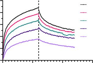
ARTICLE                                                                             NATURE COMMUNICATIONS | https://doi.org/10.1038/s41467-020-18058-8

7.    Zhu, Z. et al. Potent cross-reactive neutralization of SARS coronavirus isolates    36. Juarez, P., Virdi, V., Depicker, A. & Orzaez, D. Biomanufacturing of protective
      by human monoclonal antibodies. Proc. Natl Acad. Sci. USA 104,                          antibodies and other therapeutics in edible plant tissues for oral applications.
      12123–12128 (2007).                                                                     Plant Biotechnol. J. 14, 1791–1799 (2016).
8.    Sui, J. et al. Potent neutralization of severe acute respiratory syndrome (SARS)    37. Virdi, V., Juarez, P., Boudolf, V. & Depicker, A. Recombinant IgA production
      coronavirus by a human mAb to S1 protein that blocks receptor association.              for mucosal passive immunization, advancing beyond the hurdles. Cell Mol.
      Proc. Natl Acad. Sci. USA 101, 2536–2541 (2004).                                        Life Sci. 73, 535–545 (2016).
9.    Greenough, T. C. et al. Development and characterization of a severe acute          38. Wang, C. et al. A human monoclonal antibody blocking SARS-CoV-2
      respiratory syndrome-associated coronavirus-neutralizing human monoclonal               infection. Nat. Commun. 11, 2251 (2020).
      antibody that provides effective immunoprophylaxis in mice. J. Infect. Dis.         39. Jeffers, S. A. et al. CD209L (L-SIGN) is a receptor for severe acute
      191, 507–514 (2005).                                                                    respiratory syndrome coronavirus. Proc. Natl Acad. Sci. USA 101,
10.   Roberts, A. et al. Therapy with a severe acute respiratory syndrome-associated          15748–15753 (2004).
      coronavirus-neutralizing human monoclonal antibody reduces disease                  40. Dougherty, R. & Harris, R. Techniques in experimental virology. RJC Harris,
      severity and viral burden in golden Syrian hamsters. J. Infect. Dis. 193,               Ed. 169 (1964).
      685–692 (2006).                                                                     41. Li, F., Li, W., Farzan, M. & Harrison, S. C. Structure of SARS coronavirus
11.   Du, L. et al. MERS-CoV spike protein: a key target for antivirals. Expert Opin.         spike receptor-binding domain complexed with receptor. Science 309,
      Ther. Targets 21, 131–143 (2017).                                                       1864–1868 (2005).
12.   Letko, M. & Munster, V. Functional assessment of cell entry and receptor            42. Leidner, F., Kurt Yilmaz, N., Paulsen, J., Muller, Y. A. & Schiffer, C. A.
      usage for lineage B betacoronaviruses, including 2019-nCoV. (2020).                     Hydration structure and dynamics of inhibitor-bound HIV-1 protease. J.
13.   Li, W. et al. Angiotensin-converting enzyme 2 is a functional receptor for the          Chem. Theory Comput. 14, 2784–2796 (2018).
      SARS coronavirus. Nature 426, 450–454 (2003).                                       43. Hou, S. et al. Structural analysis of the active site and DNA binding of
14.   Becker, M. M. et al. Synthetic recombinant bat SARS-like coronavirus is                 human cytidine deaminase APOBEC3B. J. Chem. Theory Comput. 15, 637–647
      infectious in cultured cells and in mice. Proc. Natl Acad. Sci. USA 105,                (2019).
      19944–19949 (2008).                                                                 44. Harder, E. et al. OPLS3: a force field providing broad coverage of drug-like
15.   Jiang, S., Hillyer, C. & Du, L. Neutralizing antibodies against SARS-CoV-2 and          small molecules and proteins. J. Chem. Theory Comput. 12, 281–296 (2016).
      other human coronaviruses. Trends Immunol. 41, 355–359 (2020).                      45. Walls, A. C. et al. Structure, function, and antigenicity of the SARS-CoV-2
16.   Pinto, D. et al. Cross-neutralization of SARS-CoV-2 by a human monoclonal               spike glycoprotein. Cell 181, 281–292.e6 (2020).
      SARS-CoV antibody. Nature 583, 290–295 (2020).                                      46. Yuan, M. et al. A highly conserved cryptic epitope in the receptor binding
17.   Yuan, M. et al. A highly conserved cryptic epitope in the receptor-binding              domains of SARS-CoV-2 and SARS-CoV. Science 368, 630–633 (2020).
      domains of SARS-CoV-2 and SARS-CoV. Science 368, 630–633                            47. Johnson, R. M., Dahlgren, L., Siegfried, B. D. & Ellis, M. D. Acaricide,
      (2020).                                                                                 fungicide and drug interactions in honey bees (Apis mellifera). PLoS ONE 8,
18.   Bakema, J. E. & van Egmond, M. Immunoglobulin A: a next generation of                   e54092 (2013).
      therapeutic antibodies? MAbs 3, 352–361 (2011).                                     48. Probit Analysis, 3rd ed. By D. J. Finney, Cambridge University Press, 32 E.
19.   Du, L. et al. Intranasal vaccination of recombinant adeno-associated virus              57th St., New York, Ny 10022, 1971. xv + 333 pp. 14.5 × 22 cm. Price $18.50.
      encoding receptor-binding domain of severe acute respiratory syndrome                   J. Pharm. Sci. 60, 1432–1432, (1971).
      coronavirus (SARS-CoV) spike protein induces strong mucosal immune
      responses and provides long-term protection against SARS-CoV infection.
      J. Immunol. 180, 948–956 (2008).                                                    Acknowledgements
20.   Stoppato, M. et al. Oral administration of an anti-CfaE secretory IgA antibody      We thank Dr. Robert Finberg at UMMS for helpful advice on functional assays. The
      protects against enterotoxigenic Escherichia coli diarrheal disease in a            initial mouse immunization work in 2005 was supported by NIH Contract NO1-AI-
      nonhuman primate model. Vaccine 38, 2333–2339 (2020).                               65315. S.H., N.Y.K., and C.A.S. were supported by NIH R01AI150478. The production of
21.   Hu, Y. et al. Preformulation characterization and stability assessments of          stabilized ectodomain trimer was supported by NIH contract U24AI126683. The dIgA
      secretory IgA monoclonal antibodies as potential candidates for passive             and sIgA expression platform was supported by Bill & Melinda Gates Foundation.
      immunization by oral administration. J. Pharm. Sci. 109, 407–421 (2020).
22.   Wrapp, D. et al. Cryo-EM structure of the 2019-nCoV spike in the prefusion          Author contributions
      conformation. Science 367, 1260–1263 (2020).                                        M.E., Q.L, A.W. cloned, expressed, and purified MAbs, S proteins, truncations, and
23.   Shang, J. et al. Structural basis of receptor recognition by SARS-CoV-2. Nature     variants with assistance from A.A., J.R.T., A.S.R., R.S., and C.G.; M.E. and Q.L. designed
      581, 221–224 (2020).                                                                and performed affinity and binding assays and flow cytometry with assistance from Y.W.,
24.   Hwang, W. C. et al. Structural basis of neutralization by a human anti-severe       L.A.C., and Z.A.S.; S.H., carried out structural modeling and analyses with assistance
      acute respiratory syndrome spike protein antibody, 80R. J. Biol. Chem. 281,         from Q.L. N.Y.K., Y.W., and C.A.S.; M.E. and Q.L. conducted pseudovirus neutralization
      34610–34616 (2006).                                                                 with assistance from B.J.C., D.-Y.C., H.L.C., S.M., L.A.C., and Y.W.; J.A.T, K.R.B., M.J.E.,
25.   Hansen, J. et al. Studies in humanized mice and convalescent humans yield a         K.G., N.C., and M.W.C. designed and conducted live virus neutralization and analysis;
      SARS-CoV-2 antibody cocktail. Science eabd0827 (2020).                              Z.A.S. and J.A.T. conducted data and statistical analysis with assistance from M.E., Q.L,
26.   Ju, B. et al. Human neutralizing antibodies elicited by SARS-CoV-2 infection.       and Y.W.; Z.A.S. and Y.W. wrote the paper with assistance from M.E., Q.L., L.A.C.,
      Nature 584, 115–119 (2020).                                                         M.S.K., J.A.T., and C.A.S.; M.S.K., L.A.C., and Y.W. supervised the project.
27.   Shi, R. et al. A human neutralizing antibody targets the receptor binding site of
      SARS-CoV-2. Nature 584, 120–124 (2020).
28.   Wu, Y. et al. A noncompeting pair of human neutralizing antibodies block            Competing interests
      COVID-19 virus binding to its receptor ACE2. Science 368, 1274–1278 (2020).         A patent application has been filed on 5 May 2020 on monoclonal antibodies targeting
29.   Ou, X. et al. Characterization of spike glycoprotein of SARS-CoV-2 on virus         SARS-CoV-2 (U.S. Patent and Trademark Office patent application no. 63/020,483;
      entry and its immune cross-reactivity with SARS-CoV. Nat. Commun. 11,               patent applicants: Y.W., M.E., Q.L., and M.K., University of Massachusetts Medical
      1620 (2020).                                                                        School). The remaining authors declare no competing interests.
30.   Giuntini, S. et al. Identification and characterization of human monoclonal
      antibodies for immunoprophylaxis against enterotoxigenic Escherichia coli
      Infection. Infect. Immun. 86, e00355–18 (2018).
                                                                                          Additional information
                                                                                          Supplementary information is available for this paper at https://doi.org/10.1038/s41467-
31.   Yu, X. et al. Impact of IgA constant domain on HIV-1 neutralizing function of
                                                                                          020-18058-8.
      monoclonal antibody F425A1g8. J. Immunol. 190, 205–210 (2013).
32.   Boehm, M. K., Woof, J. M., Kerr, M. A. & Perkins, S. J. The Fab and Fc
                                                                                          Correspondence and requests for materials should be addressed to L.A.C., M.S.K., C.A.S.
      fragments of IgA1 exhibit a different arrangement from that in IgG: a study by
                                                                                          or Y.W.
      X-ray and neutron solution scattering and homology modelling. J. Mol. Biol.
      286, 1421–1447 (1999).
                                                                                          Peer review information Nature Communications thanks Andrew Ward and the other,
33.   Woof, J. M. & Burton, D. R. Human antibody-Fc receptor interactions
                                                                                          anonymous, reviewer for their contribution to the peer review of this work. Peer reviewer
      illuminated by crystal structures. Nat. Rev. Immunol. 4, 89–99 (2004).
                                                                                          reports are available.
34.   Muramatsu, M. et al. Comparison of antiviral activity between IgA and IgG
      specific to influenza virus hemagglutinin: increased potential of IgA for
                                                                                          Reprints and permission information is available at http://www.nature.com/reprints
      heterosubtypic immunity. PLoS ONE 9, e85582 (2014).
35.   Tudor, D. et al. Isotype modulates epitope specificity, affinity, and antiviral
                                                                                          Publisher’s note Springer Nature remains neutral with regard to jurisdictional claims in
      activities of anti-HIV-1 human broadly neutralizing 2F5 antibody. Proc. Natl
                                                                                          published maps and institutional affiliations.
      Acad. Sci. USA 109, 12680–12685 (2012).

8                     NATURE COMMUNICATIONS | (2020)11:4198 | https://doi.org/10.1038/s41467-020-18058-8 | www.nature.com/naturecommunications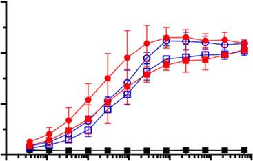
NATURE COMMUNICATIONS | https://doi.org/10.1038/s41467-020-18058-8                                                         ARTICLE

                  Open Access This article is licensed under a Creative Commons
                  Attribution 4.0 International License, which permits use, sharing,
adaptation, distribution and reproduction in any medium or format, as long as you give
appropriate credit to the original author(s) and the source, provide a link to the Creative
Commons license, and indicate if changes were made. The images or other third party
material in this article are included in the article’s Creative Commons license, unless
indicated otherwise in a credit line to the material. If material is not included in the
article’s Creative Commons license and your intended use is not permitted by statutory
regulation or exceeds the permitted use, you will need to obtain permission directly from
the copyright holder. To view a copy of this license, visit http://creativecommons.org/
licenses/by/4.0/.

© The Author(s) 2020

NATURE COMMUNICATIONS | (2020)11:4198 | https://doi.org/10.1038/s41467-020-18058-8 | www.nature.com/naturecommunications         9
You can also read


