CHARACTERIZATION OF SARS-COV-2 AND HOST ENTRY FACTORS DISTRIBUTION IN A COVID-19 AUTOPSY SERIES - NATURE
←
→
Page content transcription
If your browser does not render page correctly, please read the page content below
ARTICLE
                  https://doi.org/10.1038/s43856-021-00025-z               OPEN

                  Characterization of SARS-CoV-2 and host entry
                  factors distribution in a COVID-19 autopsy series
                  Xiao-Ming Wang1,2,6, Rahul Mannan 1,2,6, Lanbo Xiao1,2, Eman Abdulfatah1, Yuanyuan Qiao1,2, Carol Farver1,
                  Jeffrey L. Myers1, Sylvia Zelenka-Wang1,2, Lisa McMurry1, Fengyun Su2, Rui Wang2, Liron Pantanowitz1,
                  Jeffrey Jentzen1, Allecia Wilson1, Yuping Zhang2, Xuhong Cao2, Arul M. Chinnaiyan1,2,3,4,5,6 &
                  Rohit Mehra 1,2,3,6 ✉

                                                                                                                                 Plain language summary
                  Abstract                                                                                                       To understand SARS-CoV-2 infection
                                                                                                                                 of human organs, we characterized
1234567890():,;

                  Background SARS-CoV-2 is a highly contagious virus that causes the disease COVID-19. We
                                                                                                                                 the tissue distribution of SARS-CoV-2
                  have recently reported that androgens regulate the expression of SARS-CoV-2 host entry
                                                                                                                                 virus, and the presence of host factors
                  factors ACE2 and TMPRSS2, and androgen receptor (AR) in lung epithelial cells. We also                         that enable the virus to enter cells, in
                  demonstrated that the transcriptional repression of the AR enhanceosome inhibited SARS-                        postmortem tissues from six patients
                  CoV-2 infection in vitro.                                                                                      who had COVID-19. We assessed the
                  Methods To better understand the various sites of SARS-CoV-2 infection, and presence of                        presence of SARS-CoV-2 viral RNA
                                                                                                                                 and the expression of human genes
                  host entry factors, we extensively characterized the tissue distribution and localization of
                                                                                                                                 that facilitate virus entry in host cells,
                  SARS-CoV-2 virus, viral replication, and host entry factors in various anatomical sites sam-
                                                                                                                                 using several techniques. We
                  pled via autopsy. We applied RNA in-situ-hybridization (RNA-ISH), immunohistochemistry                         observed that SARS-CoV-2, and fac-
                  (IHC) and quantitative reverse transcription polymerase chain reaction (qRT-PCR) approa-                       tors that facilitate virus entry in host
                  ches. We also assessed histopathological changes in SARS-CoV-2 infected tissues.                               cells, were present in the same loca-
                  Results We detect SARS-CoV-2 virus and viral replication in pulmonary tissues by RNA-ISH                       tion in pulmonary and multiple non-
                                                                                                                                 pulmonary tissues, including lung,
                  and IHC and a variety of non-pulmonary tissues including kidney, heart, liver, spleen, thyroid,
                                                                                                                                 bronchus, trachea, kidney, heart, liver,
                  lymph node, prostate, uterus, and colon by qRT-PCR. We observe heterogeneity in viral load
                                                                                                                                 spleen, thyroid, lymph node, prostate,
                  and viral cytopathic effects among various organ systems, between individuals and within the                   uterus, and colon. We also reported
                  same patient. In a patient with a history of kidney transplant and under immunosuppressant                     changes in the microscopic appear-
                  therapy, we observe an unusually high viral load in lung tissue by RNA-ISH, IHC and qRT-                       ance of SARS-CoV-2 infected tissues
                  PCR. SARS-CoV-2 virus is also detected in this patent’s kidney, liver and uterus. We find                       at various sites. Such findings will
                                                                                                                                 guide future coronavirus biology stu-
                  ACE2, TMPRSS2 and AR expression to overlap with the infection sites.
                                                                                                                                 dies on patients with advanced
                  Conclusions This study portrays the impact of dispersed SARS-CoV-2 infection in diverse
                                                                                                                                 disease.
                  organ systems, thereby facilitating avenues for systematic therapeutic approaches.

                  1 Departmentof Pathology, University of Michigan Medical School, Ann Arbor, MI, USA. 2 Michigan Center for Translational Pathology, Ann Arbor, MI, USA.
                  3 RogelCancer Center, Michigan Medicine, Ann Arbor, MI, USA. 4 Department of Urology, University of Michigan Medical School, Ann Arbor, MI, USA.
                  5 Howard Hughes Medical Institute, Ann Arbor, MI, USA. 6These authors contributed equally: Xiao-Ming Wang, Rahul Mannan, Arul M. Chinnaiyan,

                  Rohit Mehra. ✉email: mrohit@med.umich.edu

                  COMMUNICATIONS MEDICINE | (2021)1:24 | https://doi.org/10.1038/s43856-021-00025-z | www.nature.com/commsmed                                            1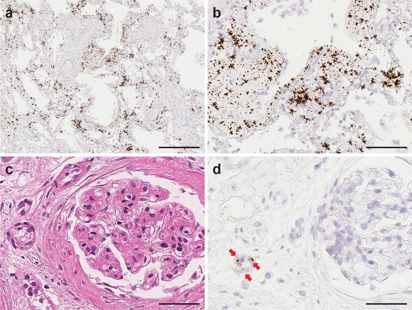
ARTICLE                                                             COMMUNICATIONS MEDICINE | https://doi.org/10.1038/s43856-021-00025-z

C
        oronavirus disease-19 (COVID-19), an infectious disease             with SARS-CoV-2 infection who died from COVID-19 disease
        caused by a novel coronavirus called severe acute                   with clinical autopsies performed at Michigan Medicine were
        respiratory syndrome coronavirus 2 (SARS-CoV-2), was                included in the study. The legal next of kin of patients provided
declared a pandemic by the World Health Organization on                     the informed consent prior to autopsy. All the patients tested
March 11, 2020. As of May 21st, 2021, 165,874,001 diagnosed                 positive for SARS-CoV-2 by quantitative reverse transcription
cases and 3,438,383 deaths have been reported worldwide                     polymerase chain reaction (qRT-PCR) performed in a clinical
(https://coronavirus.jhu.edu/), with the United States of America           laboratory. This study was conducted prior to the FDA approval
bearing the highest disease impact in terms of morbidity and                of emergency use of COVID-19 vaccines and none of the patients
mortality (33,084,872 cases and 589,222 deaths) followed by India           in this study received COVID-19 vaccination. During autopsy,
where is currently undergoing a massive surge of cases. COVID-              multiple tissue samples were harvested from various pulmonary
19 is an established multiorgan disease in humans with the                  and nonpulmonary sites for clinicopathologic characterization.
greatest involvement and derangement involving the respiratory,             For the purposes of this study, representative FFPE tissue blocks
cardiovascular, renal, and immune systems1.                                 (n = 74) were selected from 16 different anatomical sites by two
   Coronaviruses are a group of enveloped viruses with a single-            pathologists (R. Mannan and R. Mehra) based on the evaluation
stranded RNA genome. In rare instances, animal coronaviruses such           of hematoxylin and eosin (H&E) stained FFPE slides with
as severe acute respiratory syndrome (SARS) coronavirus (SARS-              brightfield microscopy.
CoV) in 2002, Middle East respiratory syndrome (MERS) cor-
onavirus (MERS-CoV) in 2012, and currently SARS-CoV-2, can
infect humans with variable clinical complications and impact. All          Histopathological evaluation. The pathological appraisal of tis-
three of these coronaviruses are transmitted zoonotically and spread        sue samples was performed for pulmonary and nonpulmonary
among humans through close contact2. SARS-CoV-2 is most com-                organs. The histopathological assessment for the respiratory
monly transmitted from person to person via respiratory droplets.           region was carried out on the trachea, bronchus, and pulmonary
Most patients experience mild symptoms. However, a significant               parenchymal tissues. For nonrespiratory sites, we evaluated
subset of patients may experience severe disease outcomes including         myocardial tissues, hematological tissues (lymph node and
long term health sequelae and death3. The SARS-CoV-2 genome                 spleen), liver, and tissues from the gastrointestinal (esophagus,
shares ~80% sequence identity with SARS-CoV and MERS-CoV,                   stomach, small intestine, and colon) tract, endocrine (thyroid,
although the sequence similarity varies among genes encoding                pancreas, and adrenal) glands, and genitourinary (kidney, pros-
structural proteins and essential enzymes. SARS-CoV-2 is considered         tate, and uterus) tract. A systematic evaluation for histo-
to be more pathogenic than the MERS-CoV and SARS-CoV4,5.                    morphological and pathological changes were noted and recor-
Structurally, SARS-CoV-2 is comprised of four structural proteins:          ded for each sample.
spike (S), envelope (E), membrane glycoprotein (M), and nucleo-
capsid phosphoprotein (N) proteins5. SARS-CoV-2 entry into host
cells depends on binding of viral spike proteins to the host receptor       RNA in situ hybridization (RNA-ISH). RNA-ISH was per-
protein angiotensin-converting enzyme 2 (ACE2) and priming by the           formed on 4 µm FFPE tissue sections using RNAscope 2.5 HD
host serine protease, the cell surface transmembrane protease serine 2      Brown kit, duplex kit and target probes against SARS-CoV-2
(TMPRSS2)6,7. In our recent study detailing transcriptional regula-         spike (S) gene (SARS-CoV-2-S probe), minus strand of SARS-
tion of SARS-CoV-2 entry factors ACE2 and TMPRSS2, we described             CoV-2-S gene (SARS-CoV-2-S-sense), SARS-CoV-2 nucleocapsid
co-expression of the androgen receptor (AR), TMPRSS2, and ACE2              (N) gene (SARS-CoV-2-N-O1-C2), human ACE2, TMPRSS2, and
in bronchial and alveolar cells in human and murine lung tissues;           AR genes (Supplementary Table 1) (Advanced Cell Diagnostics,
demonstrated that androgens regulate the expression of ACE2,                Newark, CA). The SARS-CoV-2-S-sense probe detects the minus
TMPRSS2, and AR in subpopulations of lung epithelial cells; and             strand viral RNA, which is present during active viral replication.
reported that transcriptional repression of the AR enhanceosome by          RNA quality was assessed using Hs-PPIB probe as positive con-
AR antagonists inhibited SARS-CoV2 infection in vitro8. To further          trol and assay background was monitored using DapB probe as
investigate transcriptional inhibition of critical host factors in the      negative control. In addition, tissue sections from one normal
treatment or prevention of COVID-19, we set out to characterize the         lung and one H1N1 influenza patient lung were used as negative
distribution and localization of SARS-CoV-2 viral particles, viral          control samples for SARS-CoV-2 probes. RNA-ISH assay was
replication, and the host entry factor receptor ACE2, priming pro-          performed as previously described9–11. After deparaffinization,
tease TMPRSS2 and transcriptional regulator AR by RNA in situ               FFPE sections were pretreated with hydrogen peroxide followed
hybridization (RNA-ISH), immunohistochemistry (IHC) and qRT-                by heat-induced target retrieval and protease, and subsequently
PCR in various pulmonary and nonpulmonary tissues obtained from             hybridized with target probe followed by a series of signal repli-
a series of six clinical autopsies performed on patients who suc-           cations. Finally, chromogenic detection was performed using
cumbed to COVID-19 disease. We also correlated the presence of              DAB and counterstained with 50% Gill’s hematoxylin I (Fisher
SARS-CoV-2 viruses and host entry machinery with clin-                      Scientific, Rochester, NY). To confirm that the RNA-ISH signals
icopathologic findings in the affected organ systems. We detect              were amplified from viral RNA, tissue sections were treated with
SARS-CoV-2 viral RNA and viral replication events in both pul-              5 mg/ml RNase A for 30 min at 40 °C prior to target probe
monary tissues and nonpulmonary tissues. We observe heterogeneity           hybridization.
in viral load and viral cytopathic effects among various organ sys-
tems, between individuals and within the same patient. We also find
the presence of host factors, ACE2, TMPRSS2, and AR, to overlap             Immunohistochemistry (IHC). IHC was performed on 4 µm
with the infection sites.                                                   FFPE tissue sections on the Ventana Discovery XT automated
                                                                            slide staining system (Roche-Ventana, AZ) using the Chromo-
                                                                            Map diaminobenzidine (DAB) detection kit (760-500, Roche-
Methods                                                                     Ventana, AZ). SARS-CoV-2 nucleocapsid IHC was performed
Patient selection. This study was performed under ethical                   using mouse monoclonal antibody (R&D system MAB10474) at
approval protocols of the Institutional Review Boards of the                1:100 dilutions with heat-induced epitope retrieval with TRIS
University of Michigan Medical School (IRBMED). Six patients                antigen retrieval buffer.

2                       COMMUNICATIONS MEDICINE | (2021)1:24 | https://doi.org/10.1038/s43856-021-00025-z | www.nature.com/commsmed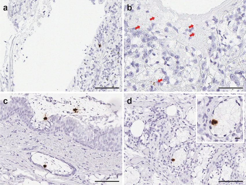
COMMUNICATIONS MEDICINE | https://doi.org/10.1038/s43856-021-00025-z                                                              ARTICLE

RNA extraction and quantitative RT-PCR (qRT-PCR). Total                       directly attributed to COVID-19, was bronchopneumonia (3/6
RNA was extracted from two 5 µm FFPE tissue sections using the                patients). One patient with underlying asthma showed associated
miRNeasy FFPE Kit (Qiagen, Hilden, Germany) according to the                  changes including mucus plugging, goblet cell metaplasia, mucus
manufacturer’s protocol and eluted into 30 µl H2O.                            gland hyperplasia, and thickening of subepithelial basement
   For SARS-CoV-2 RNA detection, 8 µl RNA template was used                   membranes, while another patient with chronic obstructive pul-
per qRT-PCR reaction using the GenePath CoViDx One kit                        monary disease (COPD) showed large emphysematous changes
(Maharashtra, India), which contains premixed RT-PCR primer                   and edema. A summary of pulmonary histopathologic findings is
sets for SARS-CoV-2 Envelop (E) gene, Nucleocapsid (N) gene,                  presented in Supplementary Table 2.
RNA-dependent RNA polymerase (RdRP) gene and the human
RNaseP, an internal extraction and amplification control.
                                                                              Nonpulmonary histopathologic findings. Histologic examina-
Samples were classified as SARS-CoV-2 positive when at least
                                                                              tion of the nonpulmonary organs revealed notable pathology
one of the three SARS-CoV-2 genes were detected with a Ct
                                                                              affecting the myocardium (5/6 patients), kidney (2/4) and liver
value < 40. Each reaction was performed in replicates. Tissues
                                                                              (3/6). A summary of nonpulmonary histopathologic findings is
from one normal lung, one H1N1 influenza patient lung and one
                                                                              presented in Supplementary Table 2. Myofibrillary hypertrophy
normal prostate were selected as negative control. For undeter-
                                                                              was identified in cardiac tissues (5/6 patients) with evidence of
mined data points, Ct value was set to 40 to calculate the ΔCt.
                                                                              focal to diffuse fibrosis in a subset of cases. Evidence of chronic
   For viral replication detection, we performed two-step strand-
                                                                              inflammation was not only identified in half of the cases, mostly
specific RT-PCR as previously described12. Frist, strand-specific
                                                                              in the pericardial region, but also observed in the perivascular
primers were used to reverse transcribe SARS-CoV-2 viral RNA
                                                                              region in one patient. Renal sampling was performed in 4/6
to cDNA in two separate reactions: a forward E gene primer to
                                                                              patients with histopathological changes mostly observed in the
generate minus strand cDNA representing viral replication and a
                                                                              medullary region of the kidney where tubules were affected. Signs
reverse E gene primer to generate plus strand cDNA representing
                                                                              of tubular injury include tubular epithelial sloughing within
the single-strand virus. In addition, a separate set of human
                                                                              lumina admixed with red blood cells and calcification. Patient 3,
RNaseP primers were added to each set of reactions to serve as
                                                                              who had a unique clinical history of diabetic nephropathy status
internal amplification control. Second, each cDNA sample was
                                                                              post-transplant, had a focal collection of large, multinucleated,
amplified by real-time PCR to detect either SARS-CoV-2 E gene
                                                                              dyscohesive cells with foamy cytoplasm in the renal medulla of
or human RNaseP gene by specific primer sets using SYBR Green
                                                                              their transplanted kidney and histological changes consistent with
(Bio-Rad, Hercules, CA).
                                                                              end-stage renal disease (ESRD) in the native kidneys. Histological
   The RT-PCR data are available in the supplementary
                                                                              examination of liver revealed mild to moderate steatosis in all
information files. All other data that support the findings of this
                                                                              patients where liver tissue was sampled. However, no evidence of
study are available from the corresponding author upon reason-
                                                                              overt inflammation or fibrosis was observed.
able request.

Reporting summary. Further information on research design is                  Detection of SARS-CoV-2 virus in pulmonary tissues by RNA-
available in the Nature Research Reporting Summary linked to                  ISH. We performed RNA-ISH using a target probe against the
this article.                                                                 SARS-CoV-2-S gene for detection of viral particles in formalin
                                                                              fixed paraffin embedded (FFPE) tissue sections. The SARS-CoV-2
                                                                              RNA-ISH signals were identified as individual brown punctate
Results                                                                       dots or clusters. As expected, SARS-CoV-2 viral particles were
Clinical and histopathologic features. The study cohort was                   detected in lung tissues of all six patients and no signal was
comprised of five males and one female patient with age ranging                observed in normal lung tissue or H1N1 influenza patient’s lung
from 37 to 80 years old (median age of 59.5 years). The most                  tissue. The viral loads, however, varied drastically among patients
common clinical presentations included pyrexia (5/6), followed                and different sampling sites from the same organ of a single
by acute hypoxemic failure refractory to supplemental oxygen                  patient. RNA-ISH signals were observed to be patchy. At ana-
(4/6), cough and dyspnea (2/6), and diarrhea (1/6). All six                   tomic level, the SARS-CoV-2 viral particles were observed in the
patients had pre-existing co-morbidities that included a history of           intact as well as desquamated alveolar epithelial cells lying in the
diabetes and hypertension (5/6 patients), asthma (2/6), obesity (1/           edema fluid of the intra-alveolar space and within the hyaline
6), hypercholesterolemia (1/6), coronary artery disease (CAD) (1/             membranes lining the alveoli. (Fig. 1a–d). SARS-CoV-2 virus was
6), and renal transplantation (1/6). All patients exhibited abnor-            also detected in the nonalveolar region such as peri-bronchial
mal pulmonary findings on radiologic imaging including the                     sero-mucinous glands in 2/4 patients (Fig. 1e, f) and peri-tracheal
presence of ground-glass opacities on computed tomography                     region in 1/4 patients, but with lower viral counts compared to
(CT). The cause of death for all six patients was attributed to               the alveolar region as indicated by RNA-ISH staining. Viral sig-
pulmonary complications associated with SARS-CoV-2 infection                  nals in lung tissue were also noted to be prominent in areas
recorded to be due to acute respiratory distress syndrome (ARDS)              exhibiting necrosis and sero-fibrinous deposits. (Fig. 1g, h).
in 5/6 patients and bronchopneumonia in one patient.                             As described in the methods, we also employed a sense probe
                                                                              against the minus strand of SARS-CoV-2-S gene to detect viral
Pulmonary histopathologic findings. Histologic examination of                  replication. Utilizing this approach, we observed active viral
lung tissues revealed acute diffuse alveolar damage (DAD) with                replication events as brown signal dots and clusters within the
prominent hyaline membranes in 5/6 patients (Fig. 1a) and dif-                intra-alveolar fibrinous deposits, the cellular proliferation seen in
fuse lymphocytic inflammatory infiltrates in all six patients.                  the alveolar septa and bronchiolar region of lung parenchyma in
Evidence of coagulopathy (i.e., microthrombi, capillaritis etc.) was          all six patients. Signals were also identified in primary/parent
identified in a majority of patients (5/6). Additional histopatho-             bronchus tissues in 2/4 patients, and tracheal tissues in 1/4
logical findings attributed to viral cytopathic effects include                patients (Fig. 2).
pronounced usual hyperplastic alveolar pneumocytes (2/6                          To demonstrate and confirm the specificity of the virus signals
patients) and intracytoplasmic vacuolation (1/6 patient). Another             detected by RNA-ISH described above, we performed duplex
histopathological feature observed in the cohort, possibly not                RNA-ISH assay for co-detection of SARS-CoV-2-N gene and S

COMMUNICATIONS MEDICINE | (2021)1:24 | https://doi.org/10.1038/s43856-021-00025-z | www.nature.com/commsmed                                     3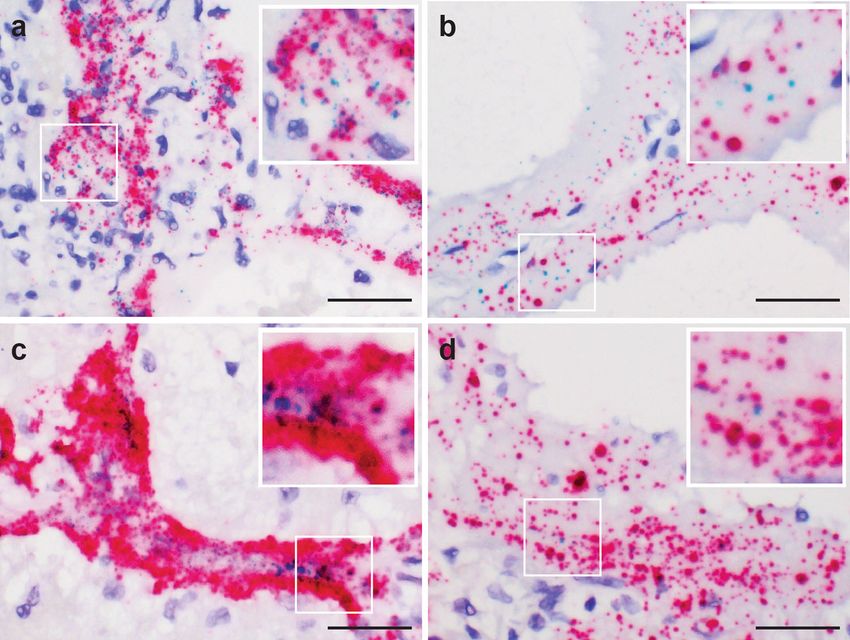
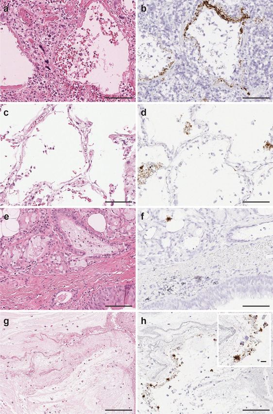
ARTICLE                                                                COMMUNICATIONS MEDICINE | https://doi.org/10.1038/s43856-021-00025-z

Fig. 1 SARS-CoV-2 virus detection in pulmonary parenchyma by SARS-CoV-2 spike RNA-ISH. The viral signals were observed within the intra-alveolar
hyaline membranes (a H&E and b SARS-CoV-2 spike RNA-ISH) and within the intact lining alveolar epithelial cells as well as in the desquamated alveolar
epithelial cells (c H&E and d SARS-CoV-2 spike RNA-ISH). Viral particles were also noted in the nonalveolar region like bronchus as a cluster of viral
signals within peri-bronchial sero-mucinous glands, as well as within the lining pseudostratified respiratory bronchial epithelium. (e H&E and f SARS-SoV-2
spike RNA-ISH). Viral signals were also detected in the necrotic and fibrinous material within damaged pulmonary tissue (g H&E and h SARS-CoV-2 spike
RNA-ISH). Inset: Viral signals shown as individual punctate brown dots and clusters. Scale bars = 100 µm. Inset scale bar = 10 µm.

gene in the lung autopsy tissue sections. We observed co-presence             viral replication event co-existing within the same cell types
of both N gene and S gene signals within the same topographical               harboring the virus genome (Fig. 3c, d).
locations of intra-alveolar region, hyaline membranes and peri-                  As an additional confirmatory validation for RNA-ISH signal
tracheal sero-mucinous glands (Fig. 3a, b). Similar strategy was              specificity, we treated the autopsy tissue sections with RNase.
utilized by combining the target probes against N gene and the                Upon RNase treatment just prior to SARS-CoV-2 target probe
minus strand of SARS-CoV-2-S gene, to show case SARS-CoV-2                    hybridization, RNA-ISH signals were no longer detectable within

4                         COMMUNICATIONS MEDICINE | (2021)1:24 | https://doi.org/10.1038/s43856-021-00025-z | www.nature.com/commsmed
COMMUNICATIONS MEDICINE | https://doi.org/10.1038/s43856-021-00025-z                                                                      ARTICLE

Fig. 2 Detection of SARS-CoV-2 viral replication in pulmonary parenchyma by SARS-CoV-2-S-sense RNA-ISH. SARS-CoV-2 viral replication events
were observed in the intra-alveolar septal region (a), hyaline membranes lining the alveolar space (b), ciliated columnar respiratory epithelium of the
bronchus (c), and the subepithelial sero-mucinous tracheal glands (d). Inset: a signal cluster representing viral replication. Scale bars = 200 µm in a, 100
microns in b–d. Inset scale bar = 10 µm. Arrows point to RNA-ISH signals.

Fig. 3 Co-detection of SARS-CoV-2 virus spike (S), nucleocapsid (N) genes, and viral replication in pulmonary parenchyma. Co-expression of the
SARS-CoV-2-S (green) and N (red) genes in the intra-alveolar (a) and hyaline membranes in lung parenchyma (b) utilizing the duplex RNA-ISH with
probes against the SARS-CoV-2-N gene and -S gene. Co-detection of the SARS-CoV-2 virus (red) and viral replication (green) in the intra-alveolar and
hyaline membranes in lung parenchyma (c, d) with probes against the SARS-CoV-2-N gene and minus strand of S gene. (a, c from patient 1, b, d from
patient 3) Scale bars = 50 µm.

COMMUNICATIONS MEDICINE | (2021)1:24 | https://doi.org/10.1038/s43856-021-00025-z | www.nature.com/commsmed                                               5
ARTICLE COMMUNICATIONS MEDICINE | https://doi.org/10.1038/s43856-021-00025-z
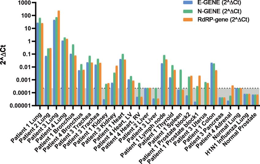
Fig. 4 qRT-PCR of SARS-CoV-2 infection in COVID-19 autopsy tissues. 2(ΔCt) for SARS-CoV-2 E gene, N gene, and RdRP gene using RNaseP
housekeeping gene as reference. The undetermined Ct was set to 40. Normal lung, H1N1 influenza lung, and normal prostate tissues were used as negative
controls. The 2(ΔCt) of normal lung was set as a cutoff point (0.000224) and the gray area below the cutoff line is considered as SARS-CoV-2 negative.
Minimum 1 out of the 3 genes should have Ct value < 40 (or 2(ΔCt) > 0.000224) to be considered as SARS-CoV-2 positive.
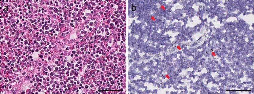
Fig. 5 SARS-CoV-2 virus detection within lymph node. Viral signal clusters were observed within lymph node germinal center as individual brown dots
(a H&E and b SARS-CoV-2 RNA-ISH, in arrow pointed area). Scale bars = 50 µm.
the tissue samples, which were earlier reported to be SARS-CoV-2 by E gene real-time PCR for lung samples positive for viral
positive by conventional RNA-ISH (Supplementary Fig. 1). replication as detected by RNA-ISH. The PCR results are
consistent with the RNA-ISH results for both S-sense RNA-
ISH-positive and -negative samples. (Supplementary Table 4)
Detection of SARS-CoV-2 virus in pulmonary tissues by IHC.
We found a concordance between SARS-CoV-2 nucleocapsid
IHC positivity and SARS-CoV-2 RNA-ISH positivity in the
Detection of SARS-CoV-2 virus in nonpulmonary tissues. To
pulmonary autopsy tissue samples. The IHC expression was
further investigate the SARS-CoV-2 tissue distribution, we
consistent with RNA-ISH assay in terms of pattern and topo-
selected 14 representative nonpulmonary tissue samples, includ-
graphical localization as it was also observed in the hyaline
ing kidney, heart, liver, lymph node, spleen, thyroid, prostate,
membrane and intra-alveolar region (Supplementary Fig. 2).
uterus, colon, adrenal, pancreas, stomach, esophagus, and small
intestine, using RNA-ISH and qRT-PCR methodologies. Low
Detection of SARS-CoV-2 virus in pulmonary tissues by qRT- level signals with high cycle threshold (Ct) values were detected in
PCR. We also performed qRT-PCR on eight SARS-CoV-2 RNA- kidney, heart, liver, lymph node, thyroid, spleen, prostate, uterus,
ISH-positive pulmonary tissue samples from four patients using colon, and adrenal by qRT-PCR. (Fig. 4 and Supplementary
primer sets for E gene, N gene, and RdRP gene. All the selected Table 3). Rare RNA-ISH signal dots were observed within the
pulmonary tissues were also qRT-PCR positive (Fig. 4). On the germinal center of the cortical lymphoid follicle of a lymph node
other hand, none of the genes were detected in normal lung tissue sample from Patient 1 (Fig. 5) and within the distal tubules of a
or H1N1 influenza patient’s lung tissue. The SARS-CoV-2 RNA- kidney sample form Patient 7 (Fig. 6c, d). Both tissues were qRT-
ISH results were highly consistent with the qRT-PCR results with PCR positive (Fig. 4, Supplementary Table 3, Patient 1 Lymph
an overall good correlation of signal intensities among pulmonary node with average Ct 30.8 and Patient 3 Kidney with average Ct
tissues (Supplementary Table 3). 36.5). Very rare signal clusters were observed in kidney, heart,
To further confirm the RNA-ISH signals of viral replication, we liver, thyroid, spleen, prostate, uterus, and colon tissue sections,
performed minus strand-specific reverse transcription followed but no individual signal dots representing viral particles were
6 COMMUNICATIONS MEDICINE | (2021)1:24 | https://doi.org/10.1038/s43856-021-00025-z | www.nature.com/commsmedCOMMUNICATIONS MEDICINE | https://doi.org/10.1038/s43856-021-00025-z ARTICLE
Fig. 6 SARS-CoV-2 infection in kidney transplant patient (patient 3) on immunosuppressant therapy. Highly abundant viral signals were observed
within the lung parenchyma exhibiting diffuse alveolar damage, reflecting the high SARS-CoV-2 virus infection load in Patient 3. The viral signals were
detected in intra-alveolar hyaline membrane and interstitial fibroblastic proliferation region (a, b. SARS-CoV-2 RNA-ISH). Scattered viral signals were
identified in the renal tubules (c H&E and d SARS-CoV-2 RNA-ISH). Scale bar = 500 µm in a, 100 µm in b, 50 µm in c, d.
detected in any of those tissues. Therefore, we did not consider thyroid, kidney, and liver (Supplementary Fig. 5). It is important
these tissues as SARS-CoV-2 positive by RNA-ISH. to mention that SARS-CoV-2 infection was detected in most
tissues and cell types expressing ACE2 and TMPRSS2, except
High abundance of SARS-CoV-2 viruses found in lung tissues adrenal, pancreas, stomach, esophagus, and small intestine, as
of a patient status post kidney transplant. Patient 3 in our described above. SARS-CoV-2 viral signals were also detected in
clinical autopsy series had a clinical history of kidney transplant tissues where ACE2 and TMPRSS2 transcripts were not detected,
for end-stage diabetic nephropathy and was receiving immuno- including spleen, colon, heart (ACE2 only), lymph node (ACE2
suppressant therapy with mycophenolate and prednisolone. only), and uterus (ACE2 only).
SARS-CoV-2 RNA-ISH staining revealed very high viral infection
in the hyaline membranes and intra-alveolar septum throughout
the lung tissue from this immunosuppressed patient (Fig. 6a, b). Discussion
Viral inflammatory changes were observed in the transplant The highly contagious and rapidly spreading COVID-19 has
kidney and RNA-ISH indicated SARS-CoV-2 viral signals within evolved into a global health threat in 2020. This disease is now
the distal tubules of the transplant kidney (Fig. 6c, d). SARS-CoV- known to manifest with a wide spectrum of severity with severe
2 viral signals were also detected in the liver and uterus tissues by disease resulting in intense and multiorgan dysfunction13,14.
qRT-PCR (Supplementary Table 3). Currently, there is a limited understanding of the pathophysiol-
ogy of COVID-19, and no targeted therapy is yet available for
Localization of ACE2, TMRPSS2, and AR expression in various treatment of established SARS-CoV-2 infection. As a follow-up to
organ systems. ACE2 and TMPRSS2 RNA-ISH signals in pul- our recent study on transcriptional regulation of SARS-CoV-2
monary tissues were noted in tracheal epithelial cells in 2/2 entry factors in lung8, herein, we studied the tissue distribution
patients, peri-bronchial glands in 2/4 patients, bronchial and and localization of SARS-CoV-2 virus, viral replication, the
bronchiolar respiratory epithelial cells and a subpopulation of transcripts of the host cell entry factors, ACE2 and TMPRSS2, and
alveolar epithelial cells in 6/6 patients. In nonpulmonary tissues, the transcriptional regulator of TMPRSS2, AR, in postmortem
ACE2 and TMPRSS2 were both expressed in biliary duct epi- human organ systems using in situ and qRT-PCR approaches,
thelium, renal distal tubule and collecting duct, prostatic acinar and correlate those results to the histopathologic findings in
cells, and glandular epithelium of small intestine. In addition, COVID-19 patient autopsy cases.
ACE2 mRNA were also detected in endothelial cells within lymph Clinical autopsy examination revealed ARDS due to SARS-
node, enterocytes lining the crypts, and glands of the intestine CoV-2 infection to be implicated in the demise of all six patients.
and myocardial endothelial and stromal cells. (Supplementary The medical history in this series was enriched for at least one
Fig. 3, Supplementary Fig. 4) The expression of AR, the tran- underlying or predisposing medical condition in each of the six
scriptional regulator of TMPRSS2, were also observed in the patients including diabetes, hypertension, coronary artery disease,
bronchial, bronchiolar and alveolar epithelial cells, subepithelial asthma, and obesity. These findings are consistent with previous
bronchial, and tracheal sero-mucinous glands of the pulmonary reports in the literature suggesting older age (>60 years), serious
tissues, as well as nonpulmonary tissues including the prostate, co-morbidities, and tobacco exposure to be associated with a
COMMUNICATIONS MEDICINE | (2021)1:24 | https://doi.org/10.1038/s43856-021-00025-z | www.nature.com/commsmed 7ARTICLE COMMUNICATIONS MEDICINE | https://doi.org/10.1038/s43856-021-00025-z higher risk of developing ARDS and mortality in COVID-19 receptor on host cells and is cleaved and activated by cell surface patients, especially in males13. transmembrane protease TMPRSS26,43. Targeting the transcrip- Histologic examination of pulmonary tissues revealed acute tional regulation or activity of these host factors could inhibit diffuse alveolar damage with prominent hyaline membranes as a SARS-CoV-2 infection and replication. TMPRSS2 has been common COVID-19 pathological feature, in concordance with widely studied in prostate cancer where it is highly expressed in the literature1,15–17. In our study, the most common pathological an androgen-dependent manner44. The recurrent oncogenic changes encountered in nonpulmonary tissues included myofi- TMPRSS2-ETS gene fusions are also found in more than 50% of brillary hypertrophy in the heart, tubular injury in the kidney, prostate cancers41. AR inhibitors have been developed for treat- and steatosis in the liver. These observations are in line with ment of prostate cancer and could be repurposed for COVID- published reports that SARS-CoV-2 infection may cause acute 1945. In our recent study, we demonstrated that ACE2, TMPRSS2 injury in heart, kidney, and other tissues through a direct cyto- and AR are co-expressed in a subset of lung epithelial cells under pathic effect14,18 In addition, catecholamine surge leads to cyto- the transcriptional regulation of androgen; and transcriptional kine storm imploding as systemic inflammatory response repression of AR inhibited SARS-CoV-2 infection in vitro8. syndrome (SIRS), leading to multiorgan failure, which has been However, the distribution of ACE2 and TMPRSS2 expression in documented as a major cause of death in COVID-19 patients1,14. human organ systems associated with viral infection has not been We further employed a single-molecule RNA-ISH technology for comprehensively characterized in the literature. Herein, we fur- sensitive and specific detection of SARS-CoV-2 in FFPE tissue ther characterized the tissue distribution of ACE2, TMPRSS2, and sections. This technology has been adapted to facilitate SARS-CoV- AR transcripts in multiorgan systems and found that the dis- 2 diagnosis and research, and has demonstrated SARS-CoV2-2 tribution of host entry factors, ACE2 and TMPRSS2, and reg- virus detection in cultured cells19–21, rodent22, non-human ulator, AR, highly overlaps with the viral infection sites in primates23,24, and human tissues autopsies25–29. Employing RNA- pulmonary tissues and numerous nonpulmonary tissues, sup- ISH, we were able to concurrently document both the tissue dis- porting the notion that targeting ACE2 and/or TMPRSS2 could be tribution of SARS-CoV-2 viral particles and the host entry factors in an effective treatment strategy to counter SARS-CoV-2 infection pulmonary and extrapulmonary tissues. The SARS-CoV-2 RNA- from a multiorgan perspective and inhibition of AR would be ISH staining pattern and signal localization resembles those of the effective against SARS-CoV-2 infection8. The expression and SARS-CoV-2 IHC signals. Our observations of RNA-ISH staining tissue distribution of entry and attachment receptors can influ- patterns in pulmonary tissues are consistent with published ence viral tropism and pathogenicity. Our study provides a studies28,30 and the viral localization matches the pulmonary his- panoramic view of such factors at a systemic level. topathological findings in our study and other postmortem and We also investigated viral replication activity by targeting the retrospective studies16,25,26 Among nonpulmonary tissues, very rare minus strand of SARS-CoV-2. Active viral replication events were signal clusters were observed in kidney, heart, liver, thyroid, spleen, detected in lung, bronchus, and trachea. The detection frequency prostate, uterus, and colon tissue sections, but no individual signal of viral replication is much less than that of viral particle signals dots were detected. Low viral signals were detected in these speci- in the tissues mentioned above. Similar observation has been mens by qRT-PCR as indicated by the high Ct values in the range of reported in lung tissue28. Replication events and kinetics may 27.4–38.8. The failure to detect SARS-CoV-2 viral particles by influence viral pathogenicity, tissue tropism, and accelerated RNA-ISH might be due to the low viral count and/or RNA clinical organ system involvement and decline. Our study docu- degradation prior to tissue fixation. ments such events at a systemic level in COVID-19 disease. Such It is noteworthy that apart from pulmonary infection, there has findings also provide valuable resources for the development of been limited experimental evidence of SARS-CoV-2 infection in SARS-CoV-2 replication inhibitors for disease management. nonpulmonary tissues previously1,31. A few research groups have Based on our observations, COVID-19 disease demonstrates attempted by means of RT-PCR, electron microscopy or IHC, to heterogeneity with respect to viral load and viral cytopathic detect SARS-CoV-2 viruses in nonpulmonary tissues and have effects among various organ systems, even within the same recorded the presence of viral particles in heart27,32, kidney16,32, patient. The highest viral load was observed in lung tissues and liver33,34, placenta35–37, gastrointestinal tract38, and skin39. SARS- concentrated within the lung alveolar hyaline membrane and CoV-2 viruses were also detected in kidney16,29 and intra-alveolar spaces, followed by bronchus and trachea in the placenta36,37,40 by RNA-ISH. The current study is by far the most respiratory system. It is currently unclear whether heterogeneous comprehensive report of SARS-CoV-2 infection in multiorgan viral abundance and distribution is associated with disease systems and the first report of SARS-CoV-2 viral detection in severity. lymph node, spleen, thyroid, colon, prostate, and uterus. The Our study cohort included a COVID-19 patient with prior identification of SARS-CoV-2 viral infection in distant, non- kidney transplant receiving immunosuppression therapy. The pulmonary tissues, like prostate and uterus, suggest the wide SARS-CoV-2 RNA-ISH staining revealed extremely high viral spread of SARS-CoV-2 in the diverse human organ systems. infection in the lung tissue of this decedent along with SARS- The SARS-CoV-2 host entry mediator, TMPRSS2, was first CoV-2 viral infection and viral inflammatory changes in the found to be highly enriched in prostate cancer41 were reported in transplant kidney, liver, and uterus. Recently studies linked high the prostate of six male COVID-19 patients33. Recent single-cell viral load with disease severity and transmissibility. A hospital- RNA expression studies mapped SARS-CoV-2 entry factors in a based study in China revealed that the severe COVID-19 cases broad range of human tissues and predicted that testicular sper- tend to have higher viral load and longer virus-shedding period matogonial cells and prostatic endocrine cells are susceptible to than mild cases46. An observational study conducted in India SARS-CoV-2 infection8,42, and we presented evidence of SARS- showed that the index cases with high viral load detected by RT- CoV-2 infection in prostatic tissue. Overall, our examination of PCR transmitted 6.25 secondary cases on average whereas the SARS-Cov-2 tissue localization by in situ and qRT-PCR approaches cases with low viral load transmitted an average of 0.8 case47. revealed a systemic fashion of viral infection, which may explain the Immunocompromised persons may experience persistent SARS- multisystemic involvement observed in COVID-19 disease. CoV-2 infection and bear accelerated viral evolution under The 2003 SARS coronavirus and 2019 SARS-CoV-2 virus immunocompromised state48. Therefore, the high viral load employ host proteins ACE2 and TMPRSS2 to gain cell entry. The observed in the COVID-19 patient under immunosuppression viral spike (S) glycoprotein recognizes and binds to ACE2 therapy could be related to the immune suppression status and 8 COMMUNICATIONS MEDICINE | (2021)1:24 | https://doi.org/10.1038/s43856-021-00025-z | www.nature.com/commsmed
COMMUNICATIONS MEDICINE | https://doi.org/10.1038/s43856-021-00025-z ARTICLE
SARS-CoV-2 infected, immunosuppressed patients may act as 20. Hou, Y. J. et al. SARS-CoV-2 reverse genetics reveals a variable infection
super-spreaders under certain circumstances. gradient in the respiratory tract. Cell 182, 429–446 e414 (2020).
Overall, our findings show co-existence of SARS-CoV-2 21. Liu, J. et al. Molecular detection of SARS-CoV-2 in formalin-fixed, paraffin-
embedded specimens. JCI Insight 5, e139042, https://doi.org/10.1172/
infection and host entry factors in multiple pulmonary and jci.insight.139042 (2020).
nonpulmonary tissues. We believe that a detailed characterization 22. Osterrieder, N. et al. Age-dependent progression of SARS-CoV-2 infection in
of such biomarkers in conjunction with histopathological Syrian Hamsters. Viruses 12, 779–784 (2020).
assessment to assess disease severity and progression will guide 23. Chandrashekar, A. et al. SARS-CoV-2 infection protects against rechallenge in
future coronavirus biology studies on patients with advanced rhesus macaques. Science 369, 812–817 (2020).
24. Woolsey, C. et al. Establishment of an African green monkey model for
disease, as well as provide a framework for identifying better, COVID-19 and protection against re-infection. Nat. Immunol. 22, 86–98
novel or specific antiviral therapeutics. (2020).
25. Best Rocha, A. et al. Detection of SARS-CoV-2 in formalin-fixed paraffin-
Data availability embedded tissue sections using commercially available reagents. Lab Invest.
The RT-PCR data are available in the supplementary information files. All other data 100, 1485–1489 (2020).
that support the findings of this study are available from the corresponding author upon 26. Borczuk, A. C. et al. COVID-19 pulmonary pathology: a multi-institutional
reasonable request. autopsy cohort from Italy and New York City. Mod. Pathol. 33, 2156–2168
(2020).
27. Lindner, D. et al. Association of cardiac infection with SARS-CoV-2 in
Received: 21 February 2021; Accepted: 23 July 2021; confirmed COVID-19 autopsy cases. JAMA Cardiol. 5, 1281–1285 (2020).
28. Massoth, L. R. et al. Comparison of RNA in situ hybridization and
immunohistochemistry techniques for the detection and localization of SARS-
CoV-2 in human tissues. Am. J. Surg. Pathol. 45, 14–24 (2020).
29. Westhoff, T. H. et al. Allograft infiltration and meningoencephalitis by SARS-
CoV-2 in a pancreas-kidney transplant recipient. Am. J. Transplant. 20,
References 3216–3220 (2020).
1. Polak, S. B., Van Gool, I. C., Cohen, D., von der Thusen, J. H. & van Paassen, J. A
30. Schaefer, I. M. et al. In situ detection of SARS-CoV-2 in lungs and airways of
systematic review of pathological findings in COVID-19: a pathophysiological
patients with COVID-19. Mod. Pathol. 33, 2104–2114 (2020).
timeline and possible mechanisms of disease progression. Mod. Pathol. 33,
31. Pascarella, G. et al. COVID-19 diagnosis and management: a comprehensive
2128–2138 (2020).
review. J. Intern. Med. 288, 192–206 (2020).
2. de Wit, E., van Doremalen, N., Falzarano, D. & Munster, V. J. SARS and
32. Szabolcs, M. et al. Identification of immunohistochemical reagents for in situ
MERS: recent insights into emerging coronaviruses. Nat. Rev. Microbiol. 14,
protein expression analysis of coronavirus-associated changes in human
523–534 (2016).
tissues. Appl. Immunohistochem. Mol. Morphol. 29, 5–12 (2021).
3. WHO. WHO Transmission of SARS-CoV-2: implications for infection
33. Wichmann, D. et al. Autopsy findings and venous thromboembolism in
prevention precautions. Scientific brief https://www.who.int/news-room/
patients With COVID-19: a prospective cohort study. Ann. Intern. Med. 173,
commentaries/detail/transmission-of-sars-cov-2-implications-for-infection-
268–277 (2020).
prevention-precautions. (July 9 2020).
34. Tian, S. et al. Pathological study of the 2019 novel coronavirus disease
4. Zhang, Y. Z. & Holmes, E. C. A genomic perspective on the origin and
(COVID-19) through postmortem core biopsies. Mod. Pathol. 33, 1007–1014
emergence of SARS-CoV-2. Cell 181, 223–227 (2020).
(2020).
5. Gussow, A. B. et al. Genomic determinants of pathogenicity in SARS-CoV-2
35. Baud, D. et al. Second-trimester miscarriage in a pregnant woman with SARS-
and other human coronaviruses. Proc. Natl Acad. Sci. USA 117, 15193–15199
CoV-2 infection. JAMA 323, 2198–2200 (2020).
(2020).
36. Facchetti, F. et al. SARS-CoV2 vertical transmission with adverse effects on
6. Hoffmann, M. et al. SARS-CoV-2 cell entry depends on ACE2 and TMPRSS2
the newborn revealed through integrated immunohistochemical, electron
and is blocked by a clinically proven protease inhibitor. Cell 181, 271–280
microscopy and molecular analyses of Placenta. EBioMedicine 59, 102951
e278 (2020).
(2020).
7. Shang, J. et al. Cell entry mechanisms of SARS-CoV-2. Proc. Natl Acad. Sci.
37. Hecht, J. L. et al. SARS-CoV-2 can infect the placenta and is not associated
USA 117, 11727–11734 (2020).
with specific placental histopathology: a series of 19 placentas from COVID-
8. Qiao, Y. et al. Targeting transcriptional regulation of SARS-CoV-2 entry
19-positive mothers. Mod. Pathol. 33, 2092–2103 (2020).
factors ACE2 and TMPRSS2. Proc. Natl Acad. Sci. USA 118, e2021450118,
38. Xiao, F. et al. Evidence for gastrointestinal infection of SARS-CoV-2.
https://doi.org/10.1073/pnas.2021450118 (2020).
Gastroenterology 158, 1831–1833 e1833 (2020).
9. Skala, S. L. et al. Next-generation RNA sequencing-based biomarker
39. Magro, C. et al. Complement associated microvascular injury and thrombosis
characterization of chromophobe renal cell carcinoma and related oncocytic
in the pathogenesis of severe COVID-19 infection: a report of five cases.
neoplasms. Eur. Urol. 78, 63–74 (2020).
Transl. Res. 220, 1–13 (2020).
10. Wang, L. et al. VSTM2A overexpression is a sensitive and specific biomarker
40. Menter, T. et al. Placental pathology findings during and after SARS-CoV-2
for mucinous tubular and spindle cell carcinoma (MTSCC) of the kidney. Am.
infection: features of villitis and malperfusion. Pathobiology 88, 69–77
J. Surg. Pathol. 42, 1571–1584 (2018).
(2020).
11. Wang, X. M. et al. TRIM63 is a sensitive and specific biomarker for MiT family
41. Tomlins, S. A. et al. Recurrent fusion of TMPRSS2 and ETS transcription
aberration-associated renal cell carcinoma. Mod. Pathol. 34, 1596–1607 (2021).
factor genes in prostate cancer. Science 310, 644–648 (2005).
12. Hogan, C. A. et al. Strand-specific reverse transcription PCR for detection of
42. Singh, M., Bansal, V. & Feschotte, C. A single-cell RNA expression map of
replicating SARS-CoV-2. Emerg. Infect. Dis. 27, 632–635 (2021).
human coronavirus entry factors. bioRxiv https://doi.org/10.1101/
13. Hu, B., Guo, H., Zhou, P. & Shi, Z. L. Characteristics of SARS-CoV-2 and
2020.05.08.084806 (2020).
COVID-19. Nat. Rev. Microbiol. 19, 141–154 (2020).
43. Hamming, I. et al. Tissue distribution of ACE2 protein, the functional receptor
14. Vasquez-Bonilla, W. O. et al. A review of the main histopathological findings
for SARS coronavirus. A first step in understanding SARS pathogenesis. J.
in coronavirus disease 2019. Hum. Pathol. 105, 74–83 (2020).
Pathol. 203, 631–637 (2004).
15. Hanley, B. et al. Histopathological findings and viral tropism in UK patients
44. Lucas, J. M. et al. The androgen-regulated protease TMPRSS2 activates a
with severe fatal COVID-19: a post-mortem study. Lancet Microbe 1,
proteolytic cascade involving components of the tumor microenvironment
e245–e253 (2020).
and promotes prostate cancer metastasis. Cancer Discov. 4, 1310–1325
16. Menter, T. et al. Postmortem examination of COVID-19 patients reveals
(2014).
diffuse alveolar damage with severe capillary congestion and variegated
45. Asangani, I. A. et al. Therapeutic targeting of BET bromodomain proteins in
findings in lungs and other organs suggesting vascular dysfunction.
castration-resistant prostate cancer. Nature 510, 278–282 (2014).
Histopathology 77, 198–209 (2020).
46. Liu, Y. et al. Viral dynamics in mild and severe cases of COVID-19. Lancet
17. Damiani, S. et al. Pathological post-mortem findings in lungs infected with
Infect. Dis. 20, 656–657 (2020).
SARS-CoV-2. J. Pathol. 253, 31–40 (2020).
47. Sarkar, B., Sinha, R. N. & Sarkar, K. Initial viral load of a COVID-19-infected
18. Gavriatopoulou, M. et al. Organ-specific manifestations of COVID-19
case indicated by its cycle threshold value of polymerase chain reaction could
infection. Clin. Exp. Med. 20, 493–506 (2020).
be used as a predictor of its transmissibility—an experience from Gujarat,
19. Carossino, M. et al. Detection of SARS-CoV-2 by RNAscope((R)) in situ
India. Indian J. Community Med. 45, 278–282 (2020).
hybridization and immunohistochemistry techniques. Arch. Virol. 165,
48. Choi, B. et al. Persistence and evolution of SARS-CoV-2 in an
2373–2377 (2020).
immunocompromised host. N. Engl. J. Med. 383, 2291–2293 (2020).
COMMUNICATIONS MEDICINE | (2021)1:24 | https://doi.org/10.1038/s43856-021-00025-z | www.nature.com/commsmed 9ARTICLE COMMUNICATIONS MEDICINE | https://doi.org/10.1038/s43856-021-00025-z
Acknowledgements Correspondence and requests for materials should be addressed to R.M.
We thank the patients and their families for participation in the autopsy program at the
University of Michigan Health System. They further thank Paul Harms, M.D., and Peer review information Communications Medicine thanks Danny Jonigk and the other,
Angela Wilson for helpful suggestions, Monique Micallef for assisting the autopsies, and anonymous, reviewer(s) for their contribution to the peer review of this work. Peer
the histology staff of the Department of Pathology at the University of Michigan Health reviewer reports are available.
System. We are thankful to Jyoti Athanikar for assistance with manuscript preparation
and submission. This work was supported by the following: Prostate Specialized Pro- Reprints and permission information is available at http://www.nature.com/reprints
grams of Research Excellence Grant P50-CA186786, National Cancer Institute Out-
standing Investigator Award R35-CA231996, National Cancer Institute P30-CA046592, Publisher’s note Springer Nature remains neutral with regard to jurisdictional claims in
and COVID-19 Administrative Supplement to this grant. A.M.C. is a Howard Hughes published maps and institutional affiliations.
Medical Institute Investigator, A. Alfred Taubman Scholar, and American Cancer Society
Professor.
Open Access This article is licensed under a Creative Commons
Author contributions Attribution 4.0 International License, which permits use, sharing,
X.M.W., R. Mannan, C.F., J.L.M., L.P., A.M.C. and R. Mehra designed research; X.M.W., adaptation, distribution and reproduction in any medium or format, as long as you give
R. Mannan, L.X., L.M., F.S., R.W., S.Z.W., Y.Z. and X.C. performed research; J.J. and appropriate credit to the original author(s) and the source, provide a link to the Creative
A.W. performed autopsy and generated clinical report; X.M.W., R. Mannan, L.X., Y.Q., Commons license, and indicate if changes were made. The images or other third party
Y.Z. and R. Mehra analyzed data; X.M.W., R. Mannan, L.X., E.A. and R. Mehra wrote material in this article are included in the article’s Creative Commons license, unless
the paper. indicated otherwise in a credit line to the material. If material is not included in the
article’s Creative Commons license and your intended use is not permitted by statutory
regulation or exceeds the permitted use, you will need to obtain permission directly from
Competing interests the copyright holder. To view a copy of this license, visit http://creativecommons.org/
The authors declare no competing interests.
licenses/by/4.0/.
Additional information
Supplementary information The online version contains supplementary material © The Author(s) 2021
available at https://doi.org/10.1038/s43856-021-00025-z.
10 COMMUNICATIONS MEDICINE | (2021)1:24 | https://doi.org/10.1038/s43856-021-00025-z | www.nature.com/commsmedYou can also read