On the Host Side of the Hepatitis E Virus Life Cycle - MDPI
←
→
Page content transcription
If your browser does not render page correctly, please read the page content below
cells
Review
On the Host Side of the Hepatitis E Virus Life Cycle
Noémie Oechslin , Darius Moradpour              and Jérôme Gouttenoire *
 Division of Gastroenterology and Hepatology, Lausanne University Hospital and University of Lausanne,
 CH-1011 Lausanne, Switzerland; Noemie.Oechslin@unil.ch (N.O.); Darius.Moradpour@chuv.ch (D.M.)
 * Correspondence: Jerome.Gouttenoire@chuv.ch

 Received: 28 April 2020; Accepted: 21 May 2020; Published: 22 May 2020

 Abstract: Hepatitis E virus (HEV) infection is one of the most common causes of acute hepatitis in
 the world. HEV is an enterically transmitted positive-strand RNA virus found as a non-enveloped
 particle in bile as well as stool and as a quasi-enveloped particle in blood. Current understanding of
 the molecular mechanisms and host factors involved in productive HEV infection is incomplete, but
 recently developed model systems have facilitated rapid progress in this area. Here, we provide an
 overview of the HEV life cycle with a focus on the host factors required for viral entry, RNA replication,
 assembly and release. Further developments of HEV model systems and novel technologies should
 yield a broader picture in the future.

 Keywords: HEV; host factor; particle production; viral replication; virus entry

1. Introduction
      Hepatitis E virus (HEV) has been identified as a cause of the waterborne hepatitis outbreaks in
the early 1980s [1,2]. The viral genome was cloned and sequenced in 1990, allowing the development of
serological tests to study its epidemiology [3,4]. The virus has been classified in the Hepeviridae family,
and most human pathogenic strains belong to species Orthohepevirus A [5]. Members of this species
can be classified into 8 genotypes (gt): gt 1 and 2 are restricted to humans and are transmitted via
the fecal-oral route, mainly through contaminated drinking water. Gt 3 and 4 cause zoonotic infections
and the transmission occurs mainly via the consumption of un(der)cooked pork, wild boar or deer
meat. Gt 5 and 6 are found in wild boar, and gt 7 as well as 8 infect dromedary and Bactrian camels,
respectively. Gt 7 has been identified in an immunosuppressed patient after consumption of camel
milk and meat [6] (reviewed in [7]) but no transmission to humans has thus far been reported for gt 5,
6 and 8. More recently, rabbit HEV (closely related to gt 3 within species Orthohepevirus A) and rat
HEV (belonging to species Orthohepevirus C) have also been found to infect humans [8–11].
      HEV is a small, non-enveloped, icosahedral virus with a diameter of 27–34 nm [2]. It contains
a 7.2 kb single-stranded, positive-sense RNA genome which possesses a m7G cap at its 50 and a poly-A
tail at its 30 end (Figure 1A). The HEV genome harbors 3 open reading frames (ORF). ORF1 encodes
the viral replicase, ORF2 the capsid and ORF3 a small protein involved in virion secretion via its
potential ion channel activity [12]. The first contact between HEV and host cells occurs through
interaction with as yet poorly characterized entry factor(s). After endocytosis, the viral genome
is released into the cytoplasm and the host translational machinery produces the ORF1 replicase,
which drives viral RNA replication (Figure 1B). During this step, two RNA species are produced
from a negative-strand RNA intermediate: a full-length genomic RNA and a subgenomic RNA
of 2.2 kb [13,14]. Translation of the subgenomic RNA yields the ORF2 and ORF3 proteins. Later
steps of the HEV life cycle include viral assembly and release of newly produced virions. Very
similar to hepatitis A virus (HAV), another hepatotropic positive-strand RNA virus, HEV is found as
a ‘quasi-enveloped’ virion (eHEV) wrapped in exosomal membranes in blood and as a naked particle
in bile and feces (reviewed in [15]) (Figure 1B).

Cells 2020, 9, 1294; doi:10.3390/cells9051294                                     www.mdpi.com/journal/cells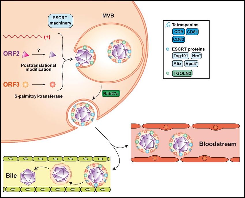
Cells 2020, 9, 1294 2 of 14
Cells 2020, 9, x FOR PEER REVIEW 2 of 14
Figure 1.
Figure 1. Genome organization and
Genome organization and life
life cycle
cycle of
of hepatitis
hepatitis EE virus
virus (HEV).
(HEV). (A) (A) The
The 7.2
7.2 kb
kb positive-strand
positive-strand
RNA genome has a 5′
0 7-methylguanylate cap (m 77G cap) and a 3′ 0
RNA genome has a 5 7-methylguanylate cap (m G cap) and a 3 polyadenylated tail (poly-A). polyadenylated tail (poly-A). ItIt
harbors 3 open reading frames (ORFs). ORF1 encodes a replicase
harbors 3 open reading frames (ORFs). ORF1 encodes a replicase of about 190 kDa comprising of about 190 kDa comprising
different
different functional
functional domains,a including
domains, including a methyltransferase
methyltransferase (Met), an RNA(Met),helicase an(Hel)
RNAand helicase (Hel) and an
an RNA-dependent
RNA-dependent
RNA RNA polymerase
polymerase (RdRp), as well as (RdRp), as well as less well-characterized
less well-characterized domains, such as the domains,
Y domain, such as the Y
a putative
papain-like cysteine protease (PCP), a hypervariable region (HVR) and the Macro domain. Macro
domain, a putative papain-like cysteine protease (PCP), a hypervariable region (HVR) and the ORF2
domain.
and ORF3ORF2encodeandtheORF3 encodeand
viral capsid the aviral
smallcapsid
protein and a smallinprotein
involved involvedrespectively,
virus secretion in virus secretion
which
respectively,
are translatedwhich
from aare2.2translated from a RNA
kb subgenomic 2.2 kbgenerated
subgenomic RNAviral
during generated during(B)
replication. viral
Thereplication.
HEV life
(B) The
cycle canHEV life cycle into
be dissected can bethedissected
following into the following
steps: steps:by(1)asviral
(1) viral entry entry by as yet
yet unidentified unidentified
receptor(s), (2)
receptor(s), (2)
endocytosis andendocytosis
release of the andviral
release of the viral positive-strand
positive-strand RNA genome (+) RNAintogenome (+) into
the cytosol, (3) the cytosol,
translation
(3)the
of translation of thetoORF1
ORF1 protein allow protein to allow
replication of the replication
full-length ofandthe full-length
generation of and generation of
the subgenomic the
RNA
subgenomic
through RNA through RNA
a negative-strand a negative-strand
intermediateRNA intermediate
(-), (4) translation of (-),the
(4) subgenomic
translation ofRNA the subgenomic
to produce
RNA
the to produce
ORF2 and ORF3the proteins
ORF2 and ORF3
and proteinspackaging,
(5) genome and (5) genome
virionpackaging,
assembly and virion assembly
release of theand release
virus into
of the
the virus into the
bloodstream andbloodstream
the bile from andthethe bile from and
basolateral the basolateral
apical sides, and apical sides,ER,
respectively. respectively.
endoplasmic ER,
endoplasmic
reticulum; reticulum;
MVB, MVB, multivesicular
multivesicular body. body.
As obligate intracellular pathogens, viruses have developed strategies to hijack and manipulate
host cell pathways in order to ensure productive infection. Moreover, RNA viruses, especially those
with relatively limited genome size and coding capacity, such as HEV, are particularly dependent on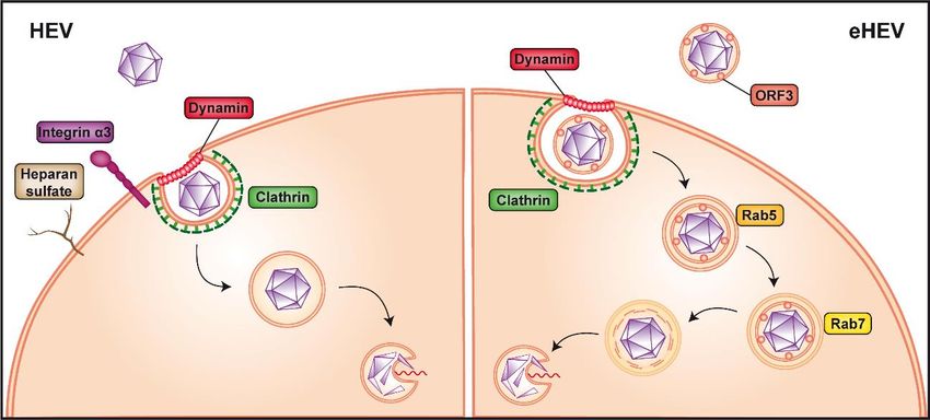
Cells 2020, 9, 1294 3 of 14
As obligate intracellular pathogens, viruses have developed strategies to hijack and manipulate
host cell pathways in order to ensure productive infection. Moreover, RNA viruses, especially those
with relatively limited genome size and coding capacity, such as HEV, are particularly dependent on
the host cell machinery. Because the tools to study HEV have been limited until recently, only little is
known about the host factors involved in the various steps of the viral life cycle. Studies performed in
heterologous settings, such as the yeast two hybrid system, identified cellular factors interacting with
Cells 2020, 9, x FOR PEER REVIEW 4 of 14
HEV proteins [16,17]. However, most of these candidates remain to be validated and further studied
using infectious cell culture systems, in vivo models and liver biopsies from patients with hepatitis
late endosomal and lysosomal Niemann-Pick disease type C1 protein, involved in cholesterol
E. In this review, we shall focus on host factors whose involvement in the viral life cycle has been
extraction, significantly reduced eHEV infection [22]. Moreover, treatment with an inhibitor of
validated in HEV infection settings.
lysosomal acid lipase, responsible for lipid degradation, resulted in a dose-dependent reduction of
eHEV
2. HEVcell entry [22].
Entry
These observations suggest that the quasi-envelope of eHEV is removed in endolysosomes. The
viral As HEVmay
capsid is present in a non-enveloped
subsequently interact with("naked") andunidentified
an as yet a quasi-enveloped form (eHEV),
host factor the entry
and undergo the
pathway of the virus may differ for these two forms. Our knowledge
conformational changes required for genome release into the cytoplasm. A similar mechanism of HEV entry remains
was
scarce
recentlybutproposed
studies using
for HAV virus-like
[31]. particles
Since both(VLP) as a model system
quasi-enveloped and have highlightedHEV
non-enveloped possible
are
host factors involved in the initial attachment to the cell and virus internalization,
internalized in vesicles belonging to the endosomal pathway, it is plausible that they use a common including
the
host78-kDa
factor glucose-regulated
to allow uncoatingprotein (GRP78),
and release ATP
of the synthase
genome subunit
into β (ATPB5)
the cytoplasm and asialoglycoprotein
(detailed in [43]).
receptor (ASGPR) [18–20]. Notably, non-enveloped HEV was shown
The ORF3 protein is present within eHEV and interacts with the capsid [44]. to interact with heparan its
However, sulfate
role
proteoglycans (HSPG), likely syndecans [21,22], which
in eHEV entry, uncoating and genome release remains to be explored. are expressed on the surface of many cell
types. Hence,
Taken treatment
together, some of HEV
susceptible hepatoma
entry factors and cell lines with
pathways haveheparinase I considerably
been identified, reduced
but key elements
VLP
are still missing. The capacity of HEV to infect a broad array of cell types [45–51], possibly relatedcell
binding as well as HEV infection [21] (Figure 2). Of note, HSPG are known to mediate to
attachment
the diverseofextrahepatic
several enveloped and nonenveloped
manifestations viruses,
of hepatitis including,
E [52–55], among
strongly others, herpes
suggests that thesimplex
entry
virus, hepatitis
receptor(s) C virus
is (are) (HCV), to
not specific norovirus and but
hepatocytes human immunodeficiency
ubiquitously expressed.virus [23–27].
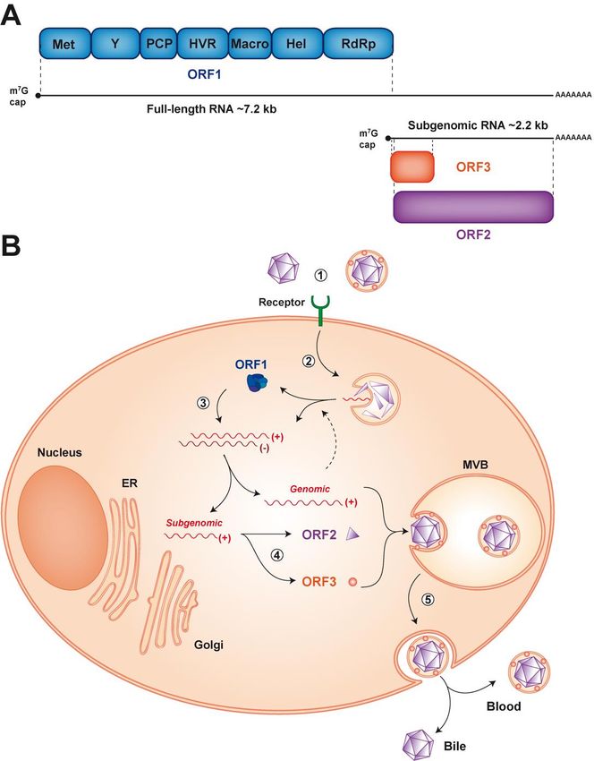
Figure 2.
Figure 2. Non-enveloped
Non-envelopedversus versusquasi-enveloped
quasi-envelopedhepatitis
hepatitisEEvirus
virusentry.
entry.Non-enveloped
Non-enveloped hepatitis E
hepatitis
virus
E (HEV)
virus (HEV)is believed to first
is believed bindbind
to first to heparan sulfate
to heparan proteoglycans
sulfate and integrin
proteoglycans α3 at the
and integrin α3cell surface
at the cell
(left panel).
surface The virus
(left panel). is then
The virus internalized
is then via via
internalized clathrin-
clathrin-and
anddynamin
dynamin2-dependent
2-dependent endocytosis,
endocytosis,
followed by release of the viral genome into the cytoplasm by an as yet unknown mechanism,
followed by release of the viral genome into the cytoplasm by an as yet unknown mechanism, possibly
possibly
involving a conformational
conformational change
changeof ofthe
thecapsid.
capsid.The The cofactor(s) and
cofactor(s) receptor(s)
and receptor(s)allowing
allowingentry of
entry
quasi-enveloped
of quasi-enveloped HEVHEV (eHEV)
(eHEV) are
areunknown
unknown(right(rightpanel).
panel). Internalization
Internalizationrequires
requires clathrin-
clathrin- and
2-dependentendocytosis
dynamin 2-dependent endocytosisasas well
well as trafficking
as trafficking through
through Rab5-Rab5-
(early)(early)
as wellasaswell as Rab7-
Rab7-positive
positive (late) endosomes and eventually lysosomes to allow release of the viral
(late) endosomes and eventually lysosomes to allow release of the viral genome into the cytoplasm, genome into the
cytoplasm,
likely likely by
by a process a process
similar similar
to that to that
of naked of naked HEV.
HEV.
A recent
3. Viral RNAmicroarray
Replicationanalysis comparing gene expression in permissive versus non-permissive cells
identified integrin α3 as an entry factor for HEV [28]. Integrins belong to a family of transmembrane
Upon release of the viral positive-strand RNA genome into the cytoplasm, the host translational
machinery, including ribosomal subunits and elongation factors, seeds on the 5′ untranslated region
to start translation of the ORF1 protein. Among the different components, the eukaryotic translation
initiation factor 4F (eIF4F) complex has been identified as being important for HEV replication. This
complex is known to be involved in the cap-dependent translation and replication of several viruses
(reviewed in [56]). In this context, an RNA interference-based loss-of-function study showed thatCells 2020, 9, 1294 4 of 14
proteins localized at the cell surface which can bind to extracellular matrix (ECM) as well as cell surface
and intracellular ligands. These interactions trigger a myriad of intracellular signals modulating
cell behavior through effects on actin microfilaments, thereby connecting the extracellular space
and the cytoskeleton (reviewed in [29]). Integrins were shown to function as receptors for Kaposi’s
sarcoma-associated herpesvirus [30] and, interestingly, also for HAV [31]. In the case of HEV,
overexpression of integrin α3 in non-permissive cells allowed non-enveloped HEV infection only
and, conversely, knockout of integrin α3 gene in permissive cells prevented entry of non-enveloped
HEV but not of eHEV [28]. Together with the physical interaction between integrin α3 and HEV,
the data strongly suggests that this ECM receptor is an entry factor for the non-enveloped viral particle
(Figure 2). Integrins are expressed in a broad array of tissues, including in the intestine [26,32], where
HEV is known to replicate and which may represent the initial site of infection [33,34]. However, in
some organs, such as the liver, integrin α3 expression is rather low [35–37], raising the possibility that
integrin α3 may act as a cofactor for HEV entry rather than as the key receptor. Further studies are
needed to clarify the entry pathway used by non-enveloped HEV.
Internalization of non-enveloped HEV is believed to occur through clathrin- and dynamin
2-dependent endocytosis [22,38,39] (Figure 2). In this context, membrane cholesterol was also shown
to be important for HEV entry, as treatment of cells with cholesterol sequestering agents significantly
reduced VLP uptake [39]. Following endocytosis, the HEV genome needs to be uncoated to be released
into the cytoplasm. It is likely that the capsid protein itself plays a crucial role in this poorly understood
process, possibly by undergoing conformational changes induced by interaction with a host protein, as
it is seen in other non-enveloped viruses, such as human papilloma virus, murine polyomavirus and
poliovirus [40–42].
Since the viral capsid is not exposed at the surface of eHEV (see Section 4), the quasi-enveloped
particle may use different, as yet unknown entry factor(s). However, as for the naked virus, eHEV is
internalized through clathrin- and dynamin 2-dependent endocytosis [38,39]. Unlike non-enveloped
HEV, eHEV entry is dependent on the small GTPases, Rab5 and Rab7, both of which are involved in
endosomal trafficking. In fact, depletion of one or the other has been shown to considerably reduce
virus infection [22] (Figure 2). In addition, eHEV entry depends on endosomal acidification [22],
indicating that transition of the endosome to a more acidic cell compartment is necessary (Figure 2).
Lysosomal lipid degradation appears to be a required step in eHEV entry. Indeed, depletion of the late
endosomal and lysosomal Niemann-Pick disease type C1 protein, involved in cholesterol extraction,
significantly reduced eHEV infection [22]. Moreover, treatment with an inhibitor of lysosomal acid
lipase, responsible for lipid degradation, resulted in a dose-dependent reduction of eHEV cell entry [22].
These observations suggest that the quasi-envelope of eHEV is removed in endolysosomes.
The viral capsid may subsequently interact with an as yet unidentified host factor and undergo
the conformational changes required for genome release into the cytoplasm. A similar mechanism was
recently proposed for HAV [31]. Since both quasi-enveloped and non-enveloped HEV are internalized
in vesicles belonging to the endosomal pathway, it is plausible that they use a common host factor to
allow uncoating and release of the genome into the cytoplasm (detailed in [43]).
The ORF3 protein is present within eHEV and interacts with the capsid [44]. However, its role in
eHEV entry, uncoating and genome release remains to be explored.
Taken together, some HEV entry factors and pathways have been identified, but key elements
are still missing. The capacity of HEV to infect a broad array of cell types [45–51], possibly related
to the diverse extrahepatic manifestations of hepatitis E [52–55], strongly suggests that the entry
receptor(s) is (are) not specific to hepatocytes but ubiquitously expressed.
3. Viral RNA Replication
Upon release of the viral positive-strand RNA genome into the cytoplasm, the host translational
machinery, including ribosomal subunits and elongation factors, seeds on the 50 untranslated region
to start translation of the ORF1 protein. Among the different components, the eukaryotic translationCells 2020, 9, 1294 5 of 14
initiation factor 4F (eIF4F) complex has been identified as being important for HEV replication. This
complex is known to be involved in the cap-dependent translation and replication of several viruses
(reviewed in [56]). In this context, an RNA interference-based loss-of-function study showed that
components of the eIF4F complex are required for efficient HEV replication, while known negative
regulatory factors of this pathway limit viral RNA synthesis [57]. Along these lines, silvestrol, a natural
compound inhibiting part of the eIF4F complex, efficiently inhibits HEV replication in vitro, and in
a mouse model in vivo, ORF2 protein production and virus spread [58,59]. Interestingly, silvestrol was
reported to also inhibit corona-, picorna-, alpha-, flavi- and filo-viruses and, therefore, displays broad
antiviral activity [60–63].
ORF1 encodes the functional domains required for viral RNA synthesis, including an
RNA-dependent RNA polymerase (RdRp) located at the C-terminal end of the polyprotein. In
addition to the RdRp, the replicase encoded by ORF1 comprises an RNA helicase, a methyltransferase
as well as less well-characterized domains, including the Macro and Y domains, a hypervariable
region and a putative papain-like cysteine protease (PCP) (Figure 1A). Of note, the PCP has been
associated with deubiquitination and the Macro domain with deribosylation activities likely involved
in the posttranslational modifications of host proteins [64,65]. Positive-strand RNA viruses commonly
process their polyproteins into individual functional proteins by viral, and in some instances, cellular
proteases. In the case of HEV, however, it is unsettled whether, and if so, by which mechanism,
the ORF1 protein is processed. While some studies reported processing of the ORF1 polyprotein,
including by the PCP or the cellular proteases thrombin and factor Xa [66], others have found that
the major form of ORF1 protein is unprocessed (reviewed in [53,67]). Further studies in complete cell
culture systems and using more sensitive techniques for the detection of ORF1 protein are required [68].
As for all positive-strand RNA viruses, replication includes synthesis of a complementary
negative-strand RNA which serves as a template for the production of positive-strand genomic and, in
the case of HEV, an additional subgenomic RNA. The key enzyme responsible for these steps is the RdRp.
The mechanisms regulating the production of these RNA species from the negative-strand RNA are still
unknown but imply cis-acting elements within the genome [69]. Regulation of transcription may also
involve host factor(s), such as heterogeneous nuclear ribonucleoproteins (hnRNP) [70,71]. hnRNPs play
a role in nuclear RNA metabolism and have been reported to re-localize to the cytoplasm of HEV-infected
cells [71], similarly to what had been observed for infection with other RNA viruses [72–76], suggesting
a potential role of hnRNPs in genomic and subgenomic HEV RNA transcription.
Replication of positive-strand RNA viruses takes place in membrane-associated replication
complexes composed of viral proteins, the replicating viral RNA, rearranged cellular membranes
and other host factors [77]. The subcellular site of HEV RNA replication has not been identified to
date but recent advances such as the development of tagged functional HEV genomes may facilitate
progress in this area [68]. Insertion of a hemagglutinin epitope tag in the ORF1 polyprotein allowed
us to visualize the HEV replicase together with viral RNA in cytoplasmic dot-like structures, likely
indicating the site of active replication. ORF1 protein was found to colocalize best with exosomal
markers as well as with the ORF2 and ORF3 proteins, suggesting that HEV RNA replication takes
place in close proximity to virion assembly sites [68]. Virus-induced membrane rearrangements may
serve several purposes, including the physical support and organization of the replication complex,
the compartmentalization and local concentration of viral and host factors required for RNA replication,
tethering of the viral RNA during unwinding and coordination of its translation, replication and
packaging, provision of lipid constituents important for replication, and physical protection from host
antiviral defenses (reviewed in [77,78]). Of note, HCV as well as picorna- and coronaviruses require
the guanine nucleotide-exchange factor Golgi brefeldin A resistance factor 1 (GBF1) for the induction
of membrane alterations making up their viral replication complexes [79–83]. Interestingly, similar
observations have been made for HEV [84]. However, no colocalization of GBF1 with HEV ORF1
and ORF2 proteins or relocalization of GBF1 upon HEV infection have been observed. One may thus
hypothesize that GBF1 is not recruited to HEV replication sites and is not involved in replicationCells 2020, 9, 1294 6 of 14
complex formation but rather plays an indirect role in HEV replication [84], as also proposed for
HCV [79,80] and mouse hepatitis virus [85].
4. Virion Assembly and Infectious Particle Release
Virion assembly consists of the packaging of genomic viral RNA in the capsid. While the subcellular
site of HEV assembly has not been identified yet, it is likely tightly connected to replication complexes
(see above). The mechanisms driving HEV assembly are poorly understood but early observations
showed that RNA and the ORF2 protein can spontaneously assemble into virus-like particles in insect
cells [86] (reviewed in [87]). These findings argue in favor of a self-assembly process involving a limited
number of host factors.
The ORF2 protein exists in several forms, of which a non-glycosylated form is involved in
virion formation [88–91]. It is currently debated whether this form, harboring a truncated and
non-functional signal peptide, results from translation at an alternative start site [90] or from proteolytic
cleavage, as suggested by mass spectrometry analyses [89]. The non-glycosylated ORF2 protein may
represent the major intracellular form detected in the cytosol as well as the cell nucleus [89,91,92].
While the role of nuclear ORF2 protein is unknown, its nucleocytoplasmic shuttling likely involves
the host nuclear import/export machineries. In contrast to the non-glycosylated form packaging
the viral genome, the glycosylated form of ORF2 protein is rapidly released extracellularly through
the secretory pathway [88–91,93,94]. This implies recognition of its signal peptide by the translocon,
translocation into the ER lumen, cleavage by signal peptidase, followed by sialylation as well as N-
and O-glycosylation by yet to be defined glycosyltransferases in the ER and Golgi [89], and secretion.
Glycosylated ORF2 protein is present in at least two forms, of which the smaller may result from
cleavage by furin-like proteases at an RRR motif [91]. Overall, these studies suggest that ORF2 protein
likely has more than one function in genome packaging and point toward the implication of different
host factors.
HEV assembly involves the non-glycosylated ORF2 protein and the viral RNA but does not require
the ORF3 protein, as genomes harboring a mutated ORF3 start codon yield infectious particles; however,
these are not secreted from the cell [95,96]. Infectious virus is believed to be released as quasi-enveloped
particles from both the basolateral and apical sides of hepatocytes, facing the liver sinusoids and
the bile canaliculi, respectively [33,97,98]. The pseudo-envelope of virions secreted into the bile is likely
delipidated by bile acids and/or pancreatic enzymes (Figure 3). Interestingly, HEV is preferentially
secreted from the apical side, explaining the high viral load detected in feces. Based on these
observations, different host factors may be involved in virus secretion from the apical and basolateral
sides of hepatocytes. Moreover, HEV ORF3 protein is essential for the secretion of infectious particles,
possibly by connecting the capsid with the host factors required for egress. Phosphorylation of ORF3
protein at a serine residue (Ser 70 and Ser 71 in gt 3 and gt 1, respectively) may trigger the interaction
with assembled non-glycosylated ORF2 protein [44]. Based on sequence information, phosphorylation
may involve the p34cdc2 kinase and mitogen-activated protein kinase [99]. Furthermore, ORF3 protein
recruits Tsg101, a member of the endosomal sorting complex required for transport-I (ESCRT-I), via
a highly conserved PSAP motif [100–102]. The presence of this motif in the C-terminal region of
the ORF3 protein is required for virus release [101]. It is known that proline-rich motifs P(S/T)AP and
PPXY (X being any amino acid), which are also called "late domains", are essential for budding of
enveloped viruses such as human immunodeficiency virus (HIV)-1 or Ebola [103,104]. The ESCRT
machinery is composed of 4 complexes, ESCRT-0 to ESCRT-III, which are involved in membrane
remodeling, leading to budding reactions or membrane involution. These complexes can further
recruit accessory proteins, such as vacuolar protein sorting 4 (Vps4) and apoptosis-linked gene-2
interacting protein X (Alix), both interacting with the ESCRT-III complex, to close newly formed
vesicles (reviewed in [105]). Further confirming the requirement for the ESCRT machinery, hepatocyte
growth factor-regulated tyrosine kinase substrate (Hrs), a member of ESCRT-0, and Vps4 were shown
to be required for HEV particle release [102,106]. In addition, the envelope wrapping the virion hasORF3 proteins were shown to harbor classical exosomal markers, including the tetraspanins CD81,
CD63 and CD9, as well as the ESCRT components Alix and Tsg101 [106,108,112]. Interestingly, trans-
Golgi network protein 2 (TGOLN2) has also been detected on eHEV [107] (Figure 3). This observation
and the fact that TGOLN2 is located in the cytoplasm confirms that quasi-enveloped particles are
produced
Cells within the cell and not at the plasma membrane.
2020, 9, 1294 7 of 14
The quasi-enveloped nature of virions circulating in blood may provide several advantages to
HEV, including protection from neutralizing antibodies. However, the presence of eHEV in blood
been
may shown to bevirus
also favor derived from exosomes,
dissemination small vesicles
to organs generated
other than fromasmultivesicular
the liver, bodies (MVB)
exosomes containing viral
by the ESCRT pathway [102,106]. These MVB, which fuse with the plasma membrane
genetic material were shown to lead to productive infection by other viruses and to modulate to release their
cellular
content
responses into the extracellular
(reviewed in [113]).milieu,
Hence,require the Rab27a
it is plausible thatprotein, which has
the production ofbeen shown to colocalize
quasi-enveloped virions
with ORF3 protein [102,106] (Figure 3).
has additional functions beyond the spread of HEV within the liver.
Figure 3.
Figure 3. Assembly
Assemblyand andrelease
releaseofofinfectious
infectioushepatitis
hepatitisEEvirus. Packaging
virus. Packaging of the viral
of the genome
viral intointo
genome the
capsid is believed to occur by spontaneous self-assembly of the non-glycosylated
the capsid is believed to occur by spontaneous self-assembly of the non-glycosylated ORF2 protein. ORF2 protein. The
non-glycosylated
The non-glycosylatedORF2 ORF2protein
proteinmay undergo
may undergo post-translational
post-translationalmodification
modificationby by yet unknown
yet unknown
enzyme(s). Formation
enzyme(s). Formation of of the quasi-enveloped particle
the quasi-enveloped particle involves
involves phosphorylation
phosphorylation and and palmitoylation
palmitoylation
of the
of the ORF3
ORF3protein
proteinandandthetheESCRT
ESCRT machinery,
machinery, of of
which
whichthethe
components
components Tsg101, Hrs,Hrs,
Tsg101, Vps4Vps4
and Alix
and
were shown to be required (see text for abbreviations). The virion is wrapped
Alix were shown to be required (see text for abbreviations). The virion is wrapped in an exosomal in an exosomal
membrane harboring
membrane harboringthe thetetraspanins
tetraspanins CD9,
CD9, CD63
CD63 andand
CD81,CD81, as as
as well well
the as the trans-Golgi
trans-Golgi networknetwork
protein
2protein 2 (TGOLN2),
(TGOLN2), Alix andAlix
Tsg101.andRelease
Tsg101.ofRelease of quasi-enveloped
quasi-enveloped HEV (eHEV) HEV (eHEV)Rab27a-dependent
involves involves Rab27a-
dependent oftrafficking
trafficking of multivesicular
multivesicular bodies (MVB) bodies (MVB)
and fusion withand fusion with
the plasma the plasma
membrane. Secretedmembrane.
particles
Secretedassociated
remain particles with
remain
the associated with the
lipid membrane lipid
in the membranewhile
bloodstream in the bloodstream
they whileinthey
are delipidated are
the bile.
Asterisks indicate host factors that were not found on the quasi-envelope of eHEV.
Current evidence indicates that none of the viral proteins are present on the surface of eHEV
but identified ORF3 protein beneath the quasi-envelope [107–109]. Palmitoylation at N-terminal
cysteine residues mediates membrane association as well as stability of the ORF3 protein and is
required for virus secretion [110]. A membrane topology where ORF3 protein is located entirely
on the cytosolic side, corresponding to the exosome lumen, has therefore been proposed [110] and
is further supported by the interaction of its PSAP motif with Tsg101. Of note, palmitoylation is
a reversible protein modification taking place in the cytosol, which increases the hydrophobicity of
a protein and contributes to its membrane association (reviewed in [111]). This implies the requirement
for one or more as yet unidentified S-palmitoyl-transferase(s) of the host cell [110].Cells 2020, 9, 1294 8 of 14
Given the origin of the quasi-envelope, host proteins, and more specifically, exosomal proteins,
can be present on eHEV. Indeed, particles released from infected cells and positive for ORF2 and ORF3
proteins were shown to harbor classical exosomal markers, including the tetraspanins CD81, CD63
and CD9, as well as the ESCRT components Alix and Tsg101 [106,108,112]. Interestingly, trans-Golgi
network protein 2 (TGOLN2) has also been detected on eHEV [107] (Figure 3). This observation and
the fact that TGOLN2 is located in the cytoplasm confirms that quasi-enveloped particles are produced
within the cell and not at the plasma membrane.
The quasi-enveloped nature of virions circulating in blood may provide several advantages to
HEV, including protection from neutralizing antibodies. However, the presence of eHEV in blood may
also favor virus dissemination to organs other than the liver, as exosomes containing viral genetic
material were shown to lead to productive infection by other viruses and to modulate cellular responses
(reviewed in [113]). Hence, it is plausible that the production of quasi-enveloped virions has additional
functions beyond the spread of HEV within the liver.
5. Conclusions and Perspectives
Although HEV research is a rapidly growing field, our current knowledge of the virus life cycle
is still limited by important gaps. We lack key information on virus entry, including the cellular
receptor(s), and on the uncoating of the viral RNA. Moreover, future efforts should concentrate on
the molecular mechanisms and subcellular compartments involved in RNA replication and assembly.
While virus release remains one of the best studied steps of the viral life cycle, many aspects need to be
clarified, in particular the contribution of host factors and the composition and role of quasi-enveloped
particles. Considering the ability of HEV to replicate in different tissues as well as to infect a wide
range of animals, one may hypothesize that HEV host dependency is not very selective, facilitating
the crossing of species barriers.
Studies on the HEV life cycle currently rely on the use of in vitro model systems, which have
certain limitations, especially with respect to host factors present in differentiated hepatocytes. As
an example, a stimulatory effect of cyclophilin inhibitors on HEV replication was reported initially
in hepatoma cells [114]. However, in stem cell-derived hepatocyte-like cells, this observation was
confirmed only with a cell culture-adapted infectious clone, but not with natural HEV isolates [115].
Future improvements of in vitro models should include the use of natural HEV isolates together with
primary and stem cell-derived hepatocyte-like cells, polarized cell culture models, as well as ex vivo
and in vivo infection model systems to confirm in vitro findings. Ultimately, key findings will have to
be validated in liver specimens from patients with hepatitis E.
Obtaining a broader and unbiased view of the host factors involved in the HEV life cycle will
likely depend on novel technologies, such as clustered regularly interspaced short palindromic
repeats (CRISPR)/Cas9-based genome-wide screening and proteomic proximity labeling approaches.
CRISPR/Cas9-based screens have advanced the understanding of, among others, virus entry [116,117]
and replication [118,119], and have also facilitated the identification of new antiviral targets [120].
Proximity labeling has been successfully used, for example, to characterize the microenvironment
of coronavirus replication complexes [121]. In the future, the combination of improved HEV model
systems and of novel technologies should improve our knowledge of the host factors required for
productive HEV infection.
Author Contributions: N.O. and J.G. conceived the article; N.O. and J.G. wrote the original draft and prepared
figures; D.M. edited and reviewed the manuscript. All authors have read and agreed to the published version of
the manuscript.
Funding: This work was funded by the Swiss National Science Foundation (31003A_179424 to D.M. and
CRSK-3_190706 to J.G.), the Novartis Foundation (18C140 to D.M.) and the Gilead Sciences International Research
Scholars Program in Liver Disease (Award 2015 to J.G.).
Conflicts of Interest: The authors declare no conflict of interest.Cells 2020, 9, 1294 9 of 14
References
1. Khuroo, M.S. Study of an epidemic of non-A, non-B hepatitis. Possibility of another human hepatitis virus
distinct from post-transfusion non-A, non-B type. Am. J. Med. 1980, 68, 818–824. [CrossRef]
2. Balayan, M.S.; Andjaparidze, A.G.; Savinskaya, S.S.; Ketiladze, E.S.; Braginsky, D.M.; Savinov, A.P.;
Poleschuk, V.F. Evidence for a virus in non-A, non-B hepatitis transmitted via the fecal-oral route. Intervirology
1983, 20, 23–31.
3. Reyes, G.R.; Purdy, M.A.; Kim, J.P.; Luk, K.C.; Young, L.M.; Fry, K.E.; Bradley, D.W. Isolation of a cDNA
from the virus responsible for enterically transmitted non-A, non-B hepatitis. Science 1990, 247, 1335–1339.
[CrossRef] [PubMed]
4. Tam, A.W.; Smith, M.M.; Guerra, M.E.; Huang, C.C.; Bradley, D.W.; Fry, K.E.; Reyes, G.R. Hepatitis E virus
(HEV): Molecular cloning and sequencing of the full-length viral genome. Virology 1991, 185, 120–131.
[CrossRef]
5. Smith, D.B.; Simmonds, P.; Jameel, S.; Emerson, S.U.; Harrison, T.J.; Meng, X.J.; Okamoto, H.; Van der
Poel, W.H.; Purdy, M.A. Consensus proposals for classification of the family Hepeviridae. J. Gen. Virol. 2015,
96, 1191–1192. [CrossRef] [PubMed]
6. Lee, G.H.; Tan, B.H.; Teo, E.C.; Lim, S.G.; Dan, Y.Y.; Wee, A.; Aw, P.P.; Zhu, Y.; Hibberd, M.L.; Tan, C.K.; et al.
Chronic infection with camelid hepatitis E virus in a liver transplant recipient who regularly consumes camel
meat and milk. Gastroenterology 2016, 150, 355.e353–357.e353. [CrossRef]
7. Nimgaonkar, I.; Ding, Q.; Schwartz, R.E.; Ploss, A. Hepatitis E virus: Advances and challenges. Nat. Rev.
Gastroenterol. Hepatol. 2018, 15, 96–110. [CrossRef]
8. Abravanel, F.; Lhomme, S.; El Costa, H.; Schvartz, B.; Peron, J.M.; Kamar, N.; Izopet, J. Rabbit hepatitis E
virus infections in humans, France. Emerg. Infect. Dis. 2017, 23, 1191–1193. [CrossRef]
9. Sahli, R.; Fraga, M.; Semela, D.; Moradpour, D.; Gouttenoire, J. Rabbit HEV in immunosuppressed patients
with hepatitis E acquired in Switzerland. J. Hepatol. 2019, 70, 1023–1025. [CrossRef]
10. Sridhar, S.; Yip, C.C.Y.; Wu, S.; Cai, J.; Zhang, A.J.; Leung, K.H.; Chung, T.W.H.; Chan, J.F.W.; Chan, W.M.;
Teng, J.L.L.; et al. Rat hepatitis E virus as cause of persistent hepatitis after liver transplant. Emerg. Infect.
Dis. 2018, 24, 2241–2250. [CrossRef]
11. Sridhar, S.; Yip, C.C.; Wu, S.; Chew, N.F.; Leung, K.H.; Chan, J.F.; Zhao, P.S.; Chan, W.M.; Poon, R.W.;
Tsoi, H.W.; et al. Transmission of rat hepatitis E virus infection to humans in Hong Kong: A clinical and
epidemiological analysis. Hepatology 2020. [CrossRef] [PubMed]
12. Ding, Q.; Heller, B.; Capuccino, J.M.; Song, B.; Nimgaonkar, I.; Hrebikova, G.; Contreras, J.E.; Ploss, A.
Hepatitis E virus ORF3 is a functional ion channel required for release of infectious particles. Proc. Natl.
Acad. Sci. USA 2017, 114, 1147–1152. [CrossRef] [PubMed]
13. Graff, J.; Torian, U.; Nguyen, H.; Emerson, S.U. A bicistronic subgenomic mRNA encodes both the ORF2 and
ORF3 proteins of hepatitis E virus. J. Virol. 2006, 80, 5919–5926. [CrossRef] [PubMed]
14. Ichiyama, K.; Yamada, K.; Tanaka, T.; Nagashima, S.; Jirintai; Takahashi, M.; Okamoto, H. Determination of
the 50 -terminal sequence of subgenomic RNA of hepatitis E virus strains in cultured cells. Arch. Virol. 2009,
154, 1945–1951. [CrossRef]
15. Feng, Z.; Hensley, L.; McKnight, K.L.; Hu, F.; Madden, V.; Ping, L.; Jeong, S.H.; Walker, C.; Lanford, R.E.;
Lemon, S.M. A pathogenic picornavirus acquires an envelope by hijacking cellular membranes. Nature 2013,
496, 367–371. [CrossRef]
16. Geng, Y.; Yang, J.; Huang, W.; Harrison, T.J.; Zhou, Y.; Wen, Z.; Wang, Y. Virus host protein interaction
network analysis reveals that the HEV ORF3 protein may interrupt the blood coagulation process. PLoS
ONE 2013, 8, e56320. [CrossRef]
17. Subramani, C.; Nair, V.P.; Anang, S.; Mandal, S.D.; Pareek, M.; Kaushik, N.; Srivastava, A.; Saha, S.; Shalimar;
Nayak, B.; et al. Host-virus protein interaction network reveals the involvement of multiple host processes
in the life cycle of hepatitis E virus. mSystems 2018, 3, e00135-17. [CrossRef]
18. Yu, H.; Li, S.; Yang, C.; Wei, M.; Song, C.; Zheng, Z.; Gu, Y.; Du, H.; Zhang, J.; Xia, N. Homology model
and potential virus-capsid binding site of a putative HEV receptor Grp78. J. Mol. Model. 2011, 17, 987–995.
[CrossRef]
19. Ahmed, Z.; Holla, P.; Ahmad, I.; Jameel, S. The ATP synthase subunit β (ATP5B) is an entry factor for
the hepatitis E virus. bioRxiv 2016. [CrossRef]Cells 2020, 9, 1294 10 of 14
20. Zhang, L.; Tian, Y.; Wen, Z.; Zhang, F.; Qi, Y.; Huang, W.; Zhang, H.; Wang, Y. Asialoglycoprotein receptor
facilitates infection of PLC/PRF/5 cells by HEV through interaction with ORF2. J. Med. Virol. 2016, 88,
2186–2195. [CrossRef]
21. Kalia, M.; Chandra, V.; Rahman, S.A.; Sehgal, D.; Jameel, S. Heparan sulfate proteoglycans are required
for cellular binding of the hepatitis E virus ORF2 capsid protein and for viral infection. J. Virol. 2009, 83,
12714–12724. [CrossRef] [PubMed]
22. Yin, X.; Ambardekar, C.; Lu, Y.; Feng, Z. Distinct entry mechanisms for nonenveloped and quasi-enveloped
hepatitis E viruses. J. Virol. 2016, 90, 4232–4242. [CrossRef] [PubMed]
23. WuDunn, D.; Spear, P.G. Initial interaction of herpes simplex virus with cells is binding to heparan sulfate. J.
Virol. 1989, 63, 52–58. [CrossRef] [PubMed]
24. Shieh, M.T.; WuDunn, D.; Montgomery, R.I.; Esko, J.D.; Spear, P.G. Cell surface receptors for herpes simplex
virus are heparan sulfate proteoglycans. J. Cell Biol. 1992, 116, 1273–1281. [CrossRef] [PubMed]
25. Mondor, I.; Ugolini, S.; Sattentau, Q.J. Human immunodeficiency virus type 1 attachment to HeLa CD4 cells
is CD4 independent and gp120 dependent and requires cell surface heparans. J. Virol. 1998, 72, 3623–3634.
[CrossRef] [PubMed]
26. Barth, H.; Schafer, C.; Adah, M.I.; Zhang, F.; Linhardt, R.J.; Toyoda, H.; Kinoshita-Toyoda, A.; Toida, T.; Van
Kuppevelt, T.H.; Depla, E.; et al. Cellular binding of hepatitis C virus envelope glycoprotein E2 requires cell
surface heparan sulfate. J. Biol. Chem. 2003, 278, 41003–41012. [CrossRef] [PubMed]
27. Tamura, M.; Natori, K.; Kobayashi, M.; Miyamura, T.; Takeda, N. Genogroup II noroviruses efficiently bind to
heparan sulfate proteoglycan associated with the cellular membrane. J. Virol. 2004, 78, 3817–3826. [CrossRef]
28. Shiota, T.; Li, T.C.; Nishimura, Y.; Yoshizaki, S.; Sugiyama, R.; Shimojima, M.; Saijo, M.; Shimizu, H.;
Suzuki, R.; Wakita, T.; et al. Integrin alpha3 is involved in non-enveloped hepatitis E virus infection. Virology
2019, 536, 119–124. [CrossRef]
29. Takada, Y.; Ye, X.; Simon, S. The integrins. Genome Biol. 2007, 8, 215. [CrossRef]
30. Akula, S.M.; Pramod, N.P.; Wang, F.Z.; Chandran, B. Integrin alpha3beta1 (CD 49c/29) is a cellular receptor for
Kaposi’s sarcoma-associated herpesvirus (KSHV/HHV-8) entry into the target cells. Cell 2002, 108, 407–419.
[CrossRef]
31. Rivera-Serrano, E.E.; González-López, O.; Das, A.; Lemon, S.M. Cellular entry and uncoating of naked and
quasi-enveloped human hepatoviruses. Elife 2019, 8, e43983. [CrossRef] [PubMed]
32. Choy, M.Y.; Richman, P.I.; Horton, M.A.; MacDonald, T.T. Expression of the VLA family of integrins in
human intestine. J. Pathol. 1990, 160, 35–40. [CrossRef] [PubMed]
33. Marion, O.; Lhomme, S.; Nayrac, M.; Dubois, M.; Pucelle, M.; Requena, M.; Migueres, M.; Abravanel, F.;
Peron, J.M.; Carrere, N.; et al. Hepatitis E virus replication in human intestinal cells. Gut 2020, 69, 901–910.
[CrossRef] [PubMed]
34. Oechslin, N.; Moradpour, D.; Gouttenoire, J. Hepatitis E virus finds its path through the gut. Gut 2020, 69,
796–798. [CrossRef]
35. de Melker, A.A.; Sterk, L.M.; Delwel, G.O.; Fles, D.L.; Daams, H.; Weening, J.J.; Sonnenberg, A. The A and
B variants of the alpha 3 integrin subunit: Tissue distribution and functional characterization. Lab. Invest.
1997, 76, 547–563.
36. Volpes, R.; van den Oord, J.J.; Desmet, V.J. Distribution of the VLA family of integrins in normal and
pathological human liver tissue. Gastroenterology 1991, 101, 200–206. [CrossRef]
37. Volpes, R.; van den Oord, J.J.; Desmet, V.J. Integrins as differential cell lineage markers of primary liver
tumors. Am. J. Pathol. 1993, 142, 1483–1492.
38. Kapur, N.; Thakral, D.; Durgapal, H.; Panda, S.K. Hepatitis E virus enters liver cells through
receptor-dependent clathrin-mediated endocytosis. J. Viral Hepat. 2012, 19, 436–448. [CrossRef]
39. Holla, P.; Ahmad, I.; Ahmed, Z.; Jameel, S. Hepatitis E virus enters liver cells through a dynamin-2, clathrin
and membrane cholesterol-dependent pathway. Traffic 2015, 16, 398–416. [CrossRef]
40. Bubeck, D.; Filman, D.J.; Cheng, N.; Steven, A.C.; Hogle, J.M.; Belnap, D.M. The structure of the poliovirus
135S cell entry intermediate at 10-angstrom resolution reveals the location of an externalized polypeptide
that binds to membranes. J. Virol. 2005, 79, 7745–7755. [CrossRef]
41. Cavaldesi, M.; Caruso, M.; Sthandier, O.; Amati, P.; Garcia, M.I. Conformational changes of murine
polyomavirus capsid proteins induced by sialic acid binding. J. Biol. Chem. 2004, 279, 41573–41579.
[CrossRef] [PubMed]Cells 2020, 9, 1294 11 of 14
42. Sapp, M.; Bienkowska-Haba, M. Viral entry mechanisms: human papillomavirus and a long journey from
extracellular matrix to the nucleus. FEBS J. 2009, 276, 7206–7216. [CrossRef] [PubMed]
43. Yin, X.; Feng, Z. Hepatitis E virus entry. Viruses 2019, 11, 883. [CrossRef]
44. Tyagi, S.; Korkaya, H.; Zafrullah, M.; Jameel, S.; Lal, S.K. The phosphorylated form of the ORF3 protein of
hepatitis E virus interacts with its non-glycosylated form of the major capsid protein, ORF2. J. Biol. Chem.
2002, 277, 22759–22767. [CrossRef] [PubMed]
45. Drave, S.A.; Debing, Y.; Walter, S.; Todt, D.; Engelmann, M.; Friesland, M.; Wedemeyer, H.; Neyts, J.;
Behrendt, P.; Steinmann, E. Extra-hepatic replication and infection of hepatitis E virus in neuronal-derived
cells. J. Viral Hepat. 2016, 23, 512–521. [CrossRef]
46. Fu, R.M.; Decker, C.C.; Dao Thi, V.L. Cell culture models for hepatitis E virus. Viruses 2019, 11, 608. [CrossRef]
47. Gouilly, J.; Chen, Q.; Siewiera, J.; Cartron, G.; Levy, C.; Dubois, M.; Al-Daccak, R.; Izopet, J.; Jabrane-Ferrat, N.;
El Costa, H. Genotype specific pathogenicity of hepatitis E virus at the human maternal-fetal interface. Nat.
Commun. 2018, 9, 4748. [CrossRef]
48. Knegendorf, L.; Drave, S.A.; Dao Thi, V.L.; Debing, Y.; Brown, R.J.P.; Vondran, F.W.R.; Resner, K.; Friesland, M.;
Khera, T.; Engelmann, M.; et al. Hepatitis E virus replication and interferon responses in human placental
cells. Hepatol. Commun. 2018, 2, 173–187. [CrossRef]
49. Meister, T.L.; Bruening, J.; Todt, D.; Steinmann, E. Cell culture systems for the study of hepatitis E virus.
Antiviral Res. 2019, 163, 34–49. [CrossRef]
50. Shukla, P.; Nguyen, H.T.; Torian, U.; Engle, R.E.; Faulk, K.; Dalton, H.R.; Bendall, R.P.; Keane, F.E.; Purcell, R.H.;
Emerson, S.U. Cross-species infections of cultured cells by hepatitis E virus and discovery of an infectious
virus-host recombinant. Proc/ Natl. Acad. Sci. USA 2011, 108, 2438–2443. [CrossRef]
51. Zhou, X.; Huang, F.; Xu, L.; Lin, Z.; de Vrij, F.M.S.; Ayo-Martin, A.C.; van der Kroeg, M.; Zhao, M.; Yin, Y.;
Wang, W.; et al. Hepatitis E virus infects neurons and brains. J. Infect. Dis. 2017, 215, 1197–1206. [CrossRef]
[PubMed]
52. Kamar, N.; Bendall, R.; Legrand-Abravanel, F.; Xia, N.S.; Ijaz, S.; Izopet, J.; Dalton, H.R. Hepatitis E. Lancet
2012, 379, 2477–2488. [CrossRef]
53. Debing, Y.; Moradpour, D.; Neyts, J.; Gouttenoire, J. Update on hepatitis E virology: Implications for clinical
practice. J. Hepatol. 2016, 65, 200–212. [CrossRef] [PubMed]
54. Geng, Y.; Zhao, C.; Huang, W.; Harrison, T.J.; Zhang, H.; Geng, K.; Wang, Y. Detection and assessment of
infectivity of hepatitis E virus in urine. J. Hepatol. 2016, 64, 37–43. [CrossRef] [PubMed]
55. Kamar, N.; Abravanel, F.; Lhomme, S.; Rostaing, L.; Izopet, J. Hepatitis E virus: Chronic infection, extra-hepatic
manifestations, and treatment. Clin. Res. Hepatol. Gastroenterol. 2015, 39, 20–27. [CrossRef] [PubMed]
56. Montero, H.; Perez-Gil, G.; Sampieri, C.L. Eukaryotic initiation factor 4A (eIF4A) during viral infections.
Virus Genes 2019, 55, 267–273. [CrossRef]
57. Zhou, X.; Xu, L.; Wang, Y.; Wang, W.; Sprengers, D.; Metselaar, H.J.; Peppelenbosch, M.P.; Pan, Q. Requirement
of the eukaryotic translation initiation factor 4F complex in hepatitis E virus replication. Antiviral Res. 2015,
124, 11–19. [CrossRef]
58. Glitscher, M.; Himmelsbach, K.; Woytinek, K.; Johne, R.; Reuter, A.; Spiric, J.; Schwaben, L.; Grunweller, A.;
Hildt, E. Inhibition of hepatitis E virus spread by the natural compound silvestrol. Viruses 2018, 10, 301.
[CrossRef]
59. Todt, D.; Moeller, N.; Praditya, D.; Kinast, V.; Friesland, M.; Engelmann, M.; Verhoye, L.; Sayed, I.M.;
Behrendt, P.; Dao Thi, V.L.; et al. The natural compound silvestrol inhibits hepatitis E virus (HEV) replication
in vitro and in vivo. Antiviral Res. 2018, 157, 151–158. [CrossRef]
60. Biedenkopf, N.; Lange-Grunweller, K.; Schulte, F.W.; Weisser, A.; Muller, C.; Becker, D.; Becker, S.;
Hartmann, R.K.; Grunweller, A. The natural compound silvestrol is a potent inhibitor of Ebola virus
replication. Antiviral Res. 2017, 137, 76–81. [CrossRef]
61. Elgner, F.; Sabino, C.; Basic, M.; Ploen, D.; Grunweller, A.; Hildt, E. Inhibition of Zika virus replication by
silvestrol. Viruses 2018, 10, 149. [CrossRef] [PubMed]
62. Henss, L.; Scholz, T.; Grunweller, A.; Schnierle, B.S. Silvestrol inhibits Chikungunya virus replication. Viruses
2018, 10, 592. [CrossRef] [PubMed]
63. Muller, C.; Schulte, F.W.; Lange-Grunweller, K.; Obermann, W.; Madhugiri, R.; Pleschka, S.; Ziebuhr, J.;
Hartmann, R.K.; Grunweller, A. Broad-spectrum antiviral activity of the eIF4A inhibitor silvestrol against
corona- and picornaviruses. Antiviral Res. 2018, 150, 123–129. [CrossRef] [PubMed]Cells 2020, 9, 1294 12 of 14
64. Karpe, Y.A.; Lole, K.S. Deubiquitination activity associated with hepatitis E virus putative papain-like
cysteine protease. J. Gen. Virol. 2011, 92, 2088–2092. [CrossRef]
65. Li, C.; Debing, Y.; Jankevicius, G.; Neyts, J.; Ahel, I.; Coutard, B.; Canard, B. Viral macro domains reverse
protein ADP-ribosylation. J. Virol. 2016, 90, 8478–8486. [CrossRef]
66. Kanade, G.D.; Pingale, K.D.; Karpe, Y.A. Activities of thrombin and factor Xa are essential for replication of
hepatitis E virus and are possibly implicated in ORF1 polyprotein processing. J. Virol. 2018, 92, e01853-17.
[CrossRef]
67. LeDesma, R.; Nimgaonkar, I.; Ploss, A. Hepatitis E virus replication. Viruses 2019, 11, 719. [CrossRef]
68. Szkolnicka, D.; Pollan, A.; Da Silva, N.; Oechslin, N.; Gouttenoire, J.; Moradpour, D. Recombinant hepatitis E
viruses harboring tags in the ORF1 protein. J. Virol. 2019, 93, e00459-19. [CrossRef]
69. Ding, Q.; Nimgaonkar, I.; Archer, N.F.; Bram, Y.; Heller, B.; Schwartz, R.E.; Ploss, A. Identification of
the intragenomic promoter controlling hepatitis E virus subgenomic RNA transcription. mBio 2018, 9,
e00769-18. [CrossRef]
70. Kanade, G.D.; Pingale, K.D.; Karpe, Y.A. Protein interactions network of hepatitis E virus RNA and
polymerase with host proteins. Front. Microbiol. 2019, 10, 2501. [CrossRef]
71. Pingale, K.D.; Kanade, G.D.; Karpe, Y.A. Heterogeneous nuclear ribonucleoproteins participate in hepatitis E
virus (HEV) replication. J. Mol. Biol. 2020, 432, 2369–2387. [CrossRef] [PubMed]
72. Burnham, A.J.; Gong, L.; Hardy, R.W. Heterogeneous nuclear ribonuclear protein K interacts with Sindbis
virus nonstructural proteins and viral subgenomic mRNA. Virology 2007, 367, 212–221. [CrossRef] [PubMed]
73. Pettit Kneller, E.L.; Connor, J.H.; Lyles, D.S. hnRNPs relocalize to the cytoplasm following infection with
vesicular stomatitis virus. J. Virol. 2009, 83, 770–780. [CrossRef] [PubMed]
74. Bourai, M.; Lucas-Hourani, M.; Gad, H.H.; Drosten, C.; Jacob, Y.; Tafforeau, L.; Cassonnet, P.; Jones, L.M.;
Judith, D.; Couderc, T.; et al. Mapping of Chikungunya virus interactions with host proteins identified nsP2
as a highly connected viral component. J. Virol. 2012, 86, 3121–3134. [CrossRef] [PubMed]
75. Brunetti, J.E.; Scolaro, L.A.; Castilla, V. The heterogeneous nuclear ribonucleoprotein K (hnRNP K) is a host
factor required for dengue virus and Junin virus multiplication. Virus Res. 2015, 203, 84–91. [CrossRef]
[PubMed]
76. Poenisch, M.; Metz, P.; Blankenburg, H.; Ruggieri, A.; Lee, J.Y.; Rupp, D.; Rebhan, I.; Diederich, K.; Kaderali, L.;
Domingues, F.S.; et al. Identification of hnRNPK as regulator of hepatitis C virus particle production. PLoS
Pathog. 2015, 11, e1004573. [CrossRef]
77. Paul, D.; Madan, V.; Bartenschlager, R. Hepatitis C virus RNA replication and assembly: Living on the fat of
the land. Cell Host Microbe 2014, 16, 569–579. [CrossRef]
78. Moradpour, D.; Penin, F.; Rice, C.M. Replication of hepatitis C virus. Nature Rev. Microbiol. 2007, 5, 453–463.
[CrossRef]
79. Farhat, R.; Seron, K.; Ferlin, J.; Feneant, L.; Belouzard, S.; Goueslain, L.; Jackson, C.L.; Dubuisson, J.; Rouillé, Y.
Identification of class II ADP-ribosylation factors as cellular factors required for hepatitis C virus replication.
Cell. Microbiol. 2016, 18, 1121–1133. [CrossRef]
80. Goueslain, L.; Alsaleh, K.; Horellou, P.; Roingeard, P.; Descamps, V.; Duverlie, G.; Ciczora, Y.; Wychowski, C.;
Dubuisson, J.; Rouille, Y. Identification of GBF1 as a cellular factor required for hepatitis C virus RNA
replication. J. Virol. 2010, 84, 773–787. [CrossRef]
81. Belov, G.A.; Feng, Q.; Nikovics, K.; Jackson, C.L.; Ehrenfeld, E. A critical role of a cellular membrane traffic
protein in poliovirus RNA replication. PLoS Pathog. 2008, 4, e1000216. [CrossRef] [PubMed]
82. Lanke, K.H.; van der Schaar, H.M.; Belov, G.A.; Feng, Q.; Duijsings, D.; Jackson, C.L.; Ehrenfeld, E.; van
Kuppeveld, F.J. GBF1, a guanine nucleotide exchange factor for Arf, is crucial for coxsackievirus B3 RNA
replication. J. Virol. 2009, 83, 11940–11949. [CrossRef] [PubMed]
83. Wang, J.; Du, J.; Jin, Q. Class I ADP-ribosylation factors are involved in enterovirus 71 replication. PLoS ONE
2014, 9, e99768. [CrossRef] [PubMed]
84. Farhat, R.; Ankavay, M.; Lebsir, N.; Gouttenoire, J.; Jackson, C.L.; Wychowski, C.; Moradpour, D.; Dubuisson, J.;
Rouille, Y.; Cocquerel, L. Identification of GBF1 as a cellular factor required for hepatitis E virus RNA
replication. Cell. Microbiol. 2018, 20, e12804. [CrossRef]
85. Verheije, M.H.; Raaben, M.; Mari, M.; Te Lintelo, E.G.; Reggiori, F.; van Kuppeveld, F.J.; Rottier, P.J.; de
Haan, C.A. Mouse hepatitis coronavirus RNA replication depends on GBF1-mediated ARF1 activation. PLoS
Pathog. 2008, 4, e1000088. [CrossRef]Cells 2020, 9, 1294 13 of 14
86. Xing, L.; Li, T.C.; Mayazaki, N.; Simon, M.N.; Wall, J.S.; Moore, M.; Wang, C.Y.; Takeda, N.; Wakita, T.;
Miyamura, T.; et al. Structure of hepatitis E virion-sized particle reveals an RNA-dependent viral assembly
pathway. J. Biol. Chem. 2010, 285, 33175–33183. [CrossRef]
87. Mori, Y.; Matsuura, Y. Structure of hepatitis E viral particle. Virus Res. 2011, 161, 59–64. [CrossRef]
88. Graff, J.; Zhou, Y.H.; Torian, U.; Nguyen, H.; St Claire, M.; Yu, C.; Purcell, R.H.; Emerson, S.U. Mutations
within potential glycosylation sites in the capsid protein of hepatitis E virus prevent the formation of
infectious virus particles. J. Virol. 2008, 82, 1185–1194. [CrossRef]
89. Montpellier, C.; Wychowski, C.; Sayed, I.M.; Meunier, J.C.; Saliou, J.M.; Ankavay, M.; Bull, A.; Pillez, A.;
Abravanel, F.; Helle, F.; et al. Hepatitis E virus lifecycle and identification of 3 forms of the ORF2 capsid
protein. Gastroenterology 2018, 154, 211.e218–223.e218. [CrossRef]
90. Yin, X.; Ying, D.; Lhomme, S.; Tang, Z.; Walker, C.M.; Xia, N.; Zheng, Z.; Feng, Z. Origin, antigenicity, and
function of a secreted form of ORF2 in hepatitis E virus infection. Proc. Natl. Acad. Sci. USA 2018, 115,
4773–4778. [CrossRef]
91. Ankavay, M.; Montpellier, C.; Sayed, I.M.; Saliou, J.M.; Wychowski, C.; Saas, L.; Duvet, S.; Aliouat-Denis, C.M.;
Farhat, R.; de Masson d’Autume, V.; et al. New insights into the ORF2 capsid protein, a key player of
the hepatitis E virus lifecycle. Sci. Rep. 2019, 9, 6243. [CrossRef] [PubMed]
92. Lenggenhager, D.; Gouttenoire, J.; Malehmir, M.; Bawohl, M.; Honcharova-Biletska, H.; Kreutzer, S.;
Semela, D.; Neuweiler, J.; Hurlimann, S.; Aepli, P.; et al. Visualization of hepatitis E virus RNA and proteins
in the human liver. J. Hepatol. 2017, 67, 471–479. [CrossRef] [PubMed]
93. Jameel, S.; Zafrullah, M.; Ozdener, M.H.; Panda, S.K. Expression in animal cells and characterization of
the hepatitis E virus structural proteins. J. Virol. 1996, 70, 207–216. [CrossRef] [PubMed]
94. Zafrullah, M.; Ozdener, M.H.; Kumar, R.; Panda, S.K.; Jameel, S. Mutational analysis of glycosylation,
membrane translocation, and cell surface expression of the hepatitis E virus ORF2 protein. J. Virol. 1999, 73,
4074–4082. [CrossRef]
95. Emerson, S.U.; Nguyen, H.; Torian, U.; Purcell, R.H. ORF3 protein of hepatitis E virus is not required for
replication, virion assembly, or infection of hepatoma cells in vitro. J. Virol. 2006, 80, 10457–10464. [CrossRef]
96. Yamada, K.; Takahashi, M.; Hoshino, Y.; Takahashi, H.; Ichiyama, K.; Nagashima, S.; Tanaka, T.; Okamoto, H.
ORF3 protein of hepatitis E virus is essential for virion release from infected cells. J. Gen. Virol. 2009, 90,
1880–1891. [CrossRef]
97. Capelli, N.; Marion, O.; Dubois, M.; Allart, S.; Bertrand-Michel, J.; Lhomme, S.; Abravanel, F.; Izopet, J.;
Chapuy-Regaud, S. Vectorial release of hepatitis E virus in polarized human hepatocytes. J. Virol. 2019, 93.
[CrossRef]
98. Dao Thi, V.L.; Wu, X.; Belote, R.L.; Andreo, U.; Takacs, C.N.; Fernandez, J.P.; Vale-Silva, L.A.; Prallet, S.;
Decker, C.C.; Fu, R.M.; et al. Stem cell-derived polarized hepatocytes. Nat. Commun. 2020, 11, 1677.
[CrossRef]
99. Zafrullah, M.; Ozdener, M.H.; Panda, S.K.; Jameel, S. The ORF3 protein of hepatitis E virus is a phosphoprotein
that associates with the cytoskeleton. J. Virol. 1997, 71, 9045–9053. [CrossRef]
100. Surjit, M.; Oberoi, R.; Kumar, R.; Lal, S.K. Enhanced alpha1 microglobulin secretion from hepatitis E virus
ORF3-expressing human hepatoma cells is mediated by the tumor susceptibility gene 101. J. Biol. Chem.
2006, 281, 8135–8142. [CrossRef]
101. Nagashima, S.; Takahashi, M.; Jirintai; Tanaka, T.; Yamada, K.; Nishizawa, T.; Okamoto, H. A PSAP motif in
the ORF3 protein of hepatitis E virus is necessary for virion release from infected cells. J. Gen. Virol. 2011, 92,
269–278. [CrossRef] [PubMed]
102. Nagashima, S.; Takahashi, M.; Jirintai, S.; Tanaka, T.; Nishizawa, T.; Yasuda, J.; Okamoto, H. Tumour
susceptibility gene 101 and the vacuolar protein sorting pathway are required for the release of hepatitis E
virions. J. Gen. Virol. 2011, 92, 2838–2848. [CrossRef] [PubMed]
103. Garrus, J.E.; von Schwedler, U.K.; Pornillos, O.W.; Morham, S.G.; Zavitz, K.H.; Wang, H.E.; Wettstein, D.A.;
Stray, K.M.; Cote, M.; Rich, R.L.; et al. Tsg101 and the vacuolar protein sorting pathway are essential for
HIV-1 budding. Cell 2001, 107, 55–65. [CrossRef]
104. Martin-Serrano, J.; Zang, T.; Bieniasz, P.D. HIV-1 and Ebola virus encode small peptide motifs that recruit
Tsg101 to sites of particle assembly to facilitate egress. Nat. Med. 2001, 7, 1313–1319. [CrossRef] [PubMed]
105. Vietri, M.; Radulovic, M.; Stenmark, H. The many functions of ESCRTs. Nat. Rev. Mol. Cell Biol. 2020, 21,
25–42. [CrossRef] [PubMed]You can also read