JBC Papers in Press. Published on June 2, 2020 as Manuscript REV120.012669 The latest version is at ...
←
→
Page content transcription
If your browser does not render page correctly, please read the page content below
JBC Papers in Press. Published on June 2, 2020 as Manuscript REV120.012669
      The latest version is at https://www.jbc.org/cgi/doi/10.1074/jbc.REV120.012669

Transcription factors activated through RIP (Regulated Intramembrane Proteolysis) and RAT (Regulated
                                        Alternative Translocation)

                                                   Jin Ye1*

From the 1Department of Molecular Genetics, University of Texas Southwestern Medical Center, 5323
Harry Hines Boulevard, Dallas, TX 75390, USA

                 Running title: Proteolytic & topological regulation of membrane proteins

*To whom correspondence should be addressed: 5323 Harry Hines Blvd., Dallas, TX. Tel.:214-648-3461;
Fax:214-648-8804; E-mail: Jin.Ye@UTSouthwestern.edu.

Keywords: ceramide, endoplasmic reticulum, Golgi, protein translocation, proteolysis, transcription factor,
transmembrane domain, transport, RIP, RAT.

                                                                                                                      Downloaded from http://www.jbc.org/ by guest on June 19, 2020
Abstract                                                     of CREB3L1. Here, I review recent insights into
         Transmembrane proteins are membrane-                RIP of membrane-bound transcription factors,
anchored proteins whose topologies are important             focusing on CREB3L1 activation through both RIP
for their functions. These properties enable                 and RAT, and discuss current open questions about
regulation of certain transmembrane proteins by              these two signaling pathways.
regulated intramembrane proteolysis (RIP) and
regulated alternative translocation (RAT). RIP               Introduction
enables a protein fragment of a transmembrane                         Regulated intramembrane proteolysis
precursor to function at a new location, and RAT             (RIP) is a signal transduction mechanism that
leads to an inverted topology of a transmembrane             generates       regulatory       molecules       from
protein by altering the direction of its translocation       transmembrane proteins through proteolysis. Such
across membranes during translation. RIP mediated            cleavage liberates cytoplasmic domains from
by Site-1 protease (S1P) and Site-2 protease (S2P)           transmembrane precursors, allowing the cleaved
are involved in proteolytic activation of membrane-          fragments to function at a new location (1,2). RIP
bound transcription factors. In resting cells, these         is catalyzed by four different families of proteases
transcription factors remain in the endoplasmic              that cleave within a transmembrane domain: Site-2
reticulum (ER) as inactive transmembrane                     protease (S2P), γ-Secretase, Signal peptide
precursors. Upon stimulation by signals within the           peptidase (SPP), and Rhomboid (2). Except for
ER, they are translocated from the ER to the Golgi.          Rhomboid, the intramembrane proteolysis
There, they are cleaved first by S1P and then by             catalyzed by these proteases does not occur until the
S2P, liberating their N-terminal domains from                bulk of the protein on the extracytoplasmic
membranes and enabling them to activate genes in             (lumenal or extracellular) side is removed by a
the nucleus. This signaling pathway regulates lipid          primary cleavage catalyzed by a different protease
metabolism, unfolded protein responses, secretion            (2-6). RIP is known to activate hundreds of
of extracellular matrix proteins, and cell                   transmembrane proteins, influencing processes as
proliferation. Remarkably, ceramide-induced RIP              diverse as cellular differentiation, lipid metabolism,
of cAMP response element–binding protein 3–like              and immune defense (2). Among these proteins, a
1 (CREB3L1) also involves RAT. In resting cells,             family of membrane–bound transcription factors
RIP of CREB3L1 is blocked by transmembrane 4                 are activated by S2P in mammalian cells to transmit
L6 family member 20 (TM4SF20). Ceramide                      signals from the endoplasmic reticulum (ER) to
inverts the orientation of newly synthesized                 nucleus to regulate gene expression
TM4SF20 in membranes through RAT, converting                          ER is the subcellular organelle where
TM4SF20 from an inhibitor to an activator of RIP             transmembrane and secretory proteins are produced

                                                         1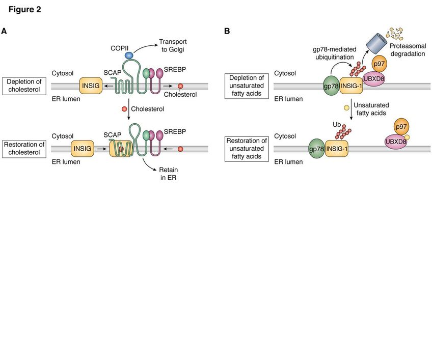
Proteolytic & topological regulation of membrane proteins

(7) and the majority of lipids are synthesized (8). To        be resolved in order to understand these signaling
carry out these functions properly, ER must                   pathways more thoroughly.
communicate with nucleus to ensure that genes
required for these functions are expressed at                 Transcription factors activated through RIP
appropriate levels. One of the mechanism to                            SREBPs
transmit signals from the ER to nucleus is through                     Sterols regulatory element binding proteins
proteolytic activation of membrane-bound                      (SREBPs) are the master regulators that control
transcription factors. These transcription factors are        cholesterol and fatty acid homeostasis in
produced as inactive membrane-bound precursors                mammalian cells (19-21). Whereas SREBP-1a and
in the ER. After stimulation by specific signals              SREBP-1c are the products of one gene generated
generated from the ER, they are transported from              from different promoters, SREBP-2 is encoded by
the ER to Golgi where their luminal domains are               a different gene. Unlike any other transcription
cleaved by Site-1-protease (S1P). This cleavage               factors activated by S1P/S2P that contain a single
enables a second cleavage catalyzed by S2P, which             transmembrane helix, SREBPs are inserted into
liberates their N-terminal transcription activation           membranes with two transmembrane helices in a
domain from membranes, allowing it to enter the               hairpin fashion, with both the N- and C-terminal
nucleus where it activates target genes (9-13) (Fig.          ends facing cytosol (Fig. 1). The N-terminal
1). All these membrane-bound transcription factors            domains of SREBPs are basic helix-loop-helix-

                                                                                                                     Downloaded from http://www.jbc.org/ by guest on June 19, 2020
share the following features: 1) They contain an N-           leucine zipper transcription factors, whereas the C-
terminal domain capable of functioning as a                   terminal domains mediate association with a
transcription factor; 2) They contain a S1P                   membrane protein called SREBP cleavage
recognition motif RXXR/L in the lumen in which                activating protein (SCAP) (22) (Fig. 2A). In cells
the last amino acid of the motif is the cleavage site         depleted of cholesterol, SCAP is incorporated into
for S1P (14,15); and 3) They contain a helical                COPII-coated vesicles, the vehicle that transports
destabilization motif required for S2P-catalyzed              proteins from ER to Golgi (23) (Fig. 2A). The
cleavage such as NP, NXXP or PXXP sequence in                 translocation of the SCAP/SREBPs complex from
the transmembrane helix C-terminal to the cleavage            ER to Golgi allows SREBPs to be cleaved by S1P
site (13,16,17). However, RIP of these transcription          followed by S2P (11,24,25) (Fig. 1). These
factors are stimulated by different signals generated         cleavages liberate the N-terminal domains of
in the ER to activate their unique set of target genes.       SREBPs from membranes, allowing them to enter
         Remarkably, recent studies on RIP of                 nucleus where they activate transcription of all
cAMP response element binding protein 3-like 1                genes required for cholesterol synthesis and uptake
(CREB3L1), one of the membrane-bound                          (26) (Fig. 1). In cells loaded with cholesterol, the
transcription factors activated by S1P/S2P,                   sterol is accumulated in ER membranes. Upon
revealed another mechanism that regulates                     exceeding 5% of total ER lipids (molar basis),
transmembrane proteins. This mechanism, which is              cholesterol in the ER binds to SCAP (27-29), a
designated as regulated alternative translocation             reaction causing association of SCAP with insulin-
(RAT), leads to inverted topology of newly                    induced gene (INSIG) proteins, a family of
synthesized Transmembrane 4 L6 family member                  transmembrane proteins localized in the ER (30,31)
20 (TM4SF20), a polytopic transmembrane protein.              (Fig. 2A). This binding blocks the interaction
This topological inversion turns TM4SF20 from an              between SCAP and components of the COPII coat.
inhibitor to an activator for RIP of CREB3L1 (18).            Consequently, the SCAP/SREBPs complex is
         In this article, I will first provide an             retained in the ER (Fig. 2A), making SREBPs
overview on several membrane-bound transcription              inaccessible to cleavage catalyzed by Golgi-
factors activated by S1P/S2P to illustrate how RIP            localized S1P and S2P (23,30,32). As a result,
transmits signals from the ER to nucleus to regulate          transcription of the genes required for cholesterol
gene expression. I will then focus on CREB3L1 as              synthesis and uptake declines. Thus, the
an example to illustrate how the membrane-bound               cholesterol-mediated inhibition of RIP of SREBPs
transcription factor is activated through RIP and             plays a critical role in feedback inhibition of
RAT. Finally, I will discuss questions that need to           cholesterol synthesis and uptake (33).

                                                          2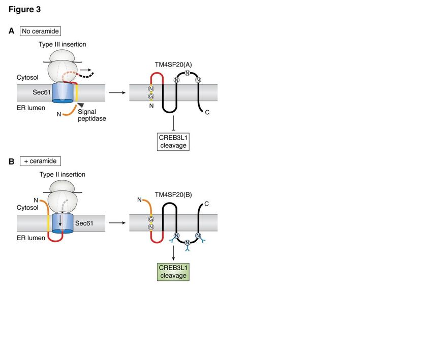
Proteolytic & topological regulation of membrane proteins

         In addition to genes involved in cholesterol       overexpression of a tail-anchored transmembrane
synthesis and uptake, SREBPs, particularly                  protein without any luminal domain or
SREBP-1a and SREBP-1c, also activate genes                  accumulation of dihydroceramide in ER
involved in synthesis of unsaturated fatty acids            membranes. Interestingly, these treatments only
(26). Thus, unsaturated fatty acids also exert              induced proteolytic activation of ATF6α but not the
feedback inhibition on SREBP-1a and SREBP-1c.               other two branches of the ER stress response,
In mice, expression of SREBP-1c, the predominant            namely splicing of X-box binding protein 1 and
isoform of SREBP-1 expressed in liver, is driven by         activation of double-stranded RNA-dependent
liver X receptor (LXR), a ligand-activated nuclear          protein kinase-like ER kinase (45,46). A mutation
receptor (34). Polyunsaturated fatty acids inhibit          in the transmembrane helix of ATF6α impaired
expression of SREBP-1c by acting as an antagonist           dihydroceramide-induced RIP of the protein but not
to LXR (35,36). Unsaturated fatty acids also inhibit        that caused by accumulation of unfolded proteins in
cleavage of SREBP-1a in cultured cells (37). In             the ER lumen. In contrast, a mutation in the luminal
cells depleted of fatty acids, INSIG-1, the                 domain of ATF6α abolished RIP of the protein
predominant isoform of INSIG proteins expressed             induced by unfolded proteins in the ER lumen but
in cultured cells, is ubiquitinated by gp78 (38) and        not that triggered by dihydroceramide (46). These
binds to ubiquitin regulatory X domain-containing           observations suggest that signals generated in the
protein 8 (UBXD8), a membrane protein that                  ER lumen and membrane are independently sensed

                                                                                                                     Downloaded from http://www.jbc.org/ by guest on June 19, 2020
associates with the ER-associated degradation               by the luminal and transmembrane domain of
(ERAD) co-factor p97 (39) (Fig. 2B).                        ATF6α, respectively.
Ubiquitination and p97 recruitment causes rapid                      In contrast to ATF6α, ATF6β is much less
degradation of INSIG-1 by proteasomes through               well characterized. While ER stress also induces
ERAD in these cells (39) (Fig. 2B). Unsaturated             RIP of ATF6β in cultured cells, the cleaved nuclear
fatty acids directly bind to UBXD8, causing                 form of the protein does not activate transcription
dissociation of UBXD8 from INSIG-1 (39,40) (Fig.            of ER chaperons. Instead, it inhibits transcription of
2B). The resultant stabilization of INSIG-1 leads to        these genes by antagonizing the activity of the
increased retention of the SCAP/SREBPs complex              cleaved nuclear form of ATF6α (47). In a mouse
in the ER, making cholesterol more effective in             model of metaphyseal chondrodysplasia type
inhibiting cleavage of SREBP-1a in cells loaded             Schmid caused by expression of a misfolded mutant
with unsaturated fatty acids (39).                          type X collagen in chondrocytes, the severity of the
                                                            disease was increased by knockout of ATF6α but
         ATF6                                               decreased by ablation of ATF6β (48). These results
         Between the two isoforms of ATF6,                  suggest that ATF6β may serve as a transcriptional
ATF6α is much better characterized. In resting              repressor to antagonize the strength and duration of
cells, ATF6α remains as an inactive transmembrane           ATF6α activity that protects cells from ER stress.
precursor in the ER. Upon accumulation of                   However, RIP of ATF6β in mouse cardio myocytes
unfolded proteins in the ER lumen, ATF6α is                 triggered by hemodynamic stress induced
incorporated into COPII-coated vesicles and                 expression of genes overlapping with that induced
translocated from the ER to Golgi (12,41) where it          by ATF6α including ER chaperones (49). Thus,
is cleaved by S1P followed by S2P (13) (Fig. 1).            ATF6β may have different functions in different
These cleavages release the N-terminal domain of            tissues.
ATF6α from membranes, allowing it to enter
nucleus where it activates transcription of BiP and                 CREB3L3
other genes whose products assist folding of ER                     CREB3L3, also known as CREB-H, is
proteins (42,43). Thus, RIP of ATF6α has been               primarily expressed in livers and intestines. Earlier
established as one of the three signaling pathways          studies demonstrated that proinflammatory
responsive to ER stress (44).                               cytokines and lipopolysaccharide (LPS) triggered
         In addition to unfolded proteins                   RIP of CREB3L3 in hepatocytes to induce
accumulated in the ER lumen, RIP of ATF6α can               expression of acute phase response genes such as
also be triggered by signals generated in ER                C-reactive protein and serum amyloid P-component
membranes. RIP of ATF6α was stimulated by                   (50). Recent studies focused on the metabolic

                                                        3
Proteolytic & topological regulation of membrane proteins
consequence of CREB3L3 activation. Expression fragment of CREB3L2 as a substrate for S2P. The
and RIP of CREB3L3 in mice is stimulated by S2P-catalyzed cleavage releases the N-terminal
fasting (51,52). During fasting, fatty acids released domain of CREB3L2 from membranes so that it can
from adipocytes enter livers, causing hepatic enter nucleus to stimulate transcription of genes
accumulation of triglycerides (TGs). These TGs encoding components of the COPII coat such as
then enter ER lumen where they are packaged into various isoforms of Sec23 and Sec24 (61) (Fig. 1).
very low density lipoprotein (VLDL) particles. The Expression of these genes enlarges COPII-coated
nascent VLDL particles produced in the ER lumen vesicles to accommodate the bulky type II collagen,
are transported to Golgi and secreted out of the secretion of which is crucial for differentiation
hepatocytes through exocytosis as a vehicle that of chondrocytes (61,62); 2) The S1P-catalyzed
delivers lipids to peripheral tissues (53). It appears cleavage releases the C-terminal domain of the
that VLDL assembly in the ER lumen is the signal protein from membranes, allowing it to be secreted
to trigger RIP of CREB3L3 (Fig. 1), as treatments out of the cells to stimulate proliferation of
inhibiting this reaction blocked fasting-induced neighboring chondrocytes by activating the
proteolytic activation of the protein (54). Since hedgehog signaling pathway (63) (Fig. 1). Thus, the
nascent VLDL particles are transported from the N- and C-terminal fragments of CREB3L2
ER to Golgi by specialized vesicles (55), it will be generated through RIP simultaneously stimulate
interesting to determine whether CREB3L3 is co- chondrocyte differentiation and proliferation in
Downloaded from http://www.jbc.org/ by guest on June 19, 2020
delivered with VLDL by these vesicles to Golgi for developing cartilage, respectively. As a result, mice
proteolytic activation by S1P/S2P. deficient in CREB3L2 developed severe
Once cleaved by S1P and S2P, the N- chondrodysplasia (61).
terminal domain of CREB3L3 activates genes CREB3L2 is frequently fused with Fused
known to be activated during fasting (Fig. 1). These in Sarcoma (FUS) in low-grade fibromyxoid
genes include fibroblast growth factor 21, a fasting- sarcoma (LGFMS), a malignant soft tissue tumor
induced hormone that stimulates hepatic fatty acid through chromosome rearrangement (64). In these
oxidation and ketogenesis (56); Insig-2, the Insig tumors, the N-terminal domain of FUS is fused in
isoform expressed in fasting livers (51); and genes frame with CREB3L2 at a position N-terminal to its
required for gluconeogenesis (57). More DNA binding domain (65). Since overexpression of
importantly, VLDL assembly-triggered RIP of transcription factors activated by RIP leads to
CREB3L3 coordinates secretion of VLDL with unregulated proteolytic activation presumably by
hydrolysis of the lipoprotein particles in circulation. overpowering the regulatory machinery that retains
In order to use fatty acids stored in VLDL by them in the ER (13,66), the FUS-CREB3L2 fusion
peripheral tissues, TGs stored in VLDL must be protein driven by the strong FUS promoter is likely
hydrolyzed by lipoprotein lipase (LPL) so that fatty to be constitutively cleaved. The cleaved N-
acids can be released from the lipoprotein particle terminal domain of CREB3L2 fused with FUS does
(58). Following activation through RIP, the N- not activate target genes of CREB3L2. Instead, the
terminal domain of CREB3L3 stimulates fusion protein activates CD24, a marker for
expression of apolipoproteins such as apoA2, LGFMS, the function of which in tumor
apoA4 and apoA5 that activate LPL activity development remains obscure (67). Inasmuch as
(59,60). Consequently, CREB3L3 deficiency in expression of genes downstream of hedgehog
both mice and humans leads to development of signaling is elevated in LGFMS (67), the increased
hypertriglyceridemia caused by insufficient LPL secretion of the C-terminal fragment of CREB3L2
activity (59). (an activator for hedgehog signaling) produced by
unregulated cleavage of the fusion protein may also
CREB3L2 contribute to development of the tumor.
CREB3L2, also known as BBF2H7, is
best-characterized in chondrocytes. During RIP of CREB3L1 activated by RAT of
chondrocyte differentiation, CREB3L2 is cleaved TM4SF20
by S1P. This cleavage has two consequences: 1) The physiological function of CREB3L1,
Like other transcription factors activated through also known as OASIS, was first determined from
RIP, it produces the membrane-bound N-terminal the study of mice deficient in the gene. Creb3l1-/-
4Proteolytic & topological regulation of membrane proteins
mice exhibited severe osteopenia owing to The two orientations of TM4SF20 are
insufficiency of type 1 collagen in bone matrix (68). illustrated in Figs. 3A and B. In the absence of
RIP of CREB3L1 in osteoblasts was shown to be ceramide, the N-terminus of TM4SF20 is located in
stimulated by bone morphogenetic protein 2 (BMP- the ER lumen, and the sequence N-terminal to the
2), a member of the transforming growth factor β first transmembrane helix is cleaved off co-
(TGF-β) family of cytokines that is critical for bone translationally by the signal peptidase. Under this
development (68). The cleaved nuclear form of configuration, the loop that contains three potential
CREB3L1 in turn activates transcription of genes sites for N-linked glycosylation is located in the
involved in assembly of the collagen matrix cytosol where glycosylation cannot occur. This
including collagen 1α1 (Col1a1) that underlies form of the protein is designated as TM4SF20(A)
bone formation (17,68,69) (Fig. 1). The crucial role (18). When cells are treated with ceramide, the N-
of CREB3L1 for bone development in humans is terminus of newly synthesized TM4SF20 is located
supported by the observation that people with in the cytosol and the Asn-containing loop is
homozygous mutations inactivating CREB3L1 located in the ER lumen where it is glycosylated.
exhibit osteogenesis imperfecta, a disease caused This form of the protein is designated as
by insufficient deposition of collagen in bone TM4SF20(B) (18). It should be emphasized that
matrix (70-73). ceramide leads to inverted topology of newly
TGF-β, a cytokine homologous to BMP-2, synthesized TM4SF20 but does not flip the
Downloaded from http://www.jbc.org/ by guest on June 19, 2020
induces collagen synthesis during tissue repair and topology of pre-existing TM4SF20. Since ceramide
fibrosis (74). Like BMP2, TGF-β triggers RIP of alters the direction through which TM4SF20 is
CREB3L1 in cultured cells (75). TGF-β induces translocated across membranes during its synthesis,
RIP of CREB3L1 by inhibiting expression of this regulatory process is designated as Regulated
TM4SF20, an inhibitor for proteolytic activation of Alternative Translocation (RAT) (18).
CREB3L1 (75). In eukaryotic cells, the topology of
RIP of CREB3L1 is also essential for polytopic membrane proteins is primarily
doxorubicin, a drug extensively used for cancer determined by the direction through which the first
chemotherapy (76), to inhibit proliferation of transmembrane helix is translocated across ER
cancer cells. Doxorubicin stimulates production of membranes (81). The translocation process has
ceramide, which in turn triggers RIP of CREB3L1, been categorized into three classes: Type I insertion
allowing the cleaved nuclear form of the protein to refers to proteins that contain a cleavable ER-
activate p21 and other genes that inhibit cell targeting signal peptide N-terminal to the first
proliferation (17,77) (Fig. 1). The importance of transmembrane helix. The nascent signal peptide
this CREB3L1-mediated signaling pathway was binds to the signal recognition particle that direct
demonstrated by the finding that at clinically the ribosome/nascent polypeptide complex to the
relevant doses, doxorubicin was much more ER membranes, enabling sequence C-terminal to
effective in cancer cells that expressed high levels the signal peptide to be transported into the ER
of CREB3L1 than in those expressing low levels of lumen through the Sec61 ER translocon. Following
the gene. This correlation was observed in cancer the translocation, the signal peptide is cleaved from
cells cultured in vitro, in xenograft tumors the mature protein by the signal peptidase (7,82).
established in mice, and in human tumor samples The other two insertions do not use a signal peptide.
(77-80). These results suggest that CREB3L1 may Instead, their insertions are initiated by recognition
serve as a biomarker to predict sensitivity to of the hydrophobic sequence present in the first
doxorubicin. transmembrane helix of nascent peptides by the
RIP of CREB3L1 is critical for ceramide to signal recognition particle, which directs the
inhibit cell proliferation (77). In contrast to TGF-β, nascent peptide/ribosome complex to the Sec61 ER
ceramide does not affect expression of TM4SF20. translocon. The crystal structure of the
Instead, ceramide leads to inverted topology of translocation complex suggests that insertion of the
newly synthesized TM4SF20 (18). This topological hydrophobic transmembrane helix into ER
inversion converts TM4SF20 from an inhibitor to membranes adjacent to the Sec61 translocon is the
an activator for RIP of CREB3L1 (18). leading event that initiates the translocation process
(81) (Fig. 3). In Type II insertion, the N-terminus of
5Proteolytic & topological regulation of membrane proteins
transmembrane proteins is in cytosol (82). In type unfolded and pulled through the translocation
III insertion, the orientation of the transmembrane channel by forces other than peptide elongation
helix is reverted so that the N-terminus of the (81). Other studies suggest that this process could
proteins is in the ER lumen (82). be Sec61-independent (85-87). One clue is that this
TM4SF20(A) cannot be inserted into process may require translocating chain-associated
membranes via the Type I insertion because the N- membrane protein 2 (TRAM2), which is highly
terminal peptide does not serve as a signal peptide. homologous to TRAM1, an accessory protein that
This conclusion is supported by the following lines functions together with the Sec61 translocon
of evidence: First, the N-terminal sequence of (81,88-90). RNAi-mediated knockdown of
TM4SF20 does not contain any of the hallmarks TRAM2 but not TRAM1 enabled production of
characteristic for a signal peptide (83); Second, the TM4SF20(B) even in the absence of ceramide (18).
N-terminal sequence of TM4SF20 did not function This observation suggests that TRAM2 may play a
as a signal peptide when it was substituted for the specific role in Type III insertion of the first
endogenous signal sequence of alkaline transmembrane helix of TM4SF20(A). It is
phosphatase (18); Third, the N-terminal sequence interesting that TRAM2, and all TRAM protein,
of TM4SF20 could be replaced by other peptides contains a TLC domain that is postulated to bind
without altering the topology of TM4SF20(A) and ceramide or related sphingolipids (91). These
ceramide-induced RAT of TM4SF20 (18). Even observations raise the possibility that TRAM2 is the
Downloaded from http://www.jbc.org/ by guest on June 19, 2020
though it is not a signal sequence, the N-terminal sensor that allows ceramide to block synthesis of
peptide of TM4SF20 is cleaved by signal peptidase TM4SF20(A).
(18), apparently because the peptide is accessible to Since both the Type II and III insertions are
the protease in the ER lumen. This type of cleavage initiated by contact of the first transmembrane helix
catalyzed by the signal peptidase has been reported with the ER translocon, the first transmembrane
in proteolytic processing of hepatitis C virus protein helix of TM4SF20 should be critical for RAT of the
in which the protease cleaves the viral polyprotein protein. Indeed, replacing the signal peptide of
precursor at multiple sites in the ER lumen distal to alkaline phosphatase with the N-terminal sequence
the N-terminal sequence (84). of TM4SF20 that contains the first transmembrane
Since the N-terminus of TM4SF20(A) is in helix led to ceramide-induced topological inversion
the ER lumen yet the protein is not inserted through of the fusion protein (18). Mutagenesis analysis
the type I insertion, the first transmembrane helix revealed that RAT of TM4SF20 required a Gly and
of TM4SF20(A) is translocated through an Asn separated by 3 residues (designated as the
membranes via the Type III insertion (Fig. 3A). In GXXXN motif) present in the first transmembrane
the presence of ceramide, the first transmembrane helix. When the Gly or Asn within the motif was
helix of TM4SF20(B) is inserted into membranes mutated to Leu, TM4SF20 failed to adopt the A
via the Type II insertion (Fig. 3B). Thus, RAT configuration, and it was constitutively in the B
changes the translocation of the first topology, even in the absence of ceramide (18,92).
transmembrane helix of TM4SF20 from the Type Similar to TM4SF20(A), the first
III to Type II insertion. While both of these transmembrane helix of the majority of GPCRs is
insertions have been observed in other membrane inserted into membranes through the Type III
proteins, TM4SF20 is the first recognized example orientation (93,94). The first transmembrane helix
in which the two insertion types are interconvertible of several of these receptors contains the GXXXN
in a regulated manner. motif (95). One of these GPCRs is C-C chemokine
Unlike the well-characterized Type II receptor 5 (CCR5). In unstimulated macrophages,
insertion through which the hydrophilic sequence this receptor adopts a topology consistent with that
C-terminal to the transmembrane helix is pushed of GPCRs so it can function as a chemokine
through the translocon by the ribosome, the receptor (96). When the macrophages were
mechanism by which the hydrophilic sequence N- stimulated with LPS, the increased production of
terminal to the transmembrane helix reaches ER dihydroceramide triggered RAT of CCR5, and the
lumen during the Type III insertion remains protein with the inverted topology no longer
obscure. If this process is mediated by Sec61, then functioned as a chemokine receptor (95). This
the N-terminal hydrophilic sequence must be finding may explain the well-known observation
6Proteolytic & topological regulation of membrane proteins
that LPS-activated macrophages are insensitive to Patient expressing this mutant of S2P developed
chemotaxis (97). The findings also suggest that osteogenesis imperfecta, the same phenotype
RAT may be a widespread mechanism to regulate exhibited by individuals deficient in CREB3L1
transmembrane proteins. (98). In contrast, subjects with mutations that
inactivate S2P completely develop IFAP
Perspective (ichthyosis follicularis atrichia photophobia), a
RIP of membrane-bound transcription syndrome that has other abnormalities in addition
factors is ultimately achieved by regulated transport to osteogenesis imperfecta, presumably caused by
of these proteins from the ER to Golgi upon deficiency in activating membrane-bound
stimulation by signals generated from the ER. transcription factors besides CREB3L1 (99).
Except for SREBPs, the exact signaling mechanism Understanding why S2P is less efficient in cleaving
that triggers RIP of the other membrane-bound CREB3L1 may provide strategies to treat these
transcription factors are not well defined. RIP of genetic diseases.
most if not all of the membrane-bound transcription Accumulation of ceramide and/or
factors can be stimulated by treatments of dihydroceramide in ER membranes triggers RIP of
pharmacological compounds such as tunicamyin or ATF6α and CREB3L1 (46,77). Whereas the
thapsigargin that induce ER stress. Thus, it is mechanism for dihydroceramide-induced RIP of
tempting to conclude that all of these transcription ATF6α remains obscure, ceramide activates RIP of
Downloaded from http://www.jbc.org/ by guest on June 19, 2020
factors are ER stress transducers. However, the CREB3L1 by inverting the topology of TM4SF20
term ER stress is too broad to categorize the signals through altering the direction by which TM4SF20
that activate RIP of the membrane-bound is translocated into ER membranes (18). While
transcription factors, as distinct signals that activate regulation of protein translocation was proposed
RIP of different membrane-bound transcription more than a decade ago (100), ceramide-induced
factors, e.g., alteration in lipid composition of ER RAT of TM4SF20 discovered recently is the first
membranes that activates SREBPs, accumulation example that this regulation indeed takes place in
of unfolded proteins in the ER lumen that activates mammalian cells. This discovery raises more
ATF6, and increased synthesis of extracellular questions on regulation of protein translocation: If
matrix proteins that activates RIP of CREB3L1 and the Type III insertion responsible for production of
CREB3L2, may all be considered as ER stress. TM4SF20(A) is Sec61-dependent, what is the
Thus, identification of the exact ER-generated driving force and unfolding mechanism that allows
signals that activate RIP of the individual the N-terminal sequence to be pulled through the
membrane-bound transcription factor will be the translocon? If not, what is the translocation
primary challenge in the future to understand the machinery responsible for the Type III insertion?
signal transduction pathways mediated by these Are TRAM proteins the ceramide sensors that
proteins. control RAT? Perhaps the most important question
Another important question regarding RIP is how many transmembrane proteins are subjected
is whether S2P is equally active in cleaving to topological regulation. The reports published so
different membrane-bound transcription factors. far suggest that transmembrane proteins regulated
Previous reports showed that the membrane-bound through RAT contain a GXXXN motif in the first
intermediate form, which represents a fragment of transmembrane helix (18,95). However, TM4SF4,
these transcription factors that has been cleaved by another transmembrane protein that contains the
S1P but not S2P (Fig. 1), was present for CREB3L1 same motif in the first transmembrane helix, does
but not for SREBPs or ATF6α in cells expressing not undergo RAT (92). These observations suggest
wild type S2P (13,25,77,98). This observation that the GXXXN motif present in the first
suggests that CREB3L1 may be a less efficient transmembrane helix may be required but not
substrate for S2P than other membrane-bound sufficient to induce RAT. It appears that a
transcription factors. Consistent with this proteome-wide approach capable of measuring
hypothesis, S2P(L505F), a point mutation that topology of transmembrane proteins globally is
partially inactivates S2P, diminished cleavage of needed to systematically identify transmembrane
CREB3L1 but not ATF6 in cultured cells (95). proteins subjected to topological regulation.
7Proteolytic & topological regulation of membrane proteins
Acknowledgements: I would like to thank Drs. Michael Brown and Joseph Goldstein for their constant
support and helpful discussion, and Nancy Heard for graphic illustration.
Conflict of interest: The authors declare that they have no conflicts of interest with the contents of this
article.
Downloaded from http://www.jbc.org/ by guest on June 19, 2020
8Proteolytic & topological regulation of membrane proteins
Reference
1. Brown, M., Ye, J., Rawson, R., and Goldstein, J. (2000) Regulated intramembrane proteolysis: a
control mechanism conserved from bacteria to humans. Cell 100, 391-398
2. Ye, J. (2013) Regulated intramembrane proteolysis. in Encyclopedia of Biological Chemistry
(Lennarz, W., and Lane, D. eds.), 2 Ed., Academic Press, Waltham. pp 50-55
3. Wolfe, M. S. (2020) Substrate recognition and processing by γ-secretase. Biochem Biophys Acta
1862, 183016
4. Spinazzi, M., and De Strooper, B. (2016) PARL: The mitochondrial rhomboid protease. Sem.Cell
Dev. Biol. 60, 19-28
5. Rawson, R. B. (2013) The site-2 protease. Biochem Biophys Acta 1828, 2801-2807
6. Mentrup, T., Loock, A.-C., Fluhrer, R., and Schröder, B. (2017) Signal peptide peptidase and SPP-
like proteases - Possible therapeutic targets? Biochim Biophys Acta 1864, 2169-2182
7. Zimmermann, R., Eyrisch, S., Ahmad, M., and Helms, V. (2011) Protein translocation across the
ER membrane. Biochim. Biophys. Acta 1808, 912-924
8. Balla, T., Sengupta, N., and Kim, Y. J. (2020) Lipid synthesis and transport are coupled to regulate
membrane lipid dynamics in the endoplasmic reticulum. Biochem Biophys Acta Mol Cell Biol
Downloaded from http://www.jbc.org/ by guest on June 19, 2020
Lipids 1865
9. Sakai, J., Rawson, R. B., Espenshade, P. J., Cheng, D., Seegmiller, A. C., Goldstein, J. L., and
Brown, M. S. (1998) Molecular identification of the sterol-regulated luminal protease that cleaves
SREBPs and controls lipid composition of animal cells. Mol. Cell 2, 505-514
10. Rawson, R. B., Zelenski, N. G., Nijhawan, D., Ye, J., Sakai, J., Hasan, M. T., Chang, T. Y., Brown,
M. S., and Goldstein, J. L. (1997) Complementation cloning of S2P, a gene encoding a putative
metalloprotease required for intramembrane cleavage of SREBPs. Mol Cell 1, 47-57
11. Nohturfft, A., Yabe, D., Goldstein, J. L., Brown, M. S., and Espenshade, P. J. (2000) Regulated
step in cholesterol feedback localized to budding of SCAP from ER membranes. Cell 102, 315-323
12. Shen, J., Chen, X., Hendershot, L., and Prywes, R. (2002) ER stress regulation of ATF6 localization
by dissociation of BiP/GRP78 binding and unmasking of Golgi localization signals. Dev Cell 3,
99-111
13. Ye, J., Rawson, R. B., Komuro, R., Chen, X., Davé, U. P., Prywes, R., Brown, M. S., and Goldstein,
J. L. (2000) ER stress induces cleavage of membrane-bound ATF6 by the same proteases that
process SREBPs. Mol. Cell 6, 1355-1364
14. Espenshade, P. J., Cheng, D., Goldstein, J. L., and Brown, M. S. (1999) Autocatalytic processing
of Site-1 protease removes propeptide and permits cleavage of sterol regulatory element-binding
proteins. J Biol Chem 274, 22795-22804
15. Duncan, E. A., Brown, M. S., Goldstein, J. L., and Sakai, J. (1997) Cleavage site for sterol-regulated
protease localized to a Leu-Ser bond in the lumenal loop of sterol regulatory element-binding
protein-2. J Biol Chem 272, 12778-12785
16. Ye, J., Davé, U. P., Grishin, N. V., Goldstein, J. L., and Brown, M. S. (2000) Asparagine-proline
sequence within membrane-spanning segment of SREBP triggers intramembrane cleavage by Site-
2 protease. Proc Natl Acad Sci USA 97, 5123
17. Denard, B., Seemann, J., Chen, Q., Gay, A., Huang, H., Chen, Y., and Ye, J. (2011) The membrane-
bound transcription factor CREB3L1 is activated in response to virus infection to inhibit
proliferation of virus-infected cells. Cell Host & Microbe 10, 65-74
18. Chen, Q., Denard, B., Lee, C.-E., Han, S., Ye, James S., and Ye, J. (2016) Inverting the topology
of a transmembrane protein by regulating the translocation of the first transmembrane helix. Mol
Cell 63, 567-578
19. Goldstein, Joseph L., and Brown, Michael S. (2015) A century of cholesterol and coronaries: From
plaques to genes to statins. Cell 161, 161-172
9Proteolytic & topological regulation of membrane proteins
20. Ye, J., and DeBose-Boyd, R. A. (2011) Regulation of cholesterol and fatty acid synthesis. Cold
Spring Harb. Perspect. Biol. 3, 10.1101/cshperspect.a004754
21. DeBose-Boyd, R. A., and Ye, J. (2018) SREBPs in lipid metabolism, insulin signaling, and beyond.
Trends Biochem Sci. 43, 358-368
22. Sakai, J., Nohturfft, A., Cheng, D., Ho, Y. K., Brown, M. S., and Goldstein, J. L. (1997)
Identification of complexes between the COOH-terminal domains of sterol regulatory element-
binding proteins (SREBPs) and SREBP cleavage-activating protein. J. Biol. Chem. 272, 20213-
20221
23. Sun, L.-P., Seemann, J., Goldstein, J. L., and Brown, M. S. (2007) Sterol-regulated transport of
SREBPs from endoplasmic reticulum to Golgi: Insig renders sorting signal in Scap inaccessible to
COPII proteins. Proc Natl Acad Sci USA 104, 6519
24. DeBose-Boyd, R. A., Brown, M. S., Li, W.-P., Nohturfft, A., Goldstein, J. L., and Espenshade, P.
J. (1999) Transport-dependent proteolysis of SREBP: Relocation of Site-1 protease from Golgi to
ER obviates the need for SREBP transport to Golgi. Cell 99, 703-712
25. Sakai, J., Duncan, E. A., Rawson, R. B., Hua, X., Brown, M. S., and Goldstein, J. L. (1996) Sterol-
regulated release of SREBP-2 from cell membranes requires two sequential cleavages, one within
a transmembrane segment. Cell 85, 1037-1046
26. Horton, J. D., Shah, N. A., Warrington, J. A., Anderson, N. N., Park, S. W., Brown, M. S., and
Downloaded from http://www.jbc.org/ by guest on June 19, 2020
Goldstein, J. L. (2003) Combined analysis of oligonucleotide microarray data from transgenic and
knockout mice identifies direct SREBP target genes. Proc. Natl. Acad. Sci. USA 100, 12027-12032
27. Radhakrishnan, A., Sun, L.-P., Kwon, H. J., Brown, M. S., and Goldstein, J. L. (2004) Direct
binding of cholesterol to the purified membrane region of SCAP: Mechanism for a sterol-sensing
domain. Mol Cell 15, 259-268
28. Motamed, M., Zhang, Y., Wang, M. L., Seemann, J., Kwon, H. J., Goldstein, J. L., and Brown, M.
S. (2011) Identification of luminal loop 1 of Scap protein as the sterol sensor that maintains
cholesterol homeostasis. J. Biol. Chem. 286, 18002-18012
29. Radhakrishnan, A., Goldstein, J. L., McDonald, J. G., and Brown, M. S. (2008) Switch-like control
of SREBP-2 transport triggered by small changes in ER cholesterol: A delicate balance. Cell Metab
8, 512-521
30. Yang, T., Espenshade, P. J., Wright, M. E., Yabe, D., Gong, Y., Aebersold, R., Goldstein, J. L.,
and Brown, M. S. (2002) Crucial step in cholesterol homeostasis: sterols promote binding of SCAP
to INSIG-1, a membrane protein that facilitates retention of SREBPs in ER. Cell 110, 489-500
31. Yabe, D., Brown, M. S., and Goldstein, J. L. (2002) Insig-2, a second endoplasmic reticulum
protein that binds SCAP and blocks export of sterol regulatory element-binding proteins. Proc.
Natl. Acad. Sci. USA 99, 12753-12758
32. Sun, L.-P., Li, L., Goldstein, J. L., and Brown, M. S. (2005) Insig required for sterol-mediated
inhibition of Scap/SREBP binding to COPII proteins in vitro. J Biol Chem 280, 26483-26490
33. Brown, M. S., and Goldstein, J. L. (2009) Cholesterol feedback: from Schoenheimer's bottle to
Scap's MELADL. J. Lipid Res. 50, S15-S27
34. Liang, G., Yang, J., Horton, J. D., Hammer, R. E., Goldstein, J. L., and Brown, M. S. (2002)
Diminished hepatic response to fasting/refeeding and liver X receptor agonists in mice with
selective deficiency of sterol regulatory element-binding protein-1c. J. Biol. Chem. 277, 9520-9528
35. Ou, J., Tu, H., Shan, B., Luk, A., DeBose-Boyd, R. A., Bashmakov, Y., Goldstein, J. L., and Brown,
M. S. (2001) Unsaturated fatty acids inhibit transcription of the sterol regulatory element-binding
protein-1c (SREBP-1c) gene by antagonizing ligand-dependent activation of the LXR. Proc Natl
Acad Sci USA 98, 6027-6032
36. Kim, C.-W., Addy, C., Kusunoki, J., Anderson, N. N., Deja, S., Fu, X., Burgess, S. C., Li, C.,
Ruddy, M., Chakravarthy, M., Previs, S., Milstein, S., Fitzgerald, K., Kelley, D. E., and Horton, J.
D. (2017) Acetyl CoA carboxylase inhibition reduces hepatic steatosis but elevates plasma
triglycerides in mice and humans: A dedside to bench investigation. Cell Metab 26, 394-406.e396
10Proteolytic & topological regulation of membrane proteins
37. Hannah, V. C., Ou, J., Luong, A., Goldstein, J. L., and Brown, M. S. (2001) Unsaturated fatty acids
down-regulate SREBP isoforms 1a and 1c by two mechanisms in HEK-293 Cells. J. Biol. Chem.
276, 4365-4372
38. Lee, J. N., Song, B., DeBose-Boyd, R. A., and Ye, J. (2006) Sterol-regulated degradation of Insig-
1 mediated by the membrane-bound ubiquitin ligase gp78. J. Biol. Chem. 281, 39308-39315
39. Lee, J. N., Zhang, X., Feramisco, J. D., Gong, Y., and Ye, J. (2008) Unsaturated fatty acids inhibit
proteasomal degradation of Insig-1 at a postubiquitination step. J. Biol. Chem. 283, 33772-33783
40. Lee, J. N., Kim, H., Yao, H., Chen, Y., Weng, K., and Ye, J. (2010) Identification of Ubxd8 protein
as a sensor for unsaturated fatty acids and regulator of triglyceride synthesis. Proc. Natl. Acad. Sci.
USA 107, 21424-21429
41. Schindler, A. J., and Schekman, R. (2009) In vitro reconstitution of ER-stress induced ATF6
transport in COPII vesicles. Proc Natl Acad Sci USA 106, 17775
42. Haze, K., Yoshida, H., Yanagi, H., Yura, T., and Mori, K. (1999) Mammalian transcription factor
ATF6 Is synthesized as a transmembrane protein and activated by proteolysis in response to
endoplasmic reticulum stress. Mol Biol Cell 10, 3787-3799
43. Yoshida, H., Matsui, T., Yamamoto, A., Okada, T., and Mori, K. (2001) XBP1 mRNA Is induced
by ATF6 and spliced by IRE1 in response to ER stress to produce a highly active transcription
factor. Cell 107, 881-891
Downloaded from http://www.jbc.org/ by guest on June 19, 2020
44. Walter, P., and Ron, D. (2011) The unfolded protein response: from stress pathway to homeostatic
regulation. Science 334, 1081-1086
45. Maiuolo, J., Bulotta, S., Verderio, C., Benfante, R., and Borgese, N. (2011) Selective activation of
the transcription factor ATF6 mediates endoplasmic reticulum proliferation triggered by a
membrane protein. Proc Natl Acad Sci USA 108, 7832
46. Tam, A. B., Roberts, L. S., Chandra, V., Rivera, I. G., Nomura, D. K., Forbes, D. J., and Niwa, M.
(2018) The UPR activator ATF6 responds to proteotoxic and lipotoxic stress by distinct
mechanisms. Dev Cell 46, 327-343.e327
47. Thuerauf, D. J., Morrison, L., and Glembotski, C. C. (2004) Opposing roles for ATF6α and ATF6β
in endoplasmic reticulum stress response gene induction. J Biol Chem 279, 21078-21084
48. Forouhan, M., Mori, K., and Boot-Handford, R. P. (2018) Paradoxical roles of ATF6α and ATF6β
in modulating disease severity caused by mutations in collagen X. Matrix Biol 70, 50-71
49. Correll, R. N., Grimes, K. M., Prasad, V., Lynch, J. M., Khalil, H., and Molkentin, J. D. (2019)
Overlapping and differential functions of ATF6α versus ATF6β in the mouse heart. Sci Rep 9, 2059
50. Zhang, K., Shen, X., Wu, J., Sakaki, K., Saunders, T., Rutkowski, D. T., Back, S. H., and Kaufman,
R. J. (2006) Endoplasmic reticulum stress activates cleavage of CREBH to induce a systemic
inflammatory response. Cell 124, 587-599
51. Wang, H., Zhao, M., Sud, N., Christian, P., Shen, J., Song, Y., Pashaj, A., Zhang, K., Carr, T., and
Su, Q. (2016) Glucagon regulates hepatic lipid metabolism via cAMP and Insig-2 signaling:
implication for the pathogenesis of hypertriglyceridemia and hepatic steatosis. Sci Rep 6, 32246
52. Nakagawa, Y., Satoh, A., Tezuka, H., Han, S.-i., Takei, K., Iwasaki, H., Yatoh, S., Yahagi, N.,
Suzuki, H., Iwasaki, Y., Sone, H., Matsuzaka, T., Yamada, N., and Shimano, H. (2016) CREB3L3
controls fatty acid oxidation and ketogenesis in synergy with PPARα. Sci Rep 6, 39182
53. Gibbons, G. F., Wiggins, D., Brown, A. M., and Hebbachi, A. M. (2004) Synthesis and function of
hepatic very-low-density lipoprotein. Biochem Soc Trans 32, 59-64
54. Cheng, D., Xu, X., Simon, T., Boudyguina, E., Deng, Z., VerHague, M., Lee, A.-H., Shelness, G.
S., Weinberg, R. B., and Parks, J. S. (2016) Very low density lipoprotein assembly is required for
cAMP-responsive element-binding protein H processing and hepatic apolipoprotein A-IV
expression. J Biol Chem 291, 23793-23803
55. Siddiqi, Shadab A. (2008) VLDL exits from the endoplasmic reticulum in a specialized vesicle, the
VLDL transport vesicle, in rat primary hepatocytes. Biochem J 413, 333-342
11Proteolytic & topological regulation of membrane proteins
56. Kim, H., Mendez, R., Zheng, Z., Chang, L., Cai, J., Zhang, R., and Zhang, K. (2014) Liver-enriched
transcription factor CREBH interacts with peroxisome proliferator-activated receptor α to regulate
metabolic hormone FGF21. Endocrinology 155, 769-782
57. Lee, M.-W., Chanda, D., Yang, J., Oh, H., Kim, S. S., Yoon, Y.-S., Hong, S., Park, K.-G., Lee, I.-
K., Choi, C. S., Hanson, R. W., Choi, H.-S., and Koo, S.-H. (2010) Regulation of hepatic
gluconeogenesis by an ER-bound transcription factor, CREBH. Cell Metab 11, 331-339
58. Olivecrona, G. (2016) Role of lipoprotein lipase in lipid metabolism. Curr Opin Lipidol 27, 233-
241
59. Lee, J. H., Giannikopoulos, P., Duncan, S. A., Wang, J., Johansen, C. T., Brown, J. D., Plutzky, J.,
Hegele, R. A., Glimcher, L. H., and Lee, A.-H. (2011) The transcription factor cyclic AMP–
responsive element–binding protein H regulates triglyceride metabolism. Nat Med 17, 812-815
60. Xu, X., Park, J.-G., So, J.-S., Hur, K. Y., and Lee, A.-H. (2014) Transcriptional regulation of
apolipoprotein A-IV by the transcription factor CREBH. J Lipid Res 55, 850-859
61. Saito, A., Hino, S.-i., Murakami, T., Kanemoto, S., Kondo, S., Saitoh, M., Nishimura, R., Yoneda,
T., Furuichi, T., Ikegawa, S., Ikawa, M., Okabe, M., and Imaizumi, K. (2009) Regulation of
endoplasmic reticulum stress response by a BBF2H7-mediated Sec23a pathway is essential for
chondrogenesis. Nat Cell Biol 11, 1197-1204
62. Ishikawa, T., Toyama, T., Nakamura, Y., Tamada, K., Shimizu, H., Ninagawa, S., Okada, T.,
Downloaded from http://www.jbc.org/ by guest on June 19, 2020
Kamei, Y., Ishikawa-Fujiwara, T., Todo, T., Aoyama, E., Takigawa, M., Harada, A., and Mori, K.
(2017) UPR transducer BBF2H7 allows export of type II collagen in a cargo- and developmental
stage–specific manner. J Cell Biol 216, 1761-1774
63. Saito, A., Kanemoto, S., Zhang, Y., Asada, R., Hino, K., and Imaizumi, K. (2014) Chondrocyte
proliferation regulated by secreted luminal domain of ER stress transducer BBF2H7/CREB3L2.
Mol Cell 53, 127-139
64. Arbajian, E., Puls, F., Antonescu, C. R., Amary, F., Sciot, R., Debiec-Rychter, M., Sumathi, V. P.,
Järås, M., Magnusson, L., Nilsson, J., Hofvander, J., and Mertens, F. (2017) In-depth genetic
analysis of sclerosing epithelioid fibrosarcoma reveals recurrent genomic alterations and potential
treatment targets. Clin Cancer Res 23, 7426
65. Mohamed, M., Fisher, C., and Thway, K. (2017) Low-grade fibromyxoid sarcoma: Clinical,
morphologic and genetic features. Ann Diagn Pathol 28, 60-67
66. Hua, X., Sakai, J., Brown, M. S., and Goldstein, J. L. (1996) Regulated cleavage of sterol regulatory
element binding proteins requires sequences on both sides of the endoplasmic reticulum membrane.
J Biol Chem 271, 10379-10384
67. Möller, E., Hornick, J. L., Magnusson, L., Veerla, S., Domanski, H. A., and Mertens, F. (2011)
FUS-CREB3L2–positive sarcomas show a specific gene expression profile with upregulation of
CD24 and FOXL1. Clin Cancer Res 17, 2646
68. Murakami, T., Saito, A., Hino, S.-i., Kondo, S., Kanemoto, S., Chihara, K., Sekiya, H., Tsumagari,
K., Ochiai, K., Yoshinaga, K., Saitoh, M., Nishimura, R., Yoneda, T., Kou, I., Furuichi, T.,
Ikegawa, S., Ikawa, M., Okabe, M., Wanaka, A., and Imaizumi, K. (2009) Signalling mediated by
the endoplasmic reticulum stress transducer OASIS is involved in bone formation. Nat Cell Biol
11, 1205-1211
69. Vellanki, R. N., Zhang, L., Guney, M. A., Rocheleau, J. V., Gannon, M., and Volchuk, A. (2010)
OASIS/CREB3L1 induces expression of genes involved in extracellular matrix production but not
classical endoplasmic reticulum stress response genes in pancreatic β-cells. Endocrinology 151,
4146-4157
70. Lindahl, K., Åström, E., Dragomir, A., Symoens, S., Coucke, P., Larsson, S., Paschalis, E.,
Roschger, P., Gamsjaeger, S., Klaushofer, K., Fratzl-Zelman, N., and Kindmark, A. (2018)
Homozygosity for CREB3L1 premature stop codon in first case of recessive osteogenesis
imperfecta associated with OASIS-deficiency to survive infancy. Bone 114, 268-277
71. Guillemyn, B., Kayserili, H., Demuynck, L., Sips, P., De Paepe, A., Syx, D., Coucke, P. J., Malfait,
F., and Symoens, S. (2019) A homozygous pathogenic missense variant broadens the phenotypic
12Proteolytic & topological regulation of membrane proteins
and mutational spectrum of CREB3L1-related osteogenesis imperfecta. Hum Mol Genet 28, 1801-
1809
72. Cayami, F. K., Maugeri, A., Treurniet, S., Setijowati, E. D., Teunissen, B. P., Eekhoff, E. M. W.,
Pals, G., Faradz, S. M., and Micha, D. (2019) The first family with adult osteogenesis imperfecta
caused by a novel homozygous mutation in CREB3L1. Mol Genet Genomic Med 7, e823
73. Symoens, S., Malfait, F., D’hondt, S., Callewaert, B., Dheedene, A., Steyaert, W., Bächinger, H.
P., De Paepe, A., Kayserili, H., and Coucke, P. J. (2013) Deficiency for the ER-stress transducer
OASIS causes severe recessive osteogenesis imperfecta in humans. Orphanet J Rare Dis 8, 154-
154
74. Massague, J. (2012) TGFβ signalling in context. Nat Rev Mol Cell Biol 13, 616-630
75. Chen, Q., Lee, C.-E., Denard, B., and Ye, J. (2014) Sustained induction of collagen synthesis by
TGF-β requires regulated intramembrane proteolysis of CREB3L1 PLoS ONE 9, e108528
76. Yang, F., Teves, S. S., Kemp, C. J., and Henikoff, S. (2014) Doxorubicin, DNA torsion, and
chromatin dynamics. Biochim Biophys Acta 1845, 84-89
77. Denard, B., Lee, C., and Ye, J. (2012) Doxorubicin blocks proliferation of cancer cells through
proteolytic activation of CREB3L1. eLife Sciences 1, 10.7554/eLife.00090
78. Denard, B., Pavia-Jimenez, A., Chen, W., Williams, N. S., Naina, H., Collins, R., Brugarolas, J.,
and Ye, J. (2015) Identification of CREB3L1 as a biomarker predicting doxorubicin treatment
Downloaded from http://www.jbc.org/ by guest on June 19, 2020
outcome. PLoS ONE 10, e0129233
79. Denard, B., Jiang, S., Peng, Y., and Ye, J. (2018) CREB3L1 as a potential biomarker predicting
response of triple negative breast cancer to doxorubicin-based chemotherapy. BMC Cancer 18, 813
80. Xiao, W., Liang, Y., Que, Y., Li, J., Peng, R., Xu, B., Wen, X., Zhao, J., Guan, Y., and Zhang, X.
(2019) Comparison of the MAID (AI) and CAV/IE regimens with the predictive value of cyclic
AMP-responsive element-binding protein 3 like protein 1 (CREB3L1) in palliative chemotherapy
for advanced soft-tissue sarcoma patients. J Cancer 10, 3517-3525
81. Rapoport, T. A., Li, L., and Park, E. (2017) Structural and mechanistic insights into protein
translocation. Annl Rev Cell Dev Biol 33, 369-390
82. Lodish, H., Berk, A., Kaiser, C., Krieger, M., Scott, M., Bretscher, A., Ploegh, H., and Matsudaira,
P. (2007) Molecular Cell Biology, W. H. Freeman; 6th edition
83. Petersen, T. N., Brunak, S., von Heijne, G., and Nielsen, H. (2011) SignalP 4.0: discriminating
signal peptides from transmembrane regions. Nat. Meth. 8, 785-786
84. Hijikata, M., Kato, N., Ootsuyama, Y., Nakagawa, M., and Shimotohno, K. (1991) Gene mapping
of the putative structural region of the hepatitis C virus genome by in vitro processing analysis.
Proc. Natl. Acad. Sci. USA 88, 5547-5551
85. Chitwood, P. J., Juszkiewicz, S., Guna, A., Shao, S., and Hegde, R. S. (2018) EMC is required to
initiate accurate membrane protein topogenesis. Cell 175, 1507-1519.e1516
86. McKenna, M., Simmonds, R. E., and High, S. (2017) Mycolactone reveals the substrate-driven
complexity of Sec61-dependent transmembrane protein biogenesis. J Cell Sci 130, 1307
87. Morel, J.-D., Paatero, A. O., Wei, J., Yewdell, J. W., Guenin-Macé, L., Van Haver, D., Impens, F.,
Pietrosemoli, N., Paavilainen, V. O., and Demangel, C. (2018) Proteomics reveals scope of
mycolactone-mediated Sec61 blockade and distinctive stress signature. Mol Cell Proteomics 17,
1750
88. Görlich, D., Hartmann, E., Prehn, S., and Rapoport, T. A. (1992) A protein of the endoplasmic
reticulum involved early in polypeptide translocation. Nature 357, 47-52
89. Voigt, S., Jungnickel, B., Hartmann, E., and Rapoport, T. A. (1996) Signal sequence-dependent
function of the TRAM protein during early phases of protein transport across the endoplasmic
reticulum membrane. J. Cell Biol. 134, 25-35
90. Do, H., Falcone, D., Lin, J., Andrews, D. W., and Johnson, A. E. (1996) The cotranslational
integration of membrane proteins into the phospholipid bilayer is a multistep process. Cell 85, 369-
378
13Proteolytic & topological regulation of membrane proteins
91. Winter, E., and Ponting, C. P. (2002) TRAM, LAG1 and CLN8: members of a novel family of
lipid-sensing domains? Trends Biochem Sci. 27, 381-383
92. Wang, J., Kinch, L. N., Denard, B., Lee, C.-E., Esmaeilzadeh Gharehdaghi, E., Grishin, N., and
Ye, J. (2019) Identification of residues critical for topology inversion of the transmembrane protein
TM4SF20 through regulated alternative translocation. J Biol chem 294, 6054-6061
93. Guan, X. M., Kobilka, T. S., and Kobilka, B. K. (1992) Enhancement of membrane insertion and
function in a type IIIb membrane protein following introduction of a cleavable signal peptide. J
Biol Chem 267, 21995-21998
94. Von Heijne, G. (2006) Membrane-protein topology. Nat Rev Mol Cell Biol 7, 909-918
95. Denard, B., Han, S., Kim, J., Ross, E. M., and Ye, J. (2019) Regulating G protein-coupled receptors
by topological inversion. eLife 8, e40234
96. Oppermann, M. (2004) Chemokine receptor CCR5: insights into structure, function, and regulation.
Cell Signal 16, 1201-1210
97. Biswas, S. K., and Lopez-Collazo, E. (2009) Endotoxin tolerance: new mechanisms, molecules and
clinical significance. Trends Immunol 30, 475-487
98. Lindert, U., Cabral, W. A., Ausavarat, S., Tongkobpetch, S., Ludin, K., Barnes, A. M., Yeetong,
P., Weis, M., Krabichler, B., Srichomthong, C., Makareeva, E. N., Janecke, A. R., Leikin, S.,
Röthlisberger, B., Rohrbach, M., Kennerknecht, I., Eyre, D. R., Suphapeetiporn, K., Giunta, C.,
Downloaded from http://www.jbc.org/ by guest on June 19, 2020
Marini, J. C., and Shotelersuk, V. (2016) MBTPS2 mutations cause defective regulated
intramembrane proteolysis in X-linked osteogenesis imperfecta. Nature Commun 7, 11920
99. Oeffner, F., Fischer, G., Happle, R., König, A., Betz, R. C., Bornholdt, D., Neidel, U., Boente, M.
d. C., Redler, S., Romero-Gomez, J., Salhi, A., Vera-Casaño, A., Weirich, C., and Grzeschik, K.-
H. (2009) IFAP syndrome is caused by deficiency in MBTPS2, an intramembrane zinc
metalloprotease essential for cholesterol homeostasis and ER stress response. Am J Hum Genet 84,
459-467
100. Hegde, R. S., and Kang, S.-W. (2008) The concept of translocational regulation. J. Cell Biol. 182,
225-232
14Proteolytic & topological regulation of membrane proteins
FOOTNOTES
Funding was provided by grants to J.Y. by National Institutes of Health (GM-116106 and HL-20948) and
the Welch Foundation (I-1832).
The content is solely the responsibility of the authors and does not necessarily represent the official views
of the National Institutes of Health.
The abbreviations used are: ATF6, activating transcription factor 6; BMP2, bone morphogenetic protein 2
CREB3L, cAMP response element binding protein 3-like; ER, endoplasmic reticulum; ERAD, ER-
associated degradation; FUS, Fused in Sarcoma; INSIG, insulin-induced gene; LGFMS, low grade
fibromyxoid sarcoma; LPL, lipoprotein lipase; LPS, lipopolysaccharide; LXR, liver X receptor; RAT,
regulated alternative translocation; RIP, regulated intramembrane proteolysis; S1P, Site-1 protease; S2P,
Site-2 protease; SCAP, SREBP cleavage activating protein; SREBPs, sterol regulatory element-binding
proteins; TG, triglyceride; TGF-β, transforming growth factor-β; TM4SF20, transmembrane 4 L six family
member 20; TRAM, translocating chain-associated membrane protein; UBXD8, ubiquitin regulatory X
domain-containing protein 8; VLDL, very low density lipoprotein.
Downloaded from http://www.jbc.org/ by guest on June 19, 2020
15Proteolytic & topological regulation of membrane proteins
Downloaded from http://www.jbc.org/ by guest on June 19, 2020
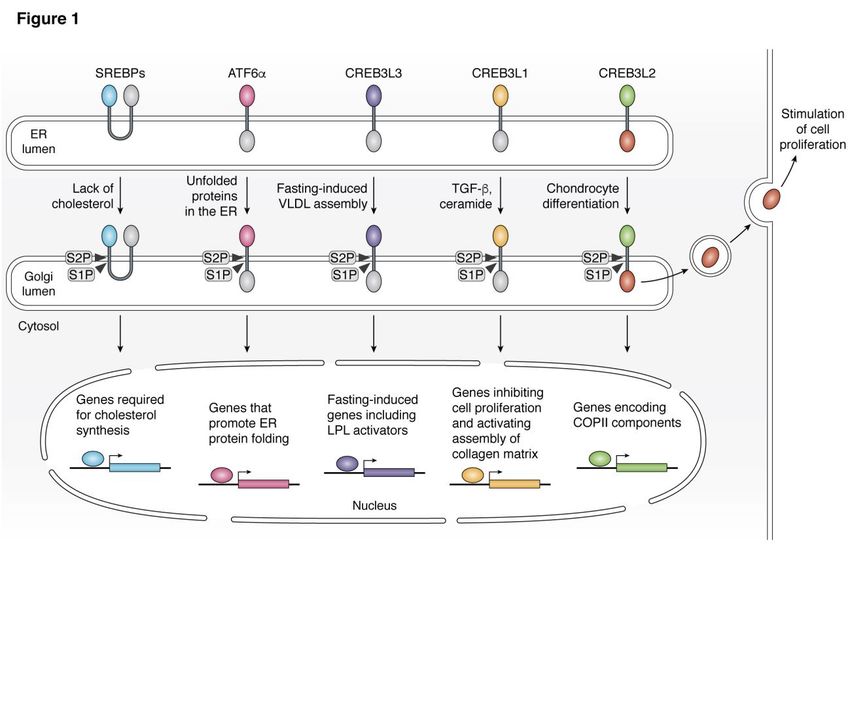
Figure 1. Graphic illustration of RIP mediated by S1P and S2P. Cholesterol deprivation triggers RIP of
SREBP to activate genes required for cholesterol synthesis and uptake. Accumulation of unfolded proteins
in the ER stimulates RIP of ATF6α to activate genes facilitating protein folding in the ER. Fasting-induced
VLDL assembly triggers RIP of CREB3L3 to activate fasting-induced genes including activators for LPL
that hydrolyze VLDL particles. Cytokines of the TGF-β family and ceramide induce RIP of CREB3L1 to
stimulate expression of genes that inhibit cell proliferation and that activate assembly of collagen-
containing matrix. Chondrocyte differentiation stimulates RIP of CREB3L2 to activate genes encoding
COPII components. The luminal C-terminal fragment of CREB3L2 released from S1P-catalyzed cleavage
is secreted out of cells to stimulate proliferation of neighboring cells.
16You can also read