The obese liver environment mediates conversion of NK cells to a less cytotoxic ILC1-like phenotype - bioRxiv
←
→
Page content transcription
If your browser does not render page correctly, please read the page content below
bioRxiv preprint first posted online Mar. 14, 2019; doi: http://dx.doi.org/10.1101/576538. The copyright holder for this preprint
   (which was not peer-reviewed) is the author/funder, who has granted bioRxiv a license to display the preprint in perpetuity.
                                    It is made available under a CC-BY 4.0 International license.

             The obese liver environment mediates
            conversion of NK cells to a less cytotoxic
                     ILC1-like phenotype
    Antonia O. Cuff1 , Francesca Sillito1,2 , Simone Dertschnig1,2 , Andrew Hall3 , TuVinh Luong3 , Ronjon Chakraverty1,2 , and
                                                          Victoria Male1,
                                           1
                                              Institute of Immunity and Transplantation, University College London, London, U.K
                                                        2
                                                          UCL Cancer Institute, University College London, London, U.K.
                             3
                                 Institute for Liver and Digestive Health, Royal Free Hospital and University College London, London, U.K.

Non-alcoholic fatty liver disease (NAFLD) is an obesity-                                steatohepatitis (NASH), which in turn progresses to fibrosis
associated disease in which the fatty liver becomes chronically                         and ultimately cirrhosis, which increases the risk of develop-
inflamed. Here, we report that in the livers of both humans                             ing hepatocellular carcinoma. NASH patients have increased
and mice suffering from NAFLD, NK cells are less able to de-                            numbers of CD3− CD56+ CD57+ cells, which are likely to
granulate. In mice, this is associated with a decreased ability to                      represent circulating NK cells, in their livers (Kahraman et
kill cancerous targets both in vitro and in vivo. On the other
                                                                                        al, 2010). The expression of NK cell-activating ligands is
hand, perforin-deficient mice suffer from less severe NAFLD,
suggesting that perforin-mediated killing is harmful in the obese
                                                                                        also increased in the livers of NASH patients. But the role of
liver and that the reduction in NK cell cytotoxicity may there-                         NK cells in NASH pathogenesis is not yet clear.
fore be protective. The decrease in cytotoxicity is associated with                     NK cells can limit fibrosis in liver diseases of various eti-
a shift towards a transcriptional profile characteristic of ILC1,                       ologies by controlling the activity of hepatic stellate cells
increased expression of inhibitory receptors expressed by ILC1,                         (Radaeva et al, 2006; Male et al, 2017). In a mouse study in
and an altered metabolic profile mimicking that of ILC1. This                           which a NASH-like disease was induced using a methionine
conversion of NK cells to a less cytotoxic ILC1-like phenotype is                       and choline deficient diet, IFNγ production by NKp46+ NK
at least partially mediated by high levels of TGFβ produced in                          cells and ILC1 protected against disease (Tosello-Trampont
the obese liver.                                                                        et al, 2016). On the other hand, NK cells can be harmful
NK cells | ILC1 | obesity | NAFLD | immunometabolism | TGFβ                             in chronic inflammatory liver disease, where they contribute
Correspondence: v.male@ucl.ac.uk                                                        to pathology by killing hepatocytes (Dunn et al, 2007; Be-
                                                                                        raza et al, 2009). In support of this being the case in NASH,
                                                                                        mice lacking either TRAIL (an apoptosis-inducing TNF fam-
Introduction
                                                                                        ily ligand expressed by NK cells) or its receptor were par-
Natural killer (NK) cells are innate lymphoid cells that recog-                         tially protected against obesity-associated NASH (Idrissova
nize and kill virally infected and cancerous cells. In mice, NK                         et al, 2014; Hirsova et al, 2017).
cells were long defined as Lineage-negative NK1.1+ . How-                               Here, we investigate the activity of NK cells and ILC1 in
ever, it has recently come to light that, in tissues, cells defined                     NAFLD patients, and in a mouse model of NASH. We report
in this way contain at least two distinct populations: circulat-                        that in diseased livers, NK cells are less able to degranulate in
ing, or conventional, NK cells that are CD49a− CD49b+ and                               both humans and mice. We further show that this is accompa-
tissue-resident NK-like cells that are CD49a+ CD49b− (Peng                              nied by a decreased ability of the NK cells to kill cancerous
and Tian, 2017). A population of cells that is in many ways                             targets, both in vivo and in vitro. This decrease in cytotoxic-
equivalent to mouse tissue-resident CD49a+ NK cells is also                             ity is associated with a shift towards an ILC1-like phenotype,
present among CD3− CD56+ cells in human livers (Male,                                   which seems to be at least partially mediated by high levels
2017). These tissue-resident cells have variously been called                           of TGFβ produced in the obese liver.
tissue-resident NK cells or innate lymphoid cells, type 1
(ILC1): here, we call them ILC1 (Peng and Tian, 2015). The
finding that Lin− NK1.1+ cells in mice and CD3− CD56+
                                                                                        Results
cells in humans are in fact heterogeneous populations means                             NK cells in the livers of NAFLD patients are less able to de-
that it is now necessary to re-examine the roles of these cells,                        granulate than those from healthy controls
distinguishing between NK and ILC1 subpopulations. This is                              A number of research groups have recently identified liver-
particularly true in the liver, which contains a large number                           resident cells in humans, which are in many ways equivalent
of ILC1.                                                                                to mouse liver ILC1 (Male, 2017). We sought to determine
Non-alcoholic fatty liver disease (NAFLD) is a spectrum                                 how the activity of these cells, as well as conventional NK
of disease, usually associated with obesity, in which excess                            cells in the liver, is altered in NAFLD and NASH patients.
fat builds up in the liver. Simple steatosis can progress to                            We isolated intrahepatic leukocytes from nine healthy livers,
a chronic inflammatory condition known as non-alcoholic                                 six livers with simple steatosis (livers that showed steatosis

                                                                                                                   Cuff et al.   |   bioRχiv   |   March 13, 2019   |   1–10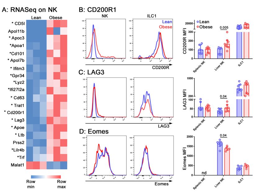
bioRxiv preprint first posted online Mar. 14, 2019; doi: http://dx.doi.org/10.1101/576538. The copyright holder for this preprint
     (which was not peer-reviewed) is the author/funder, who has granted bioRxiv a license to display the preprint in perpetuity.
                                      It is made available under a CC-BY 4.0 International license.

                                                                                                           Fig. 1. NK cells in the livers of obese hu-
                                                                                                           mans and mice are less able to degranulate.
                                                                                                           A. Immune cells were isolated from human liv-
                                                                                                           ers. NK cells were identified by scatter, and as
                                                                                                           live CD45+ CD56+ CD3- Tbethi Eomeslo cells.
                                                                                                           B and C. Intrahepatic leukocytes were cultured for
                                                                                                           4h in the presence of anti-CD107a and Brefeldin.
                                                                                                           Representative CD107a staining of NK cells from
                                                                                                           a healthy (B) and a NAFLD (C) liver is shown.
                                                                                                           D. Inverse correlation between CD107a staining
                                                                                                           and histological score in human livers. H: healthy;
                                                                                                           SS: simple steatosis; NAS: NASH Activity Score;
                                                                                                           C: cirrhosis. Significance was determined using
                                                                                                           Spearman’s Rank Correlation. E. Immune cells
                                                                                                           were isolated from mouse livers. NK cells were
                                                                                                           identified by scatter, and as live CD45+ Lineage-
                                                                                                           negative NK1.1+ CD49b+ cells. ILC1 were iden-
                                                                                                           tified as live CD45+ Lineage-negative NK1.1+
                                                                                                           CD49a+ cells. F and G. Intrahepatic leukocytes
                                                                                                           were cultured for 4h in the presence of anti-
                                                                                                           CD107a and Brefeldin. Representative CD107a
                                                                                                           staining of NK cells from a lean (F) and an obese
                                                                                                           (G) mouse is shown. H and I. CD107a staining in
                                                                                                           splenic NK, liver NK and liver ILC1 in unstimulated
                                                                                                           cells (H) and cells stimulated with PMA and iono-
                                                                                                           mycin (I) from lean and obese mice. J. Represen-
                                                                                                           tative perforin staining in liver NK from a lean (blue
                                                                                                           trace) and an obese (red trace) mouse. K. MFI of
                                                                                                           perforin in splenic NK, liver NK and liver ILC1 from
                                                                                                           lean and obese mice. E - K. n = 6 mice per group;
                                                                                                           significance was determined using Mann Whitney
1.png                                                                                                      U Tests; medians and IQRs are shown.

but no inflammation) and six livers displaying various levels                  NAFLD in which mice were maintained on an obesogenic
of inflammation and fibrosis in addition to steatosis. NK cells                diet. Similar to our observations in human livers, we found
release cytotoxic granules containing perforin and granzymes                   that NK cells isolated from the livers of obese mice degran-
in a process known as degranulation, which can be measured                     ulated less than those from the livers of their lean littermates
using CD107a staining. By doing this, we found that the                        (Figure 1e-h). This was also the case for NK cells isolated
ability of NK cells (defined as CD3− CD56+ Tbethi Eomeslo ;                    from spleens, although the reduction was smaller (a differ-
Cuff, 2016, JI; Figure 1a) to degranulate was inversely corre-                 ence in the medians of 4.3% in splenic NK, compared to
lated with disease severity (Figure 1b-d).                                     10.0% in liver NK; Figure 1h). Following stimulation with
                                                                               PMA and ionomycin, the degranulation of the NK cells from
NK cells in the livers of obese mice are less able to degranu-                 obese mice partially recovered, indicating that the cells re-
late than those in lean mice                                                   tained the ability to degranulate (Figure 1i). However, we
To confirm and extend these findings, we next looked                           also found a significant reduction in the expression of per-
at the ability of NK cells (defined as Lineage-negative                        forin by these cells (Figure 1j,k). This may suggest that even
NK1.1+ CD49a− CD49b+ ) to degranulate in a model of

2    |   bioRχiv                                                                                                Cuff et al.   |   NK convert to ILC1 in obesity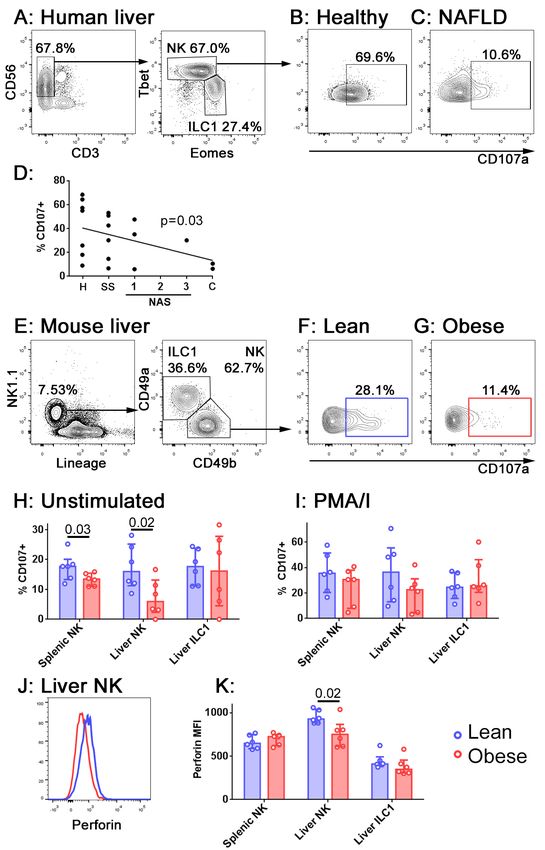
bioRxiv preprint first posted online Mar. 14, 2019; doi: http://dx.doi.org/10.1101/576538. The copyright holder for this preprint
    (which was not peer-reviewed) is the author/funder, who has granted bioRxiv a license to display the preprint in perpetuity.
                                     It is made available under a CC-BY 4.0 International license.

  2.png
Fig. 2. NK cells from obese mice are less able to kill cancerous targets. A.
CTV-labelled RMA/S (NK cell targets) CTB-labelled RMA (recovery control) cells
were mixed in a 1:1 ratio and intravenously injected into lean or obese mice. B
and C. Cells recovered from the spleens of a representative lean (B) and obese (C)
mouse at 4h. D. RMA/S:RMA ratio in recovered splenocytes, normalised to input
ratio. E and F. NK cells were sorted from the spleens (E) and livers (F) of lean and
obese mice, and cultured with the NK cell target line YAC-1. Cell death at 24h is
shown. n = 6 mice per group; significance was determined using Mann Whitney U
Tests; medians and IQRs are shown.

if NK from the livers of obese mice can degranulate in re-
sponse to certain stimuli, they are likely to be less cytotoxic
than those from lean animals.
                                                                                        3.png
NK cells from obese mice are less able to kill cancer cells
                                                                                       Fig. 3. Perforin-mediated killing promotes fibrosis in NAFLD. A. Growth curves
The reduced ability of NK cells in obese mice to degranu-                              for wild type and Perforin knockout mice on the obesogenic diet. B. Liver weights
late suggests that their ability to kill cancerous target cells                        for wild type and Perforin knockout mice after 24 weeks on the obesogenic diet.
will be impaired. To determine whether this was the case                               C. Representative H&E and Picrosirius red staining in a wild type and a perforin
                                                                                       knockout mouse. D. Histological score in wild type and a perforin knockout mouse
in vivo, we injected mice intravenously with equal numbers                             livers, after 24 weeks on the obesogenic diet. E. Plasma ALT levels. F. Picrosirius
of fluorescently-labelled RMA/s cells (which are MHC class                             red positive area in representative histological fields. G and H. Acta2 and Col1a1
I-negative NK targets) and RMA cells (which are not NK                                 mRNA in livers of wild type and a perforin knockout mouse livers, after 24 weeks
                                                                                       on the obesogenic diet. n = 8 mice per group; significance was determined using
targets and act as a recovery control) (Kärre et al, 1986) (Fig-                       Mann Whitney U Tests; medians and IQRs are shown.
ure 2a). We collected spleens from the recipient mice after 4
hours and determined the number of cells remaining in each
population by flow cytometry (Figure 2b,c). The lean mice                              and since the reduction in perforin expression seemed to par-
had eliminated the RMA/s cells with approximately 40% ef-                              ticularly affect NK cells in the liver, we sought to define the
ficiency. In contrast, the obese mice had eliminated them                              likely effect of these changes in NAFLD. Given that TRAIL-
with only 5% efficiency (Figure 2d).                                                   mediated cytotoxicity exacerbates NAFLD (Idrissova et al,
                                                                                       2014; Hirsova et al, 2017), we speculated that there might be
To determine the relative defects in NK cell killing in the
                                                                                       some advantage to the reduced perforin-mediated cytotoxi-
spleens and livers of obese compared to lean mice, we sorted
                                                                                       city we observed in the obese liver. We hypothesized that
NK cells from the two organs and assessed their ability to kill
                                                                                       perforin-mediated killing is harmful in the liver during obe-
YAC-1 cancerous target cells in vitro. NK cells from both
                                                                                       sity, and that the reduction in NK cell degranulation, perforin
the spleens (Figure 2e) and livers (Figure 2f) of obese mice
                                                                                       expression and cytotoxicity could therefore be protective. In
were less able to kill YAC-1 cells than those from their lean
                                                                                       this case, we would expect mice lacking perforin to suffer
littermates, although the defect in killing was only significant
                                                                                       less severe liver disease as a result of obesity. We therefore
(at p < 0.05) in NK cells isolated from the liver. Similar to
                                                                                       took a number of measures of liver damage in wild type and
our observations of degranulation, the reduction in target cell
                                                                                       perforin-deficient mice that had been kept on the obesogenic
killing was greater in NK cells isolated from livers than those
                                                                                       diet for 24 weeks. This later timepoint was chosen so that
isolated from spleens (a difference in the medians of 2.7% in
                                                                                       we could compare the development of fibrosis, which is not
splenic NK cells, compared to 10.1% in liver NK cells).
                                                                                       usually pronounced at 12 weeks, between the two genotypes.
Perforin-mediated cytotoxicity promotes fibrosis in NAFLD                              There was no difference in weight gain or hepatomegaly
Clearly, the diminished ability of NK cells to kill target cells                       between the wild type and perforin knockout mice (Fig-
in obese animals is likely to limit cancer surveillance. How-                          ure 3a,b). However, livers from the perforin-deficient mice
ever, since we observed the largest reductions in both degran-                         displayed a significantly lower histopathological NAS score
ulation and cytotoxicity in NK cells isolated from the liver,                          than wild type controls (Figure 3c,d) and less liver damage

Cuff et al.   |   NK convert to ILC1 in obesity                                                                                                            bioRχiv   |   3
bioRxiv preprint first posted online Mar. 14, 2019; doi: http://dx.doi.org/10.1101/576538. The copyright holder for this preprint
(which was not peer-reviewed) is the author/funder, who has granted bioRxiv a license to display the preprint in perpetuity.
It is made available under a CC-BY 4.0 International license.
4.png
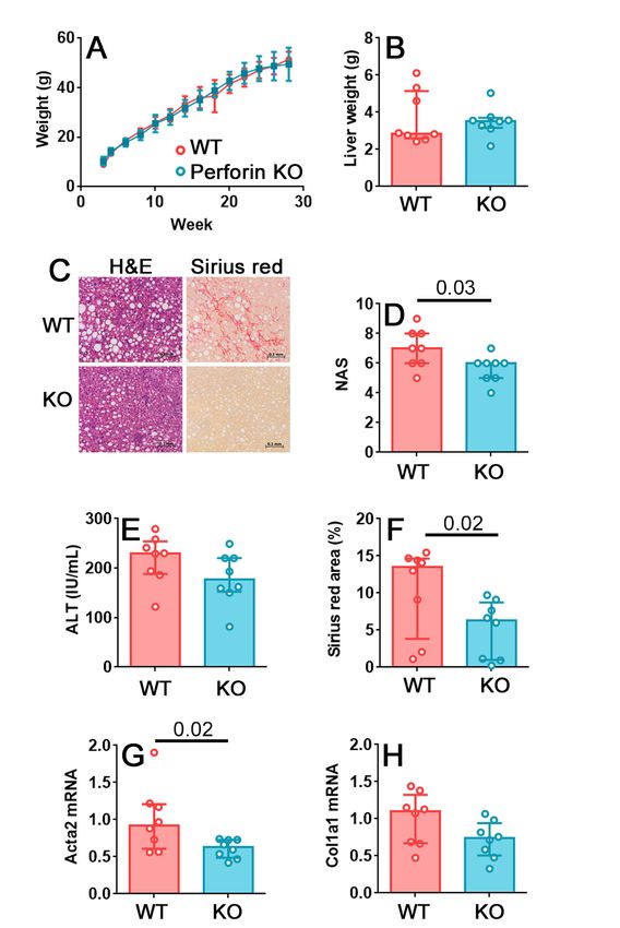
Fig. 4. NK cells in the livers of obese mice acquire an ILC1-like transcriptional profile. A. NK cells and ILC1 were sorted from the livers of lean and obese mice
and examined by RNASeq. Genes that were differentially expressed by > 2-fold, with padj < 0.05, in NK cells of lean versus obese mice are shown. Genes that are also
differentially expressed in ILC1 compared to NK (> 2-fold, with padj < 0.05) are marked with an asterisk. B - D. Expression of the inhibitory receptors CD200R1 (B) and
LAG3 (C) in NK cells and ILC1 of lean (blue) compared to obese (red) mice. D. Expression of Eomes in NK cells and ILC1 of lean (blue) compared to obese (red) mice. n = 6
mice per group; significance was determined using Mann Whitney U Tests; medians and IQRs are shown.
as determined by circulating alanine transaminase (ALT) al- ceptors CD200R1, LAG3 and CD101, all of which are in-
though the difference between the two genotypes was not sig- volved in limiting immune pathology in inflammatory dis-
nificant (Figure 3e). Fibrosis, determined by Picrosirius red eases (Hoek et al, 2000; Fernandez et al, 2007; Snelgrove et
staining, was lower in perforin-deficient mice (Figure 3f) and al, 2008; Anderson et al, 2016) were increased in NK cells
real time PCR for markers of fibrosis revealed that the liv- from obese mice compared to lean, as well as in ILC1 com-
ers of the perforin-deficient mice expressed less Acta2 (alpha pared to NK cells. We confirmed the increase in expression
smooth muscle actin; Figure 3g) and Col1a1 (collagen type I at the protein level for CD200R1 (Figure 4b) and LAG3 (Fig-
alpha 1; Figure 3h) than those of controls, although the dif- ure 4c). CD101 was increased in ILC1 compared to NK cells,
ference was only statistically significant for Acta2. but we did not detect a difference in its expression in NK cells
NK cells in obese mice display a transcriptional profile char- from obese compared to lean animals (not shown).
acteristic of ILC1 NK cells and ILC1 express the transcription factor Tbet at
We next sought to define the mechanism by which the reduc- roughly similar levels, although they rely on it to different
tion in NK cell cytotoxicity occurs. To achieve this, we an- extents (Sojka et al, 2014). On the other hand, the transcrip-
alyzed the transcriptomes of NK cells and ILC1 in the livers tion factor Eomes is expressed only by NK cells and speci-
of lean and obese mice by RNASeq. Raw RNASeq data and fies the NK cell lineage (Daussy et al, 2014). In the light of
differentially expressed gene lists are available from the Na- the ILC1-like transcriptional profile expressed by NK cells in
tional Center for Biotechnology Information Gene Expres- obese mice, we examined the expression of these two tran-
sion Omnibus under accession no. GSE122828. scription factors and found that Eomes was underexpressed
18 of the 20 genes identified as being significantly overex- in NK cells in obese mice, again reminiscent of ILC1 (Figure
pressed (FC > 2, padj < 0.05) in NK cells from obese mice, 4c).
compared to those from their lean littermates, were also over- NK cells in obese mice are metabolically reprogrammed
expressed in ILC1, compared to NK cells (Figure 4a, genes Gene ontogeny analysis on the differentially expressed tran-
characteristic of ILC1 are marked with an asterisk). We par- scripts (Figure 4a) further revealed that the most overrepre-
ticularly noted that transcripts encoding the inhibitory re- sented pathway in the NK cells of obese mice, compared
4 | bioRχiv Cuff et al. | NK convert to ILC1 in obesity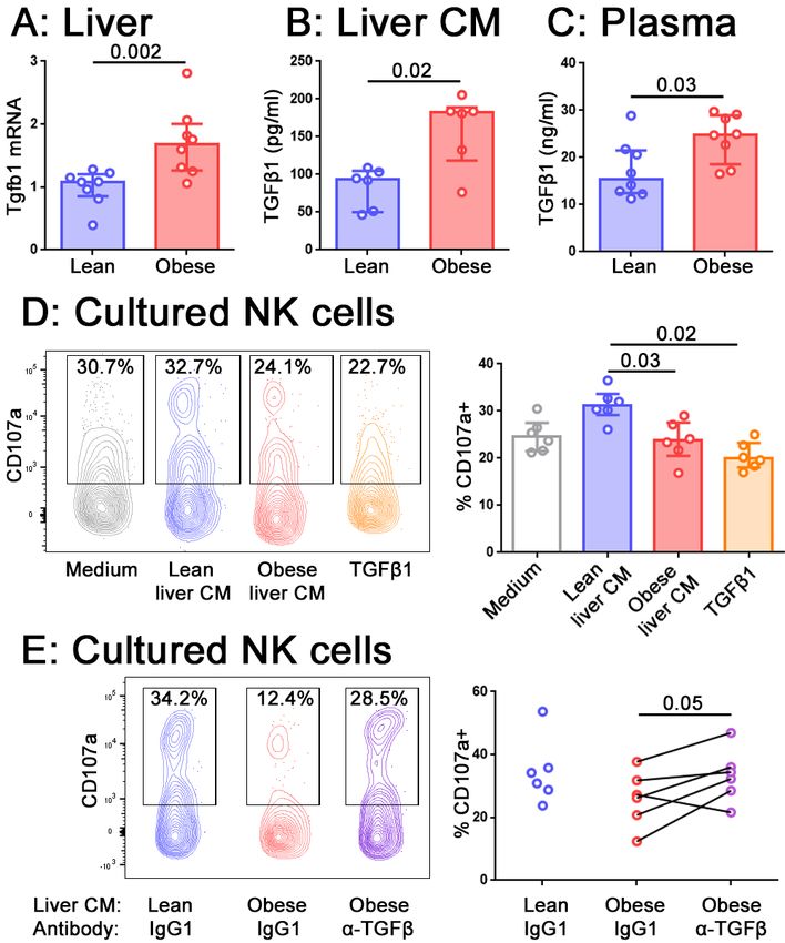
bioRxiv preprint first posted online Mar. 14, 2019; doi: http://dx.doi.org/10.1101/576538. The copyright holder for this preprint
    (which was not peer-reviewed) is the author/funder, who has granted bioRxiv a license to display the preprint in perpetuity.
                                     It is made available under a CC-BY 4.0 International license.

                                                                                                         Fig. 5. NK cells in the livers of obese mice
                                                                                                         are metabolically reprogrammed. Forward and
                                                                                                         side scatter (A), pS6 staining (B) and Glut1 stain-
                                                                                                         ing (C) of freshly isolated NK cells and ILC1 from
                                                                                                         lean (blue) and obese (red) mice. n = 12 mice
                                                                                                         per group (A), 6 mice per group (B) and 7 mice
                                                                                                         per group (C); significance was determined using
                                                                                                         Mann Whitney U Tests; medians and IQRs are
5.png                                                                                                    shown.

to those of lean littermates, was Triglyceride catabolic pro-                the acquisition of ILC1-like features that we observed. Tgfb1
cesses (p = 3.65 x 10⁻³ ). We therefore examined the cells                   transcript was increased in the livers of obese compared to
for signs of metabolic reprogramming.                                        lean mice (Figure 6a) and we also observed increased TGFβ 1
Scatter has been used as an indicator of metabolic alteration                protein in conditioned medium from obese compared to lean
in NK cells (Castro et al, 2018) and we found that side scatter              livers (Figure 6b) and in the plasma of obese mice (Figure
increased in NK cells from the livers of obese mice (Figure                  6c).
5a).                                                                         When NK cells from the spleens of lean animals were cul-
NK cells use mTOR for nutrient sensing and it also has an                    tured for 24 hours in conditioned medium from either lean
important role in immune regulation (O’Brien and Finlay,                     or obese mouse livers, we observed that their ability to de-
2019). The phosphorylation of ribosomal S6 protein (pS6) is                  granulate was impaired following culture in obese, compared
used as an indicator of mTORC1 signaling and we found that                   to lean, liver conditioned medium and we were able to reca-
pS6 was increased in NK cells freshly isolated from the liv-                 pitulate this phenotype by culturing the NK cells in recom-
ers of obese compared to lean mice (Figure 5b). Notably, in                  binant TGFβ 1 (Figure 6d). Furthermore, the addition of an
both these cases, the metabolic alterations that we observed                 anti-panTGFβ antibody to the cultures partially rescued the
in the NK cells served to make them more ILC1-like, similar                  ability of the NK cells to degranulate following culture with
to our findings at the transcriptional level. We also observed               obese liver conditioned medium (Figure 6e), suggesting that
an increase in the expression of the glucose transporter Glut1               TGFβ in the obese liver is at least partially responsible for
(Figure 5c).                                                                 limiting the ability of NK cells to degranulate.

TGFβ in the obese liver inhibits NK cell degranulation
The conversion of NK cells to a less cytotoxic ILC1-like phe-
                                                                             Discussion
notype has been reported in a number of situations (Cuff et                  It is becoming increasingly apparent that NK cells are dys-
al, 2016; Gao et al, 2017; Cortez et al, 2017). In all these                 functional in obesity (O’Shea et al, 2010; Tobin et al, 2017;
cases, TGFβ was shown to be a key cytokine driving the con-                  Michelet et al, 2018), and that this could be one mecha-
version. We therefore hypothesized that TGFβ in the obese                    nism that accounts for the link between obesity and cancer
liver might be mediating the reduction in cytotoxic ability and              (Michelet et al, 2018). These studies have largely focused on

Cuff et al.   |   NK convert to ILC1 in obesity                                                                                             bioRχiv    |   5
bioRxiv preprint first posted online Mar. 14, 2019; doi: http://dx.doi.org/10.1101/576538. The copyright holder for this preprint
(which was not peer-reviewed) is the author/funder, who has granted bioRxiv a license to display the preprint in perpetuity.
It is made available under a CC-BY 4.0 International license.
NK cells in the blood, but in this study we looked at how NK
cells in the liver change during obesity, and the impact that
this may have not only on cancer immunosurveillance, but
also on NAFLD pathogenesis.
We found that, in obesity, NK cells in the livers of both hu-
mans and mice are less able to degranulate and kill target
cells. This is consistent with a number of reports that NK
cells taken from the blood of obese humans (O’Shea et al,
2010; Tobin et al, 2017; Michelet et al, 2018) or the spleens
of obese mice (Michelet et al, 2018) are less cytotoxic than
those taken from their lean counterparts. We further show
that the NK cells of obese mice are less able to kill cancerous
target cells in vivo. In a recent high profile study, NK cells
that had been cultured in fatty acids before being adoptively
transferred into a B16 melanoma-bearing host were less able
to control the cancer than control NK cells (Michelet et al,
2018). Obese mice were also less able to control B16 metas-
tases than their lean counterparts, a phenotype that was asso-
ciated with reduced NK cell infiltration into metastatic foci.
Our finding that obese mice are less able to control RMA/S
cells confirms these findings using another type of cancerous
target.
In humans, NK cell dysfunction in obesity is associated with
metabolic reprogramming (Tobin et al, 2017; Michelet et al,
2018). Similarly, we observed metabolic changes in NK cells
taken from the livers of obese mice. In a pediatric cohort,
obesity was associated with increased Glut1 and pS6 expres-
sion (Tobin et al, 2017), whereas in adults, obesity was asso-
ciated with decreased 2-NBDG uptake (a proxy for glucose
uptake) and pS6 expression (Michelet et al, 2018). This sug-
gests that the way in which NK cells are metabolically dys-
6.png regulated in obesity may change over time. Interestingly, the
NK cells from our obese mice more closely resembled those
Fig. 6. TGFβ in the livers of obese mice limits the ability of NK cells to de- from the pediatric cohort and this may reflect the fact that
granulate. A. Tgfb1 mRNA in the livers of lean and obese mice, normalised so
that mean Tgfb1 transcript expression in the livers of lean mice = 1. B,C. TGFβ1
both these children and our mice had both been obese for a
in lean and obese liver conditioned medium (B) and lean and obese mouse plasma relatively short time, compared to those in the adult human
(C) measured by ELISA. n = 8 mice per group (A, C) and 6 conditioned media per cohort.
group (B); significance was determined using Mann Whitney U Tests; medians and
IQRs are shown. D. Splenic NK cells were cultured for 24h in medium alone, lean The altered metabolic profile of the NK cells in the obese
or obese liver conditioned medium, or 10 ng/ml TGFβ1. For the last 4h of culture, mice was somewhat reflective of the metabolic profile of
CD107a antibody and Brefeldin A were added to media. n = 6 culture conditions per
group; significance was determined using Mann Whitney U Tests with Holm’s cor-
ILC1. We also observed an ILC1-like transcriptional profile
rection for multiple comparisons; medians and IQRs are shown. E. Splenic NK cells and expression of certain inhibitory receptors that are char-
were cultured as described in D, with the addition of 10 µg/ml anti-TGFβ antibody or acteristic of ILC1, including CD200R1, which has recently
isotype control. n = 6 culture conditions per group; significance was determined us-
ing a Wilcoxon signed ranks test, where each pair is obese liver conditioned medium
been proposed to be a better marker of ILC1 than CD49a
from the same mouse, with anti-TGFβ antibody or isotype control. Degranulation in (Weizman et al, 2017). NK cells are converted into less cy-
NK cells cultured in lean liver conditioned medium with isotype control are shown totoxic ILC1-like cells in the tumor microenvironment, a tu-
for comparison, but were not statistically tested.
mor immunevasion strategy mediated by TGFβ (Gao et al,
2017). Similarly, TGFβ promotes the conversion of NK cells
to ILC1 in the mouse salivary gland (Cortez et al, 2017) and
in recently-transplanted human livers (Cuff et al, 2016). We
confirmed previous reports that TGFβ is highly expressed in
the obese liver (Yadav et al, 2011; Mouralidarane et al, 2013;
Hart et al, 2017) and further showed that conditioned medium
from obese livers could limit the ability of NK cells to de-
granulate, mirroring the phenotype that we observe in NK
cells taken from obese livers, in a TGFβ-dependent manner.
This is consistent with previous reports that TGFβ limits NK
cell cytotoxicity (Viel et al, 2016; Zaiatz-Bittencourt et al,
6 | bioRχiv Cuff et al. | NK convert to ILC1 in obesitybioRxiv preprint first posted online Mar. 14, 2019; doi: http://dx.doi.org/10.1101/576538. The copyright holder for this preprint
(which was not peer-reviewed) is the author/funder, who has granted bioRxiv a license to display the preprint in perpetuity.
It is made available under a CC-BY 4.0 International license.
2018), although in contrast to one of these reports (Viel et Hospital Biobank (National Health Service Research Ethics
al, 2016) we observed an increase, rather than a decrease, in Committee approval no. 11/WA/0077, study no. 9455).
pS6 expression. This may reflect differences in the kinetics of
Mice
altered mTORC1 signaling in short-term versus longer term
Female C57BL/6J (bred in house) or Prf1−/− mice (Charles
TGFβ exposure (Zaiatz-Bittencourt et al, 2018) but is also
River; RRID MGI:5576721) were randomized at weaning
likely to be attributable to differences in the liver environ-
onto standard chow (RM1) or a highly palatable obesogenic
ment other than TGFβ production. For example high levels
diet consisting of 22.6% fat, 23.0% protein and 40.2% car-
of fatty acids may interact with TGFβ to produce a different
bohydrate (w/w); diet code 824018 - ’45% AFE fat’ Special
metabolic phenotype (Michelet et al, 2018).
Dietary Services, Essex, UK), supplemented with sweetened
The reduction in NK cell cytotoxicity in obesity is likely to
condensed milk (Nestle) ad libitum (Oben et al, 2010).
be detrimental to tumor immunosurveillance, but one key
question is whether it is helpful or harmful in the context After 12 or 24 weeks on the diet, mice were sacrificed by di-
of NAFLD. We observed greater reductions in degranula- rect cervical dislocation. To determine the severity of liver
tion and cytotoxicity in the liver than in the spleen. Further, disease, blood was collected by cardiac puncture and cen-
TRAIL-mediated cytotoxicity is known to promote patho- trifuged at 10,000 xg for 10 minutes at room temperature
genesis in NAFLD (Idrissova et al, 2014; Hirsova et al, to isolate serum for ALT measurement and pieces of liver
2017). Therefore, we postulated that the decrease in degran- were fixed in 10% neutral buffered formalin, fixed, sectioned
ulation, and thus perforin-mediated cytotoxicity, that we ob- and stained with H& E or Picrosirius red. Sections were
served in the obese liver might protect against liver disease. blinded and scored by a hepatopathologist, using the Brunt-
In support of this idea, perforin-deficient mice suffered from Kleiner NASH activity score (NAS; Brunt et al, 2011) (Sup-
less severe NAFLD. Since these animals lack perforin glob- plementary figure 1). Spleens were collected and spleno-
ally, we cannot say with certainty that the protection is not cytes isolated as previously described (Cuff and Male, 2017).
mediated at least in part by defective T cell cytotoxicity. In- Intrahepatic lymphocytes were isolated using an adaptation
deed if, as we suggest, the liver is acting to protect itself of the method from Cuff and Male, 2017. Briefly, finely
against immunopathology by producing TGFβ, it is likely minced liver tissue was collected in RPMI 1640 medium
that T cell cytotoxicity will also be reduced. Nevertheless, (Life Technologies brand; Thermo Fisher Scientific, Hud-
the protection from liver disease that we observe in the ab- son, NH) and passed through a 70 µm cell strainer. The
sence of perforin does suggest that defect in NK cell de- suspension was spun down (500 × g, 4°C, 10 minutes) and
granulation we observe in the obese liver may act to protect the pellet resuspended in RPMI 1640 medium. The cell
it. NK cell-derived ILC1-like cells in tumors produce angio- suspension was layered over 24% Optiprep (Sigma-Aldrich)
genic and growth factors (Gao et al, 2017), so it is tempting and centrifuged without braking (700 × g, RT, 20 minutes).
to speculate that the ILC1-like cells we see emerging from The interface layer was taken and washed in HBSS without
the NK cell population in the obese liver may have a similar Ca2+ Mg2+ (Lonza, distributed by VWR, Lutterworth, UK)
reparative effect. supplemented with 0.25% bovine serum albumin (Sigma-
It has been suggested that NK cell metabolism could be tar- Aldrich, Hammerhill, U.K) and 0.001% DNase I (Roche, dis-
geted to prevent cancer (O’Brien and Finlay, 2019). In the tributed by Simga-Aldrich).
light of recent findings that NK cells are less cytotoxic in obe- Animal husbandry and experimental procedures were per-
sity, and that this is at least partially mediated by metabolic formed according to UK Home Office regulations and insti-
changes, it might be tempting to consider using such treat- tute guidelines, under project license 70/8530.
ments as a way to break the well-established link between In vivo NK cytotoxicity assays
cancer and obesity (Park et al, 2014). However, our finding RMA and RMA/s thymoma cells (cultured in Iscove’s mod-
that perforin-mediated killing is harmful in NAFLD sounds a ified Dulbecco’s medium supplemented with 10% FCS; Life
note of caution about such approaches, which may result in Technologies) were labeled with Cell Trace Blue or Cell
adverse effects in the liver. Trace Violet, respectively, according to the manufacturer’s
instructions (Thermo Fisher Scientific). 107 cells of each cell
Materials and Methods type were injected intravenously into control or obese mice.
After 4h, recipients were sacrificed by direct cervical dislo-
Human liver biopsies
cation and the spleens were examined for fluorescent target
Liver biopsies were taken from livers that were destined
cells.
for transplantation, but that were discarded because of long
warm ischemic time, vascular abnormalities, or tumors else- Ex vivo NK cell activity assays
where in the patient (n = 9), or because they displayed signs For degranulation and cytokine production assays, total intra-
of NAFLD or NASH on histopathologic examination (n = hepatic leukocytes or splenocytes were cultured for 4 hours
10). Biopsies were also taken from livers explanted dur- at 107 cells/mL in RPMI 1640 medium supplemented with
ing transplantation for NASH (n = 2). Cells were isolated 10% FCS, 25 mM HEPES, 1 mM sodium pyruvate, 50 µM
from human liver samples and degranulation assessed as pre- 2-ME, MEM nonessential amino acids, penicillin, and strep-
viously described (Cuff et al, 2016). Ethical approval for use tomycin (all Life Technologies brand; Thermo Fisher Sci-
of human liver samples was obtained through the Royal Free entific). Brefeldin A (1 µg/mL; Sigma-Aldrich) and anti-
Cuff et al. | NK convert to ILC1 in obesity bioRχiv | 7bioRxiv preprint first posted online Mar. 14, 2019; doi: http://dx.doi.org/10.1101/576538. The copyright holder for this preprint
(which was not peer-reviewed) is the author/funder, who has granted bioRxiv a license to display the preprint in perpetuity.
It is made available under a CC-BY 4.0 International license.
CD107a (1:100, eBioscience, San Diego, CA) were added Essex, U.K). Differential expression analysis was carried out
to all conditions. Experiments were carried out in either the using SARTools (Varet et al, 2016), filtering at padj < 0.05
presence or absence of PMA and ionomycin (25 ng/mL and and FC > 2.
1 µg/mL, respectively; Sigma-Adrich). TGFβ1 ELISA
For cytotoxicity assays, 15,000 sorted NK cells were cul- Total TGFβ1 protein concentrations in plasma and liver con-
tured with 75,000 YAC-1 target cells in 100 µL RPMI sup- ditioned medium were determined using a mouse TGF-beta1
plemented as before for 24h. DuoSet ELISA (R&D Systems), according to the manufac-
Cell death was measured using an LDH cytotoxicity assay turer’s instructions.
(Abcam, Cambridge, U.K.), according to the manufacturer’s
instructions. Readings were normalized between the medium Statistics
only (0% cell death) and lysis buffer (100% cell death) con- The data was analyzed and found not to be normally dis-
trols. tributed. Significance was therefore determined using non-
parametric tests: Mann Whitney U Tests (for unpaired data),
Cell culture experiments Wilcoxon signed rank tests (for paired data) or Spearman’s
Liver conditioned medium was produced by culturing 500 tests (for correlations). P values < 0.05 are reported. Bars
mg of finely minced liver tissue in serum-free M199 medium represent medians and interquartile ranges; individual data
(Gibco brand, Thermo Fisher Scientific) for 48h. The points are also plotted.
medium was cleared of debris by being passed through a 70
µm strainer and by centrifugation at 500 xg for 5 minutes at Supplementary material
4. Splenic NK cells were cultured for 24h in RPMI supple- Supplementary Table 1 gives full details of all the antibodies
mented as before mixed 3:1 with liver conditioned medium used for flow cytometry in the study. Supplementary Figure
or M199 medium. TGFβ1 (10 ng/mL, PeproTech, Rocky 1 demonstrates that mice kept on the obesogenic diet become
Hill, NJ), anti-mouse TGFβ (10 µg/mL, clone 1D11 R&D obese and develop NAFLD.
Systems, Minneapolis, MN) or isotype control antibody (10 ACKNOWLEDGEMENTS
µg/mL, clone 11711 R&D Systems) were added as indicated. This work was funded by Royal Society/Wellcome Trust Sir Henry Dale Fellowship
WT105677 to VM. The authors declare no competing financial interests.
Flow cytometry
Details of antibodies used in the study are given in Supple-
References
mentary table 1. The lineage cocktail for mouse cells con-
sisted of CD3, CD8α, CD19 and Gr1. Dead cells were ex- Anderson A.C., Joller N., Kuchroo V.K. Lag-3, Tim-3,
cluded using fixable viability dye eFluor 450 (eBioscience) and TIGIT: Co-inhibitory Receptors with Specialized Func-
(4°C, 15 minutes). Surface staining was carried out in PBS tions in Immune Regulation. Immunity. 44:989-1004
supplemented with 1% FCS (4°C, 15 minutes). Intracellu- 10.1016/j.immuni.2016.05.001
lar staining was carried out using Human FoxP3 Buffer (BD Beraza N., Malato Y., Sander L.E., Al-Masaoudi M.,
Biosciences, Oxford, UK), except for pS6 staining, which Freimuth J., Riethmacher D., Gores G.J., Roskams T.,
was carried out using Cytofix/cytoperm (BD Biosciences). Liedtke C., Trautwein C. 2009. Hepatocyte-specific NEMO
Data were acquired on an LSRFortessa II (BD Biosciences) deletion promotes NK/NKT cell- and TRAIL-dependent liver
and analyzed using FlowJo v.X.0.7 (Tree Star, Ashland, OR, damage. J Exp Med. 206:1727-1737 10.1084/jem.20082152
USA). Sorting was carried out on an Aria (BD Biosciences).
Brunt E.M., Kleiner D.E., Wilson L.A., Belt P.,
Real time PCR Neuschwander-Tetri B.A.; NASH Clinical Research
Liver sections were dissected directly into RNAlater and Network (CRN). 2011. Nonalcoholic fatty liver disease
RNA was extracted using an RNeasy Lipid Tissue Mini (NAFLD) activity score and the histopathologic diagnosis in
kit (both from Qiagen, Manchester, U.K.). cDNA was NAFLD: distinct clinicopathologic meanings. Hepatology.
made using a Transcriptor First Strand cDNA Synthe- 53:810-820 10.1002/hep.24127
sis kit (Roche, Welwyn Garden City, U.K.) and real-
time PCR was performed using TaqMan (Applied Biosys- Castro W., Chelbi S.T., Niogret C., Ramon-Barros C., Wel-
tems, Warrington, U.K.) primer/probe sets recognizing Hprt1 ten S.P.M., Osterheld K., Wang H., Rota G., Morgado L.,
(Mm00446968_m1), Acta2 (Mm01546133_m1), Col1a1 Vivier E., et al. 2018. The transcription factor Rfx7 limits
(Mm00801666_g1) and Tgfb1 (Mm01178820_m1). metabolism of NK cells and promotes their maintenance and
immunity. Nat Immunol. 19:809-820 10.1038/s41590-018-
RNASeq 0144-9
Total RNA was extracted from sorted cells using an RNeasy
Micro kit (Qiagen) and libraries were prepared from 2 ng of Cortez V.S., Ulland T.K., Cervantes-Barragan L., Bando J.K.,
total RNA using the NEBNext low input kit (New England Robinette M.L., Wang Q., White A.J., Gilfillan S., Cella M.,
Biolabs, Hitchen, U.K.). Libraries were assessed for correct Colonna M. 2017. SMAD4 impedes the conversion of NK
size distribution on the Agilent 2200 TapeStation and quan- cells into ILC1-like cells by curtailing non-canonical TGF-β
tified by Qubit DNA High Sensitivity assay (Thermo Fisher signaling. Nat Immunol. 18:995-1003 10.1038/ni.3809
Scientific) before being pooled at an equimolar concentra- Cuff A.O., Robertson F.P., Stegmann K.A., Pallett L.J., Maini
tion. Samples were sequenced on a NextSeq 500 (Illumina, M.K., Davidson B.R., Male V. 2016. Eomeshi NK Cells
8 | bioRχiv Cuff et al. | NK convert to ILC1 in obesitybioRxiv preprint first posted online Mar. 14, 2019; doi: http://dx.doi.org/10.1101/576538. The copyright holder for this preprint
(which was not peer-reviewed) is the author/funder, who has granted bioRxiv a license to display the preprint in perpetuity.
It is made available under a CC-BY 4.0 International license.
in Human Liver Are Long-Lived and Do Not Recirculate B (MIC A/B): a novel role in nonalcoholic steatohepatitis.
but Can Be Replenished from the Circulation. J Immunol. Hepatology 51:92-102 10.1002/hep.23253.
197:4283-4291 10.4049/jimmunol.1601424
Kärre K., Ljunggren H.G., Piontek G., Kiessling R. 1986.
Cuff A.O., Male V. 2017. Conventional NK cells and ILC1 Selective rejection of H-2-deficient lymphoma variants sug-
are partially ablated in the livers of Ncr1 iCreTbx21 fl/fl gests alternative immune defence strategy. Nature. 319:675-
mice. Wellcome Open Res. 2:39 10.12688/wellcomeopen- 678 10.1038/319675a0
res.11741.2 Male. V. 2017. Liver-Resident NK Cells: The Human Factor.
Daussy C., Faure F., Mayol K., Viel S., Gasteiger G., Char- Trends Immunol. 38:307-309 10.1016/j.it.2017.02.008
rier E., Bienvenu J., Henry T., Debien E., Hasan U.A., et al. Male V., Stegmann K.A., Easom N.J., Maini M.K. 2017. Nat-
2014. T-bet and Eomes instruct the development of two dis- ural Killer Cells in Liver Disease. Semin Liver Dis. 37:198-
tinct natural killer cell lineages in the liver and in the bone 209 10.1055/s-0037-1603946
marrow. J Exp Med. 211:563-577 10.1084/jem.20131560
Michelet X., Dyck L., Hogan A., Loftus R.M., Duquette D.,
Dunn C., Brunetto M., Reynolds G., Christophides T., Wei K., Beyaz S., Tavakkoli A., Foley C., Donnelly R., et
Kennedy P.T., Lampertico P., Das A., Lopes A.R., Borrow al. 2018. Metabolic reprogramming of natural killer cells in
P., Williams K., et al. 2007. Cytokines induced during obesity limits antitumor responses. Nat Immunol. 19:1330-
chronic hepatitis B virus infection promote a pathway for 1340 10.1038/s41590-018-0251-7
NK cell-mediated liver damage. J Exp Med. 204:667-680
10.1084/jem.20061287 Mouralidarane A., Soeda J., Visconti-Pugmire C., Samuels-
son A.M., Pombo J., Maragkoudaki X., Butt A., Saraswati
Fernandez I., Zeiser R., Karsunky H., Kambham N., Beil- R., Novelli M., Fusai G., et al. 2013. Maternal obesity
hack A., Soderstrom K., Negrin R.S., Engleman E. 2007. programs offspring nonalcoholic fatty liver disease by in-
CD101 surface expression discriminates potency among nate immune dysfunction in mice. Hepatology. 58:128-138
murine FoxP3+ regulatory T cells. J Immunol. 179:2808- 10.1002/hep.26248
2814 10.4049/jimmunol.179.5.2808
Oben J.A., Mouralidarane A., Samuelsson A.M., Matthews
Gao Y., Souza-Fonseca-Guimaraes F., Bald T., Ng S.S., P.J., Morgan M.L., McKee C., Soeda J., Fernandez-Twinn
Young A., Ngiow S.F., Rautela J., Straube J., Waddell N., D.S., Martin-Gronert M.S., Ozanne S.E., et al. 2010. Ma-
Blake S.J., et al. 2017. Tumor immunoevasion by the con- ternal obesity during pregnancy and lactation programs the
version of effector NK cells into type 1 innate lymphoid cells. development of offspring non-alcoholic fatty liver disease in
Nat Immunol. 18:1004-1015 10.1038/ni.3800 mice. J Hepatol. 52:913-920 10.1016/j.jhep.2009.12.042
Hart K.M., Fabre T., Sciurba J.C., Gieseck R.L. 3rd, Borth- O’Brien K.L., Finlay D.K. 2019. Immunometabolism and
wick L.A., Vannella K.M., Acciani T.H., de Queiroz Prado natural killer cell responses. Nat Rev Immunol. ePub ahead
R., Thompson R.W., et al. 2017. Type 2 immunity is pro- of print 10.1038/s41577-019-0139-2
tective in metabolic disease but exacerbates NAFLD collab-
oratively with TGF-β. Sci Transl Med. 9:pii: eaal3694 O’Shea D., Cawood T.J., O’Farrelly C., Lynch L. 2010. Nat-
10.1126/scitranslmed.aal3694 ural killer cells in obesity: impaired function and increased
susceptibility to the effects of cigarette smoke. PLoS One.
Hirsova P., Weng P., Salim W., Bronk S.F., Griffith T.S., 5:e8660 10.1371/journal.pone.0008660
Ibrahim S.H., Gores G.J. 2017. TRAIL Deletion Pre-
vents Liver, but Not Adipose Tissue, Inflammation during Park J., Morley T.S., Kim M., Clegg D.J., Scherer P.E.
Murine Diet-Induced Obesity. Hepatol Commun. 1:648-662 2014. Obesity and cancer–mechanisms underlying tumour
10.1002/hep4.1069 progression and recurrence. Nat Rev Endocrinol. 10:455-
465 10.1038/nrendo.2014.94
Hoek R.M., Ruuls S.R., Murphy C.A., Wright G.J., God-
dard R., Zurawski S.M., Blom B., Homola M.E., Streit W.J., Peng, H., Tian, Z. 2015. Re-examining the origin and func-
Brown M.H., et al. Down-regulation of the macrophage tion of liver-resident NK cells. Trends Immunol. 36:293-299
lineage through interaction with OX2 (CD200). Science. 10.1016/j.it.2015.03.006.
290:1768-1771 10.1126/science.290.5497.1768 Peng H., Tian Z. 2017. Diversity of tissue-resident NK cells.
Idrissova L., Malhi H., Werneburg N.W., LeBrasseur N.K., Semin Immunol. 31:3-10 10.1016/j.smim.2017.07.006
Bronk S.F., Fingas C., Tchkonia T., Pirtskhalava T., White Radaeva S., Sun R., Jaruga B., Nguyen V.T., Tian
T.A., Stout M.B., et al. 2015. TRAIL receptor deletion in Z., Gao B. 2006. Natural killer cells ameliorate liver
mice suppresses the inflammation of nutrient excess. J Hep- fibrosis by killing activated stellate cells in NKG2D-
atol. 62:1156-1163 10.1016/j.jhep.2014.11.033 dependent and tumor necrosis factor-related apoptosis-
inducing ligand-dependent manners. Gastroenterology
Kahraman A., Schlattjan M., Kocabayoglu P., Yildiz-
130:435-452 10.1002/hep.23253.
Meziletoglu S., Schlensak M., Fingas C.D., Wedemeyer I.,
Marquitan G., Gieseler R.K., Baba H.A., et al. 2010. Ma- Snelgrove R.J., Goulding J., Didierlaurent A.M., Lyonga D.,
jor histocompatibility complex class I-related chains A and Vekaria S., Edwards L., Gwyer E., Sedgwick J.D., Barclay
Cuff et al. | NK convert to ILC1 in obesity bioRχiv | 9bioRxiv preprint first posted online Mar. 14, 2019; doi: http://dx.doi.org/10.1101/576538. The copyright holder for this preprint
(which was not peer-reviewed) is the author/funder, who has granted bioRxiv a license to display the preprint in perpetuity.
It is made available under a CC-BY 4.0 International license.
A.N., Hussell T. 2008. A critical function for CD200 in lung
immune homeostasis and the severity of influenza infection.
Nat Immunol. 9:1074-1083 10.1038/ni.1637
Sojka D.K., Plougastel-Douglas B., Yang L., Pak-Wittel
M.A., Artyomov M.N., Ivanova Y., Zhong C., Chase J.M.,
Rothman P.B., Yu J., et al. 2014. Tissue-resident natu-
ral killer (NK) cells are cell lineages distinct from thymic
and conventional splenic NK cells. Elife. 3:e01659
10.7554/eLife.01659
Tobin L.M., Mavinkurve M., Carolan E., Kinlen D., O’Brien
E.C., Little M.A., Finlay D.K., Cody D., Hogan A.E., O’Shea
D. 2017. NK cells in childhood obesity are activated,
metabolically stressed, and functionally deficient. JCI In-
sight. 2:pii: 94939 10.1172/jci.insight.94939
Tosello-Trampont A.C., Krueger P., Narayanan S., Landes
S.G., Leitinger N., Hahn Y.S. 2016. NKp46(+) natural killer
cells attenuate metabolism-induced hepatic fibrosis by regu-
lating macrophage activation in mice. Hepatology 63:799-
812 10.1002/hep.28389
Varet H., Brillet-Guéguen L., Coppée J.Y., Dillies M.A.
2016. SARTools: A DESeq2- and EdgeR-Based R Pipeline
for Comprehensive Differential Analysis of RNA-Seq Data.
PLoS One. 11:e0157022 10.1371/journal.pone.0157022
Viel S., Marçais A., Guimaraes F.S., Loftus R., Rabilloud J.,
Grau M., Degouve S., Djebali S., Sanlaville A., Charrier E.,
et al. 2016. TGF-/beta inhibits the activation and functions
of NK cells by repressing the mTOR pathway. Sci Signal.
9:ra19 10.1126/scisignal.aad1884
Weizman O.E., Adams N.M., Schuster I.S., Krishna C., Pri-
tykin Y., Lau C., Degli-Esposti M.A., Leslie C.S., Sun J.C.,
O’Sullivan T.E. 2017. ILC1 Confer Early Host Protec-
tion at Initial Sites of Viral Infection. Cell. 171:795-808
10.1016/j.cell.2017.09.052
Yadav H., Quijano C., Kamaraju A.K., Gavrilova O., Malek
R., Chen W., Zerfas P., Zhigang D., Wright E.C., Stuelten C.,
et al. 2011. Protection from obesity and diabetes by block-
ade of TGF-/beta /Smad3 signaling. Cell Metab. 14:67-79
10.1016/j.cmet.2011.04.013
Zaiatz-Bittencourt V., Finlay D.K., Gardiner C.M. 2018.
Canonical TGF-/beta Signaling Pathway Represses Hu-
man NK Cell Metabolism. J Immunol. 200:3934-3941
10.4049/jimmunol.1701461.
10 | bioRχiv Cuff et al. | NK convert to ILC1 in obesityYou can also read