Calcilytic NPS2143 promotes proliferation and inhibits apoptosis of spontaneously hypertensive rat vascular smooth muscle cells via activation of ...
←
→
Page content transcription
If your browser does not render page correctly, please read the page content below
818                                EXPERIMENTAL AND THERAPEUTIC MEDICINE 20: 818-829, 2020

           Calcilytic NPS2143 promotes proliferation and inhibits
         apoptosis of spontaneously hypertensive rat vascular smooth
          muscle cells via activation of the renin‑angiotensin system
                          YONGLI ZHAO1*, NA TANG1*, DONGMEI XI1, ZHEN HUANG1, TIAN ZHANG1,
                           YONGMIN LIU1, LAMEI WANG2, YAN TANG3, HUA ZHONG1 and FANG HE1

                     1
                      Department of Pathophysiology; 2The Centre of Medical Functional Experiments, Medical
                      College of Shihezi University; 3Department of Geriatrics, The First Affiliated Hospital of
                           Medical College of Shihezi University, Shihezi, Xinjiang 832002, P.R. China

                                        Received March 4, 2019; Accepted January 17, 2020

                                                     DOI: 10.3892/etm.2020.8759

Abstract. Vascular smooth muscle cell (VSMC) proliferation            inhibitor MDL12330A, angiotensin converting enzyme
and apoptosis and the renin‑angiotensin system (RAS) play             inhibitor captopril, angiotensin I receptor (AT1R) inhibitor
critical roles in the development of essential hypertension. The      losartan, NPS2143 + U73122, NPS2143 + 2‑APB, NPS2143
activation of calcium‑sensing receptor (CaSR), functionally           + MDL12330A, NPS2143 + captopril and NPS2143 + losartan.
expressed in VSMCs, inhibits cyclic adenosine monophos-               The results suggested that NPS2143 promoted cell prolifera-
phate (cAMP) formation by elevating intracellular calcium             tion, inhibited cell apoptosis, decreased [Ca2+]i and increased
([Ca 2+]i) and then suppressing renin release. The present            the expression of RAS compared with control treatments.
study aimed to investigate the effects of NPS2143‑mediated            NPS2143 + U73122 and NPS2143 + 2‑APB enhanced the
inhibition of CaSR on VSMC proliferation and apoptosis in             effects of NPS2143, while NPS2143 + MDL12330A, NPS2143
spontaneously hypertensive rat (SHR) VSMCs and to assess              + captopril, NPS2143 + losartan attenuated the effected of
whether these effects were mediated by alterations to RAS             NPS2143 in SHR VSMCs. Furthermore, the knockdown of
signaling. Primary VSMCs were isolated from the aortas of             AT1R by AT1R‑short hairpin RNA also attenuated the effects
SHRs and Wistar‑Kyoto rats. SHR VSMCs were treated with               of NPS2143 compared with NPS2143 alone. Collectively,
CaSR antagonist NPS2143 and cell proliferation and CaSR               these data indicated that NPS2143 promoted proliferation and
and RAS‑related protein expression levels were measured               inhibited apoptosis of VSMCs in SHRs, the effect of which
to assess the effect. The results indicated that NPS2143              was achieved by activation of RAS signaling.
treatment promoted SHR VSMC proliferation, lower CaSR
expression levels and higher RAS‑related proteins levels when         Introduction
compared with control treatment. Additional measurement
of the expression levels of proteins related to proliferation,        Approximately one billion people worldwide are affected
remodeling, apoptosis and RAS related proteins, as well as            by hypertension. Essential hypertension (EH) accounts for
cell viability, cell cycle, cell apoptosis ratio, [Ca2+]i, and the    90‑95% of these cases, the progression of which is affected by
concentration of cAMP was performed after treatment with              both genetic and environmental factors (1‑3). The underlying
NPS2143, PLC inhibitor U73122, IP3 receptor antagonist                mechanisms of hypertension are complex. It is known that
2‑aminoethoxydiphenylborane (APB), adenylyl cyclase‑V                 vascular smooth muscle cells (VSMCs) are located mainly
                                                                      in the arterial tunica media and act as critical determinants
                                                                      in vascular‑related diseases (4). Excessive VSMC proliferation
                                                                      is involved in hypertension‑induced maladaptive vascular
                                                                      remodeling (5,6). An imbalance between proliferation and
Correspondence to: Professor Hua Zhong or Professor Fang He,          apoptosis may be important in the structural changes associ-
Department of Pathophysiology, Medical College of Shihezi             ated with vascular injury. In addition, the renin‑angiotensin
University, 59 North 2nd Road, Shihezi, Xinjiang 832002, P.R. China
                                                                      system (RAS) plays a pivotal role in regulating blood pressure
E‑mail: 156696992@qq.com
                                                                      and promoting the proliferation of VSMCs (7). Renin is the
E‑mail: fangf2002shz@126.com
                                                                      first rate‑limiting enzyme of the RAS system and is closely
*
    Contributed equally                                               linked to hypertension. Cyclic adenosine monophosphate
                                                                      (cAMP) plays a key role in this system (8). Another enzyme,
Key words: calcium‑sensing receptor, renin‑angiotensin system,        tonin, directly catalyzes the formation of the plasma protein
proliferation, apoptosis                                              angiotensin II (Ang II) using renin tetradecapeptide as a
                                                                      substrate and angiotensin I (Ang I) (9). Ang II is a multi-
                                                                      functional peptide that exerts effects on VSMCs, including
ZHAO et al: NPS2143 PROMOTES PROLIFERATION VIA RAS IN SHR VSMCs                                   819

regulation of vasoconstriction, wall tension, cell growth and     Cell culture and treatment. Primary VSMCs were isolated from
apoptosis, through its vasoconstrictive properties (10).          thoracic aortas of 20 male Wistar‑Kyoto (WKY) rats and 50
    The relationship between Ca2+ and hypertension has been       SHRs. Rats (age, 8 weeks; weight, 150‑180 g) were purchased
clinically well documented (11). A large number of studies        from Vital River Laboratory Animal Science and Technology
have shown that a low calcium diet is a significant factor in     Co., Ltd (license number: SCXK2012‑0001) and were kept
hypertension and that calcium intake can effectively reduce       in an alternating 12‑h light/dark cycle at a temperature of
blood pressure (BP), both effects which have been identi-         20‑25˚C, constant humidity with free access to food and water.
fied in animal models of hypertension (12,13), though their       This animal study was approved by the Institutional Animal
underlying mechanisms are not well understood. Calcium            Research Committee of Shihezi Medical University, and all
sensitive receptor (CaSR), an extracellular Ca2+ receptor, is a   animals received humane care in compliance with the Guide
member of the C subfamily of the G‑protein‑coupled receptor       for the Care and Use of Laboratory Animals published by the
superfamily. Despite extensive current interest, the emer-        National Institutes of Health (24). On the day of the experi-
gence of CaSR in the field of cardiovascular diseases was         ment, rats were anaesthetized by the intraperitoneal injection
actually fairly recent. In 2003, the expression of CaSR in rat    of 30 mg/l pentobarbital sodium (Merck KGaA) at a dose of
myocardium was first confirmed (14). Subsequently, scholars       50 mg/kg and the chest was opened to extract the thoracic
worldwide have confirmed the importance of CaSR's role in         aorta. After surgery, the rats were euthanized by the intraperi-
cardiovascular diseases, such as myocardial hypertrophy,          toneal injection of pentobarbital sodium (150 mg/kg). Rat aorta
ischemia‑reperfusion injury, atherosclerosis and pulmonary        was cleaned from adherent tissue, and the endothelium was
hypertension (15,16). Further research has determined that        denuded. Pieces of aorta (1 mm2) were cultured in Dulbecco's
CaSR was functionally expressed in the outer membrane of          modified Eagle's medium/F12 (DMEM/F12; Gibco®; Thermo
the blood vessel wall, fibroblast cells and VSMCs (17). CaSR's    Fisher Scientific, Inc.) supplemented with 20% FBS (Gibco®;
main function is to maintain the homeostasis of Ca2+ and other    Thermo Fisher Scientific, Inc.) and 1% penicillin‑streptomycin
metal ions throughout the body. Several studies have shown        at 37˚C in a 5% CO2 humidified incubator in accordance with
that CaSR may regulate BP, but the underlying mechanism           a previously published protocol (25). Cells migrated from the
is unknown. Rybczynska et al (18) reported that treatment         pieces of aorta after 7‑9 days. Aortic pieces were then removed
with NPS2143, an allosteric inhibitor of CaSR, elevated BP        and VSMCs were left to proliferate. Experiments used VSMCs
in normotensive rats; however, in rats subjected to parathy-      from passages 3‑6 at 80‑90% confluence. Cell growth was
roidectomy or treated with an Ang I receptor (AT1R) blocker       arrested using serum‑free DMEM‑F12 to incubate the cells for
(such as losartan) in the presence of a calcium‑channel           24 h before intervention. VSMCs were divided into 13 groups
blocker or antagonist, elevated BP was not observed (19).         and treated as follows: i) Untreated WKY VSMC; ii) untreated
Ogata et al (20) found that NPSR568 (R‑568) decreased BP          SHR VSMC; iii) SHR VSMC treated with NPS2143 (1 µM);
in uremic and spontaneously hypertensive rats (SHRs), but         iv) SHR VSMC treated with U73122 (10 µM (21); PLC‑specific
had no effect on normotensive rats. Atchison et al (12) and       inhibitor); v) SHR VSMC treated with 2‑aminoethoxydiphe-
Ortiz‑Capisano et al (21) suggested that CaSR was expressed       nylborane (2‑APB; 100 µM; IP3 receptor antagonist) (21);
in juxtaglomerular cells and its activation in turn activated     vi) SHR VSMC treated with MDL12330A (10 µM; adenylyl
the ryanodine receptor (RyR) via the phospholipase C              cyclase (AC)‑V inhibitor) (26); vii) SHR VSMC treated with
(PLC)/inositol 1,4,5‑triphosphate (IP3) pathway to augment        captopril [100 µM; an angiotensin‑converting enzyme (ACE)
intracellular calcium ([Ca 2+]i) and inhibit cAMP formation,      inhibitor] (27); viii) SHR VSMC treated with losartan (10 µM,
thereby suppressing renin release. These researchers hypoth-      AT1R inhibitor) (28); ix) SHR VSMC treated with NPS2143 +
esized that the mechanism by which CaSR regulates blood           U73122; x) SHR VSMC treated with NPS2143 + 2‑APB group;
pressure may be related to intervention by the RAS.               xi) SHR VSMC treated with NPS2143 + MDL12330A; xii) SHR
    Previous studies have demonstrated that lower levels of       VSMC treated with NPS2143 + captopril; xiii) SHR VSMC
the CaSR were associated with increased vascular remod-           treated with NPS2143 + losartan group. All the aforementioned
eling, promoting the development of EH by activating the          treatment regimens were cultured at 37˚C in a 5% CO2 humidi-
cAMP‑RAS pathway; moreover, the CaSR agonist R568 was             fied incubator for 48 h prior to subsequent experimentation.
a viable suppressant of local RAS activity to relieve these
symptoms in vivo (22,23). However, the potential molecular        MTT assay‑guided optimization. VSMCs (5x10³ cells/well)
mechanisms underlying this phenomenon remain to be eluci-         were plated in 96‑well plates and cultured for 12, 24, 48 and 72
dated. The present study was therefore conducted in order         h with NPS2143 of different concentrations (0.01, 0.03, 0.1, 0.3
to determine whether NPS2143 promotes the proliferation           and 1 µM) (29), then treated with the designated drugs. MTT
of SHR VSMCs and whether this molecular mechanism is              (10 µl, 5 mg/ml) was added into each well and then incubated
involved in activating the RAS signaling pathway.                 at 37˚C for 4 h. The supernatant was removed and 150 µl of
                                                                  DMSO was added to each well. The absorbance at 490 nm
Materials and methods                                             was measured using a model 680 microplate reader (Bio‑Rad
                                                                  Laboratories, Inc.). Cell proliferation ratio was calculated using
Drugs. NPS2143 was purchased from Tocris Bioscience               the following formula: Ratio=(A490 value in test group‑A490
Co., Ltd. U73122, captopril and losartan were obtained            value in control group)/A490 value in control group x100%.
from Selleck Chemicals. 2‑APB was purchased from
Abcam and MDL12330A was obtained from ApexBio                     Flow cytometry analysis of cell cycle distribution. Cell
Technology LLC.                                                   cycle distribution was determined using propidium iodide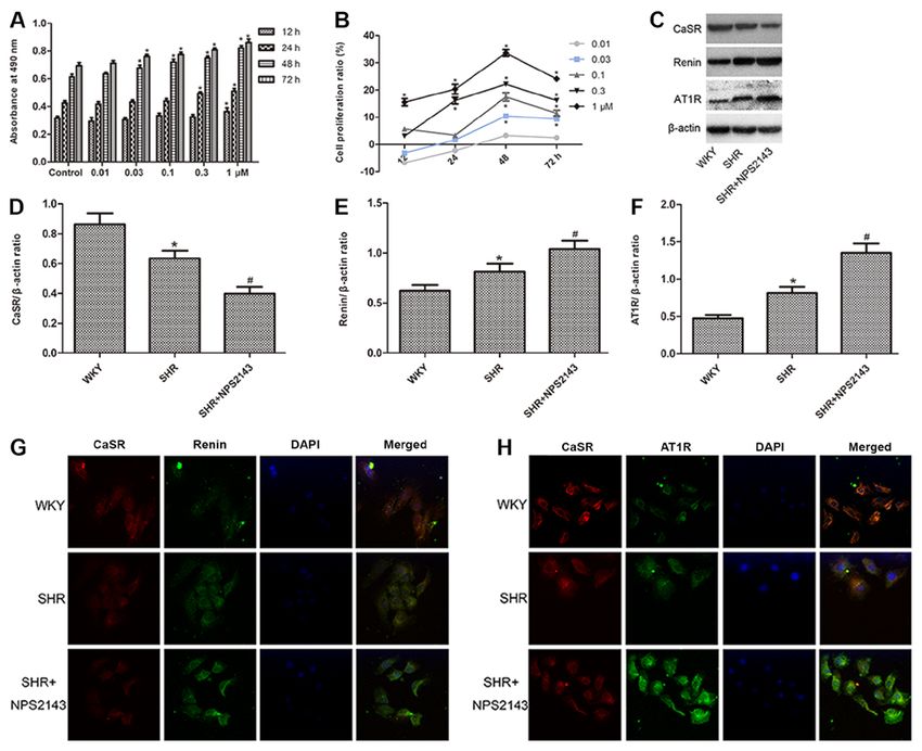
820 EXPERIMENTAL AND THERAPEUTIC MEDICINE 20: 818-829, 2020
(PI) staining according to the manufacturer's instructions Next, the cells were examined and images were captured using
[MultiSciences (Lianke) Biotech Co., Ltd.]. Following confocal microscopy.
optimal time treatments with the designated drug interven-
tions, cells were harvested and fixed with 70% ethanol ELISA. ELISA kits (Elabscience Biotechnology Co.,
overnight at 4˚C. Fixed cells were collected by centrifugation Ltd.) were used to determine the levels of Renin (cat.
(1,000 x g for 5 min at 4˚C), washed twice using PBS, and no. E‑EL‑R0030c) and Ang II (cat. no. E‑EL‑R1430c) in the
incubated with 1 ml of PI staining buffer (20 µg/ml PI and cell culture medium and cAMP (cat. no. E‑EL‑0056c) from the
50 µg/ml RNaseA) at room temperature for 30 min in the cell lysate, each specific measurement method was performed
dark. Thereafter, cellular fluorescence was measured by flow according to the manufacturer's protocols. Optical absorbance
cytometry (BD FACSAria™ III Cell Sorter; cat. no. 648282; values were read at 450 nm (Bio‑Rad Model 3550‑UV;
BD Biosciences). Flow cytometry data analysis was conducted Bio‑Rad Laboratories, Inc.).
performed using the FlowJo 7.6.1 software (FlowJo LLC).
Western blot analysis. After specific drug treatments, cells were
Apoptosis assays. Apoptosis was detected using an annexin incubated in lysis buffer (PMSF:RIPA, 1:100; Sigma‑Aldrich;
V‑FITC/PI apoptosis detection kit or annexin V‑APC/PI apop- Merck KGaA) for 20 min on ice. After insoluble debris was
tosis detection kit according to the manufacturer's instructions pelleted by centrifugation at 12,000 x g for 15 min at 4˚C,
[MultiSciences (Lianke) Biotech Co., Ltd.]. Briefly, VSMCs the supernatants were collected and protein concentrations
were plated into six‑well plates and incubated with drugs were assessed by the bicinchoninic acid method. After boiling
for optimal intervals using the results from MTT assay. The the samples for 10 min, the protein samples (10 µg/lane)
cells were harvested, washed twice with pre‑chilled PBS, and were fractionated by SDS‑PAGE (10‑12% polyacrylamide
centrifuged (1,000 x g for 5 min at 4˚C), followed by staining gels), transferred to polyvinylidene fluoride membranes
with annexin V‑FITC/PI or annexin V‑APC/PI at 4˚C for (EMD Millipore) and blocked with 5% non‑fat milk for 2 h
10 min in the dark and then stained cells were analyzed by flow at room temperature. The membranes were then incubated
cytometry (BD FACSAria™ III Cell Sorter; cat. no. 648282; overnight at 4˚C using appropriate dilutions of primary anti-
BD Biosciences). Flow cytometry data analysis of the propor- bodies against CaSR (1:1,000; cat. no.; Abcam), proliferating
tion of apoptotic cells was performed using the FlowJo 7.6.1 cell nuclear antigen (PCNA, 1:250; cat. no. BM0104; Boster
software (Flowjo LLC). Biological Technology), smooth muscle actin α (α‑SMA;
1:500; cat. no. BM0002; Boster Biological Technology,
Fluo‑3/acetoxymethyl (Fluo‑3/AM) measurements of [Ca2+]i. Wuhan, China), calponin (1:400; cat. no. BM4088; Boster
VSMCs were cultured where the cell density was adjusted to Biological Technology), osteopontin (OPN; 1:1,000; cat.
1x10⁵ cells/ml with the indicated drugs for optimal time no. ab8448; Abcam), renin (1:250; cat. no. bs‑6184R; BIOSS),
intervals, then loaded with 5 µM fluo‑3/AM (Beijing Solarbio AT1R (1:1,000; cat. no. ab9391; Abcam, UK), Bcl‑2 (1:200;
Science & Technology Co., Ltd.) for 40 min at 37˚C in the cat. no. BA0412; Boster Biological Technology), Bax (1:200;
dark. The VSMCs were then washed twice with Ca2+ (0.14 g/l) cat. no. BA0315; Boster Biological Technology) and β‑actin
HEPES‑buffered saline (HBS) to remove the extracellular (1:1,000; cat. no. TA‑09; ZSGB‑Bio; OriGene Technologies,
fluo‑3/AM and incubated further in Ca 2+ HBS for 20 min Inc.). Membranes were then washed three times with
at 37˚C. Next, confocal microscopy (model: Zeiss LSM 510 Tris‑buffered saline containing 0.1% Tween 20 and incubated
META; Zeiss AG) was performed to analyze the fluorescence with horseradish peroxidase‑conjugated secondary antibodies
intensity induced by fluo‑3. (1:20,000; Boster Biological Technology) for 2 h at room
temperature. Detection was performed with an enhanced
Immunofluorescence analysis. VSMCs were fixed in 4% chemiluminescence system (Pierce; Thermo Fisher Scientific,
paraformaldehyde for 20 min at room temperature and Inc.). Band intensity was quantified using Bio‑Rad Quantity
blocked with 5% BSA (Sigma‑Aldrich; Merck KGaA) for One software (version 4.3.0; Bio‑Rad Laboratories, Inc.) with
30 min at 37˚C. The cells were then co‑incubated with either β‑actin as an internal control.
CaSR (1:200; cat. no. ab19347; Abcam) and Renin (1:100; cat.
no. bs‑6184R; BIOSS) primary antibodies or CaSR (1:200; Adenovirus‑mediated downregulation of AT1R in VSMCs.
cat. no. ab223360) and AT1R (1:50; cat. no. ab9391; Abcam) VSMCs (1x10⁵/ml) were prepared and infected at a multi-
primary antibodies overnight at 4˚C. On the following day, the plicity of infection of 100 with shRNA scrambled or AT1R
cells were brought to room temperature over 1 h and washed down‑regulating AT1R‑shRNA adenovirus 67, 68, 69
3 times (5 min each time) with PBS. Goat tetramethylrhoda- (Shanghai GeneChem Co., Ltd.) for 48 h at 37˚C in a 5% CO2
mine (TRITC)‑conjugated anti‑mouse (1:50; cat. no. BA1089; humidified incubator, following which interference efficiency
Boster Biological Technology), goat FITC‑conjugated was assessed by either reverse transcription‑quantitative PCR
anti‑rabbit secondary antibody (1:50; cat. no. BA1105; Boster (RT‑qPCR) or western blot analysis. The target sequences
Biological Technology), goat TRITC‑conjugated anti‑rabbit were as follows: shRNA scrambled, 5'‑TTCTCCGAACGT
(1:50; cat. no. BA1090; Boster Biological Technology) or goat GTCACGT‑3'; shAT1R‑67, 5'‑GGCCAGCGTCTTTCTTCT
FITC‑conjugated anti‑mouse secondary antibody (1:50; cat. CAA‑3'; shAT1R‑68, 5'‑CAGCGTGAGCTTCAACCTCTA‑3'
no. BA1101; Boster Biological Technology) diluted using 5% and shAT1R‑69, 5'‑GGGCTGGGCCTTACCAAGAAT‑3'.
BSA were added in the dark and the cells were incubated
at room temperature for 1 h. After being washed with PBS, RT‑qPCR. Total RNA was extracted from cultured VSMCs
the cells were treated with 0.5 µM DAPI for 10 min at 37˚C. using TRIzol® Reagent (Invitrogen; Thermo Fisher Scientific,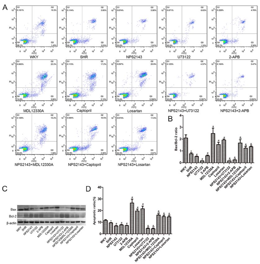
ZHAO et al: NPS2143 PROMOTES PROLIFERATION VIA RAS IN SHR VSMCs                               821

Inc.) according to manufacturer's protocol. First‑strand cDNA      NPS2143 combined with PLC‑IP3/AC‑V/cAMP/RAS signaling
synthesis was performed using a TIANScript RT kit according        pathway inhibitors. Western blot analysis indicated that the
to manufacturer's protocol (Tiangen Biotech Co., Ltd.). The        protein levels of two contractile/differentiated VSMC pheno-
temperature step protocol for the reverse transcription reaction   type marker proteins (α‑SMA and calponin) were decreased
70˚C for 5 min, on ice for 2 min, 42˚C for 50 min and lastly       in SHR VSMCs compared with WKY VSMCs, while the
95˚C for 5 min. The consequent cDNA products were used as          expression levels of OPN, a synthetic/dedifferentiated pheno-
the PCR templates. Subsequent qPCR was performed using             type marker protein, and PCNA were significantly increased
SYBR™ Green PCR Master Mix (Thermo Fisher Scientific,              in SHR VSMCs compared with WKY VSMCs (P
822                                      EXPERIMENTAL AND THERAPEUTIC MEDICINE 20: 818-829, 2020

Figure 1. Determination of cell proliferation levels and the expression of CaSR, renin and AT1R in SHRs VSMCs. (A) Effects of NPS2143 at different
concentrations of NPS2143 (0.01, 0.03, 0.1, 0.3 and 1 µM) for 12, 24, 48 and 72 h on the absorbance at 490 nm of SHR VSMCs was tested by MTT assay.
(B) Effects of NPS2143 at different concentrations (0.01, 0.03, 0.1, 0.3 and 1 µM) for 12, 24, 48 and 72 h on the cell proliferation ratio of SHR VSMCs as tested
by MTT assay. *P
ZHAO et al: NPS2143 PROMOTES PROLIFERATION VIA RAS IN SHR VSMCs                                                  823

Figure 2. Western blot analysis of proliferation and remodeling‑associated protein expression and MTT and cell cycle regulation analysis of proliferation
in SHR VSMCs. (A) Western blot analysis of α‑SMA, calponin, OPN and PCNA protein expression. (B) Densitometric analysis of α‑SMA expression.
(C) Densitometric analysis of calponin expression. (D) Densitometric analysis of OPN expression. (E) Densitometric analysis of PCNA expression. (F) MTT
assay was used to test VSMC proliferation. (G) Flow cytometry was used to examine cell cycle distribution in the different treatment groups (green, G 0/G1
phase, yellow, S phase, blue, G2/M phase) and (H) Quantitative analysis of flow cytometry data. VSMCs in the intervention groups were all derived from
spontaneously hypertensive rats. Data are presented as the mean ± SEM, n=6. *P
824                                     EXPERIMENTAL AND THERAPEUTIC MEDICINE 20: 818-829, 2020

Figure 3. Determination of the apoptotic rate and levels of apoptosis‑related proteins in SHR VSMCs. (A) Annexin‑V/PI staining and flow cytometry was used
to determine the level of apoptosis. (B) Cells in Q2 and Q3 were classed as apoptotic and the results were analyzed quantitatively. (C) Bax and Bcl‑2 protein
expression was assessed by western blot analysis and (D) the ratio of Bax/Bcl‑2 quantified. VSMCs in the intervention groups were all derived from spontane-
ously hypertensive rats. The data are expressed as the mean ± SEM, n=3. *P
ZHAO et al: NPS2143 PROMOTES PROLIFERATION VIA RAS IN SHR VSMCs                                                  825

Figure 4. Determination of [Ca2+]i, AT1R protein expression and cAMP, renin and Ang II levels in SHR VSMCs. (A) Changes in the intensity of fluorescence
of [Ca2+]i were recorded with a laser scanning confocal microscope under different treatment conditions. Magnification, x400. (B) Quantitative analysis of
fluorescence intensity. (C) AT1R protein expression assayed by western blot analysis. (D) Densitometric analysis of AT1R expression. (E) ELISA detection
of cAMP concentration. (F) ELISA detection of renin concentration. (G) ELISA detection of Ang II concentration. VSMCs in the intervention group were all
derived from spontaneously hypertensive rats. The data are expressed as the mean ± SEM. n=3 for (A‑D), n=6 for (E‑G). *P
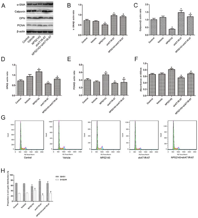
826                                     EXPERIMENTAL AND THERAPEUTIC MEDICINE 20: 818-829, 2020

Figure 5. NPS2143 promotes proliferation by downregulating AT1R expression in SHR VSMCs. (A) Western blot analysis of α‑SMA, calponin, OPN and
PCNA protein expression. (B) Densitometric analysis of α‑SMA expression. (C) Densitometric analysis of calponin expression. (D) Densitometric analysis of
OPN expression. (E) Densitometric analysis of PCNA expression. (F) MTT assay was used to test VSMC viability. (G) Flow cytometry was used to examine
cell cycle distribution (green, G 0/G1 phase, yellow, S phase, blue, G2/M phase). (H) Quantitative analysis of flow cytometry data. Data are expressed as the
mean ± SEM, n=3, *P
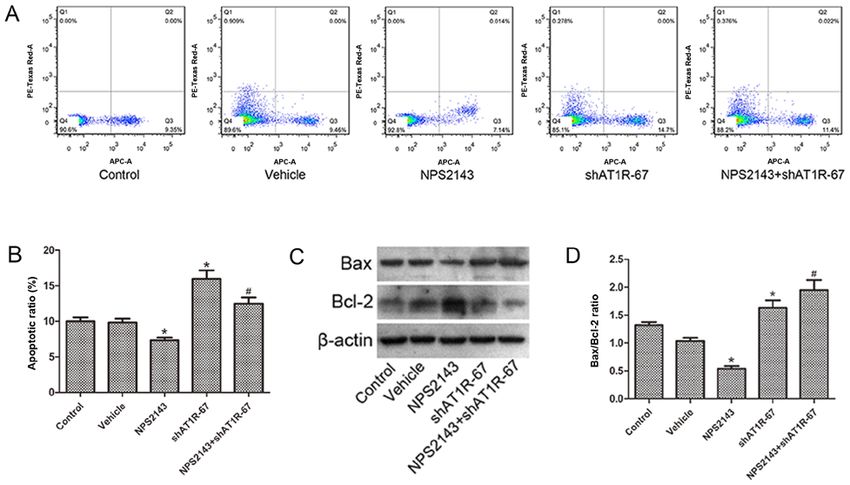
ZHAO et al: NPS2143 PROMOTES PROLIFERATION VIA RAS IN SHR VSMCs                                                     827

Figure 6. NPS2143 inhibits apoptosis by downregulating AT1R expression in SHR VSMCs. (A) The level of apoptosis was examined using flow cytometry and
the upper right area and the lower right area were regarded as apoptotic areas. (B) Quantitative analysis of flow cytometry results. (C) Bax and Bcl‑2 protein
expression assayed by western blot analysis. (D) The ratio of the Bax/Bcl‑2. The data are expressed as the mean ± SEM, n=3. *P
828                               EXPERIMENTAL AND THERAPEUTIC MEDICINE 20: 818-829, 2020

in normotensive rats. In the current study, the expression of        Ethics approval and consent to participate
cAMP, renin, Ang II and AT1R was increased in SHR VSMCs
compared with WKY VSMCs, and was further increased                   The study was approved by the Ethics Committee of Shihezi
by NPS2143 treatment. NPS2143 combined with PLC‑IP3                  Medical University (approval no. A2017‑175‑01; Shihezi,
pathway blockers enhanced the effect of NPS2143 on SHR               China).
VSMCs, while NPS2143 combined with AC‑V/cAMP/RAS
pathway blockers inhibited the effect of NPS2143 on SHR              Patient consent for publication
VSMCs. These results suggested that the CaSR antagonist
NPS2143 promoted SHR VSMC proliferation and inhibited                Not applicable.
apoptosis by activating the AC‑V/cAMP/RAS pathway.
    Finally, in order to further elucidate the molecular mecha-      Competing interests
nism by which the RAS contributes to the proliferation and
apoptosis induced by NPS2143, a loss‑of‑function mutation            The authors declare that they have no competing interests.
in AT1R was established through adenovirus infection. Prior
transfection with AT1R shRNA by adenovirus infection                 References
resulted in α‑SMA and calponin upregulation and PCNA and
OPN downregulation, as well as inhibition of the prolifera-           1. Kearney PM, Whelton M, Reynolds K, Muntner P, Whelton PK
tion and promotion of apoptosis induced by NPS2143. These                and He J: Global burden of hypertension: Analysis of worldwide
                                                                         data. Lancet 365: 217‑223, 2005.
results are consistent with the suggestion that regulation of         2. Writing Group Members; Mozaffarian D, Benjamin EJ, Go AS,
RAS pathways is the molecular mechanism by which CaSR                    Arnett DK, Blaha MJ, Cushman M, Das SR, de Ferranti S,
contributes to proliferation and apoptosis in SHR VSMCs.                 Després JP, et al: Heart Disease and Stroke Statistics‑2016
                                                                         Update: A Report From the American Heart Association.
    The present study has limitations. The RAS includes                  Circulation 133: e338‑e360, 2016.
the classical and non‑classical RAS, while the current only           3. Svetkey LP, Harris EL, Eden M, Vollmer WM, Meltesen GT,
focused on classical RAS. Whether non‑classical RAS also                 Ricchiuti V, Williams G, Appel LJ, Bray GA, Moore TJ, et al:
                                                                         Modulation of the BP response to diet by genes in the
has a role in CaSR‑mediated cell proliferation and apoptosis             renin‑angiotensin system and the adrenergic nervous system. Am
remains to be elucidated.                                                J Hypertens 24: 209‑217, 2011.
    In conclusion, the results of the present study suggested that    4. Cai X: Regulation of smooth muscle cells in development and
                                                                         vascular disease: Current therapeutic strategies. Expert Rev
a reduction in the expression of CaSR can lead to the prolifera-         Cardiovasc Ther 4: 789‑800, 2006.
tion and apoptosis of SHR VSMCs by activation of the RAS.             5. Haudenschild CC, Grunwald J and Chobanian AV: Effects of
CaSR antagonist NPS2143 promoted cell proliferation and                  hypertension on migration and proliferation of smooth muscle in
                                                                         culture. Hypertension 7: I101‑1104, 1985.
inhibited apoptosis, while the use of a CaSR agonist combined         6. Belo VA, Guimarães DA and Castro MM: Matrix metallopro-
with a RAS blocker interfered with abnormal proliferation and            teinase 2 as a potential mediator of vascular smooth muscle cell
apoptosis of VSMCs. This method of treatment may have a                  migration and chronic vascular remodeling in hypertension.
                                                                         J Vasc Res 52: 221‑231, 2016.
positive role in lowering blood pressure and reversing vascular       7. Bavishi C, Bangalore S and Messerli FH: Renin angiotensin
remodeling in the treatment of hypertension.                             aldosterone system inhibitors in hypertension: Is there evidence
                                                                         for benefit independent of blood pressure reduction? Prog
                                                                         Cardiovasc Dis 59: 253‑261, 2016.
Acknowledgements                                                      8. Churchill PC: Second messengers in renin secretion. Am J
                                                                         Physiol 249: 175‑184, 1985.
Not applicable.                                                       9. Genest J, Nowaczynski W, Boucher R, Kuchel O and
                                                                         Rojo‑Ortega JM: Aldosterone and renin in essential hyperten-
                                                                         sion. Can Med Assoc J 113: 421‑431, 1975.
Funding                                                              10. Touyz RM: The role of angiotensin II in regulating vascular struc-
                                                                         tural and functional changes in hypertension. Curr Hypertens
                                                                         Rep 5: 155‑164, 2003.
The present study was supported by the National Natural              11. Allender PS, Cutler JA, Follmann D, Cappuccio FP, Pryer J and
Science Foundation of China (grant no. 31560287) and the                 Elliott P: Dietary calcium and blood pressure: A meta‑analysis of
Xinjiang Graduate Student Research Innovation Project (grant             randomized clinical trials. Ann Intern Med 124: 825‑831, 1996.
                                                                     12. Atchison DK, Ortiz‑Capisano MC and Beierwaltes WH: Acute
no. XJ2019G105).                                                         activation of the calcium‑sensing receptor inhibits plasma renin
                                                                         activity in vivo. Am J Physiol Regul Integr Comp Physiol 299:
Availability of data and materials                                       R1020‑R1026, 2010.
                                                                     13. Ayachi S: Increased dietary calcium lowers blood pressure in
                                                                         the spontaneously hypertensive rat. Metabolism 28: 1234‑1238,
The datasets used and/or analyzed during the current study               1979.
are available from the corresponding author on reasonable            14. Wang R, Xu C, Zhao W, Zhang J, Cao K, Yang B and Wu L:
                                                                         Calcium and polyamine regulated calcium‑sensing receptors in
request.                                                                 cardiac tissues. Eur J Biochem 270: 2680‑2688, 2003.
                                                                     15. Yamamura A, Yamamura H, Guo Q, Zimnicka AM, Wan J,
Authors' contributions                                                   Ko EA, Smith KA, Pohl NM, Song S, Zeifman A, et al:
                                                                         Dihydropyridine Ca(2+) channel blockers increase cytosolic
                                                                         [Ca(2+)] by activating Ca(2+)‑sensing receptors in pulmonary
YZ, FH and HZ conceived and designed the experiments. YZ,                arterial smooth muscle cells. Circ Res 112: 640‑650, 2013.
NT, DX and ZH performed the experiments. YZ, TZ, YL, LW              16. Guo J, Li HZ, Zhang WH, Wang LC, Wang LN, Zhang L,
                                                                         Li GW, Li HX, Yang BF, Wu L, et al: Increased expression of
and YT analyzed the data. YZ, HZ and FH wrote or modified                calcium‑sensing receptors induced by ox‑LDL amplifies apop-
the paper. All authors contributed to and approved the final             tosis of cardiomyocytes during simulated ischaemia‑reperfusion.
draft of the manuscript.                                                 Clin Exp Pharmacol Physiol 37: e128‑e135, 2010.
ZHAO et al: NPS2143 PROMOTES PROLIFERATION VIA RAS IN SHR VSMCs 829
17. Weston AH, Geraghty A, Egner I and Edwards G: The vascular 32. Smajilovic S, Hansen JL, Christoffersen TE, Lewin E, Sheikh SP,
extracellular calcium‑sensing receptor: An update. Acta Physiol Terwilliger EF, Brown EM, Haunso S and Tfelt‑Hansen J:
(Oxf) 203: 127‑137, 2011. Extracellular calcium sensing in rat aortic vascular smooth
18. Rybczynska A, Jurska‑Jasko A, Boblewski K, Lehmann A and muscle cells. Biochem Biophys Res Commun 348: 1215‑1223,
Orlewska C: Blockade of calcium channels and AT1 receptor 2006.
prevents the hypertensive effect of calcilytic NPS 2143 in rats. 33. Maillard MP, Tedjani A, Perregaux C and Burnier M:
J Physiol Pharmacol 61: 163‑170, 2010. Calcium‑sensing receptors modulate renin release in vivo and
19. Rybczynska A, Lehmann A, Jurska‑Jasko A, Boblewski K, in vitro in the rat. J Hypertens 27: 1980‑1997, 2009.
Orlewska C, Foks H and Drewnowska K: Hypertensive effect 34. Ström A, Franzén A, Wängnerud C, Knutsson AK, Heinegård D
of calcilytic NPS 2143 administration in rats. J Endocrinol 191: and Hultgårdh‑Nilsson A: Altered vascular remodeling in osteo-
189‑195, 2006. pontin‑deficient atherosclerotic mice. J Vasc Res 41: 314‑322,
20. Ogata H, Ritz E, Odoni G, Amann K and Orth SR: Beneficial 2004.
effects of calcimimetics on progression of renal failure and 35. Lai YM, Fukuda N, Su JZ, Suzuki R, Ikeda Y, Takagi H, Tahira Y
cardiovascular risk factors. J Am Soc Nephrol 14: 959‑967, 2003. and Kanmatsuse K: Novel mechanisms of the antiproliferative
21. Ortiz‑Capisano MC, Reddy M, Mendez M, Garvin JL and effects of amlodipine in vascular smooth muscle cells from spon-
Beierwaltes WH: Juxtaglomerular cell CaSR stimulation taneously hypertensive rats. Hypertens Res 25: 109‑115, 2002.
decreases renin release via activation of the PLC/IP(3) pathway 36. Yi B, Cui J, Ning JN, Wang GS, Qian GS and Lu KZ:
and the ryanodine receptor. Am J Physiol Renal Physiol 304: Over‑expression of PKGIα inhibits hypoxia‑induced prolif-
F248‑F256, 2013. eration, Akt activation, and phenotype modulation of human
22. Qu YY, Hui J, Wang LM, Tang N, Zhong H, Liu YM, Li Z, PASMCs: The role of phenotype modulation of PASMCs in
Feng Q and He F: Reduced expression of the extracellular pulmonary vascular remodeling. Gene 492: 354‑360, 2012.
calcium‑sensing receptor (CaSR) is associated with activation of 37. Dickhout JG and Lee RM: Apoptosis in the muscular arteries
the renin‑angiotensin system (RAS) to promote vascular remod- from young spontaneously hypertensive rats. J Hypertens 17:
eling in the pathogenesis of essential hypertension. PLoS One 11: 1413‑1419, 1999.
e0157456, 2016. 38. Song L, Gao LN, Wang J, Thapa S, Li Y, Zhong XB,
23. Sun R, Zhang W, Zhong H, Wang L, Tang N, Liu Y, Zhao Y, Zhao H W, Xiang XR, Zhang FG and Ji P: Stromal
Zhang T and He F: Calcimimetic R568 reduced the blood Cell‑derived factor‑1α alleviates calcium‑sensing receptor
pressure and improved aortic remodeling in spontaneously activation‑mediated ischemia/reperfusion injury by inhibiting
hypertensive rats by inhibiting local renin‑angiotensin system caspase‑3/caspase‑9‑induced cell apoptosis in rat free flaps.
activity. Exp Ther Med 16: 4089‑4099, 2018. Biomed Res Int 2018: 8945850, 2018.
24. U.S. National Institutes of Health: Laboratory animal welfare: 39. Mcconkey DJ and Orrenius S: The role of calcium in the regulation
Public Health Service policy on humane care and use of labo- of apoptosis. Biochem Biophys Res Commun 239: 357‑366, 1997.
ratory animals by awardee institutions; notice. Fed Regist 50: 40. McConkey DJ and Orrenius S: Signal transduction pathways to
19584‑19585, 1985. apoptosis. Trends Cell Biol 4: 370‑375, 1994.
25. Chi J, Meng L, Pan S, Lin H, Zhai X, Liu L, Zhou C, Jiang C 41. Schiffrin EL and Touyz RM: From bedside to bench to bedside:
and Guo H: Primary culture of rat aortic vascular smooth muscle Role of renin‑angiotensin‑aldosterone system in remodeling of
cells: A New Method. Med Sci Monit 23: 4014‑4020, 2017. resistance arteries in hypertension. Am J Physiol Heart Circ
26. Valero MS, Pereboom D, Barcelo‑Batllory S, Brines L, Garay RP Physiol 287: H435‑H446, 2004.
and Alda JO: Protein kinase A signalling is involved in the 42. Gomes RA, Teodoro Ld, Lopes IC, Bersanetti PA, Carmona AK
relaxant responses to the selective β‑oestrogen receptor agonist and Hial V: Angiotensin‑converting enzyme in pericardial fluid:
diarylpropionitrile in rat aortic smooth muscle in vitro. J Pharm Comparative study with serum activity. Arq Bras Cardiol 91:
Pharmacol 63: 222‑229, 2011. 156‑161, 2008 (In English, Portuguese).
27. Yamaguchi T, Ida T, Hiraga M, Oishi K, Uchida MK and 43. Intengan HD and Schiffrin EL: Vascular remodeling in
Echizen H: Effects of angiotensin II receptor blockers, hypertension: Roles of apoptosis, inflammation, and fibrosis.
angiotensin converting enzyme inhibitors, 3‑hydroxy‑3‑methyl Hypertension 38: 581‑587, 2001.
glutaryl (HMG) CoA reductase inhibitors, amlodipine and epalr- 44. deBlois D, Orlov SN and Hamet P: Apoptosis in cardiovascular
estat on cultured basilar artery smooth muscle cell proliferation. remodeling‑effect of medication. Cardiovasc Drugs Ther 15:
Yakugaku Zasshi 124: 159‑163, 2004. 539‑545, 2001.
28. Kim JE and Choi HC: Losartan inhibits vascular smooth muscle 45. Tea BS, Der Sarkissian S, Touyz RM, Hamet P and deBlois D:
cell proliferation through activation of AMP‑activated protein Proapoptotic and growth‑inhibitory role of angiotensin II type
kinase. Korean J Physiol Pharmacol 14: 299‑304, 2010. 2 receptor in vascular smooth muscle cells of spontaneously
29. Yamamura A, Ohara N and Tsukamoto K: Inhibition of excessive hypertensive rats in vivo. Hypertension 35: 1069‑1073, 2000.
cell proliferation by calcilytics in idiopathic pulmonary arterial
hypertension. PLoS One 10: e0138384, 2015.
This work is licensed under a Creative Commons
30. Livak KJ and Schmittgen TD: Analysis of relative gene expres-
sion data using real‑time quantitative PCR and the 2(‑Delta Delta Attribution-NonCommercial-NoDerivatives 4.0
C(T)) method. Method 25: 402‑408, 2001. International (CC BY-NC-ND 4.0) License.
31. Kobayashi‑Torii M, Takahashi Y, Sunanaga J, Fujita M, Lee EY,
Ichimaru Y, Fujita T, Kanmura Y and Kuwaki T: Possible
participation of extracellular calcium‑sensing receptor in blood
presure regulation in rats. Brain Res 1367: 181‑187, 2011.You can also read