C-type lectin receptor Dectin3 deficiency balances the accumulation and function of FoxO1-mediated LOX-1+ M-MDSCs in relieving lupus-like symptoms ...
←
→
Page content transcription
If your browser does not render page correctly, please read the page content below
www.nature.com/cddis

ARTICLE                 OPEN

C-type lectin receptor Dectin3 deficiency balances the
accumulation and function of FoxO1-mediated LOX-1+
M-MDSCs in relieving lupus-like symptoms
                                                                                                                           1✉                   2✉
Dan Li1, Li Lu1, Wei Kong2, Xiaoyu Xia1, Yuchen Pan1, Jingman Li1, Jiali Wang1, Tingting Wang                                   , Jun Liang          ,
                 ✉                  ✉
Huan Dou 1,3 and Yayi Hou 1,3

© The Author(s) 2021

   Recent studies indicate that Toll-like receptors (TLRs) and C-type lectin receptors (CLRs) can function as the signal of pattern
   recognition receptors, which play a pivotal role in the pathogenesis of the autoimmune disease. Systemic lupus erythematosus
   (SLE) is a classic autoimmune disease. Previous reports mainly focused on the potential role of TLRs in regulating the development
   of SLE, but little is known about the role of CLRs in the progression of SLE. Our previous studies showed that the inflammation-
   mediated accumulation of myeloid-derived suppressor cells (MDSCs) including granulocytic (G-MDSCs) and monocytic (M-MDSCs)
   participated in the pathogenesis of lupus. Mice deficient in Card9 (the downstream molecule of CLRs) were more susceptible to
   colitis-associated cancer via promoting the expansion of MDSCs. Whether the abnormal activation of CLRs regulates the expansion
   of MDSCs to participate in the pathogenesis of lupus remains unknown. In the present study, the expressions of CLRs were
   examined in both SLE patients and mouse models, revealing the expression of Dectin3 was positively correlated with SLEDAI.
   Dectin3 deficiency retarded the lupus-like disease by regulating the expansion and function of MDSCs. The mechanistic analysis
   revealed that Dectin3 deficiency promoted FoxO1-mediated apoptosis of MDSCs. Syk-Akt1-mediated nuclear transfer of FoxO1
   increased in Dectin3-deficient MDSCs. Notedly, the accumulation of M-MDSCs mainly decreased in Dectin3−/− lupus mice, and the
   nuclear transfer of FoxO1 negatively correlated with the expression of LOX-1 on M-MDSCs. The silencing of FoxO1 expression in
   Dectin3−/− mice promoted the expansion of LOX-1+ M-MDSCs in vivo, and LOX-1+ M-MDSCs increased the differentiation of Th17
   cells. Both LOX-1 expression on M-MDSCs and Dectin3 expression on MDSCs increased in patients with SLE. These data indicated
   that increased LOX-1+ M-MDSCs were related to the exacerbation of SLE development and might be potential target cells for the
   treatment of SLE.

   Cell Death and Disease (2021)12:829 ; https://doi.org/10.1038/s41419-021-04052-5

INTRODUCTION                                                                           modulating the innate immune response to participate in the
Previous studies indicated that pattern recognition receptors                          development of SLE remains ambiguous.
might participate in the pathogenesis of chronic inflammatory                              Myeloid-derived suppressor cells (MDSCs) are immature hetero-
conditions and autoimmune diseases [1]. Toll-like receptors (TLRs)                     geneous myeloid-derived cells characterized by immature state
and some members of C-type lectin receptors (CLRs) superfamily                         and significant ability to suppress the T-cell response. MDSCs
play a pivotal role in the autoimmune disease [2–5]. Systemic                          significantly expand during inflammation, tumor, and infection,
lupus erythematosus (SLE) is a complex, multi-system autoim-                           which are comprised of polymorphonuclear (G-MDSCs) and
mune disease caused by a combination of genetic and environ-                           monocytic (M-MDSCs) [8–10]. G-MDSCs are phenotypically and
mental factors [6]. Previous reports mainly focused on the                             morphologically similar to neutrophils, and M-MDSCs are more
potential role of TLRs in regulating the development of SLE [7];                       similar to monocytes [9]. Recently, MDSCs were suspected to play
however, recent studies indicated that CLRs played a vital role in                     a vital role in the pathogenesis of SLE. MDSCs were reported to be
the progression of SLE. One study suggested that the defective                         increased in peripheral blood of SLE patients and promoted Th17
expression and function of Dectin1 on monocytes contributed to                         polarization by secreting Arg-1 in vitro [11]. We previously found
the progression of SLE [2, 4, 5]. Meanwhile, another study                             that MDSCs promoted IL-1β-mediated Th17 polarization and
indicated that the expression of Dectin1 on dendritic cells from                       inhibited Treg differentiation by reactive oxygen species (ROS)
SLE patients increased, enhancing the production of IL-1β and                          production in MRL/lpr mice [12]. Meanwhile, we found that MDSCs
promotes Th17 differentiation [5]. However, the role of CLRs in                        induced podocyte injury by increasing ROS in lupus nephritis and

1
 The State Key Laboratory of Pharmaceutical Biotechnology, Division of Immunology, Medical School, Nanjing University, Nanjing, PR China. 2Department of Rheumatology and
Immunology, Nanjing Drum Tower Hospital, The Affiliated Hospital of Nanjing University Medical School, Nanjing, PR China. 3Jiangsu Key Laboratory of Molecular Medicine,
Medical School, Nanjing University, Nanjing, PR China. ✉email: wangtt@nju.edu.cn; 13505193169@139.com; douhuan@nju.edu.cn; yayihou@nju.edu.cn
Edited by H.-U. Simon

Received: 27 January 2021 Revised: 18 May 2021 Accepted: 1 June 2021

Official journal of CDDpress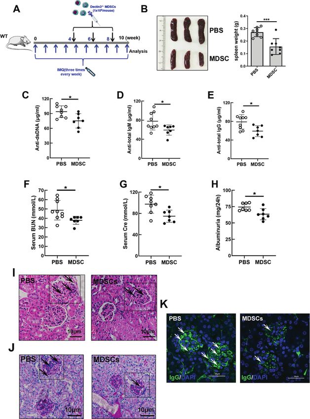
D. Li et al.
                  2
                      mammalian target of rapamycin (mTOR) inhibitor INK128 relieved                ester (CFSE) was brought from Thermo Fisher Scientific. The PI and
                      the symptoms of pristane-induced lupus via downregulating the                 Annexin-V were obtained from FMS.
                      expansion of MDSCs [13, 14]. The depletion of MDSCs in
                      humanized NOD/SCID mice significantly alleviated the symptoms                  Isolation of MDSCs and MDSC suppression assay
                      of SLE [11]. However, the molecular mechanism to regulate the                 Spleen tissue- and bone marrow (BM)-derived MDSCs in mice were purified
                      accumulation and function of MDSCs in SLE remains unclear.                    using an MDSC isolation kit. CD4+ T cells were pretreated with CFSE
                         The adaptor protein Card9, the downstream signal molecules                 following the manufacturer’s protocols. The CD4+ T cells (2 × 10⁵ cells/
                      of CLRs, was reported to attenuate the progression of colitis-                well) were co-cultured with purified MDSCs at 1:2 ratio in a 96-well round-
                      associated colon cancer by restricting the expansion of MDSCs                 bottom plate pretreated with 4 μg/mL anti-CD3 monoclonal antibody and
                                                                                                    2 μg/mL anti-CD28 monoclonal antibody for 72 h. Then, the cells were
                      [15]. CARD9 also protected against lung cancer development by
                                                                                                    collected to detect the proliferation of CD4 +T cells using flow cytometry.
                      reducing the expansion of MDSCs and indoleamine 2,3-dioxy-
                      genase (IDO) production [16]. The accumulation of fungus-
                      mediated MDSCs was dependent on the activation of Dectin1                     In vitro generation of mouse MDSCs
                      [17]. β-Glucan induced the differentiation and function of                    The tibiae and femora were removed from 8–10-week-old C57BL/6 J mice,
                      M-MDSCs via the Dectin1 pathway to enhance antitumor immune                   and BM cells were flushed. Then, BM cells were supplemented with Roswell
                                                                                                    Park Memorial Institute Medium (RPMI) 1640, and 40 ng/mL murine
                      response [18]. Recently, some studies implicated some CLRs as                 IL-6 and 40 ng/mL granulocyte-macrophage colony-stimulating factor
                      risk genes for the progression of autoimmune diseases [19].                   (GM-CSF) were added for 4 days.
                      However, it remains unknown which CLRs affect MDSCs and
                      involve in the progression of SLE.
                         This study aimed to investigate the expression of CLRs in the              ELISA
                                                                                                    Anti-dsDNA, IgG, IgM, BUN, creatinine, and urine proteins were measured
                      development of SLE. We found that symptoms of lupus were                      using ELISA kits following the manufacturer’s protocols.
                      relieved in Dectin3-deficient mice via regulating the accumulation
                      and function of MDSCs. Moreover, Dectin3 promoted the
                      expression of LOX-1 on M-MDSCs to increase the Th17/Treg cell                 Quantitative reverse transcription-PCR
                      imbalance, indicating that LOX-1+ M-MDSCs could be regarded as                Total RNA was isolated using TRIzol reagent following the manufacturer’s
1234567890();,:

                                                                                                    protocols. Real-time PCR was performed using SYBR green dye on Step
                      new target cells for treating lupus.                                          One sequence detection system (Applied Biosystems, MA, USA). The
                                                                                                    relative abundance of genes was calculated using the 2−ΔΔCT method, and
                                                                                                    GAPDH or PBGD as the internal control. The primers used in the study are
                      MATERIALS AND METHODS                                                         listed in Supplementary Table 1.
                      Mice
                      Female wild-type (WT) C57BL/6 J mice (6–8 weeks old) were brought from        Western blot analysis
                      the Model Animal Research Center of Nanjing University (Nanjing, China).      The cells were lysed with cell lysis buffer (Beyotime P0013B) supplemented
                      Dectin3−/− mice were generated as previously described [20] were crossed      with the protease inhibitor complex (Beyotime P1006). The nuclear and
                      five generations onto C57BL/6 J background (96.88%) [15]. WT mice and          cytoplasmic proteins were extracted using a nuclear and cytoplasmic
                      Dectin3−/− mice were housed under pathogen-free conditions in a 12 h          protein extraction kit (Beyotime P0027) following the manufacturer’s
                      light and dark cycle. All procedures involving mice were based on the         protocols. The protein concentrations in the extracts were detected
                      institutional guidelines for animal care and approved by the Animal Care      using the BCA assay (Beyotime P0012S). Equal amounts of the protein
                      Committee at Nanjing University (SCXK-Jiangsu-2019-0056). All animals         sample were loaded to sodium dodecyl sulfate–polyacrylamide gel
                      were acclimatized for 2 weeks before experiments. For establishing the        electrophoresis gels. Then, the protein sample was electrotransferred to
                      pristane-induced lupus mouse model, the mice were injected with pristane      polyvinylidene difluoride membranes. The membranes were blocked
                      (500 µL) by intraperitoneal injection for the following 7 months. For         with 5% BSA dissolved in TBST (50 mM Tris/HCl, pH 7.6, 150 mM NaCl,
                      generating imiquimod-induced mice with lupus, the right ears of the mice      and 0.1% Tween-20) for 2 h at room temperature and incubated with the
                      were treated with 1.25 mg of 5% imiquimod cream every other day for the       indicated primary antibody overnight at 4 °C, followed by incubation with
                      following 10 weeks.                                                           appropriate enzyme horseradish peroxidase-linked secondary antibody for
                                                                                                    2 h at room temperature. Protein bands were visualized using ECL plus
                                                                                                    western blotting detection reagents (Millipore, MA, USA). The blot images
                      Antibody and reagents                                                         were captured using the FluorChem8000 imaging system (Alpha-Innotech,
                      Antibodies against phosphorylated FoxO1 (Ser256, 9641), phosphorylated        CA, USA). The gray values were analyzed using ImageJ gel analysis
                      Akt1 (Ser473, 9018), phosphorylated Syk (Tyr525/526, 2710), FoxO1             software [21].
                      (C29H4, 2880s), Akt1 (C73H10, 2938), Syk (D3Z1E, 13198), Bim (2933),
                      Bcl2 (3498), Bax (2772), PCNA (13110), and β-actin (4970) for western blot
                      analysis were bought from Cell Signaling Technology. The antibody against     Histological analysis
                      LOX-1 (DF6522) for western blot analysis was obtained from Affinity. The       The kidneys were fixed with 4% paraformaldehyde in phosphate buffer.
                      anti-IgG antibody for immunofluorescence staining was bought from              Paraffin-embedded samples were sectioned at 3 μm and then stained with
                      Abcam. The enzyme-linked immunosorbent assay (ELISA) kits for mouse           hematoxylin and eosin (HE) or periodic acid-Schiff (PAS).
                      dsDNA, total IgG, and IgM were purchased from FMS. The ELISA kits for
                      mouse creatinine and blood urea nitrogen were obtained from Wako Pure
                      Chemical Industries. The ELISA kit for mouse urine protein was procured       Immunofluorescence staining
                      from Bethyl Laboratories. The anti-CD11b-fluorescein isothiocyanate (FITC)     Paraffin-embedded kidney tissues were used in the study. Then, the heat-
                      antibody, Gr1-allophycocyanin (APC) antibody, CD4-FITC antibody, CD69-        mediated antigen retrieval was used to treat the slides with citrate buffer.
                                                                                                    The slides were treated with the primary antibody against mouse IgG-FITC
                      APC antibody, B220-FITC antibody, CD25-APC antibody, CD45-PerCp, Ly6G-
                                                                                                    (Servicebio) overnight at 4 °C. The slides were counterstained with DAPI for
                      phycoerythrin (PE), Ly6C-APC, FoxP3-PE antibody, and IL-17-PE antibody
                      for mouse detection were obtained from Biolegend. The mouse LOX-1-PE-         5 min. Fluorescence images were captured using a laser scanning confocal
                      cy7 was bought from the R&D system. The anti-CD45-PerCp antibody, HLA-        microscope (FV3000, Olympus Corporation, Japan).
                      DR-PE Vio770, CD14-Alexa Fluor488, CD11b-PE-Cy5, CD66b-APC, and
                      CD33-PE for human sample detection were obtained from Biolegend.              Flow cytometry analysis
                      The human LOX-1-Alexa Fluor 750 and Dectin3-Alexa Fluor 750 were              The BM, kidney, and spleen cells were used to prepare single-cell
                      bought from Miltenyi. The human FoxO1-Alexa Fluor 750 was obtained            suspensions. Then, the red cells were lysed, filtered through 70-μm cell
                      from Novus. The cytokines of GM-CSF and IL-6 were obtained from Miltenyi      strainers, and collected by centrifugation at 300 × g for 5 min at 4 °C. After
                      Biotec. The MDSCs and CD4+ T cells were sorted using magnetic beads           washing, the cells were immediately prepared for flow cytometry. For
                      bought from Miltenyi Biotec. The purified CD3 and CD28 antibodies were         detecting cell surface markers, the cells were pre-incubated with the
                      obtained from eBioscience. The probe of carboxyfluorescein succinimidyl        primary antibody for 30 min at 4 °C in the dark and then washed with

                                                                                                                           Cell Death and Disease (2021)12:829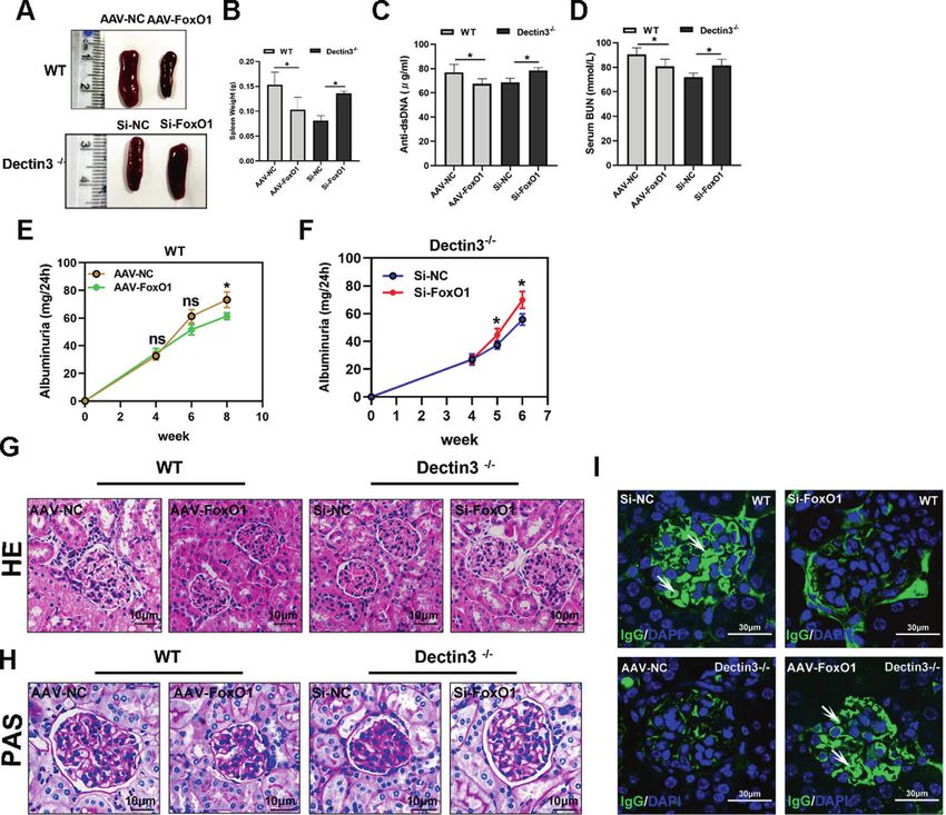
D. Li et al.
                                                                                                                                                                 3
                                                                                 prospectively enrolled. All SLE patients were diagnosed according to the
 Table 1.   The basic information of SLE patients.                               criteria set out by American College of Rheumatology revised criteria in
                                   Active group             Inactive group       1997 [22]. Patients who had other autoimmune diseases; had a history of
                                                                                 familial hyperlipidemia and/or thyroid disease, diabetes mellitus, and/or
                                   n = 35                   n = 24               other rheumatic diseases; and took lipid-lowering agents or thyroid
Age (year, mean ± SD)              45 ± 8                   41.2 ± 5.1           medications were excluded. The disease activity of these patients was
Gender (female/male)               30/5                     21/3                 measured using the Systemic Lupus Erythematosus Disease Activity Index
                                                                                 (SLEDAI) [22]. Disease activity was evaluated using the SLEDAI and a cutoff
SLEDAI < 8                                                  24 (40.6%)           of ≥8 was used to define active disease (Table 1). This study was approved
SLEDAI ≥ 8                         35 (59.4%)                                    by the ethics committee at The Affiliated Drum Tower Hospital of Nanjing
                                                                                 University Medical School (ID: SC201700201) and undertaken according to
                                                                                 the guidelines of the Declaration of Helsinki. At entry, patients completed a
phosphate-buffered saline (PBS). For detecting intracellular markers (IL-        standardized medical history, laboratory tests, and analyses. All the
17A), the cells were re-stimulated with phorbol myristate acetate (PMA;          detections were carried out at the clinical laboratory of Nanjing Drum
25 ng/mL) and ionomycin (250 ng/mL) for 6 h in the presence of brefeldin         Tower Hospital.
A prior to the staining of cell surface markers. The Treg cells were surface
labeled with CD4 and CD25 for 30 min at 4 °C in the dark, followed by
fixation, permeabilization, and intracellular staining with Foxp3 for another     Screening of differentially expressed genes
30 min at 4 °C in the dark. The mouse Th17 cells were surface labeled with       The samples of MDSCs were analyzed by microarray hybridization in
CD4 for 30 min at 4 °C in the dark, followed by fixation, permeabilization,       Beijing Oligo Biotech Co. (Beijing, China). The preliminary progression was
and intracellular staining with IL-17 for another 30 min at 4 °C in the dark.    analyzed after obtaining the raw data. The fold value represented the
Flow cytometry was performed on a FACSCalibur flow cytometer (BD                  degree of differential expression between MDSCs of WT mice with lupus
Biosciences). The data were analyzed using FlowJo software (Tree Star, OR,       and Dectin3-deficient mice with lupus. The standard used to judge
USA) [21]. The whole blood was obtained from mice, and peripheral blood          differential expression was as follows: the gene expression from the MDSCs
mononuclear cells (PBMCs) were isolated following the manufacturer’s             of WT mice with lupus was used as a valid gene. Compared with the
protocols for density reagent. The sorting of LOX-1+ M-MDSCs and LOX-1−          MDSCs of WT mice with lupus, a fold change of 1.0 indicated an upregulated
                                                                                 gene. Genes with a fold change >1.5 or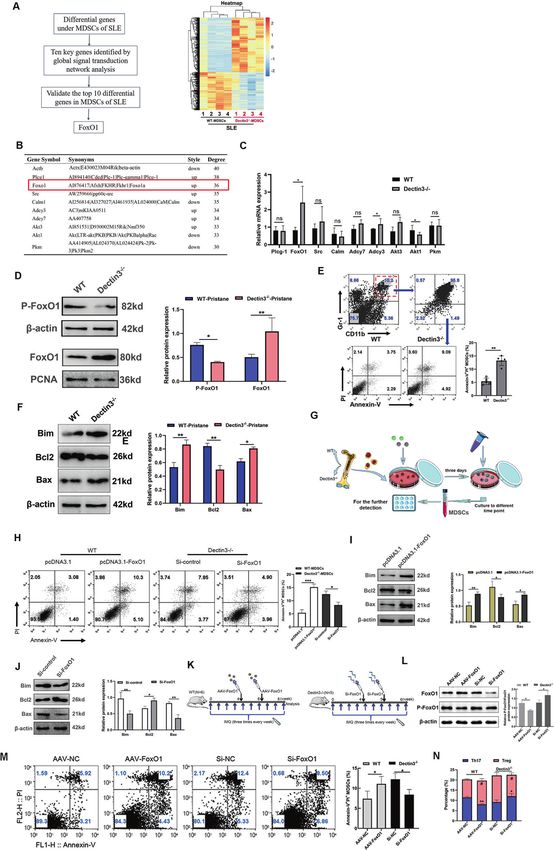
D. Li et al.
4

    Fig. 1 Dectin3 deficiency prevented pristane-induced lupus-like disease. A Schematic diagram of a pristine-induced lupus mouse model.
    B Representative photographs of spleens and spleen weights. C Serum level of anti-dsDNA was measured using ELISA. D, E ELISA of the serum
    levels of total IgG and IgM. F, G ELISA of the serum levels of BUN and Cre. H The level of mouse urine protein was detected using ELISA. I, J HE
    and PAS staining of kidney sections (scale bar = 10 μM). K IgG deposits in glomeruli were detected by immunofluorescence analysis (scale
    bar = 30 μM). Data represent the mean scores ± SEM. *P ≤ 0.05, *P ≤ 0.01, ***P ≤ 0.001; n = 7–9 mice in each group.

                                                                                                   Cell Death and Disease (2021)12:829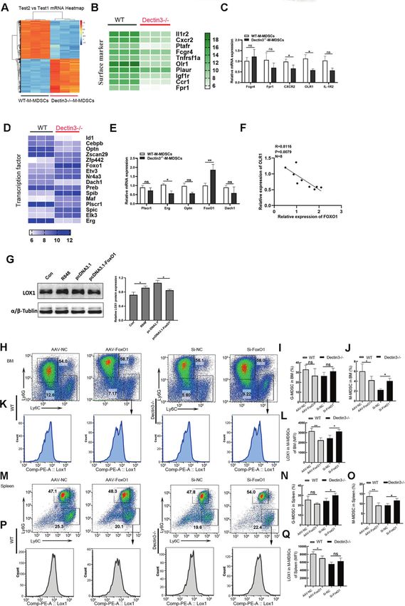
D. Li et al.
5
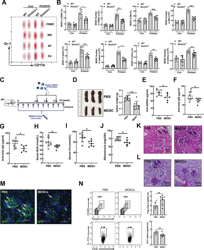
Fig. 2 Adoptive transfer of MDSCs from Dectin3−/− mice with lupus alleviated the induction of lupus-like disease. A Representative flow
cytometry results of MDSCs in PBMC, BM, SP, and Kd of each group. B Statistical data of percentages and absolute counts of MDSCs in PBMC,
BM, SP, and Kd. C Schematic diagram of the adoptive transfer of MDSCs. D Representative photographs of the spleen and spleen weights in
the MDSC and PBS groups. E–I ELISA of the serum levels of anti-dsDNA, anti-total IgM, IgG, BUN, and Cre in the MDSC and PBS groups. J The
level of 24-h mouse urine protein was measured using ELISA. K, L HE and PAS staining of kidney sections in the MDSC and PBS groups (scale
bar = 10 μM). M Amounts of IgG deposits in glomeruli were measured using immunofluorescence analysis (scale bar = 30 μM). N Flow
cytometry analysis detected the percentages of Th17 and Treg cells. Data represent the mean scores ± SEM. *P ≤ 0.05, **P ≤ 0.01, ***P ≤ 0.001;
n = 7–9 mice in each group.
Cell Death and Disease (2021)12:829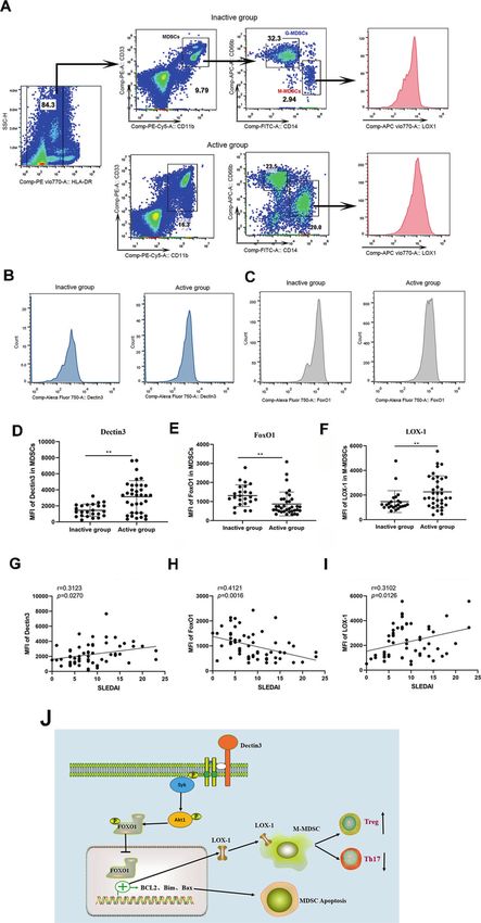
D. Li et al.
6
    B). Meanwhile, the function of MDSCs was improved in Dectin3−/−          Syk-Akt1-mediated nuclear transfer of FoxO1 reduced in
    mice with pristane-induced lupus (Fig. S4).                              MDSCs of Dectin3−/− mice with lupus
       Purified MDSCs (1 × 10⁶ cells/mouse) were injected into WT             To explore further the molecular mechanism of regulating the
    mice with lupus according to the schematic diagram (Fig. 2C). The        nuclear transfer of FoxO1 in Dectin3-deficient MDSCs, a co-
    splenomegaly was relieved in mice with the adoptive transfer of          expression network of differentially expressed genes with a hub of
    MDSCs (MDSC-group mice) compared with mice in the PBS group              FoxO1 was used. FoxO1 is regulated by the PI3K/Akt1 pathway and
    (Fig. 2D). The serum levels of anti-dsDNA, anti-total IgM, IgG, BUN,     associated with cell survival and apoptosis progression (Fig. 4A).
    Cre, and albuminuria significantly decreased in the MDSC-group            The phosphorylation of Akt1 and Syk reduced in MDSCs of
    mice compared with the PBS-group mice (Fig. 2E–J). The lupus             Dectin3−/− mice with lupus compared with WT mice with lupus
    nephritis was detected by HE and PAS staining and the amount of          (Fig. 4B, C). BM-MDSCs with Syk and Akt1 interference fragments
    glomeruli IgG deposits reduced in the MDSC group compared                were transfected for 12 and 24 h (Fig. 4D, E) and then treated with
    with the PBS group (Fig. 2K–M). The balance of Th17 and Treg cells       R848 for 24 h to confirm whether Dectin3 regulated the nuclear
    improved in the MDSC group compared with the PBS group                   transfer of FoxO1 via the Syk-Akt1 signal. BM-MDSCs transfected
    (Fig. 2N). The expansion of MDSCs in PBMC, BM, SP, and Kd                with Akt1 RNA fragment significantly reduced the nuclear transfer
    significantly reduced in the MDSC group compared with the PBS             of FoxO1 after R848 stimulation (Fig. 4F, G). The data showed that
    group (Fig. S5A–H). In addition, the activation of B and T               Dectin3 inhibited Syk-Akt1-mediated nuclear transfer of FoxO1.
    lymphocytes significantly reduced in the MDSC group compared
    with the PBS group (Fig. S5A–H). These data indicated that               Interference of FoxO1 promoted the induction of lupus-like
    Dectin3−/− mice depended mainly on the regulation of MDSCs to            disease in Dectin3−/− mice
    relieve lupus progression.                                               To further confirm whether Dectin3 promoted lupus progression
                                                                             via inhibiting FoxO1 expression, FoxO1-high-expression adeno-
    Dectin3 deficiency promoted FoxO1-mediated apoptosis and                  virus (0.2 mL of 1 × 10¹¹) and 0.2 mL of empty adenovirus were
    reduced the accumulation of MDSCs                                        injected into WT mice with lupus intravenously (Fig. 3K, left). Si-
    To explore the molecular mechanism underlying the reduction in           FoxO1 RNA fragment (0.2 mL of 15 nmol) and 0.2 mL of negative
    MDSC accumulation, MDSCs were isolated from pristane-induced             control fragment were injected into Dectin3−/− mice with lupus
    WT and Dectin3−/− mice with lupus to perform transcriptome               intravenously (Fig. 3K, right). The results showed that the
    microarray assays (Fig. 3A). According to the analysis pipeline,         splenomegaly in AAV-FoxO1 WT mice with lupus was significantly
    we analyzed the differential expression genes of MDSCs from the          relieved compared with that in AAV-NC-group mice. However, the
    spleen between WT lupus mice and Dectin3−/− lupus mice, and              splenomegaly in Si-FoxO1 Dectin3−/− mice with lupus was higher
    the results were shown on a heatmap (Fig. 3A). Then, these               than in Si-NC-group mice (Fig. 5A, B). The serum Anti-dsDNA and
    candidates were addressed by performing a secondary screening            BUN were higher in Si-FoxO1 Dectin3−/− mice with lupus than in
    by using the global signal-transduction network based on the             Si-NC-group mice (Fig. 5C, D). The symptoms of lupus nephritis
    significantly regulated GOs and pathways to determine the core            were significantly aggravated in Si-FoxO1 Dectin3−/− mice with
    different genes, and the top 10 genes were found to possess the          lupus compared with Si-NC-group mice, including decreased
    score of a degree above 30 (Fig. 3B). To further narrow down             infiltration of crescentic and renal interstitial inflammatory cells
    the candidates, we then used a QPCR assay to screen for the ones         and reduced amounts of glomeruli IgG deposits (Fig. 5E–I). These
    that highly correlated with the SLE process in MDSCs isolated from       data indicated that the downregulation of FoxO1 exacerbated the
    the spleen of WT lupus mice and Dectin3−/− lupus mice. Among             progression of Dectin3−/− lupus-deficient mice.
    these 10 genes, FoxO1, which is involved in regulating cell
    apoptosis and survival, showed the most change fold in MDSCs             FoxO1 negatively regulated LOX-1 expression in M-MDSCs
    from Dectin3−/− lupus mice compared with WT lupus mice,                  from Dectin3−/− mice
    whereas the other candidates showed a little or moderate change          Compared with WT lupus mice, the expansion of M-MDSCs
    (Fig. 3C). In addition, the nuclear transfer of FoxO1 increased in       significantly decreased in Dectin3−/− lupus mice was revealed
    MDSCs of Dectin3−/− mice with lupus (Fig. 3D). The flow                   (Fig. S7). To perform transcriptome microarray assays so as to
    cytometry analysis results indicated that the percentage of MDSC         explore the molecular mechanism of how Dectin3 influenced M-
    apoptosis was higher in Dectin3−/− lupus mice than that in WT            MDSC, M-MDSCs were isolated from pristane-induced WT mice
    lupus mice (Fig. 3E). And the protein expressions of Bim and Bax         and Dectin3−/− mice with lupus (Fig. 6A). MDSCs own the
    increased in MDSCs from Dectin3−/− lupus mice compared with              heterogeneity, and the differential expression of surface markers
    WT lupus mice; however, the expression of Bcl2 in MDSCs of               on MDSCs has a different role in SLE. Thus, we analyzed different
    Dectin3−/− mice with lupus was lower than that in WT mice with           genes of surface markers. The expression of OLR1 (LOX-1 protein
    lupus (Fig. 3F). The apoptosis level of MDSCs in Dectin3−/− mice         gene) significantly decreased in M-MDSCs of Dectin3−/− lupus
    increased following FoxO1 interference (Fig. 3G–J).                      mice compared with WT lupus mice (Fig. 6B, C). The data
       To confirm whether Dectin3 promotes FoxO1-mediated MDSC                suggested that the expression of LOX-1 on M-MDSCs of SP, BM,
    accumulation and abnormal immunomodulatory function in vivo,             and Kd significantly decreased in Dectin3−/− lupus mice
    FoxO1-high-expressed adenovirus (0.2 mL of 1 × 10¹¹) and 0.2 mL          compared with WT lupus mice (Fig. S8).
    of empty adenovirus were injected into WT mice with lupus                   To further determine the molecular mechanism of regulating
    intravenously (Fig. 3K, left). Meanwhile, Si-FoxO1 RNA fragment          the expression of OLR1 on Dectin3-deficient M-MDSCs, the
    (0.2 mL of 15 nmol) and 0.2 mL of negative control fragment were         expression of FoxO1 significantly increased in M-MDSCs of
    injected into Dectin3−/− mice with lupus intravenously (Fig. 3K,         Dectin3−/− mice with lupus compared with WT mice with lupus
    right). The results showed that percentages of MDSCs increased in        (Fig. 6D, E). The mRNA expression of FoxO1 negatively correlated
    BM, spleen, and kidney of Si-FoxO1 Dectin3−/− mice with lupus            with OLR1 in Dectin3-deficient M-MDSCs (Fig. 6F). When FoxO1
    compared with Si-NC-group mice (Fig. S6). The nuclear transfer of        was further overexpressed in BM-M-MDSCs and then stimulated
    FoxO1 in Si-FoxO1 Dectin3−/− mice with lupus was lower than              with R848, the protein expression of LOX-1 increased (Fig. 6G). The
    that in Si-NC-group mice. The percentage of apoptotic MDSCs              expression of LOX-1 on M-MDSCs increased in BM, spleen, and
    decreased in Si-FoxO1 Dectin3−/− mice with lupus compared with           kidney of Si-FoxO1 Dectin3−/− mice with lupus compared with Si-
    Si-NC-group mice (Fig. 3M). These data indicated that Dectin3            NC-group mice (Fig. 6H–Q) in vivo. Above all, the results
    deficiency promoted FoxO1-mediated apoptosis to decrease the              suggested that FoxO1 negatively regulated the expression of
    expansion of MDSCs in lupus development in vitro and in vivo.            LOX-1 in M-MDSCs in vitro and in vivo.

                                                                                                 Cell Death and Disease (2021)12:829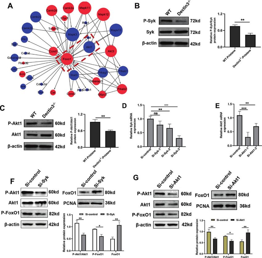
D. Li et al.
                                                                                                                                               7

LOX-1+ M-MDSCs promoted the differentiation of Th17 cells                  with CD4+ T cells from normal mice in different conditional media
To explore the effect of LOX-1 expression on the immunoregulatory          (Fig. 7A–C). The results showed that the inhibitory effect on the
function of M-MDSCs. LOX-1+ M-MDSCs promoted the accumulation              proliferation of CD4+ T cells was lower in LOX-1+ M-MDSCs than in
of Th17 cells and inhibited the differentiation of Treg cells, LOX-1+ M-   LOX-1− M-MDSCs (Fig. 7D). The data indicated that LOX-1+ M-MDSCs
MDSCs, and LOX-1− M-MDSCs from mice with lupus were co-cultured            promoted the accumulation of proinflammatory Th17 cells.

Cell Death and Disease (2021)12:829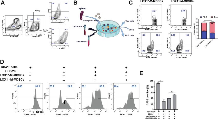
D. Li et al.
8
    Fig. 3 FoxO1 was significantly upregulated in MDSCs of Dectin3−/− mice with lupus and promoted MDSC apoptosis. A Analysis pipeline to
    identify Foxo1 as the downstream markers (left) and gene expression profiling of MDSCs (right). The mRNA of MDSCs was extracted and
    analyzed by gene expression profiling using a Mouse Transcriptome Array (v.1.0) (Affymetrix). B Top 10 genes ranked by degree after analysis
    of signal-net. C QPCR validation of microarray data. D Phosphorylated FoxO1 and nuclear transfer FoxO1 protein expression were evaluated
    by western blot analysis in MDSCs isolated from WT mice and Dectin3−/− mice with lupus. E Flow cytometry analysis of the apoptosis level of
    MDSCs from WT and Dectin3−/− mice with lupus. F Bim, Bcl2, and Bax protein expression were evaluated by western blot analysis in MDSCs
    isolated from WT mice and Dectin3−/− mice with lupus. G Schematic diagram of BM-MDSC induction. H Flow cytometry analysis of the
    apoptosis level of MDSCs after interfering with si-FoxO1 in Dectin3−/− BM-MDSCs and overexpression with pcDNA3.1-FoxO1 in WT BM-
    MDSCs. I, J Bim, Bcl2, and Bax protein expression were detected by western blot analysis in BM-MDSCs after interference or overexpression of
    FoxO1. K Schematic diagram of injection of AAV-FoxO1 and siRNA fragments. L P-FoxO1 and FoxO1 protein expression were detected by
    western blot analysis. M Flow cytometry analysis of the apoptosis level of MDSCs. N Flow cytometry analysis of Th17 and Treg cell
    differentiation. Data represent the mean scores ± SEM. *P ≤ 0.05, **P ≤ 0.01, ***P ≤ 0.001; n = 3–5 in each group.

    Fig. 4 Syk-Akt1-mediated nuclear transfer of FoxO1 was inhibited in Dectin3-deficient MDSCs. A Gene co-expression network analysis
    showed that FoxO1 was significantly related to Akt1. B P-Syk and Syk protein expression was evaluated by western blot analysis in MDSCs
    isolated from WT mice and Dectin3−/− mice with lupus. C P-Akt1 and Akt1 protein expression were evaluated by western blot analysis in
    MDSCs isolated from WT mice and Dectin3−/− mice with lupus. D Interference efficiency of RNA fragments of Syk evaluated using qPCR.
    E Interference efficiency of RNA fragments of Akt1 by qPCR. F P-Akt1, Akt1, P-FoxO1, and FoxO1 protein expression was detected by western
    blot analysis in BM-MDSCs after interference with Syk. G Western blot detection of P-Akt1, Akt1, P-FoxO1, and FoxO1 protein expression levels
    after interference with Akt1. Data represent the mean scores ± SEM. *P ≤ 0.05, **P ≤ 0.01, ***P ≤ 0.001; n = 3 per group.

    Increased Dectin3 expression had a positive correlation with             from SLE patients was analyzed by flow cytometry (Fig. 8A) and
    the accumulation of LOX-1+ M-MDSCs in patients with SLE                  the results showed that the relative expression of LOX-1 was
    Disease activity was evaluated using the SLEDAI and a cutoff of ≥8       increased on M-MDSCs of the active SLE group compared with the
    was used to define active disease (Table 1). Flow cytometric              inactive SLE group (Fig. 8F). Furthermore, the correlation between
    analysis of Dectin3 and statistical analysis of relative MFI of          SLEDAI scores and Dectin3, FoxO1, and LOX-1 levels in M-MDSCs
    Dectin3 on M-MDSCs of peripheral blood from SLE patients                 was analyzed by linear regression (stepwise). The data suggested
    (Fig. 8A, B). The results showed that Dectin3 was overexpressed on       that Dectin3 and LOX-1 levels on M-MDSCs from SLE patients were
    M-MDSCs of the active SLE group compared with the inactive SLE           positively correlated with SLEDAI scores (Fig. 8G, I), whereas the
    group (Fig. 8D). Moreover, the expression FoxO1 in M-MDSCs of            correlation between FoxO1 in M-MDSCs from SLE patients and
    peripheral blood from SLE patients was detected by flow                   SLEDAI score was negative (Fig. 8H). All the clinical data indicated
    cytometry (Fig. 8A, C). The downregulation of FoxO1 in                   that Dectin3 expression probably was positively correlated with
    M-MDSCs of the active group was detected compared with the               LOX-1 level on M-MDSCs in SLE patients, which involved in the
    inactive group (Fig. 8E). LOX-1 on M-MDSCs of peripheral blood           FoxO1 pathway.

                                                                                                 Cell Death and Disease (2021)12:829
D. Li et al.
9
Fig. 5 Silencing of FoxO1 expression in Dectin3-deficient mice aggravated the lupus-like disease. A, B Representative photographs of the
spleen and spleen weights. C–F ELISA analysis of the serum levels of anti-dsDNA, BUN, and 24-h mouse urine protein. G, H HE and PAS staining
of kidney sections (scale bar = 10 μM). I Amounts of IgG deposits in glomeruli were measured by immunofluorescence analysis (scale bar =
30 μM). Data represent the mean scores ± SEM. *P ≤ 0.05, **P ≤ 0.01, ***P ≤ 0.001; n = 5 mice per group.
DISCUSSION The data indicated that the spleens of the serum levels of dsDNA
Dectin3 is mainly expressed on the surface of myeloid cells and is antibody, total IgG, and IgM antibody of Dectin3−/− MDSC
an important type of pattern recognition receptor that can transplantation group were significantly lower than those in the
recognize sugar components on the cell wall of pathogens PBS group (Fig. 2C–J). In addition, the kidney damage was
[24, 25]. The role of Dectin3 has been shown to promote antifungal significantly relieved in the MDSC transplantation group
immunity against Candida spp., Fonsecaea pedrosoi, and Blastomyces compared with the PBS group (Fig. 2K–M). The data indicated
dermatitidis infections [18]. Studies reported that Dectin2 might be that the role of Dectin3 in alleviating lupus symptoms via
related to fungal-induced autoimmune diseases [26, 27], and the regulating MDSCs.
formation of Dectin3/Dectin2 heterodimer complexes had a greater To further determine the molecular mechanism of Dectin3 in
ability of antifungal immune response. But the role of Dectin3 in regulating MDSCs accumulation and function, we analyzed the
autoimmune diseases remains unknown. This study was novel in mRNA expression profile of MDSCs in WT and Dectin3−/− lupus
exploring the role of Dectin3 in the pathogenesis of lupus. mice. The results of the transduction network of the differential
The lupus symptoms of Dectin3−/− mice were relieved expressed genes were analyzed by bioinformatics, combined with
compared with those in WT lupus mice (Figs. 1–2). But the the GO and pathway analysis, which revealed that FoxO1
mechanism of Dectin3 to regulate the SLE progress remains expressed in MDSCs of Dectin3−/− lupus mice lower than that in
uncertain. Our previous studies suggested that MDSCs played a WT lupus mice.
vital role in the pathogenesis of SLE [24–26, 28]. However, our In addition, studies showed that FoxO1 was mainly involved in
previous study indicated Card9 (the downstream adaptor protein pathophysiological processes such as cell proliferation, apoptosis,
of Dectin3) reduced the incidence of colorectal cancer by and oxidative stress [29–32]. Researches showed that FoxO1 mRNA
inhibiting MDSCs recruitment [15]. Meanwhile, Card9 relieved expression in PBMCs of SLE patients was lower than that in
the incidence of lung cancer by reducing IDO production in healthy controls, which negatively correlated with SLEDAI. Our
MDSCs [16]. Whether the role of Dectin3 in regulating the lupus results indicated that Dectin3 negatively correlated with FoxO1
process depends on MDSCs remains unknown. gene expression in MDSCs isolated from lupus mice.
Therefore, adoptive transfer experiments were addressed to The bioinformatics analysis showed that the FoxO1 signal was
explore whether Dectin3 regulates the lupus process via MDSCs. mainly concentrated in the biological processes of MDSC
Cell Death and Disease (2021)12:829D. Li et al.
10
apoptosis, oxidative stress, cell proliferation, and so forth. In Bim and Bax in MDSCs of Dectin3−/− mice with lupus
MDSCs of Dectin3−/− mice with lupus, the phosphorylation level significantly increased, and the expression of anti-apoptotic
of FoxO1 was significantly downregulated, and the level of protein Bcl2 significantly decreased (Fig. 3D). The aforemen-
nuclear metastasis of FoxO1 significantly increased (Fig. 3C). At tioned results indicated that Dectin3 regulated the number of
the same time, the expression levels of pro-apoptotic proteins MDSCs by regulating the nuclear transfer level of FoxO1. FoxO1
Cell Death and Disease (2021)12:829D. Li et al.
11
Fig. 6 LOX-1 expression negatively correlated with the nuclear transfer of FoxO1 in M-MDSCs. A Gene expression profiling of M-MDSCs.
The mRNA of M-MDSCs was extracted and analyzed by gene expression profiling using a Mouse Transcriptome Array (v.1.0) (Affymetrix).
B Gene expression profiling of different genes on the surface of M-MDSCs. C mRNA expression of Fcgr4, Fpr1, CXCR2, IL-1R2, and OLR1 in
M-MDSCs of WT mice and Dectin3−/− mice with lupus. D Gene expression profiling of differential transcription factors in M-MDSCs. E mRNA
expression of Plscr1, Erg, Optn, Dach1, and FoxO1 in M-MDSCs of WT mice and Dectin3−/− mice with lupus mice. F Analysis of the correlation
between FoxO1 and OLR1 genes. G LOX-1 protein expression was detected by western blot analysis. H–J Representative flow cytometry
results of G-MDSCs and M-MDSCs in each group and the statistical data of percentages of G-MDSCs and M-MDSCs in BM. K, L Representative
flow cytometry results of the LOX-1 expression level in M-MDSCs in the BM of each group. M–O Representative flow cytometry results
of G-MDSCs and M-MDSCs in each group and the statistical data of percentages of G-MDSCs and M-MDSCs in the spleen. P, Q Representative
flow cytometry results of the LOX-1 expression level in M-MDSCs in the spleen of each group. Data represent the mean scores ± SEM. *P ≤ 0.05,
**
P≤ 0.01, and ***P ≤ 0.001; n = 3–5 in each group.
Fig. 7 LOX-1+ M-MDSCs promoted the differentiation of Th17 cells. A Schematic diagram of the sorting of LOX-1+ M-MDSCs and LOX-1−
M-MDSCs in the spleen. B Schematic diagram of co-cultivation of LOX-1+ M-MDSCs and LOX-1− M-MDSCs with CD4+ T cells. C Flow
cytometry analysis of Th17 and Treg cell differentiation. D, E Flow cytometry analysis of the ability of MDSCs to inhibit T-cell proliferation. Data
represent the mean scores ± SEM. *P ≤ 0.05, **P ≤ 0.01, and ***P ≤ 0.001; n = 3–5 in each group.
(Thr24, Ser256, and Ser319) was phosphorylated by Akt, which regulatory process of MDSCs. The magnetic bead sorting
was transported from the nucleus to the cytoplasm, resulting in method was used to sort and purify the M-MDSCs in the spleen
the loss of transcriptional activity, thereby inhibiting the of WT mice and Dectin3−/− mice with lupus followed by gene
expression of downstream genes regulated by FoxO1. The chip analysis.
acetylation of FoxO1 weakened the ability to bind to homo- Studies confirmed that G-MDSCs with the high expression of
logous DNA sequences while enhancing the phosphorylation of LOX-1 in the tumor environment had a stronger immunosup-
FoxO1, further reducing its transcriptional activity [29–31, 33, 34]. pressive function on T cells and promoted tumorigenesis [39–41].
Dectin3 can induce Card9/Bcl-10/Malt1 to form a complex by Also, low-density granulocytes with high LOX-1 expression in the
activating Syk, which in turn activates NF-κB and other pathways, lupus environment had no significant immunosuppressive ability
initiating the innate immune response, and mediates adaptive on T cells, but promoted the production of inflammatory T cells
immune response [32, 35]. In addition, studies pointed out that and the development of lupus [42]. LOX-1+ M-MDSCs in the
the Syk gene could bind to the promoter region of Akt1 to lupus environment inhibited the proliferation of T cells to a lesser
promote its transcriptional activation, thereby promoting cell extent compared with LOX-1− M-MDSCs and promoted Th17/
proliferation [36, 37]. Dectin3 inhibited the nuclear metastasis of Treg imbalance.
FoxO1 in lupus MDSCs through the Syk-Akt1 signal axis and The M-MDSCs in the spleens of different groups of mice
participated in the development of lupus. with lupus were further sorted and enriched, revealing that
Recent studies showed that a large number of accumulated the apoptosis level of M-MDSCs after knocking down
M-MDSCs were involved in the development of lupus [38]. The FoxO1 significantly decreased, and the increase in the number
exact mechanism of the increase in the number of MDSCs in of inflammatory Th17 cells was promoted in Dectin3−/− lupus
patients and mice with lupus is still unclear. This may be mice. These data indicated that LOX-1+ M-MDSCs were related to
related to the lack of specific surface markers of MDSCs, which, the exacerbation of the lupus process and might be potential
to some extent, hinders the understanding of the complex target cells for lupus.
Cell Death and Disease (2021)12:829D. Li et al.
12
Fig. 8 Dectin3 expression positively correlated with the level of LOX-1 on M-MDSCs in patients with SLE. A Schematic diagram of flow
cytometric detection of peripheral blood MDSCs and M-MDSCs in patients with SLE, and LOX-1 expression detection diagram in M-MDSCs
(10 samples in the inactive stage of the disease and 10 samples in the active stage of the disease). B, C Flow cytometry analysis of Dectin3 (up)
and FoxO1 (down) expression in peripheral blood MDSCs. D–F Statistical data of the expression of Dectin3, FoxO1, and LOX-1 in peripheral
blood MDSCs. G–I The correlations of Dectin3, FoxO1, and LOX-1 expressions with the score of SLEDAI. J Schematic diagram of the regulation
of the accumulation in Dectin3-deficient mice with lupus. Data represent the mean scores ± SEM. *P ≤ 0.05, **P ≤ 0.01, and ***P ≤ 0.001; active
group: n = 35, inactive group: n = 24.
Cell Death and Disease (2021)12:829D. Li et al.
13
DATA AND MATERIALS AVAILABILITY 25. Wang T, Pan D, Zhou Z, You Y, Jiang C, Zhao X, et al. Dectin-3 deficiency pro-
All data generated or analyzed during the study are included in the article. motes colitis development due to impaired antifungal innate immune responses
in the gut. PLoS Pathog. 2016;12:e1005662.
26. Brown BR, Lee EJ, Snow PE, Vance EE, Iwakura Y, Ohno N, et al. Fungal-derived
REFERENCES cues promote ocular autoimmunity through a Dectin-2/Card9-mediated
1. Takeuchi O, Akira S. Pattern recognition receptors and inflammation. Cell. mechanism. Clin Exp Immunol. 2017;190:293–303.
2010;140:805–20. 27. Graham LM, Brown GD. The Dectin-2 family of C-type lectins in immunity and
2. Salazar-Aldrete C, Galán-Díez M, Fernández-Ruiz E, Niño-Moreno P, Estrada- homeostasis. Cytokine. 2009;48:148–55.
Capetillo L, Abud-Mendoza C, et al. Expression and function of dectin-1 is 28. Li D, Shi G, Wang J, Zhang D, Pan Y, Dou H, et al. Baicalein ameliorates pristane-
defective in monocytes from patients with systemic lupus erythematosus and induced lupus nephritis via activating Nrf2/HO-1 in myeloid-derived suppressor
rheumatoid arthritis. J Clin Immunol. 2013;33:368–77. cells. Arthritis Res Ther. 2019;21:105.
3. Lee YH, Lee HS, Choi SJ, Ji JD, Song GG. Associations between TLR polymorph- 29. Jiang S, Li T, Yang Z, Hu W, Yang Y. Deciphering the roles of FOXO1 in human
isms and systemic lupus erythematosus: a systematic review and meta-analysis. neoplasms. Int J Cancer. 2018;143:1560–8.
Clin Exp Rheumatol. 2012;30:262–5. 30. Sundaresan S, Puthanveetil P. Is FoxO1 the culprit, partner in crime, or a protector
4. Fagone P, Mangano K, Mammana S, Quattrocchi C, Magro G, Coco M, et al. in systemic inflammation? Am J Physiol Cell Physiol. 2017;313:C239–c241.
Acceleration of SLE-like syndrome development in NZBxNZW F1 mice by beta- 31. Kousteni S. FoxO1: a molecule for all seasons. J Bone Min Res. 2011;26:912–7.
glucan. Lupus. 2014;23:407–11. 32. Zhao XQ, Zhu LL, Chang Q, Jiang C, You Y, Luo T, et al. C-type lectin receptor
5. Monrad SU, Rea K, Thacker S, Kaplan MJ. Myeloid dendritic cells display down- dectin-3 mediates trehalose 6,6’-dimycolate (TDM)-induced Mincle expression
regulation of C-type lectin receptors and aberrant lectin uptake in systemic lupus through CARD9/Bcl10/MALT1-dependent nuclear factor (NF)-κB activation. J Biol
erythematosus. Arthritis Res Ther. 2008;10:R114. Chem. 2014;289:30052–62.
6. Lee SJ, Silverman E, Bargman JM. The role of antimalarial agents in the treatment 33. Xing YQ, Li A, Yang Y, Li XX, Zhang LN, Guo HC. The regulation of FOXO1 and its
of SLE and lupus nephritis. Nat Rev Nephrol. 2011;7:718–29. role in disease progression. Life Sci. 2018;193:124–31.
7. Jenks SA, Cashman KS, Woodruff MC, Lee FE, Sanz I. Extrafollicular responses in 34. Shi F, Li T, Liu Z, Qu K, Shi C, Li Y, et al. FOXO1: another avenue for treating
humans and SLE. Immunol Rev. 2019;288:136–48. digestive malignancy? Semin Cancer Biol. 2018;50:124–31.
8. Gabrilovich DI, Nagaraj S. Myeloid-derived suppressor cells as regulators of the 35. Campuzano A, Castro-Lopez N, Martinez AJ, Olszewski MA, Ganguly A, Leopold
immune system. Nat Rev Immunol. 2009;9:162–74. Wager C, et al. CARD9 is required for classical macrophage activation and the
9. Veglia F, Perego M, Gabrilovich D. Myeloid-derived suppressor cells coming of induction of protective immunity against pulmonary cryptococcosis. mBio.
age. Nat Immunol. 2018;19:108–19. 2020;11:e03005–03019.
10. Pang B, Zhen Y, Hu C, Ma Z, Lin S, Yi H. Myeloid-derived suppressor cells shift 36. Szydlowski M, Kiliszek P, Sewastianik T, Jablonska E, Bialopiotrowicz E, Gorniak P,
Th17/Treg ratio and promote systemic lupus erythematosus progression through et al. FOXO1 activation is an effector of SYK and AKT inhibition in tonic BCR
arginase-1/miR-322-5p/TGF-β pathway. Clin Sci (Lond). 2020;134:2209–22. signal-dependent diffuse large B-cell lymphomas. Blood. 2016;127:739–48.
11. Wu H, Zhen Y, Ma Z, Li H, Yu J, Xu ZG, et al. Arginase-1-dependent promotion of 37. Hou X, Lin L, Xing W, Yang Y, Duan X, Li Q, et al. Spleen tyrosine kinase regulates
TH17 differentiation and disease progression by MDSCs in systemic lupus ery- mammary epithelial cell proliferation in mammary glands of dairy cows. J Dairy
thematosus. Sci Transl Med. 2016;8:331ra340. Sci. 2016;99:3858–68.
12. Ji J, Xu J, Zhao S, Liu F, Qi J, Song Y, et al. Myeloid-derived suppressor cells 38. Ji J, Li P, Shen C, Dou H, Wang T, Shi L, et al. MDSCs: friend or foe in systemic
contribute to systemic lupus erythaematosus by regulating differentiation of lupus erythematosus. Cell Mol Immunol. 2019;16:937–9.
Th17 cells and Tregs. Clin Sci (Lond). 2016;130:1453–67. 39. Nan J, Xing YF, Hu B, Tang JX, Dong HM, He YM, et al. Endoplasmic reticulum
13. Zhang D, Xu J, Ren J, Ding L, Shi G, Li D, et al. Myeloid-derived suppressor cells stress induced LOX-1(+) CD15(+) polymorphonuclear myeloid-derived sup-
induce podocyte injury through increasing reactive oxygen species in lupus pressor cells in hepatocellular carcinoma. Immunology. 2018;154:144–55.
nephritis. Front Immunol. 2018;9:1443. 40. Condamine T, Dominguez GA, Youn JI, Kossenkov AV, Mony S, Alicea-Torres K,
14. Shi G, Li D, Li X, Ren J, Xu J, Ding L, et al. mTOR inhibitor INK128 attenuates et al. Lectin-type oxidized LDL receptor-1 distinguishes population of human
systemic lupus erythematosus by regulating inflammation-induced CD11b(+)Gr1 polymorphonuclear myeloid-derived suppressor cells in cancer patients. Sci
(+) cells. Biochim Biophys Acta Mol Basis Dis. 2019;1865:1–13. Immunol. 2016;1.
15. Wang T, Fan C, Yao A, Xu X, Zheng G, You Y, et al. The adaptor protein CARD9 41. Chai E, Zhang L, Li C. LOX-1+ PMN-MDSC enhances immune suppression which
protects against colon cancer by restricting mycobiota-mediated expansion of promotes glioblastoma multiforme progression. Cancer Manag Res. 2019;11:7307–15.
myeloid-derived suppressor cells. Immunity. 2018;49:504–14. e504 42. Rahman S, Sagar D, Hanna RN, Lightfoot YL, Mistry P, Smith CK, et al. Low-density
16. Qu J, Liu L, Xu Q, Ren J, Xu Z, Dou H, et al. CARD9 prevents lung cancer devel- granulocytes activate T cells and demonstrate a non-suppressive role in systemic
opment by suppressing the expansion of myeloid-derived suppressor cells and lupus erythematosus. Ann Rheum Dis. 2019;78:957–66.
IDO production. Int J Cancer. 2019;145:2225–37.
17. Rieber N, Singh A, Öz H, Carevic M, Bouzani M, Amich J, et al. Pathogenic fungi
regulate immunity by inducing neutrophilic myeloid-derived suppressor cells. ACKNOWLEDGEMENTS
Cell Host Microbe. 2015;17:507–14. We would like to thank Dr. Lin Xin at Tsinghua University for providing the Dectin3−/−
18. Tian J, Ma J, Ma K, Guo H, Baidoo SE, Zhang Y, et al. β-Glucan enhances antitumor mice. This work was supported by the National Natural Science Foundation of China
immune responses by regulating differentiation and function of monocytic (no. 31872732, 32070883, and 81801555) and Jiangsu Province Six Talent Peaks
myeloid-derived suppressor cells. Eur J Immunol. 2013;43:1220–30. Project (no.YY-021), Provincial Key Research and Development Program of Jiangsu
19. Del Fresno C, Iborra S, Saz-Leal P, Martínez-López M, Sancho D. Flexible signaling Province (BE2019706).
of myeloid C-type lectin receptors in immunity and inflammation. Front Immunol.
2018;9:804.
20. Zhu LL, Zhao XQ, Jiang C, You Y, Chen XP, Jiang YY, et al. C-type lectin receptors AUTHOR CONTRIBUTIONS
dectin-3 and dectin-2 form a heterodimeric pattern-recognition receptor for host
Substantial contributions to conception and design: Y. H., H. D., D. Li, J. L., and T. W.
defense against fungal infection. Immunity. 2013;39:324–34.
Acquisition of data, analysis, and interpretation of data: D. Li, L. Lu, W. K., X. X., Y. P.,
21. Li D, Qi J, Wang J, Pan Y, Li J, Xia X, et al. Protective effect of dihydroartemisinin in
J. Li, and J. W. Drafting the article: Y. H., H. D., D. Li, and L. Lu. Revising it critically for
inhibiting senescence of myeloid-derived suppressor cells from lupus mice via
important intellectual content: Y. H., H. D., D. Li, L. Lu, J. L., and W. K.
Nrf2/HO-1 pathway. Free Radic Biol Med. 2019;143:260–74.
22. Hochberg MC. Updating the American College of Rheumatology revised criteria
for the classification of systemic lupus erythematosus. Arthritis Rheum.
1997;40:1725. ETHICS
23. Lian M, Wang H, Fang J, Zhai J, Wang R, Shen X, et al. Microarray gene expression This research was approved by the Ethics Committee at The Affiliated Drum Tower
analysis of chemosensitivity for docetaxel, cisplatin and 5-fluorouracil (TPF) Hospital of Nanjing University Medical School (ID: SC201700201) and was undertaken
combined chemotherapeutic regimen in hypopharyngeal squamous cell carci- according to the guidelines of the Declaration of Helsinki. All recruited patients
noma. Chin J Cancer Res. 2017;29:204–12. signed informed consent forms.
24. Huang HR, Li F, Han H, Xu X, Li N, Wang S, et al. Dectin-3 recognizes glucur-
onoxylomannan of cryptococcus neoformans serotype AD and cryptococcus
gattii serotype B to initiate host defense against cryptococcosis. Front Immunol. COMPETING INTERESTS
2018;9:1781. The authors declare no competing interests.
Cell Death and Disease (2021)12:829D. Li et al.
14
ADDITIONAL INFORMATION Open Access This article is licensed under a Creative Commons
Supplementary information The online version contains supplementary material Attribution 4.0 International License, which permits use, sharing,
available at https://doi.org/10.1038/s41419-021-04052-5. adaptation, distribution and reproduction in any medium or format, as long as you give
appropriate credit to the original author(s) and the source, provide a link to the Creative
Correspondence and requests for materials should be addressed to Tingting Wang, Commons license, and indicate if changes were made. The images or other third party
Jun Liang, Huan Dou or Yayi Hou. material in this article are included in the article’s Creative Commons license, unless
indicated otherwise in a credit line to the material. If material is not included in the
Reprints and permission information is available at http://www.nature.com/ article’s Creative Commons license and your intended use is not permitted by statutory
reprints regulation or exceeds the permitted use, you will need to obtain permission directly
from the copyright holder. To view a copy of this license, visit http://creativecommons.
Publisher’s note Springer Nature remains neutral with regard to jurisdictional claims org/licenses/by/4.0/.
in published maps and institutional affiliations.
© The Author(s) 2021
Cell Death and Disease (2021)12:829You can also read