Lymphocytic Choriomeningitis Virus-Induced Mortality in Mice Is
←
→
Page content transcription
If your browser does not render page correctly, please read the page content below
JOURNAL OF VIROLOGY, Jan. 2010, p. 312–320                                                                                    Vol. 84, No. 1
0022-538X/10/$12.00 doi:10.1128/JVI.00727-09
Copyright © 2010, American Society for Microbiology. All Rights Reserved.

     Lymphocytic Choriomeningitis Virus-Induced Mortality in Mice Is
               Triggered by Edema and Brain Herniation䌤
  Christine M. Matullo,1,2† Kevin J. O’Regan,1† Harvey Hensley,1 Mark Curtis,3 and Glenn F. Rall1,2*
     Program in Immune Cell Development and Host Defense, Fox Chase Cancer Center, 333 Cottman Avenue, Philadelphia,
          Pennsylvania 191111; Department of Microbiology and Immunology, Thomas Jefferson University, Philadelphia,
              Pennsylvania 191072; and Department of Pathology and Neuropathology, Thomas Jefferson University,
                                              Philadelphia, Pennsylvania 191073
                                             Received 8 April 2009/Accepted 1 October 2009

                                                                                                                                               Downloaded from http://jvi.asm.org/ on January 12, 2021 by guest
            Although much is known about lymphocytic choriomeningitis virus (LCMV) infection and the subsequent
         immune response in its natural murine host, some crucial aspects of LCMV-mediated pathogenesis remain
         undefined, including the underlying basis of the characteristic central nervous system disease that occurs
         following intracerebral (i.c.) challenge. We show that the classic seizures and paresis that occur following i.c.
         infection of adult, immunocompetent mice with LCMV are accompanied by anatomical and histological
         changes that are consistent with brain herniation, likely of the uncal subtype, as a causative basis for disease
         and precipitous death. Both by water weight determinations and by magnetic resonance imaging of infected
         brain tissues, edema was detected only at the terminal stages of disease, likely caused by the leakage of
         cerebrospinal fluid from the ventricles into the parenchyma. Furthermore, death was accompanied by unilat-
         eral pupillary dilation, which is indicative of uncal herniation. While immunohistochemical analysis revealed
         periventricular inflammation and a loss of integrity of the blood-brain barrier (BBB), these events preceded
         seizures by 2 to 3 days. Moreover, surviving perforin knockout mice showed barrier permeability equivalent to
         that of moribund, immunocompetent mice; thus, BBB damage does not appear to be the basis of LCMV-
         induced neuropathogenesis. Importantly, brain herniation can occur in humans as a consequence of injuries
         that would be predicted to increase intracranial pressure, including inflammation, head trauma, and brain
         tumors. Thus, a mechanistic dissection of the basis of LCMV neuropathogenesis may be informative for the
         development of interventive therapies to prevent this typically fatal human condition.

  Lymphocytic choriomeningitis (LCM) virus (LCMV), a mouse                    One attribute that makes LCMV so useful is that vastly
pathogen and prototypical member of the arenavirus family,                 different pathogenic outcomes can be achieved in mice by
has been invaluable for key discoveries in both immunology                 varying both host and viral parameters (e.g., the route of in-
and viral pathogenesis (reviewed in references 4 and 5). For               oculation, dose and viral strain, mouse strain, age, and immu-
example, LCMV was used to define T-cell receptor–class I                   nocompetence). As a result, LCMV infection can result in
major histocompatibility complex interactions, to establish the            asymptomatic clearance and immunity, lifelong persistent in-
relative immunodominance of viral epitopes, and to dissect the             fection, or rapid death (LCM). While much is known about the
events leading to CD8⫹ T-cell-mediated cytotoxicity (reviewed              first two of these outcomes, less is known about the basis of the
in reference 3). More recently, the generation, diversity, and             lethal, immune-mediated disease that occurs following intra-
exhaustion of memory T cells were established by using                     cerebral (i.c.) challenge of immunocompetent mice. In this
LCMV-challenged mice (2, 30). In addition to revealing sem-                instance, the delivery of as few as 1 PFU of LCMV into the
inal aspects of host immunity, LCMV has also been used to                  brain results in infection of the meninges, leptomeninges, and
define novel ways by which viruses trigger disease. For exam-              ependyma as well as the cerebrospinal fluid (CSF)-producing
ple, transgenic mice expressing an LCMV protein were used to               choroid plexus cells within the ventricles (3, 5). A rapid expan-
show that autoimmune disease can result from cross-reactivity              sion of virus-specific CD8⫹ T cells occurs in secondary lym-
between viral antigens and self-antigens following infection               phoid organs, which then migrate through the CSF to the
(“molecular mimicry”) (20), a process that was later shown to              infected central nervous system (CNS) (7, 33); the peak of
occur in humans following herpes simplex virus type 1 ocular               infiltration (6 to 7 days postinfection [dpi]) coincides with
infection (32). Moreover, LCMV-mediated suppression of                     characteristic seizures that immediately precede death.
cellular genes, including those encoding growth hormones                      While CD8⫹ T cells are essential for lethal disease (as CD8-
(15), neurotransmitters (17), and synaptic proteins (9), im-               deficient mice survive i.c. LCMV challenge [12, 19, 26, 29, 31]),
plicated novel roles for persisting, noncytopathic viruses in              the events that contribute to fatal neuropathology are not fully
chronic disease.                                                           established. Studies using knockout (KO) mice lacking key
                                                                           immune mediators (e.g., perforin [PFN], gamma interferon,
                                                                           granzyme B, Fas, and tumor necrosis factor alpha) indicated
  * Corresponding author. Mailing address: Fox Chase Cancer Cen-           that no single deficiency of any of these effector molecules
ter, 333 Cottman Avenue, Philadelphia, PA 19111. Phone: (215) 728-
3617. Fax: (215) 728-2412. E-mail: glenn.rall@fccc.edu.
                                                                           could fully prevent disease (14). Interestingly, disease onset
  † C.M.M. and K.J.O. contributed equally to this work.                    occurs ⬃2 to 3 days later in PFN KO mice than in wild-type
  䌤
     Published ahead of print on 14 October 2009.                          animals, which has been attributed to the reduced capacity of

                                                                     312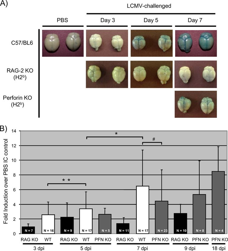
VOL. 84, 2010 BRAIN HERNIATION AND EDEMA IN LCMV-INFECTED MICE 313
PFN KO effector CD8⫹ T cells to secrete proinflammatory sets of 24 slices were acquired, with T2-weighted images made with four different
cytokines, resulting in a delayed recruitment of other effector echo times (TE ⫽ 13, 26, and 52 ms) for axial images and two echoes for each
coronal (TE ⫽ 13 and 52 ms) and sagittal (TE ⫽ 13 and 26 ms) image. The
cells to the CNS. Nevertheless, the CD8⫹ T-cell effector func- imaging time was approximately 17 min for each image set. MATLAB software,
tion(s) that causes death in LCMV-infected mice remains un- with the image processing toolbox, from MathWorks, Inc. (Natick, MA), was
known. used to determine the average pixel intensity for user-defined regions of interest.
Indeed, even the issue of whether CD8⫹ T cells act directly BBB permeability measurements. At various days after i.c. inoculation with
either 1,000 PFU LCMV-Arm or an equal volume of PBS, mice were deeply
or indirectly has been questioned: a recent report using two-
anesthetized with 400 l 3.8% chloral hydrate in PBS delivered intraperitoneally.
photon microscopy to visualize events that occur in the menin- Once animals were confirmed to be nonresponsive, 200 l of a 2% Evans blue
ges following infection showed that the infiltration of CD8⫹ T solution was then administered transcardially; 2 min postinjection, the mice were
cells coincided with the entry of neutrophils and monocytes, perfused with 30 ml PBS. Brains were carefully removed from the skull and
which those authors speculated play a role in fatal disease (14). either placed into test tubes, snap-frozen in liquid N2 and stored at ⫺80°C, or
photographed to determine gross changes in BBB permeability. Frozen brain
The depletion of both neutrophils and monocytes, but not tissues were slowly thawed at room temperature, homogenized in 1 ml N,N-
either cell population alone, delayed lethality somewhat, argu- dimethyl formamide, and incubated at 50°C for 48 h. Following centrifugation at
ing that while CD8⫹ T cells are important, they may serve 2,700 ⫻ g for 10 min, the absorbance of the supernatant containing extracted
Downloaded from http://jvi.asm.org/ on January 12, 2021 by guest
primarily a chemotactic role for other hematogenous effector Evans blue was measured in a spectrophotometer at 620 nm and normalized to
the weight of the brain. The Evans blue uptake into the tissues of each infected
populations.
animal was divided by the average uptake detected in similar tissues from PBS
Independent of the cells that are responsible for pathogen- control mice, and the results are expressed as the change in induction. Statistical
esis, the timing and order of events that lead to lethal LCM, as significance was determined by using the Wilcoxon signed-rank test.
well as the specific basis of mortality, remain poorly defined. Brain water weight measurements. Mice were inoculated i.c. with 1,000 PFU
Previous reports suggested that the loss of integrity of blood- LCMV-Arm or an equal volume of PBS. At various dpi, brains were harvested
after sacrifice by isoflurane inhalation. Brains were removed, placed into test
brain barrier (BBB) permeability may be important (1, 23, 27), tubes, and incubated in a vacuum oven for 24 h at ⬃80°C under a vacuum of 15
although it is not known how early postinfection this occurs or mm Hg. Brain weights were determined both prior to and immediately following
what role increased barrier permeability plays in LCM disease desiccation, with the percent water content calculated by the following formula:
(1, 6, 10, 23, 28). Alternatively, we hypothesized that the de- [(wet weight ⫺ dry weight)/wet weight] ⫻ 100. The water weight of the brain of
each infected animal was divided by the average water weight detected in similar
struction of the cells lining the ventricles could result in edema
tissues from PBS control mice, and the results are expressed as percent changes.
and increased intracranial pressure, triggering events that then Statistical significance was determined by using the Wilcoxon signed-rank test.
lead to the sudden onset of seizures and death in virus-chal-
lenged mice.
RESULTS
Here, we identify novel anatomical changes that are coinci-
dent with seizures and death in LCMV-infected mice; these Anatomical changes following i.c. LCMV challenge. i.c. chal-
events are consistent with uncal herniation resulting from ven- lenge of immunocompetent mice with a low dose of LCMV
tricular leakage and edema. Thus, while BBB damage is ap- (1,000 PFU) results in early signs of illness by ⬃5 to 6 dpi,
parent, we propose that ventricular failure, which is more tem- including ruffled fur, ataxia, and tremors. At the terminal
porally associated with fatal choriomeningitis, is the causative (“late”) stages of disease, between 6.5 and 7 dpi, mice experi-
basis of this classic immunopathological disease. ence pronounced seizures that can be triggered by movement
or loud noises. These seizures each last ⬃5 to 10 s and are
typified by limb extension; death is usually coincident with a
MATERIALS AND METHODS
seizure episode. As a result, mortality is accompanied by a
Mice, virus, and infections. All experiments were reviewed and approved by classic decerebrate posture, as shown in Fig. 1A. This postmor-
the Fox Chase Cancer Center Institutional Animal Care and Use Committee.
Inbred C57BL/6, recombinase-activating gene 2 (RAG-2)-deficient (24), and
tem appearance is unique to LCMV infection and is not ob-
PFN-deficient (29) mice were maintained in the closed breeding colony at Fox served in mice that succumb to other CNS infections, including
Chase. All animals were maintained on the H-2b background. LCMV Armstrong poliovirus or measles virus (data not shown). Inoculation of T-
(LCMV-Arm; ATCC) was passaged in BHK-21 fibroblasts and plaque purified; and B-cell-deficient RAG-2 KO (RAG KO) mice with LCMV
titers were determined with Vero fibroblasts. Mice were infected i.c. along the
did not result in any evident disease, and all RAG KO mice
midline with 103 PFU LCMV-Arm, or with phosphate-buffered saline (PBS) as
a control, in a total volume of 30 l by using a sterile 27-gauge needle. All mice became persistently infected, indicating that morbidity and
were anesthetized with Metofane prior to inoculation and were monitored daily mortality are due to immunopathology rather than direct viral
for signs of illness. damage.
Histological analysis of mouse tissues. Brains from four to five mice per time Two unique anatomical features were noted for mice that
point postinfection were removed at the specified times. For histology, brains
were immersed in 10% formalin for paraffin embedding and subsequent section-
succumbed to LCMV-mediated neuropathogenesis. First, uni-
ing and staining with hematoxylin and eosin. Uninfected tissues served as neg- lateral pupillary dilation (mydriasis) was seen for all LCMV-
ative controls. For all histological analyses, at least three sections per brain were infected mice, as shown in Fig. 1B, in contrast to the normal
examined from three different horizontal levels in a blinded fashion, and at least ocular appearance of PBS-challenged mice at the same time
four mice per experimental group were assessed.
postinfection (Fig. 1D). Moreover, upon the removal of the
Magnetic resonance imaging (MRI). Mice were imaged 7 dpi in a vertical
wide-bore magnet at a field strength of 7 T, equipped with a Bruker DRX300 scalp, an apparent compression of brain matter against the
spectrometer with a microimaging accessory and a 25-mm birdcage radiofre- skull was observed for all late-stage LCMV-infected mice, as
quency coil. The brain of the mouse was fixed at the imaging isocenter with a shown in the posterior view of the cerebellum in Fig. 1C.
homebuilt head restraint. After scout scans in the axial and coronal orientations, Again, this was not observed for PBS-challenged mice (Fig.
images were acquired in the axial, coronal, and sagittal planes with a two-
dimensional multispin echo pulse sequence (coronal sections are shown). Scan
1E) or for LCMV-infected RAG KO mice sacrificed at 7 dpi
parameters were as follows: slice thickness of 0.75 mm, field of view of 25.6 mm, (data not shown). Notably, these features—decerebrate pos-
acquisition matrix of 128 by 128, TR of 2,000 ms, and signal averages of 4. Image ture, unilateral pupillary dilation, and apparently increased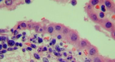
314     MATULLO ET AL.                                                                                                                J. VIROL.

                                                                                                                                                   Downloaded from http://jvi.asm.org/ on January 12, 2021 by guest
   FIG. 1. Immunocompetent, LCMV-infected mice exhibit signs of uncal herniation. Postmortem examinations were performed on C57BL/6
mice inoculated i.c. with 1,000 PFU LCMV-Arm (A to C) or PBS (D and E). Specific features that were assessed included posture (A), pupillary
dilation (B and D), and compression of white matter against the posterior portion of the skull (C and E). Photographs are representative of 5 to
10 mice per group.

intracranial pressure—are hallmarks of uncal herniation that              were identical to those seen for wild-type animals (data not
can occur in humans, prompting us to systematically address               shown). Other researchers (14, 26) previously showed a similar
whether known features of herniation were consistent with                 delay in LCMV pathogenesis in PFN KO mice, although in
LCMV neuropathology.                                                      these studies, all mice eventually succumbed; while we do not
   For these experiments, three genotypes of mice were uti-               know the basis for the difference, modest changes in the virus
lized: wild-type C57BL/6, RAG KO, and PFN KO mice, which                  strain or dose were previously shown to affect the extent of
lack the pore-forming proteins that are utilized by cytotoxic             LCMV disease (28) and may be operative here.
lymphocytes to mediate granzyme-driven lysis. All animals                    An increase in cerebral edema accompanies seizures and
were on the H-2b background. PFN KO mice were included                    death and coincides with pronounced leakage of CSF from
because, as previously reported (26), they show an intermedi-             ventricles. The apparent brain swelling visible through the
ate phenotype following LCMV challenge: while all C57BL/6                 skull suggested that LCMV disease is associated with intra-
mice died between 6.5 and 7 dpi and all RAG-2 KO mice                     cranial edema (increased fluid volume). To address this,
survived, in our hands, PFN KO mice lived to 9 dpi, after which           brains were harvested from LCMV-infected immunocompe-
approximately 50% of these mice died over the ensuing 3 to 4              tent mice, RAG KO mice, and PFN KO mice at 3, 5, and 7
days (Fig. 2). Of note, all infected PFN KO mice showed early             dpi; from seizing and nonseizing PFN KO mice between
signs of LCMV-induced disease from ⬃7 to 12 dpi (hunched                  days 8 and 11; and from surviving PFN KO mice at 18 dpi.
posture, ruffled fur, and closed eyes), but death occurred only
                                                                          Notably, while these surviving mice continued to show mild
in seizing animals. Postmortem characteristics (as in Fig. 1)
                                                                          signs of illness (ruffled fur and hunched posture) as late as
                                                                          30 dpi, none progressed to lethal LCM disease past the
                                                                          window between 8 and 11 dpi.
                                                                             Brain weight was calculated before and after dehydration to
                                                                          determine the percent fluid (hereafter referred to as “water
                                                                          weight”) at each time postinfection, with groups of five or more
                                                                          mice for each time point, as indicated in Fig. 3. Data are
                                                                          expressed as the percent change relative to PBS-inoculated
                                                                          controls. As expected, water weight values for RAG KO mice
                                                                          did not vary over the time course. In contrast, while water
                                                                          weight values for C57BL/6 mice did not appreciably change at
                                                                          3 and 5 dpi, at 7 dpi, values increased by ⬃5% compared to
                                                                          PBS controls, which is equivalent to a mean of 80.4% (com-
                                                                          pared to 75.5% in controls), with values in some moribund
                                                                          mice being as high as 89%. The correlation between death and
                                                                          a precipitous change in water volume was further supported by
   FIG. 2. LCMV-mediated pathogenesis in immunocompetent and              results from PFN KO mice: while values were baseline for all
immunodeficient mice. Mice of the indicated genotypes were chal-
                                                                          PFN KO mice at day 7 and for nonseizing mice between days
lenged i.c. with 1,000 PFU LCMV-Arm and monitored daily for sur-
vival. Black squares, C57BL/6 (Bl6) mice (n ⫽ 10); black triangles,       8 and 11, moribund mice with seizures showed statistically
RAG-2 KO mice (n ⫽ 8); white diamonds, PFN KO mice (n ⫽ 18).              significant increases in water volume. Moreover, surviving PFN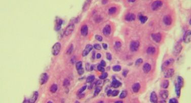
VOL. 84, 2010                                                    BRAIN HERNIATION AND EDEMA IN LCMV-INFECTED MICE                                  315

                                                                                                                                                          Downloaded from http://jvi.asm.org/ on January 12, 2021 by guest
  FIG. 3. Edema coincides with mortality in LCMV-infected mice. Mice of the indicated genotypes were inoculated with LCMV-Arm as described in
the text. At various dpi, brains were harvested and weighed before and after desiccation, and the percent water content was calculated by the following
formula: [(wet weight ⫺ dry weight)/wet weight] ⫻ 100. The value obtained for each mouse was then divided by the average water weight in brains from
PBS control mice. Results are expressed as the percent change relative to PBS controls, with the number of animals and standard deviations indicated
for each group. For PFN KO mice between 8 and 11 dpi, animals were divided into two categories: those exhibiting mild signs of LCMV disease with
no seizures and those showing severe symptoms including seizures. *, P ⫽ 0.001; **, P ⫽ 0.004 (by Wilcoxon signed-rank test). WT, wild type.

KO mice collected at 18 dpi did not have significantly increased                To test this putative association more rigorously and to gain
water weight values. The temporal association between the                     a more mechanistic understanding of the basis of this increased
appearance of edema with seizures and death suggested that                    edema, we turned to MRI approaches to evaluate intact brains
edema played a potentially causative role in LCMV neuro-                      of mice with and without seizures. At 6 dpi, a time when
pathogenesis.                                                                 immunocompetent C57BL/6 mice began to show signs of ill-

   FIG. 4. Seizing LCMV-infected mice exhibit ventricular leakage. Six days after i.c. challenge with 1,000 PFU LCMV-Arm or PBS, C57BL/6
mice were observed for signs of illness and divided into “early” and “late” groups. (Note that while all of these mice would eventually succumb
to LCMV infection, the appearance of physical signs of illness spans a range of 6 to 10 h.) Mice were imaged immediately after sacrifice by
isoflurane inhalation or following seizure-induced death. Coronal/frontal-plane brain images from similar locations within the brain are shown for
two representative mice inoculated with PBS (A and B), with mild LCMV signs (C and D), or with premortem seizures (E and F). All images were
processed identically so that changes in contrast reflect differences in fluid within these tissues.
316 MATULLO ET AL. J. VIROL.
chyma adjacent to the left ventricle shown in Fig. 4C (white
arrow), evidence of periventricular CSF leakage was observed.
These images are in sharp contrast to mice that were imaged
immediately after LCMV-triggered death, when a marked in-
crease in the fluid content of the cerebral parenchyma was
observed, characterized by increased brightness throughout
the brain (Fig. 4E and F). (All settings for capturing these
images were identical; thus, the images can be directly com-
pared.) Note that the ventricular margins in Fig. 4E and F are
less clearly defined than those in Fig. 4A to D, which is further
indicative of increased periventricular edema in moribund an-
imals. Taken together, these data implicate a sudden increase
in ventricular leakage coincident with LCMV-mediated death.
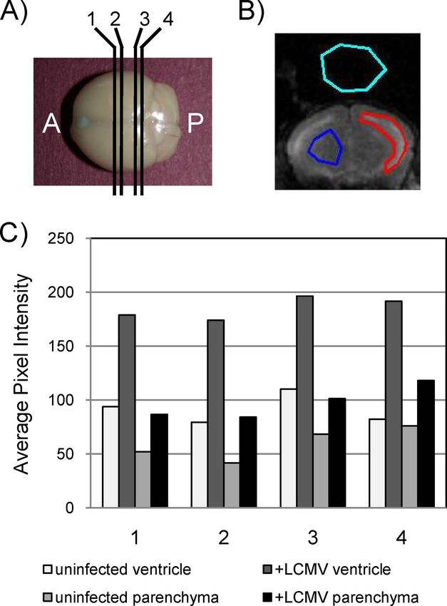
To quantify the extent of these changes, magnetic resonance
Downloaded from http://jvi.asm.org/ on January 12, 2021 by guest
images collected from four coronal planes of each mouse were
evaluated (Fig. 5A). For each image, regions were demarcated
for the ventricle (Fig. 5B), the parenchyma, and background as
a control. Average pixel intensity values are shown in Fig. 5C
for each of the four planes for a representative PBS-inoculated
mouse and an LCMV-challenged mouse. Three points can be
inferred from these data. First, as expected, the average pixel
intensity (correlating with water volume) was greater in ven-
tricles than in the parenchyma. Second, for both the ventricles
and the parenchyma, the pixel intensity was increased by 50%
or greater in infected mice. Finally, these differences were
FIG. 5. Quantitation of fluid volume changes in ventricles and pa- apparent regardless of the coronal plane that was evaluated.
renchyma. (A) Coronal images were captured by MRI at four discrete Cellular damage within the ependyma and the subependy-
planes throughout the brain, labeled 1 to 4, progressing from the mal layer in LCMV-infected mice. Given the loss of integrity of
anterior (A) to the posterior (P). (B) By using software described in
Materials and Methods, regions of ventricle (red shape), parenchyma
the brain-CSF barrier, we next histologically evaluated tissues
(blue shape), and background (aqua shape) were defined for each level from mice at late stages postinfection to identify lesions that
of each brain, and the average pixel intensity was calculated. (C) Val- might account for ventricular damage. Paraffin-embedded sec-
ues calculated from a representative mock-infected control mouse and tions were made from brains of LCMV-infected immunocom-
a representative infected mouse that showed severe (“late”) signs of
petent C57BL/6 mice at 6 to 7 dpi, stained with hematoxylin
LCMV disease. For each mouse, both ventricular and parenchymal
data are provided. and eosin, and evaluated by a neuropathologist in a blinded
manner.
Despite the presence of extensive periventricular lympho-
ness (ataxia, hunched posture, and tremors), animals were cytic infiltration, the architecture of the ventricle remained
divided into two groups: those with early signs of illness and generally normal in size and shape (data not shown). Substan-
those with late disease (e.g., noise-triggered seizures). Mice tial inflammation in both the CSF-producing choroid plexus
from both groups were imaged immediately, as were healthy, (Fig. 6A) and the subependymal lining adjacent to the ventricle
PBS-challenged mice. As shown in the representative images (Fig. 6B) was observed. This was accompanied by notable cell
in Fig. 4A and B, CSF in uninfected brains is restricted to the death (seen as nuclear condensation) (Fig. 6B and C) and focal
lateral ventricles. A generally similar picture was observed for hemosiderin deposition in the regions where inflammation was
LCMV-infected C57BL/6 mice with mild disease (Fig. 4C and noted (data not shown), which is indicative of bleeding into the
D), although in some instances, such as in the neuroparen- tissue. Thus, direct ependymal damage was noted where both
FIG. 6. Histological changes in the choroid plexus and periventricular zones. Brains of immunocompetent mice infected with LCMV (6 dpi)
were examined histologically. Evidence of inflammation in the choroid plexus (A), ependyma (B), and subependymal lining (C) was noted for all
mice examined (n ⫽ 7), as was prominent cellular damage that included apoptotic ependymal cells (white arrows in B and C). Original
magnification, ⫻800.VOL. 84, 2010 BRAIN HERNIATION AND EDEMA IN LCMV-INFECTED MICE 317
Downloaded from http://jvi.asm.org/ on January 12, 2021 by guest
FIG. 7. BBB permeability is compromised early after infection of immunocompetent mice. (A) Gross changes in BBB permeability. C57BL/6,
RAG-2 KO, and PFN KO mice were infected as described in Materials and Methods. At the indicated days postchallenge, deeply anesthetized
mice were injected transcardially with Evans blue, followed by saline perfusion. Brains were subsequently dissected and photographed. At least 10
mice/group were evaluated; two representative brains are shown for each group at each time point. (B) BBB damage was quantified by determining
the amount of Evans blue present in homogenized brains from the indicated genotypes throughout infection. Values for each animal were then
normalized to the weight of the brain. The Evans blue uptake into the tissues of each infected animal was divided by the average uptake detected
in brains from PBS control mice, and the results are expressed as changes in induction. Averages, standard deviations, and the numbers of mice
evaluated within each group are shown. #, P ⬎ 0.05 (not statistically significant); *, P ⫽ 0.001; **, P ⫽ 0.028. Significance was calculated by using
the Wilcoxon signed-rank test. WT, wild type.
infection and immune cell infiltration were found in all in- genesis, infected mice were injected on various dpi with Evans
fected brains that were evaluated. As expected, uninfected blue, a dye that binds to serum albumin and is normally ex-
tissues showed no signs of inflammation or tissue damage. cluded from the brains of mice with an intact barrier (22).
In addition to the damage in periventricular regions and the Evans blue staining was readily visible in C57BL/6 brains at 5
readily detected meningitis that is a hallmark of this disease, dpi (Fig. 7A) and could be detected by quantitative methods as
immune cell infiltration was also observed within the brain early as 3 dpi. Furthermore, Evans blue values were statistically
parenchyma (data not shown). The presence of these cells in increased from day 3 to day 5 and from day 5 to day 7 (Fig. 7B).
the neuroparenchyma is puzzling, as LCMV infection of resi- Healthy, infected RAG-2 KO mice showed no significant signs
dent CNS cells does not occur at this time postinfection (13). of barrier damage at any time point. (Parallel experiments
BBB damage is an early event in neuropathogenesis follow- using sodium fluorescein as a marker of barrier damage re-
ing LCMV infection. Previously, it was proposed that changes vealed an identical pattern [data not shown].) Importantly,
in BBB integrity contribute to lethal LCM disease. To address despite the absence of classical signs of LCM disease, PFN KO
the potential connection between barrier damage and patho- mice also showed evidence of increasing barrier damage at 7318 MATULLO ET AL. J. VIROL.
Downloaded from http://jvi.asm.org/ on January 12, 2021 by guest
FIG. 8. Proposed sequence of events that contribute to LCMV-mediated death.
dpi (Fig. 7A and B) similar to that of sick C57BL/6 mice. In 2 to 4 days later) had levels of barrier damage comparable to
addition, in surviving PFN KO mice, Evans blue levels contin- those of moribund, immunocompetent mice. Finally, and most
ued to increase, indicating that while BBB damage is a conse- strikingly, surviving PFN KO mice (at 18 dpi) had even greater
quence of LCMV infection, as previously reported, it is not barrier damage than did the animals at 7 dpi. The observation
associated with mortality. Moreover, barrier damage appears that PFN KO mice nevertheless had changes equivalent to, or
to be a PFN-independent event. greater than, those observed for wild-type mice suggests that
barrier damage is a PFN-independent process (Fig. 8). Taken
DISCUSSION together, while we cannot rule out a potential contributory role
for the loss of BBB integrity in LCMV disease, the lack of a
The lethal choriomeningitis that results from i.c. LCMV temporal correlation between barrier damage and death sug-
infection occurs in a well-defined sequence of events: virtually gested to us that other neuroanatomical changes in the brain
all infected mice develop overt signs of illness by 6 dpi, leading were responsible for LCMV-induced mortality.
to pronounced seizures by 6.5 to 7 dpi, which shortly precede
Using multiple strategies, we showed that the level of
death. The consistency of this pattern and complete penetrance of
edema, likely a consequence of ventricular damage resulting in
lethality afford a unique opportunity to define the sequence of
CSF perfusion into the brain parenchyma, is significantly ele-
neuroanatomical insults that occur during the infection win-
vated only at the terminal stages of disease. Moreover, edema
dow, with the eventual goal of elucidating the relationships
appears to be at least partially facilitated by PFN, because
among these events that lead to virus-triggered, immune-me-
diated death. While much is known about the cellular players mortality occurs in only ⬃50% of PFN KO animals, and in
that are involved in fatal choriomeningitis, the precise cause of these mice, terminal signs of LCMV disease are delayed by 2
mortality is not known. In this study, to reconstruct the events to 4 days. In humans, hydrocephalus resulting from impaired
that lead to LCMV-triggered neuropathogenesis, we found CSF circulation and transependymal leakage of CSF is noted
that brain herniation, likely of the uncal subtype, was the most by periventricular areas of unclear margins visible by computed
likely cause of mortality in infected mice. tomography or MRI due to increased intracranial pressure
Other researchers suggested that increased BBB permeabil- (11). As shown in the model presented in Fig. 8, we hypothe-
ity is the basis of LCMV disease (1, 18). Indeed, the earliest size that a disruption of ventricular integrity occurs late in
anatomical lesion that we detected was damage to the BBB at infection, coincident with peak viral load, maximal CD8⫹ T-
3 dpi, perhaps triggered by the presence of early-responder cell entry, and immune-mediated damage to the ependyma.
innate immune cells. However, animals at this time point The leakage of CSF into the brain parenchyma results in ele-
showed no signs of LCMV disease. Moreover, at 7 dpi, all PFN vated intracranial pressure, which then causes an unequal com-
KO mice (which do not succumb to LCMV pathogenesis until pression of the nerves that regulate the constriction of theVOL. 84, 2010 BRAIN HERNIATION AND EDEMA IN LCMV-INFECTED MICE 319
pupils as well as the neurons in the medulla that control au- infection mediated by virus-specific CD8⫹ T cells. J. Neuroimmunol. 31:
155–163.
tonomic functions, such as breathing and cardiac function. 2. Barber, D. L., E. J. Wherry, D. Masopust, B. Zhu, J. P. Allison, A. H. Sharpe,
Once this cascade of events is initiated, mortality follows soon G. J. Freeman, and R. Ahmed. 2006. Restoring function in exhausted CD8 T
thereafter. cells during chronic viral infection. Nature 439:682–687.
3. Borrow, P., and M. B. A. Oldstone. 1997. Lymphocytic choriomeningitis
Importantly, lessons from this mouse model may be appli- virus, p. 593–627. In N. Nathanson (ed.), Viral pathogenesis. Lippincott-
cable to human disease. Herniation often accompanies trau- Raven Publishers, Philadelphia, PA.
matic brain injury, which comprises ⬃10% of the 1.4 million 4. Buchmeier, M. J., R. M. Welsh, F. J. Dutko, and M. B. A. Oldstone. 1980.
The virology and immunology of lymphocytic choriomeningitis virus infec-
cases reported annually in the United States, although this is tion. Adv. Immunol. 30:275–331.
likely a gross underestimate since in many cases of head injury, 5. Buchmeier, M. J., and A. J. Zajac. 1999. Lymphocytic choriomeningitis virus,
death occurs before herniation can be assessed. Herniation p. 575–605. In R. Ahmed and I. Chen (ed.), Persistent viral infections. John
Wiley & Sons, Chichester, NY.
occurs when brain tissue shifts from an area of higher pressure 6. Camenga, D. L., D. H. Walker, and F. A. Murphy. 1977. Anticonvulsant
to an area of lower pressure, resulting in the movement of prolongation of survival in adult murine lymphocytic choriomeningitis. II.
brain matter through the tentorial opening and, potentially, Ultrastructural observations of pathogenic events. J. Neuropathol. Exp. Neu-
rol. 36:21–40.
passage through the foramen magnum if the downward dis- 7. Ceredig, R., J. E. Allan, Z. Tabi, F. Lynch, and P. C. Doherty. 1987. Pheno-
Downloaded from http://jvi.asm.org/ on January 12, 2021 by guest
placement continues. Any cause of severe cerebral edema, typic analysis of the inflammatory exudate in muris lymphocytic choriomen-
ingitis. J. Exp. Med. 165:1539–1551.
such as meningitis or stroke, or a mass lesion such as a tumor
8. Clemmesen, J. O., F. S. Larsen, J. Kondrup, B. A. Hansen, and P. Ott. 1999.
can result in the increased intracranial pressure that triggers Cerebral herniation in patients with acute liver failure is correlated with
these events. Different patterns of tissue shifting are distin- arterial ammonia concentration. Hepatology 29:648–653.
9. de la Torre, J. C., M. Mallory, M. Brot, L. Gold, G. Koob, M. B. A. Oldstone,
guished by their originations of herniation and include central and E. Masliah. 1996. Viral persistence in neurons alters synaptic plasticity
transtentorial, uncal, and tonsillar herniation, among others. and cognitive functions without destruction of brain cells. Virology 220:508–
The early clinical signs of central herniation differ from those 515.
10. Doherty, P. C., and R. M. Zinkernagel. 1974. T-cell-mediated immunopa-
of uncal herniation in that uncal herniation presents with uni- thology in viral infections. Transplant. Rev. 19:89–120.
lateral pupillary dilation, while central transtentorial hernia- 11. Greenlee, J. E., and K. C. Carroll. 2004. Infections of the central nervous
tion presents with small pupils that dilate briskly. With contin- system, 3rd ed. Lippincott Williams & Wilkins, Philadelphia, PA.
12. Kang, S. S., and D. B. McGavern. 2008. Lymphocytic choriomeningitis in-
ued downward displacement, both of these herniation fection of the central nervous system. Front. Biosci. 13:4529–4543.
syndromes result in hemiparesis, followed by decorticate or 13. Kappes, D. J., D. M. P. Lawerence, M. M. Vaughn, V. P. Dave, A. R. Belman,
decerebrate posturing, coma, and, ultimately, cardiorespira- and G. F. Rall. 2000. Protection of CD3 delta knockout mice from lympho-
cytic choriomeningitis virus-induced immunopathology: implications for viral
tory collapse. The course of events in uncal herniation bears a neuroinvasion. Virology 269:248–256.
remarkable resemblance to that in LCMV infection of immu- 14. Kim, J. V., S. S. Kang, M. L. Dustin, and D. B. McGavern. 2008. My-
nocompetent mice and may therefore have direct relevance to elomonocytic cell recruitment causes fatal CNS vascular injury during acute
viral meningitis. Nature 457:191–195.
humans that die of brain herniation as a consequence of in- 15. Klavinskis, L. S., and M. B. A. Oldstone. 1989. LCMV selectively alters
creased intracranial pressure (8, 11, 21, 25). For example, chil- differentiated but not housekeeping functions: block in expression of growth
hormone gene is at the level of transcriptional initiation. Virology 168:232–
dren with cerebral malaria often develop decerebrate postures 235.
reminiscent of those seen for LCMV-challenged mice, and 16. Koenig, M. A., M. Bryan, J. L. Lewin, M. A. Mirski, R. G. Geocadin, and
adults suffering from cerebral malaria will often show both R. D. Stevens. 2008. Reversal of transtentorial herniation with hypertonic
saline. Neurology 70:1023–1029.
seizures and brain herniation. 17. Lipkin, W. I., E. L. F. Battenberg, F. E. Bloom, and M. B. A. Oldstone. 1988.
In summary, this study has identified novel neuroanatomical Viral infection of neurons can depress neurotransmitter mRNA levels with-
changes that are triggered following LCMV infection of im- out histologic injury. Brain Res. 451:333–339.
18. Marker, O., F. C. Nielsen, and N. H. Diemer. 1984. The permeability of the
munocompetent mice and has defined which of these events is blood-brain barrier in mice suffering from fatal lymphocytic choriomeningitis
likely responsible for LCMV-triggered mortality. As a result of virus infection. Acta Neuropathol. 63:229–239.
these studies, a potential pathogenic link between LCMV in- 19. Nansen, A., J. P. Christensen, C. Ropke, O. Marker, A. Scheynius, and A. R.
Thomsen. 1998. Role of interferon-gamma in the pathogenesis of LCMV-
fection of mice and human brain herniation is proposed. Of induced meningitis: unimpaired leucocyte recruitment, but deficient macro-
note, while the delivery of a large bolus of mannitol is the phage activation in IFN-gamma knock-out mice. J. Neuroimmunol. 86:202–
standard of care in emergency rooms when brain herniation is 212.
20. Oldstone, M. B. A. 1998. Molecular mimicry and immune-mediated disease.
suspected (16), such efforts typically fail. Thus, if future studies FASEB J. 12:1255–1265.
substantiate this putative correlation, the LCMV model may 21. Pourmand, R. 2008. Practicing neurology. What you need to know, what you
once again prove useful in understanding human disease and need to do, 2nd ed. Humana Press, Totowa, NJ.
22. Rall, G. F., L. Mucke, and M. B. A. Oldstone. 1995. Consequences of
could be a valuable tool to test novel interventive strategies to cytotoxic T lymphocyte interaction with major histocompatibility complex
prevent or reverse this condition in humans. class I-expressing neurons in vivo. J. Exp. Med. 182:1201–1212.
23. Schwendemann, G., J. Lohler, and F. Lehmann-Grube. 1983. Evidence for
cytotoxic T-lymphocyte-target cell interaction in brains of mice infected
ACKNOWLEDGMENTS intracerebrally with lymphocytic choriomeningitis virus. Acta Neuropathol.
61:183–195.
G.F.R. was supported by NIH grants RO1-NS40500, RO1- 24. Shinkai, Y., G. Rathbun, and K. Lam. 1992. RAG-2 deficient mice lack
NS060701, and P30-CA006927; a Pilot Project grant from Autism mature lymphocytes owing to the inability to initiate V(D)J. rearrangement.
Speaks; and a gift from the F. M. Kirby Foundation. C.M.M. was Cell 68:855–864.
supported by a Thomas Jefferson University Training grant, and K.J.O. 25. Stiver, S. I., and G. T. Manley. 2008. Prehospital management of traumatic
was supported by the Greenwald Fellowship at Fox Chase Cancer brain injury. Neurosurg. Focus 25:E5–E15.
Center. 26. Storm, P., C. Bartholdy, M. R. Sorensen, J. P. Christensen, and A. R.
Thomsen. 2006. Perforin-deficient CD8⫹ T cells mediate fatal lymphocytic
choriomeningitis despite impaired cytokine production. J. Virol. 80:1222–
REFERENCES 1230.
1. Andersen, I. H., O. Marker, and A. R. Thomsen. 1991. Breakdown of blood- 27. Thomsen, A. R. 2009. Lymphocytic choriomeningitis-induced central nervous
brain barrier function in the murine lymphocytic choriomeningitis virus system disease: a model for studying the role of chemokines in regulating the320 MATULLO ET AL. J. VIROL.
acute antiviral CD8⫹ T-cell response in an immune-privileged organ. J. Vi- von Andrian, and R. Ahmed. 2003. Lineage relationship and protective
rol. 83:20–28. immunity of memory CD8 T cell subsets. Nat. Immunol. 4:225–234.
28. Walker, D. H., F. A. Murphy, S. G. Whitefield, and S. P. Bauer. 1975. 31. Zajac, A. J., J. M. Dye, and D. G. Quinn. 2003. Control of lymphocytic chori-
Lymphocytic choriomeningitis: ultrastructure pathology. Exp. Mol. Pathol. omeningitis virus infection in granzyme B deficient mice. Virology 305:1–9.
23:245–265. 32. Zhou, Z. S., F. Granucci, L. Yeh, P. A. Schaffer, and H. Cantor. 1998.
29. Walsh, C. M., M. Matloubian, C.-C. Liu, R. Ueda, C. G. Kurahara, J. L. Molecular mimicry by herpes simplex virus type I: autoimmune disease after
Christensen, M. T. F. Huang, J. D.-E. Young, R. Ahmed, and W. R. Clark. viral infection. Science 279:1344–1347.
1994. Immune function in mice lacking the perforin gene. Proc. Natl. Acad. 33. Zinkernagel, R. M., and P. C. Doherty. 1973. Cytotoxic thymus-derived
Sci. U. S. A. 91:10854–10858. lymphocytes in cerebrospinal fluid of mice with lymphocytic choriomeningi-
30. Wherry, E., V. Teichgraber, T. Becker, D. Masopust, S. Kaech, R. Antia, U. tis. J. Exp. Med. 138:1266–1269.
Downloaded from http://jvi.asm.org/ on January 12, 2021 by guestYou can also read