BIRDS Common Diseases of Urban Wildlife - Australian Registry of ...
←
→
Page content transcription
If your browser does not render page correctly, please read the page content below
Common Diseases of Urban Wildlife

                                BIRDS
                                  PART 1

                          MISSION STATEMENT:
The Australian Registry of Wildlife Health is committed to contributing to the
preservation of Australia’s biodiversity through increased understanding of the
  interactions among animals, the environment, and disease causing agents.
Common Diseases of Urban Wildlife: BIRDS
K. Rose, June 2005

1 Common Diseases of Birds
1.1 Introduction
A wide variety of diseases have been documented within free ranging Australian
birds. The following segment focuses on those diseases that occur often within a
particular species or taxonomic group of birds.
1.2 Parasitic Disease
For additional information see also: (Bain and Mawson, 1981).

1.2.1 Cheilospirura gymnorhinis
Cheilospirura gymnorhinis is also referred to as the throat worm of juvenile magpies.
The same parasite, or a similar
parasite, occurs in the oral cavity
and pharynx of currawongs,
butcherbirds, magpie larks, and
black-faced cuckoo shrikes. C.
gymnorhinis burrows its head into
the mucosa of the oral cavity and
pharynx. The host then responds
                                           Cheilospirura gymnorhinis, oropharynx, Magpie
by creating a fibrous nodule around
the parasite. Although small numbers of parasites result in self-limiting infections,
large numbers can impair prehension of food, or partially obstruct the glottis.
Treatment trials using a variety of anti-helminthic agents have not been successful in
eliminating the parasites (Larry Vogelnest, personal communication). Repeated
manual removal of the parasites with haemostats is recommended and this is assisted
by application of moxidectin directly to the nematodes. Euthanasia may be the most
humane option for severely debilitated young birds with heavy burdens of C.
gymnorhinis.

1.2.2 Syngamus trachea
Syngamus trachea is commonly found in the trachea of a variety of birds.

                                              -2-
Common Diseases of Urban Wildlife: BIRDS
K. Rose, June 2005

 Occasionally       magpies       that      die
suddenly are found to have complete
tracheal obstruction with masses of
these parasites.

1.2.3 Trichomoniasis
Oral     trichomoniasis         has        been
observed in debilitated free ranging
                                                  Syngamus trachea
birds, but is most common in captive
wildlife that are undergoing treatment for various injuries.                   Birds of prey,
columbiforms, little penguins, and psittacines are sporadically affected by
trichomoniasis.      Trichomonads are common commensal agents within the avian
alimentary tract. Trichomonads are ovoid protozoa that have four anterior flagella and
an undulating membrane. These organisms are spread through either direct or indirect
contact. The factors that predispose a bird to develop trichomoniasis are unknown.

Caseous oral plaques are created when the
organisms cause tissue necrosis. Lesions are
often subject to secondary bacterial infections.
A diagnosis of trichomoniasis is best made by
examining a wet mount preparation of the
caseous debris. Flagellates can be seen moving
within the wet preparations under light
microscopy.      The organisms are much more                Trichomonas spp.

difficult (often impossible) to see within cytologic and histologic preparations of
affected tissues.

Treatment of trichomoniasis includes debridement of the caseous plaques, supportive
care and administration of antiprotozoal agents.

                                                  -3-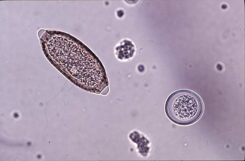
Common Diseases of Urban Wildlife: BIRDS
K. Rose, June 2005

1.2.4 Haemoproteus
                                                                   Megaloschizonts                 of
                                                                   Haemoproteus                 cause
                                                                   clinically             significant
                                                                   myopathy          in          pied
                                                                   currawongs        within       the
                                                                   Sydney       region     of   New
                                                                   South        Wales.          This
                                                                   organism        was      initially
                                                                   reported as Leucocytozoon
Lesions due to Heamoproteus sp, pectoral muscle, Pied Currawong.
                                                                   sp.      Haemoproteus spp.
infection occurred in juvenile, sub-adult and adult birds of both sexes, at any time of
year, but it has not been seen in recent years. Infection in some birds may have been
incidental; however, heavy parasite burdens in some birds resulted in lethargy,
weakness and debility. If the breast feathers are parted, pale oval foci were evident
throughout the pectoral musculature in affected birds. There is no known treatment
for this protozoal infection and birds often die shortly after initial examination. The
lifecycle of this protozoal agent is unknown.

Upon post mortem examination of affected birds, discrete, pale oval foci measuring
up to 1.5 cm long and 0.5 cm wide are scattered throughout the skeletal muscles,
tongue, myocardium and ventricular muscularis externa. Histopathologic examination
demonstrates that pale foci consist of central megaloshizonts, surrounded by necrotic
muscle and an intense inflammatory response. Haemorrhage, necrosis, and
inflammation are most severe around ruptured megaloschizonts.                     Pigmented oval
Haemoproteus gamonts may or may not be evident within circulating erythrocytes of
affected birds.

1.2.5 Toxoplasma gondii
Toxoplasmosis is a potentially fatal disease in native birds (Hartley and Dubey 1990,
ARWH). Birds with toxoplasmosis are depressed, fluffed, or are found dead. Gross

                                                    -4-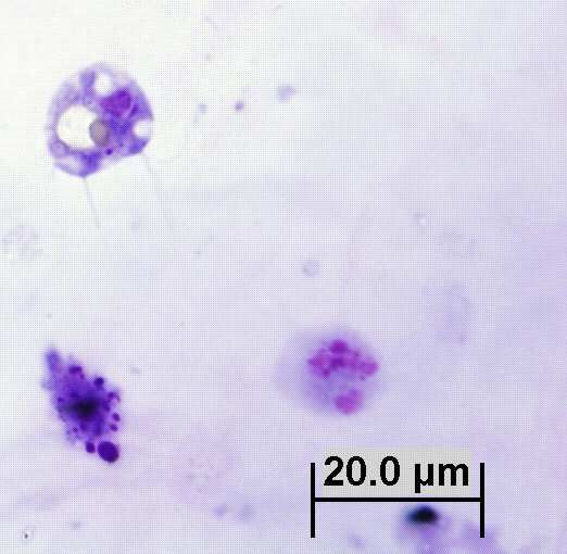
Common Diseases of Urban Wildlife: BIRDS
K. Rose, June 2005

post mortem findings consist of pulmonary oedema, pulmonary congestion, and pale
foci within the liver, spleen and intestinal mucosa. Histologic examination reveals
pulmonary oedema and congestion, fibrin within the distal airways, and non-
suppurative inflammation or necrosis within the liver, spleen, brain, skeletal muscle,
ventriculus, adrenal gland, and intestine.         Numerous protozoa, morphologically
consistent with T. gondii may be observed in the interstitium of the lung or within foci
of necrosis in other tissues. Definitive diagnosis of T. gondii protozoal encephalitis
has been established in some native birds using immunohistochemistry (Hartley and
Dubey, 1991).

Protozoal cysts resembling those of T. gondii are observed in the absence of
inflammation during routine histologic examination of nervous tissue of a variety of
native birds, especially the tawny frogmouth.             These cysts are consistent
morphologically with T. gondii and they appear to be a common incidental finding
(ARWH).

1.2.6 Other Parasites

Ectoparasites
Birds can be parasitised by ticks. Ixodes holocyclus, the paralysis tick, is found
occasionally on birds.          Anecdotal reports of tick paralysis in birds have been
documented. Three species of the genus Ornithodorus are known in Australia, O.
macmillani on wild birds, O. capensis on sea birds and O.gurneyi primarily on
kangaroos (David Spratt, personal communication).

Lice commonly infest wild birds, but rarely cause disease, such as anaemia. Heavy
infestations may be treated with topical antiparasitic powders.

Cnemidocoptes sp. infestations are capable of causing severe debility in pied
currawongs.       Infestations are associated with significant epidermal hyperplasia,
primarily involving the skin of the toes. Mites can be demonstrated by microscopic
examination of scrapings of the thickened skin. Oral or subcutaneous administration

                                             -5-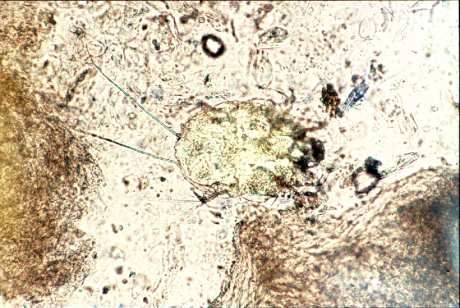
Common Diseases of Urban Wildlife: BIRDS
     K. Rose, June 2005

     of ivermectin-like drugs will control Cnemidocoptes sp. infestation, but more severe
     infestations are often associated with foot deformation and general debility, which
     may not be amenable to treatment.

     a) Pedal hyperkeratosis from Cnemidocoptes sp, pied     b) Cnemidocoptes sp, whole mount.
     currawong.

Hyperkeratosis, Cnemidocoptes spp. H & E.                  Cnemidocoptes spp. Note internal organs surrounded
                                                           by hyaline exoskeleton. H & E.

     Hippobosca are a genus of fly that occurs commonly within the plumage of birds of

                                                     -6-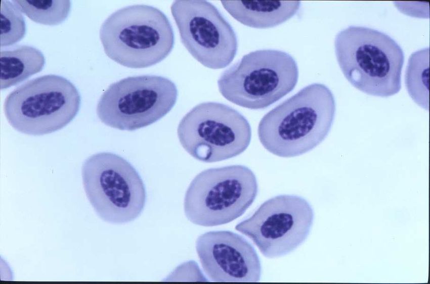
Common Diseases of Urban Wildlife: BIRDS
K. Rose, June 2005

prey,     especially      the     tawny    frogmouth.
Hippoboscid flies can bite and are capable of
acting as a vector in the transmission of disease.
These flies will infest and bite humans, but do
not seem to remain on human hosts for
prolonged periods.

Sternostoma tracheacolum is the tracheal and air
sac mite of Gouldian finches. Heavy burdens of
these mites are capable of causing coughing,
sneezing, and open mouth breathing. Disease              Hippoboscid fly

associated with this parasite is most common in captive finches. Ivermectin-like drugs
can be used to treat affected birds.

Trematodes
Australobilharzia spp. are schistosome parasites that live in the blood vessels of water
birds. These parasites are most commonly reported in silver gulls. The parasites have
an affinity for the blood vessels of the gastrointestinal tract and kidney. The presence
of adult trematodes and trematode eggs may result in the formation of multiple
granulomas within the intestine and liver. Australobilharzia sp. infection, however, is
most often an incidental finding in birds.          The cercaria of Australobilharzia sp.
penetrate the skin of humans and are the cause of “swimmer’s itch”.

Mawsontrema eudyptulae is a trematode that lives in the bile ducts of little penguins.
Infection with this parasite may be incidental, or may be associated with liver
enlargement, necrosis, and possibly haemorrhage from the liver (Harrigan 1992,
Normal et al., 1992).           For the population of little penguins in Bass Strait in
southeastern Australia to remain stable, the scale of annual, natural mortality is
astonishing and has been estimated at 100,000 birds (Norman et al., 1992). In
addition, large numbers of immature birds also perish and many become beach
washed (loc. cit.). In these so-called “wrecks”, adult mortality generally occurs in
winter due mainly to starvation. Wrecks involving immature birds occur

                                              -7-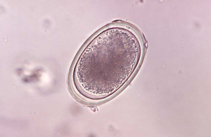
Common Diseases of Urban Wildlife: BIRDS
K. Rose, June 2005

mainly in late summer and early autumn and are directly or indirectly the result of
parasitic infection (Obendorf and McColl, 1980; Harrigan, 1992).

Nematodes
Gastrointestinal nematodiasis is usually an incidental finding in wild birds. Parasite
                                                    burdens in wild birds are usually mild.
                                                    Captive birds, however, may experience
                                                    excessive        parasite   burdens   that   can
                                                    contribute to debility.

                                                    Capillaria spp. are often evident within

Heavy ascarid burden, captive long-billed corella
                                                    serpiginous tracts created as the nematode
                                                    burrows through the mucosa and lamina
propria of the oesophagus, proventriculus and liver. Capillariasis occurs primarily in
captive birds.          Infected birds may suffer from extensive hyperplasia of the
oesophageal mucosa and marked inflammation surrounding the parasites in the
mucosa and lamina propria.                 Emaciation and dehydration can result from these
infections. Capillaria sp. infections in wild birds are primarily an incidental finding
during post mortem examination.

Ascarid ovum                                              Strongyle ovum

                                                    -8-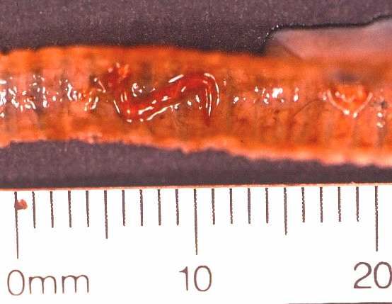
Common Diseases of Urban Wildlife: BIRDS
K. Rose, June 2005

Cestode ovum                                           Capillaria ovum and Coccidial oocyst

Contracaecum spp. are nematodes that parasitise the oesophagus and proventriculus
of piscivorous birds. Small numbers of parasites pose no threat to the host. There are
reports of large parasite burdens of C. spiculigerum being associated with
proventricular ulceration, haemorrhage, emaciation and death in little penguins in
Victoria (loc. cit.).

Dispharynx nasuta is a nematode parasite that burrows into the mucosa of the
proventriculus in a variety of birds. Infection with this parasite can result in either
granulomatous inflammation or the development of fibrous nodules surrounding the
parasites within the wall of the proventriculus. Echinuria uncinata is a parasite that is
capable of causing similar lesions in waterfowl.

Serratospiculum anaculata resides within the airsacs of birds of prey, and infection is
most common in falcons. Two species are known in Australia: S. guttatum from
Falco longipennis and F. peregrinus and S. tendo from F. peregrinus. Overseas, S.
anaculata occurs primarily as an unapparent infection. Clinical respiratory disease
has been described in Australian birds infected with S. anaculata that are subject to
stress or concurrent disease.              Diagnosis of S. anaculata requires endoscopic
examination of the air sacs. Ivermectin-like drugs can be used to treat affected birds.

Microfilariae are occasionally found during examination of peripheral blood smears of

                                                 -9-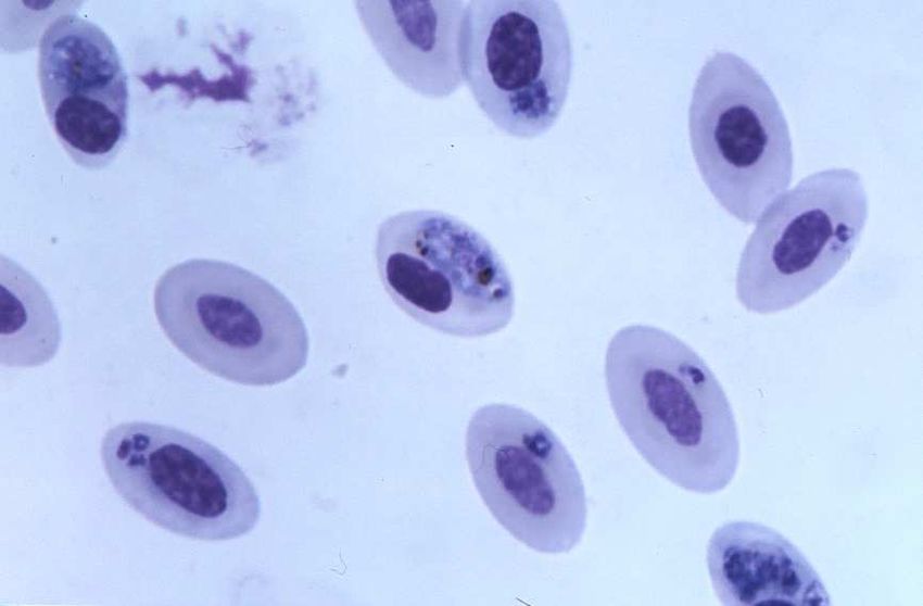
Common Diseases of Urban Wildlife: BIRDS
K. Rose, June 2005

wild birds.      Adult filarial nematodes may
reside within the air sacs, coelomic cavity,
subcutaneous tissues, heart, greater vessels,
or lungs. Infection is diagnosed during
microscopic examination of peripheral blood
smears or buffy coat smears. Microfilariae
                                                      Microfilaria, peripheral blood smear, barking
are     transmitted        by      haematophagous     owl.
arthropods.       Microfilarial infections are
incidental to the host.

Oxysprirura spp. are nematode parasites that can be found within the conjunctiva and
nictitating membrane of a number of species. Infection with this parasite is usually
asymptomatic, but may be associated with conjunctivitis in a small proportion of birds
(Pass, 1993).

Angiostrongylus cantonensis, the rat lungworm has been found to neurological
dysfunction associated with eosinophilic or non-suppurative encephalomyelitis in
yellow-tailed black cockatoos, and more commonly in tawny frogmouths. Infection in
tawny frogmouths is now a very common occurrence and seems to have a seasonal
prevalence (Monks et al., in press, Montali et al., 2004, Prociv, 1999). Birds become
infected with the parasite by eating snails and slugs, the intermediate host. Diagnosis
of the infection can be very difficult, since birds do not usually develop eosinophilia.
Cerebrospinal fluid taps collected from infected animals are also often non-
suppurative rather than eosinophilic, making it difficult to differentiate
angiostrongylosis from viral or protozoal infection. Treatment of the infection in
birds is also difficult. The parasite’s cuticle retains many antigens and killing the
worms can result in release of antigens with subsequent severe host immune response.

Cestodes
Birds are parasitised by many species of cestode. None of these is considered to be
highly pathogenic in free-living birds. It is possible, however, that large burdens of
cestodes will add to the debility of a captive or compromised bird. If

                                             - 10 -
Common Diseases of Urban Wildlife: BIRDS
K. Rose, June 2005
necessary, cestodiasis is treated with praziquantel. The treatment is usually repeated
10 days after the first dosage.
Protozoa
A wide variety of protozoa have been reported within the gastrointestinal tract,
cardiovascular system, musculature and renal tissues of free-flying birds. The
following discussions regarding protozoa are limited to those protozoal infections
known to be clinically significant.
Spironucleus (formerly Hexamita) -like organisms have been associated with
numerous outbreaks and individual cases of emaciation, diarrhoea and fatal enteritis in
Australian king parrots (Philbey et al., 2002, Vogelnest, 1994). Similar parasites have
been identified in an emaciated, wild sulphur crested cockatoo from western NSW
(ARWH). These birds become emaciated and have very thin walled intestinal tracts,
often filled with fetid brown fluid. The intestinal tissues of affected birds seem to
decompose very rapidly making it very difficult to identify organisms within tissues
on histologic examination. Saline wet-mount preparations can be used to demonstrate
the organism within the gastrointestinal tract during gross post mortem examination.
Protozoal enteritis, king parrot Protozoa (arrows) in the intestinal crypts and along the muco
basement membrane of a sulfer crested cockatoo
Gastrointestinal Giardia spp. infections have been documented in a variety of wild
and aviary birds in Australia. Giardia spp. have been recovered from the intestinal
- 11 -Common Diseases of Urban Wildlife: BIRDS
K. Rose, June 2005
lumen of straw-necked ibis in Western Australia, and a sulphur-crested cockatoo in
Victoria (Foreshaw et al., 1992, Gallagher et al., 1995). Giardiasis in captive young
budgerigars can result in decreased growth rates, dehydration, and diarrhoea
(Filippich, 1998). Diagnosis of giardiasis is based upon direct microscopic
examination of faeces or intestinal content. Giardia sp. trophozoites are pear shaped,
binucleate, and have eight flagella. A cyst form, with four nuclei is occasionally shed
in the faeces. Treatment of budgerigars with metronidazole decreased shedding of
these protozoa in the faeces (Filippich, 1998). Treatment for giardiasis is the same as
for trichomoniasis. Careful attention to hygiene will prevent clinical infection in most
captive birds.
Cryptosporidia spp. have been observed within the intestinal brush border of wild
Pacific black ducks, a red-tailed black cockatoo, an Australian magpie, and a rock
parrot (ARWH). The significance of this parasite as an avian pathogen is poorly
understood.
Eimerian and isosporan coccidial oocysts are commonly identified within the faeces of
healthy captive and free-flying birds. Coccidiosis may cause necrotising enteritis in
young captive birds of a variety of species. Disease associated with coccidial
infection in free ranging birds is rare. When large numbers of faecal oocysts
accompany diarrhoea, treatment of coccidiosis is advisable.
Renal coccidiosis is a common incidental finding within little penguins, Australian
gannets, and short-tailed shearwater (also known as the Tasmanian mutton bird).
Limey disease is the term used to describe clinically apparent renal coccidiosis in
nestling short-tailed shearwater. Chicks with limey disease are thin and have urate
and faecal soiling of the pericloacal feathers (Munday et al., 1971). Renal
enlargement and multiple pale foci throughout the kidney are evident on gross post
mortem examination. The ureter and cloaca may also be distended with urates.
Microscopic examination of the affected renal tissue reveals inflammation within the
interstitium surrounding the large collecting ducts. Coccidial oocysts are often
- 12 -Common Diseases of Urban Wildlife: BIRDS
K. Rose, June 2005
evident within multiloculated granulomas within the collecting duct mucosa and in the
surrounding interstitium.
Caryospora spp are coccidian parasites that can be found within the intestinal lamina
propria and mucosa of carnivorous birds of prey, but these are generally incidental
findings. Caryospora spp. can be found infecting reptiles, birds and rodents and can
have a single host, or two host (predator-prey) lifecycle. The intestinal forms of
Caryospora spp. are characterised by a single sporocyst containing eight elliptical
sporozoites.
Haemoproteus, Leucocytozoon, Plasmodium, Atoxoplasma, and a Babesia-like
organism are genera of the family Plasmodiidae that are commonly found within the
peripheral blood of wild Australian birds. Each of these organisms is arthropod borne.
A bird may be infected with two or more of these organisms concurrently without any
clinical signs. Young or debilitated birds may develop anaemia, anorexia, and
depression as a result of large parasite burdens.
Avian malaria, Albatross (Blood smear) Avian malaria, Blue Faced Parrot Finch (Blood smear)
Trypanosomes are occasionally found within the peripheral blood of native birds.
These parasites are extracellular flagellates that are transmitted by biting midges.
Trypanosomes are reported most commonly in little penguins, and they do not appear
to be pathogenic.
Systemic coccidiosis associated with Lankestrella spp. and Isospora serin have been
- 13 -Common Diseases of Urban Wildlife: BIRDS
K. Rose, June 2005
identified within circulating monocytes of the brown treecreeper, house sparrows, and
aviary birds within NSW. O’Donoghue and Adlard (2000) in the “Catalogue of
Protozoan Parasites Recorded in Australia” employ the generic name Lankesterella
rather than Atoxoplasma. Of Australian Lankesterella spp, one species occurs in tree-
creepers and one in white eyes and sparrows (David Spratt, personal communication).
These systemic coccidian parasites undergo sexual reproduction (gametogony) within
the mucosa of the gastrointestinal tract, and asexual reproduction (schizogony) within
extra-intestinal tissues such as the liver and spleen. Sporozoites of this organism are
evidently transported among these sites within mononuclear cells. Sporozoites are
round basophilic organisms that have a small basophilic nucleus. These sporozoites
are visible, usually as individual organisms, within the cytoplasm of mononuclear
cells, which have an indented nucleus.
A Babesia-like piroplasm is occasionally evident within the erythrocytes of little
penguins. The organism is most likely transmitted by ticks, and infection has been
associated with regenerative anaemia (ARWH).
Leukocytozoon, pied currawong.. Ring form, Babesia spp, little penguin.
1.3 Bacterial Disease
Sporadic outbreaks of mortality in native birds have been attributed to infection with
E. coli, Salmonella spp., Pasteurella spp., Mycobacterium spp., Erysipelothrix
rhusiopathiae , Listeria monocytogenes Streptococcus spp., Staphylococcus aureus,
Haemophilus spp., Myocplasma spp., and Clostridium spp (ARWH). Ideally
- 14 -Common Diseases of Urban Wildlife: BIRDS
K. Rose, June 2005
treatment of bacterial infection is based upon isolation of the organism within lesions,
and antimicrobial sensitivity testing.
1.3.1 Yersiniosis
Yersinia pseudotuberculosis infections can result in either acute enteritis and
septicaemia, or multisystemic abscesses.
1.3.2 Necrotic Enteritis
Aetiology
Wild rainbow lorikeets, scaly-breasted lorikeets, and king parrots in coastal eastern
Australia are seasonally affected with necrotising enteritis. A variety of organisms,
primarily coliforms, have been isolated within the necrotic intestinal tissue.
Clostridium perfringens and E. coli, however, are most commonly isolated within the
intestine and other tissues of birds with necrotic enteritis.
Carbohydrate overload has been suggested as a means of causing intestinal
overgrowth with Clostridium perfringens, and subsequent necrotic enteritis (Pass,
1993, McOrist and Reece, 1992). Numerous artificial feeding stations are established
for lorikeets in urban areas. Unfortunately, many of these stations provide only sugar
water. The presence of an underlying viral infection in birds suffering from necrotic
enteritis, however, has not been thoroughly investigated.
Necrotic enteritis occurs in male and female birds, juvenile animals and adults. Free-
ranging birds are most commonly diagnosed with necrotic enteritis; however, the
disease has been observed in captive lorikeets. Necrotic enteritis is most often
observed in July and August (ARWH).
An investigation into the occurrence of necrotic enteritis identified 58 dead rainbow
lorikeets, red-collared lorikeets, and scaly-breasted lorikeets originating from 18
different flocks in eastern Australia over a ten year period (McOrist and Reece, 1992).
Cl. perfringens was isolated from the intestinal tissues of many birds, and beta toxin
was demonstrated within the bacterial colonies and within intestinal content using gas
- 15 -Common Diseases of Urban Wildlife: BIRDS
K. Rose, June 2005
liquid chromatography (McOrist and Reece, 1992).
In 1998, eight birds were presented to the ARWH with necrotic enteritis. E. coli was
isolated in pure culture from the necrotic segments of intestine, and in the lung and
liver of seven of these birds. These tissues were also submitted for anaerobic culture;
however, Cl. perfringens was not identified. Thus, necrotic enteritis may be caused
by a variety of bacteria and its pathogenesis may be multifactorial.
Clinical Signs
Birds with necrotic enteritis exhibit a variety of clinical signs. Most of these birds are
in good body condition, but are weak, depressed, dehydrated, regurgitate clear fluid,
and have soiled vent feathers as a result of watery diarrhoea. The bird’s abdomen may
be palpably distended. Alternatively, birds with necrotic enteritis are found dead or
moribund. The species affected by necrotic enteritis normally have wet faeces, and
the detection of diarrhoea may be difficult.
Pathology
During the gross post mortem examination of these birds, the intestinal tract is
distended by gas or reddish-brown fluid. A diphtheritic membrane coats the mucosa,
or the mucosa is found to be friable and haemorrhagic. Microscopic examination of
affected segments of intestine reveals the following lesions: mucosal to transmural
necrosis, intense mononuclear cell infiltration, oedema and congestion throughout the
lamina propria and submucosa, and colonies of bacteria scattered throughout a
superficial layer of necrotic debris and fibrinous exudate.
Diagnosis
Necrotic enteritis is identified based upon the clinical signs and microbial culture of
faeces. Many birds with necrotic enteritis are found dead. Post mortem examination
and microbial culture of segments of intestine are used to establish a diagnosis.
Treatment
- 16 -Common Diseases of Urban Wildlife: BIRDS
K. Rose, June 2005
Although sensitivity testing of the E. coli isolated within the intestine of birds with
necrotic enteritis indicates that the organism is sensitive to a variety of commonly
used antibiotics, treatment of these birds is rarely successful. Presumably, the birds
are suffering from either enterotoxaemia, or bacteraemia by the time they demonstrate
clinical signs.
1.3.3 Chlamydiosis
Aetiology
Chlamydophila psittaci is a bacterium of the family Chlamydiaceae. These bacteria
are obligate intracellular parasites that are capable of causing severe disease in free-
living birds, aviary birds and humans.
Outside of the body C. psittaci take the form of elementary bodies, which have a rigid
cell wall. Elementary bodies are weakly gram-negative, non-motile bacteria, which
are phagocytosed by host cells. Once enveloped within a phagosome, elementary
bodies expand to become reticulate bodies, which have a more flexible cell wall and
are capable of growth and multiplication. After a period of division, these revert to
elementary bodies, which are released with the death of the host cell.
C. psittaci is endemic throughout Australia. It is a notifiable disease. Psittacine and
columbiform birds are most susceptible to C. psittaci infection. Chlamydiosis is a
common disease of lorikeets, cockatoos, budgerigars, rosellas, and aviary psittacines.
C. psittaci is transmitted either through the faecal-oral route or through respiratory
secretions. Elementary bodies may remain infective within dried faeces for several
months. Chlamydiosis should be considered among the differential diagnoses in any
emaciated wild bird, and barrier methods should be employed to prevent potential
spread of infection to other wildlife or humans.
Clinical Signs
Birds with active chlamydiosis may exhibit a broad range of symptoms associated
with either acute or chronic disease. Many birds will function as asymptomatic
- 17 -Common Diseases of Urban Wildlife: BIRDS
K. Rose, June 2005
carriers of the organism, while others may suffer severe or fatal infection.
Chlamydiosis is most often manifested as respiratory or gastrointestinal illness.
Clinical signs associated with C. psittaci infection include: weight loss, depression,
lethargy, anorexia, diarrhoea, bile stained faeces, ocular or nasal discharge, and
dyspnoea.
Pathology
Post mortem findings can be highly variable in birds suffering from chlamydiosis.
Some birds may die acutely with very few morphologic lesions, while some will
merely have splenomegaly and hepatomegaly, and others may have fibrinous air
sacculitis, pericarditis and enteritis.
Diagnosis
Definitive diagnosis of chlamydiosis relies upon isolation of the organism within cell
culture or embryonated chicken eggs. Marked leucocytosis, monocytosis, and an
elevated AST may be suggestive of C. psittaci infection, however, there is significant
species and individual variability in the haemogram of birds with chlamydiosis.
Antigen can be detected within conjunctival, nasal, or faecal swabs using antigen
capture ELISA tests or direct immunofluorescence testing. Diagnostic tests based
upon antigen capture are highly sensitive, but may not be highly specific. Some gram
negative bacteria will cross react with the antibody used in the ELISA test, thus,
conjunctival and choanal swabs will provide far fewer false positive reactions
compared with faecal swabs. ELISA based antigen capture test kits are commercially
available for in-house identification of Chlamydia sp. antigen (Clearview® test kits).
These kits are marketed for the detection of human C. trachomatis within urine
samples, but they are effective in the identification of C. psittaci.
Post mortem diagnosis of chlamydiosis is usually based on finding multisystemic
histiocytic inflammation on histologic examination and identification of the organism
within lesions. Impression smears of spleen, lung, and liver can be stained using
modified Machiavello’s staining protocols. This protocol can also be used to identify
the organism within paraffin embedded tissue. Fresh tissues, such as
- 18 -Common Diseases of Urban Wildlife: BIRDS
K. Rose, June 2005
liver, spleen and lung may be submitted to a microbiology laboratory for culture, or
swabs from fresh tissues can be tested with antigen capture ELISA tests or PCR.
Immunohistochemical demonstration of the organism is possible in fixed tissues.
1.4 Viral Disease
1.4.1 Psittacine Beak & Feather Disease (PBFD)
Aetiology
Psittacine beak and feather disease is a common disease in wild and aviary psittacines
throughout Australia. The disease is caused by psittacine circovirus, and is manifested
by lesions in the feathers, beak and occasionally the claws.
Psittacine circovirus is an icosahedral, non-enveloped virus, which has a single, round
strand of DNA. Presence of the virus can be demonstrated with feather epithelium,
follicular epithelium, macrophages within the feather pulp and dermis, macrophages
in the bursa of Fabricius, Kupffer cells in the liver, and within faeces. Psittacine
circovirus has an affinity for epithelial cells and lymphoid cells.
Clinical Signs
Clinical signs of infection with psittacine circovirus
are highly variable depending on the age and species
of the bird, and the quantity of virus in the infective
exposure. The progression of disease is also highly
variable, ranging from acute to chronic. Young birds
most often exhibit the acute form of infection.
Clinical signs of acute psittacine circovirus infection
include diarrhoea, weight loss, anorexia, depression
Galah with severe PBFD infection.
and either death or residual feather damage. The
chronic form of psittacine circovirus infection in
cockatoos begins with loss of the powder keratin in the plumage, and the production
of dystrophic down feathers over the hips. Powder down feathers become short and
lose the plumaceous barbs. The loss of powder down feathers results in a dull and
- 19 -Common Diseases of Urban Wildlife: BIRDS
K. Rose, June 2005
dirty look of contour and flight feathers, and imparts a glossy black appearance to the
beak.
When damaged by psittacine circovirus, the beak may become elongated, softened,
broken, cracked, or it may have uneven wear. These changes are most commonly
seen in cockatoos in the late stages of infection. If the germinal epithelium of the
beak is exposed by fractures or cracks in the keratin the bird will often stop eating due
to pain.
Young lorikeets of the genus Trichoglossus that
are infected with psittacine circovirus will often
present with the last two to four primary feathers
of the wings missing. If pulled from their
follicles, the calamus of the remaining tail
feathers and flight feathers will often exhibit
characteristic morphologic lesions. These
lorikeets are called “runners” since they are
unable to fly, yet healthy enough to forage and
run on the ground. Young lorikeets are identified
by their dark brown beaks. Occasionally, these
Rainbow lorikeet “runner”. Loss of primary
young lorikeets will have a blotchy yellow feathers from psittacine circovirus
pattern on the tail feathers that are usually green. infection..
Beak lesions rarely occur in lorikeets infected with psittacine circovirus.
Feathers damaged by psittacine circovirus are curled, clubbed, easily broken, or they
have retained feather sheaths, haemorrhages within the calamus (shaft), or annular
constrictions of the calamus. Replacement feathers grow slowly, or fail to regrow.
Pathology
Histologic lesions associated with psittacine circovirus infection occur primarily in the
growing feather, but may also be evident within the follicle. Necrosis occurs in the
germinal layer of the follicular and feather epithelium, and basophilic
- 20 -Common Diseases of Urban Wildlife: BIRDS
K. Rose, June 2005
cytoplasmic inclusion bodies may be evident within the epithelium, and in
reticuloendothelial cells in the dermis, feather pulp, and bursa of Fabricius.
Psittacine circovirus, large intracytoplasmic inclusions Psittacine circovirus, large intracytoplasmic inclusions
Psittacine circovirus infects the thymus and bursa of Fabricius and is associated with
lymphoid necrosis and premature atrophy of these tissues. Birds with psittacine beak
and feather disease often succumb to secondary viral, bacterial or fungal infections.
Diagnosis
The presumptive diagnosis of psittacine beak and feather disease is based upon gross
and microscopic lesions in the feather and feather follicle.
Crest feathers, Major Mitchell cockatoo, PBFD. Note pinching of shaft, retained sheaths.
Psittacine circovirus is very difficult to isolate in culture. Definitive diagnosis of
- 21 -Common Diseases of Urban Wildlife: BIRDS
K. Rose, June 2005
infection with this virus can be established through serological testing, which is
available through commercial laboratories. Haemaglutination inhibition (HI) testing
detects antibodies to psittacine circovirus in blood, serum and yolk, while
haemagglutination (HA) testing detects the virus in faecal samples or feathers. Birds
that suffer from severe psittacine beak and feather disease may not mount an effective
immune response to the virus and their titres measured via HI tests may not be
elevated.
Elevated HI titres merely indicate that antibodies have been formed in response to
exposure to psittacine circovirus. Birds with either the acute or chronic form of
circovirus infection often have low titres. Budgerigars, lorikeets, and king parrots,
however, usually have high psittacine circovirus titres and continue to shed the virus.
Immunohistochemistry, PCR, and DNA in-situ hybridisation tests for psittacine
circovirus are available overseas.
Treatment
Some species of psittacine can spontaneously recover from the acute form of beak and
feather disease. Rainbow lorikeets, budgerigars, eclectus parrots and king parrots may
recover from this infection with only mild residual feather changes. Birds with the
chronic form of psittacine circovirus infection rarely recover. The cause of death most
often relates to secondary infection with other viral, bacterial or fungal agents as a
result of immunosuppression.
There is no known cure for psittacine beak and feather disease. Nursing care to keep
the bird warm and eating will prolong the life of cockatoos. Lorikeets may
spontaneously recover from psittacine beak and feather disease, but can shed the virus
for a prolonged period.
Prevention
Since there is no effective treatment for birds suffering from psittacine beak and
feather disease, controlling the spread of the virus relies of strict hygiene
- 22 -Common Diseases of Urban Wildlife: BIRDS
K. Rose, June 2005
and euthanasia of affected birds. Birds that have clinically apparent psittacine beak
and feather disease can shed psittacine circovirus for extended periods, functioning as
a source of infection for other birds. If euthanasia is not an option, these birds should
be maintained under strict quarantine. Viracidal disinfectants used to kill parvovirus
should inactivate psittacine circovirus. A killed vaccine for the prevention of
psittacine beak and feather disease is currently under investigation.
1.4.2 Poxvirus
Aetiology
Australian magpies, native pigeons and raptors are occasionally clinically affected by
poxvirus infection.
Poxvirus is a member of the genus Avipox, which has a worldwide distribution.
Poxvirus is shed in saliva, nasal secretions, faeces and wound exudates or scabs. The
virus is transmitted primarily by haematophagous arthropods, such as mosquitos;
however, other vectors and fighting can also result in transmission. Infection results
in viraemia and then localisation within the skin or mucosa.
Clinical signs
Clinical signs of poxvirus infection vary from blistering and small nodules in the skin
to large dermal nodules with markedly hyperplastic epithelium, which may have foci
of ulceration. These lesions primarily occur on the skin of the feet, legs and head, and
around the eyes, mouth and cloaca. Secondary bacterial infection is a common
finding in poxvirus lesions. Some birds will recover spontaneously, while others will
become debilitated due to difficulty walking or obtaining food.
Pathology
The microscopic lesions associated with poxvirus infection include marked epidermal
thickening. Hyperplastic epithelial cells may contain cytoplasmic vacuoles that house
large, eosinophilic inclusion bodies. These inclusion bodies are called Bollinger
bodies.
- 23 -Common Diseases of Urban Wildlife: BIRDS
K. Rose, June 2005
Diagnosis
A diagnosis of poxvirus infection is based on finding the characteristic
intracytoplasmic eosinophilic inclusion bodies within epithelial cells upon
microscopic examination of formalin fixed tissue sections. Some diagnosticians can
use Diff Quik® stained scrapings of the proliferative lesions to identify intracellular
inclusions, but this is very difficult. Alternatively, biopsies of the proliferative
wounds can be submitted for electron microscopy to look for viral particles.
Treatment
Many birds will respond to cage rest and nursing care. Surgical debulking of large
lesions may provide relief for some birds. Nutritional support and prevention of
secondary infections will aid in the recovery of many birds suffering from poxvirus
infection.
1.4.3 Adenovirus
Adenovirus associated hepatic necrosis is a fairly common finding in tawny
frogmouths along coastal eastern Australia. Tawny frogmouths with adenovirus
hepatitis may have evidence of traumatic injury, or may be depressed and weak.
Diagnosis of adenovirus infection is based on post mortem examination. The liver is
mottled, friable, enlarged and has rounded margins upon gross post mortem
examination. Foci of acute hepatocellular necrosis are scattered throughout the
parenchyma of the liver on histological examination. Individual hepatocytes at the
margins of the necrotic foci will have peripheralisation of nuclear chromatin and
contain intranuclear eosinophilic to basophilic inclusion bodies (Reece et al., 1985).
Inclusion body hepatitis associated with adenovirus infection has also been described
in a cockatiel (Scott et al., 1986).
1.4.4 Other Viruses
An enterovirus-like agent has been identified within the faeces and enterocytes of
galahs and sulphur-crested cockatoos that were depressed, anorexic, and had profuse,
green, mucoid diarrhoea. Over a period of approximately three weeks affected birds
- 24 -Common Diseases of Urban Wildlife: BIRDS
K. Rose, June 2005
became dehydrated, emaciated and died. Dilation of the intestinal tract with mucoid
fluid, gas, and a thickened mucosa were evident during gross post mortem
examination. Histological examination of affected segments of intestine demonstrated
atrophy of villi, and hyperplasia of the crypts of Lieberkuhn. Concurrent psittacine
circovirus infection was evident within some of the ill birds (Wylie and Pass, 1989).
2 Animals mentioned in text
2.1 Aves
Little penguin (Eudyptula minor)
Short-tailed shearwater (Puffinus tenuirostris)
Australian gannet (Morus serrator)
Straw-necked ibis (Threskiornis spinicollis)
Pacific black duck (Anas superciliosa)
Silver gull (Larus novaehollandiae)
Sulphur-crested cockatoo (Cacatus galerita)
Galah (Cacatua roseicapilla)
Red-tailed black cockatoo (Calyptorhynchus magnificus)
Yellow-tailed black cockatoo (Calyptorhynchus funereus)
Rainbow lorikeet (Trichoglossus haematodus)
Scaly-breasted lorikeet (Trichoglossus chlorolepidotus)
Red-collared lorikeet (Trichoglossus haematodus rubritorquis)
Australian king parrot (Alisterus scapularis)
Cockatiel (Nymphicus hollandicus)
Budgerigar (Melopsittacus undulatus)
Laughing kookaburra (Dacelo novaeguineae)
Tawny frogmouth (Podargus strigoides)
Black-faced cuckoo-shrike (Coracina novaehollandiae)
Brown treecreeper (Climacteris picumnus)
Red wattlebird (Anthochaera carunculata)
Gouldian finch (Erythrura gouldiae)
- 25 -Common Diseases of Urban Wildlife: BIRDS
K. Rose, June 2005
House sparrow (Passer domesticus)
Magpie-lark (Grallina cyanoleuca)
Pied butcherbird (Cracticus nigrogularis)
Pied currawong (Strepera graculina)
Australian magpie (Gymnorhina tibicen)
Long-billed corella (Cacatua tenuirostris)
Peregrine falcon (Falco peregrinus)
Australian hobby (Falco longipennis)
Barking owl (Ninox connivens)
Rock parrot (Neophema petrophila)
Shy albatross (Diomedea cauta)
Blue-faced parrot finch (Erythrura trichroa)
White-eyes (Zosteropidae)
Eclectus parrot (Eclectus roratus)
3 References
Bain O and Mawson PM (1981) On some oviparous nematodes mainly from
Australian birds. Rec. S. Aust. Mus. 18: 265-284.
Blus LJ, Wiemyer SN, Henny CJ (1996) Organochlorine pesticides. . In: Fairbrother
A, Locke LN, Hogg GL (eds). Non-infectious diseases of wildlife. Iowa State
University Press, Ames, Iowa. Pp. 61 - 70.
Charles JA (1995) Organochlorine toxicity in tawny frogmouths. In: Proceedings of
the Australian Committee of the Association of Avian Veterinarians, Dubbo. Pp. 135
- 141.
Cooke-Yarborough CM, Kornberg AJ, Hogg GG, Spratt DM, Forsyth JRL (1999) A
fatal case of angiostrongyliasis in an 11-month-old infant. Medical Journal of
Australia 170: 541-543.
Fairbrother A (1996) Cholinesterase inhibiting pesticides. In: Fairbrother A, Locke
LN, Hogg GL (eds). Non-infectious diseases of wildlife. Iowa State University Press,
- 26 -Common Diseases of Urban Wildlife: BIRDS
K. Rose, June 2005
Ames, Iowa. Pp. 52 - 60.
Filippich LJ, McDonnell PA, Munoz E, Upcroft JA (1998) Giardia infection in
budgerigars. Aust. Vet. J. 76(4): 246 - 251.
Fleay P (1981) Looking at animals. Boolarong Publications, Brisbane, Queensland.
Pp 38 - 41.
Foreshaw D, Palmer DG, Halse SA, et al. (1992) Giardia in a straw-necked ibis
(Theskiornis spiricollis) in Western Australia. Vet. Rec. 131: 267 - 268.
Gallagher AN, Gartrell BD, Upcroft JA (1995) Pathogenic Giardia isolated from a
wild trapped sulphur-crested cockatoo (Cacatua galerita). In: Proceedings of the
Australian Committee of the Association of Avian Veterinarians, Dubbo. Pp. 23 - 27.
Harrigan KE (1992) Cause of mortality of little penguins Eudyptula minor in Victoria.
Emu 91, 273 – 277.
Hartley W (1999) Personal communication.
Hartley WJ, Dubey JP (1990) Fatal toxoplasmosis in some native Australian birds. J.
Vet. Diag. Invest. 3: 167 - 169.
Hartley WJ, Reece RL (1997) Nervous diseases of Australian native and aviary birds.
Aust. Vet. Prac. 27(2): 91 - 96.
Locke LN, Thomas NJ (1996) Lead poisoning of waterfowl and raptors. In:
Fairbrother A, Locke LN, Hoff GL (eds) Non-infectious diseases of wildlife. Iowa
State University Press, Ames, Iowa. Pp. 52 - 60.
McOrist S, Penny RA (1986) Encephalomyelitis in free-living Rainbow lorikeets
(Trichoglossus haematodus). Avian Pathology. 15: 783 - 789.
McOrist S, Reece RL (1992) Clostridial enteritis in free living lorikeets
- 27 -Common Diseases of Urban Wildlife: BIRDS
K. Rose, June 2005
(Trichoglossus spp.). Avian Pathology 21: 503 - 507.
Monks DJ, Carlisle MS, Carrigan M, Rose K, Spratt D, Gallagher A, Prociv P (in
press) Angiostrongylus cantonensis infection in birds: cerebrospinal disease in a
yellow-tailed black cockatoo (Calyptorhynchus funereus) and two tawny frogmouths
(Podargus strigoides). In press: J Avian Medicine and Surgery.
Montali R, Spratt D, Rose K (2004) Angiostrongylus cantonensis in tawny
frogmouths in Sydney. Presented at the Wildlife Disease Association, Australasia,
Conference 2004 – Kinchega.
Munday BL, Mason RW, Wells RJH et al. (1971) Further studies on “Limey-disease”
of Tasmanian mutton birds (Puffinus tenuirostris). J. Wildlife Disease. 7: 126.
Norman FI, DU Guesclin PB, Dann P (1992) The 1986 “Wreck” of little penguins
Eudyptula minor in western Victoria. Emu 91, 369 – 376.
Obendorf DL and McColl K (1980) Mortality in little penguins (Eudyptula minor)
along the coast of Victoria, Australia. J. Wildlife Disease. 16: 2, 251 – 259.
O’Donoghue PJ and Adlard RD (2000) Catalogue of protozoan parasites recorded in
Australia. In: Memoirs of the Queensland Museum. Queensland Museum, South
Brisbane, Australia: 2000. 45: 1, 1 – 163. 1285 ref.
Pass DA (1993) Diseases of free-ranging birds in Australia. In: Fowler ME (ed).
Zoo and wild animal medicine. Current therapy 3. W.B. Saunders Company,
Philadelphia. Pp. 172 - 177.
Paton DC, Dorward DF, Fell P (1983) Thiamine deficiency and winter mortality in
red wattlebirds, Anthochaera carunculata (Aves: Melphagidae) in suburban
Melbourne. Aust. J. Zool. 31: 147.
- 28 -Common Diseases of Urban Wildlife: BIRDS
K. Rose, June 2005
Philbey AW, Andrew AL, Gestier AW, Reece RL, Arzey KE (2002) Spironucleosis
in Australian king parrots (Alisterus scapularis). Aust. Vet. J. 80: 3, 154 – 160.
Prociv P (1999) Parasitic meningitis (editorial) Medical Journal of Australia. 170:
517-518.
Reece RL, Pass DA, Butler R (1985) Inclusion body hepatitis in a tawny frogmouth
(Podargus strigoides: Carimulgiformes). Aust. Vet. J. 62: 426.
Reiss AE, Badcock NR (1998) Itraconazole levels in serum, skin and feathers of
gouldian finches (Chloebia gouldiae) following in seed medication. Proceedings of the
American Association of Zoological Medicine, Omaha.
Scott PC, Condron RJ, Reece RL (1981) Inclusion body hepatitis associated with
adeno-like particles in a cockatiel (Psittaciformes; Nymphicus hollandicus). Aust.
Vet. J. 63(10): 337.
Slatter P, Slater P, Slatter R (1994) The Slater field guide to Australian birds.
Landsdowne Printing Pty. Ltd., Sydney.
Vogelnest L (1999) Personal communication.
Vogelnest L (1994) Veterinary care of wild Australian Native birds - medicine. Proc
233. The Post Graduate Committee in Veterinary Science. Pp. 141 - 190.
Wylie SL, Pass DA (1989) An entero-like virus infection of cockatoos. Aust. Vet. J.
66: 321.
- 29 -You can also read