Homozygous variant p. Arg90His in NCF1 is associated with early-onset Interferonopathy: a case report
←
→
Page content transcription
If your browser does not render page correctly, please read the page content below
Schnappauf et al. Pediatric Rheumatology            (2021) 19:54
https://doi.org/10.1186/s12969-021-00536-y

 CASE REPORT                                                                                                                                        Open Access

Homozygous variant p. Arg90His in NCF1
is associated with early-onset
Interferonopathy: a case report
Oskar Schnappauf1* , Liane Heale2, Dilan Dissanayake2, Wanxia L. Tsai3, Massimo Gadina3, Thomas L. Leto4,
Daniel L. Kastner1, Harry L. Malech4, Douglas B. Kuhns5, Ivona Aksentijevich1 and Ronald M. Laxer2

  Abstract
  Background: Biallelic loss-of-function variants in NCF1 lead to reactive oxygen species deficiency and chronic
  granulomatous disease (CGD). Heterozygosity for the p.Arg90His variant in NCF1 has been associated with
  susceptibility to systemic lupus erythematosus, rheumatoid arthritis, and Sjögren’s syndrome in adult patients. This
  study demonstrates the association of the homozygous p.Arg90His variant with interferonopathy with features of
  autoinflammation and autoimmunity in a pediatric patient.
  Case presentation: A 5-year old female of Indian ancestry with early-onset recurrent fever and headache, and
  persistently elevated antinuclear, anti-Ro, and anti-La antibodies was found to carry the homozygous p.Arg90His
  variant in NCF1 through exome sequencing. Her unaffected parents and three other siblings were carriers for the
  mutant allele. Because the presence of two NCF1 pseudogenes, this variant was confirmed by independent
  genotyping methods. Her intracellular neutrophil oxidative burst and NCF1 expression levels were normal, and no
  clinical features of CGD were apparent. Gene expression analysis in peripheral blood detected an interferon gene
  expression signature, which was further supported by cytokine analyses of supernatants of cultured patient’s cells.
  These findings suggested that her inflammatory disease is at least in part mediated by type I interferons. While her
  fever episodes responded well to systemic steroids, treatment with the JAK inhibitor tofacitinib resulted in
  decreased serum ferritin levels and reduced frequency of fevers.
  Conclusion: Homozygosity for p.Arg90His in NCF1 should be considered contributory in young patients with an
  atypical systemic inflammatory antecedent phenotype that may evolve into autoimmunity later in life. The complex
  genomic organization of NCF1 poses a difficulty for high-throughput genotyping techniques and variants in this
  gene should be carefully evaluated when using the next generation and Sanger sequencing technologies. The
  p.Arg90His variant is found at a variable allele frequency in different populations, and is higher in people of South
  East Asian ancestry. In complex genetic diseases such as SLE, other rare and common susceptibility alleles might be
  necessary for the full disease expressivity.
  Keywords: Autoinflammation, Autoimmunity, Interferons, Systemic lupus erythematosus, NCF1

* Correspondence: oskar.schnappauf@nih.gov
1
 National Human Genome Research Institute, National Institutes of Health,
Bethesda, USA
Full list of author information is available at the end of the article

                                        © The Author(s). 2021 Open Access This article is licensed under a Creative Commons Attribution 4.0 International License,
                                        which permits use, sharing, adaptation, distribution and reproduction in any medium or format, as long as you give
                                        appropriate credit to the original author(s) and the source, provide a link to the Creative Commons licence, and indicate if
                                        changes were made. The images or other third party material in this article are included in the article's Creative Commons
                                        licence, unless indicated otherwise in a credit line to the material. If material is not included in the article's Creative Commons
                                        licence and your intended use is not permitted by statutory regulation or exceeds the permitted use, you will need to obtain
                                        permission directly from the copyright holder. To view a copy of this licence, visit http://creativecommons.org/licenses/by/4.0/.
                                        The Creative Commons Public Domain Dedication waiver (http://creativecommons.org/publicdomain/zero/1.0/) applies to the
                                        data made available in this article, unless otherwise stated in a credit line to the data.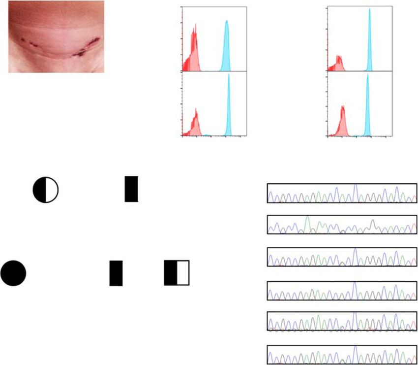
Schnappauf et al. Pediatric Rheumatology (2021) 19:54 Page 2 of 8
Background and severe headache with each fever episode. During
NCF1 (p47phox) is a component of the phagocytic NADPH one episode, her cerebrospinal fluid and brain MRI find-
oxidase complex type 2 (NOX2) that upon sensing of ings were consistent with aseptic meningitis. She showed
pathogenic stimuli releases reactive oxygen species (ROS) signs of failure to thrive, iron deficiency anemia, atrophic
into phagosomes and the extracellular compartment. skin lesions (Fig. 1a).
During this process, cytosolic NCF1 gets phosphorylated Autoantibody testing revealed positive ANA (1:640),
and interacts with NCF2 and NCF4. This ternary structure anti-Ro (> 100 U/mL) and anti-La (> 12 U/mL) anti-
gets translocated to the plasma membrane where it asso- bodies. The remainder of her specific autoantibodies, in-
ciates with the cytochrome complexes CYBB and CYBA cluding anti-dsDNA and rheumatoid factor, were
to form NOX2. Subsequently, the NOX2 complex trans- negative. Both during and between fever episodes, the
ports electrons from NADPH to oxygen, resulting in the patient had marked elevation of the erythrocyte sedi-
release of a variety of ROS. Loss-of-function variants in mentation rate (ESR), but normal to only mildly elevated
NCF1 and other genes coding for components of the levels of C-reactive protein (CRP). With two of the fever
NOX2 complex are associated with chronic granuloma- episodes, she developed a mild macrophage activation
tous disease (CGD), a primary immunodeficiency that is syndrome (MAS) with raised serum ferritin, neutropenia,
characterized by granulomatous inflammation and re- and thrombocytopenia. Otherwise, her complete blood
current infections due to defects in ROS-dependent count was normal. Abdominal ultrasound identified
destruction of phagocytized microorganisms. The rare small lymph nodes in peripancreatic and splenic hilum
missense variant p.Arg90His (rs 201802880, gnomAD regions and chest x-ray showed mild bilateral perihilar
MAF = 0.007) in NCF1 was reported as a complex- peribronchovascular linear opacities. The Schirmer test
disease susceptibility factor for systemic lupus erythe- for ocular dryness and Rose Bengal ocular staining did
matosus (SLE) and other autoimmune diseases [1, 2]. not show any ocular sicca. She had no clinical stigmata
In these studies, the p.Arg90His variant was associated of SLE, Sjögren syndrome or other autoimmune disease
with impaired extracellular ROS production and hyper- until the age of seven, when she developed the first of
activation of the interferon (IFN) type 1 signaling but not two episodes of parotitis that resolved spontaneously
with a full CGD clinical phenotype. Aside from its (Table 1). A complete set of investigations for recurrent
role in phagosome-mediated pathogen clearance, ROS fever did not reveal any infectious or malignant etiology.
also exhibit intra- and intercellular signaling properties Due to the absence of infections and a normal neutro-
and play an important role in the regulation of inflamma- phil oxidative burst capacity, her clinical features were
tion and immune responses [3–5]. Interferon (IFN) not consistent with typical CGD (Fig. 1b).
signaling is the main mediator of antimicrobial mecha- Her fever episodes responded well to systemic steroids
nisms and recent studies have suggested that neutrophil- (Dexamethasone, 0,25–4.5 mg) and recurred upon wean-
derived ROS suppress the activity of type I IFN that is ing. A trial of hydroxychloroquine did not alter the fre-
produced by plasmacytoid dendritic cells (pDCs) [6]. quency or severity of disease flares. Given features of
pDCs are a unique subset of dendritic cells and the main MAS with her febrile episodes, and the responsiveness
producers of IFN cytokines in patients with SLE [7]. pDC- of MAS in other situations to IL-1 inhibition, she re-
mediated IFN induces IL-15 production by conventional ceived treatment with anakinra (100 mg [6 mg/kg]), at
DCs (cDCs) which in turn activates IFN type II signaling the onset of the fever, which reduced the height of the
in natural killer cells [8]. fever peaks to some extent but did not completely abort
Here we characterize a female patient, homozygous for the episodes. An attempt at daily prophylactic anakinra
p.Arg90His in NCF1, who presented with autoinflamma- also did not reduce the frequency of episodes, suggesting
tory and autoimmune features accompanied by a strong that her disease was not primarily mediated by dysregu-
upregulation of IFN-regulated genes. Overall, her clinical lated interleukin-1 activity. In view of results that
features were most consistent with a periodic fever syn- showed upregulation of predominantly interferon-
drome, while her laboratory findings were suggestive of stimulated genes (ISGs) (see below), our patient was
an autoimmune disorder. treated with the JAK inhibitor tofacitinib (5–10 mg),
which resulted in decreased serum ferritin levels and fre-
Case presentation quency of fevers (Suppl. Fig. 1C), but only a partial clin-
The patient was born at 39 weeks of gestation with a ical effect was seen. Our patient was subsequently
birth weight of 2.7 kg and normal Apgar score. At age started on sirolimus (rapamycin, 2 mg), with which we
18 months, she developed episodes of fever (up to have been able to wean off her corticosteroids while
104.0 °F), anorexia and lethargy that recurred every 6–8 maintaining complete resolution of fevers. Exome se-
weeks lasting for 7–10 days. Two years into the course quencing (ES) was performed on the patient and her
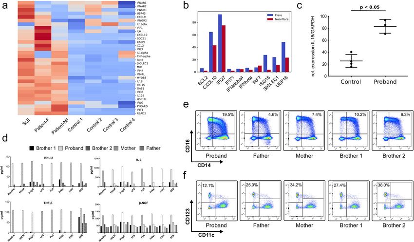
of her illness, she began experiencing nausea, vomiting parents, who are of Indian ancestry. The proband wasSchnappauf et al. Pediatric Rheumatology (2021) 19:54 Page 3 of 8 Fig. 1 Patient presentation and NCF1 p.Arg90His genotyping in the proband and her family. a: Atrophic skin lesion on lower abdomen of patient homozygous for NCF1, p.Arg90His. b: Residual oxidase activity of neutrophils and NCF1 expression in patient and healthy control. Left: Residual oxidase activity of neutrophils was determined using dihydrorhodamine (DHR) oxidation by flow cytometry. Right: NCF1 expression in patient and healthy control. NCF1 expression in neutrophils is presented as mean fluorescence intensity. Both panels: Red histograms represent neutrophils treated with buffer under basal conditions; blue histograms represent neutrophils in response to PMA (400 ng/mL). c: Pedigree of the family with the recessively inherited homozygous pathogenic variant p.Arg90His in the NCF1 gene. d: Sanger sequencing validation in proband and family. Sanger sequencing confirmed the homozygous variant NCF1, c.269G > A, p.Arg90His in the patient. Her parents and two brothers are heterozygous for the same variant found to be homozygous for the rare missense variant pathogenic variant is ΔGT in NCF1. Individuals who are p.Arg90His in the NCF1 gene, while her parents were carriers for this CGD-associated p47phox variant are healthy carriers for this variant (Fig. 1c). Three other predicted to have 1 GTGT copy and 5 ΔGT copies, or healthy siblings were either carriers for the variant or 1/6. In contrast, p47phox CGD patients are predicted to wildtype. No other plausible candidate gene variants have 0 GTGT copies and 6 ΔGT copies, or 0/6. The were identified under the assumption of either a domin- proportional ratio of GTGT/(GTGT+ΔGT) of the antly or a recessively inherited disease (Suppl. Table 1). patient and the healthy control sample were 2/6 which is Since the presence of two pseudogenes, NCF1B/C, might equivalent to the expected 2 GTGT copies and 4 ΔGT interfere with the alignment algorithms, the GTGT copies (Suppl. Fig. 1B). A custom designed Nanostring- sequence at the start of exon 2 of NCF1 was used to RNA expression array of 32 IFN-regulated and other discriminate between NCF1B/C and NCF1 [1]. This inflammatory genes showed moderate to strong up- genotype was confirmed by Sanger sequencing in the regulation of predominantly interferon-stimulated genes patient and her family members (Fig. 1d and Suppl. Fig. (ISGs) in peripheral blood of the patient during (Patient-F) 1A). To determine the copy number of NCF1B/C and and in between flares (Patient-NF) compared to heathy NCF1, a ddPCR assay containing probes specific for controls. A patient with SLE due to complement C1R either the GTGT in NCF1 or the ΔGT sequence in deficiency served as a positive control (Fig. 2a) [10]. NCF1B/C was performed [9]. Since a total of 6 copies of Quantitative RT-PCR for 10 IFN-induced genes con- NCF1/NCF1B/NCF1C are expected, healthy controls are firmed the Nanostring-RNA analysis. The strongest predicted to have 2 GTGT copies vs 4 ΔGT copies upregulation during and in between flares was seen for expressed as 2/6. In p47phox CGD, the most common IFI27, CXCL10, USP18, and ISG15 (Fig. 2b). Of note is
Schnappauf et al. Pediatric Rheumatology            (2021) 19:54                                                                                   Page 4 of 8

Table 1 Comparison of clinical features of patient with homozygous NCF1, p.Arg90His to pediatric SLE patients
Disease Features             Pediatric SLE Patientsa                                   NCF1 Variant Patient
Age of disease onset         Average 12 years                                          18 months
Fever                        35–100%                                                   +
Pattern                      With active disease                                       Recurrent episodes
Cutaneous Involvement        60–90%                                                    +
Manifestations               Malar rash; photosensitivity; discoid rash; mucosal       Inflamed linear lesion with atrophic scar
                             ulceration
Alopecia                     10–30%                                                    +
Arthritis                    60–90%                                                    –
Neuropsychiatric             15–95%                                                    +
involvement
Manifestations               Headaches; cognitive dysfunction; seizures;               Severe headache with fever
                             psychosis
Pericarditis                 20–30%                                                    –
Pleuritis                    20–30%                                                    –
Renal Disease                48–100%                                                   –
Gastrointestinal Disease     24–40%                                                    +
Manifestations               Peritonitis (sterile); abnormal liver function;           Focal minimal triaditis
                             pancreatitis; colitis
Hematological disorders      33–75%                                                    +
Manifestations               Anemia; lymphopenia > neutropenia;                        Chronic anemia; intermittent thrombocytopenia, neutropenia and
                             thrombocytopenia                                          lymphopenia
Inflammatory Markers         ESR correlates with active disease; CRP often normal ESR elevated disproportionate to CRP with fever
Autoantibodies
  ANA                        > 99%                                                     +
  Anti-ds DNA                84–100%                                                   –
  Anti-Sm                    23–48%                                                    –
  Anti-Ro                    38–54%                                                    +
  Anti-La                    16–32%                                                    +
SLE systemic lupus erythematosus; ESR erythrocyte sedimentation rate; CRP C-reactive protein; ANA antinuclear antibody; ds double-stranded; Sm Smith
a
 Adapted from Cassidy JT, Petty RE, Laxer RM, Lindsley CB. Textbook of Pediatric Rheumatology, 6th edition. 2011. Saunders Elsevier; Philadelphia, PA

that the expression of receptors for type I and type II                            upregulated cytokines were previously associated with in-
IFN signaling (IFNAR1, IFNAR2, and IFNGR) was                                      creased disease activity and clinical severity of SLE [11–14].
mostly downregulated in the patient, which raises the                                Flow cytometry immunophenotyping analysis in the
question whether type III IFN signaling pathway may                                patient revealed an increase in intermediate monocytes
be contributory to the interferon signature. Additionally,                         (CD14++CD16+) compared to her healthy family members
by qRT-PCR we showed that RNA expression of the type                               (Fig. 2e). Intermediate monocytes play an important role in
I IFN-induced cytokine IL-15 was significantly elevated in                         disease progression and severity in SLE, rheumatoid arthritis
the patient (p-value = 0.001; Fig. 2c). Together these                             and autoinflammatory diseases [15–17]. Furthermore, a re-
data corroborate that the enhanced inflammatory                                    duction in the patient’s pDCs (CD123+/CD11c−) was ob-
phenotype in this patient is mediated by an upregulation                           served (Fig. 2f). This finding is in agreement with previous
in interferon signaling pathways.                                                  reports on reduced numbers of pDCs in peripheral blood in
   Whole blood cell cytokine analysis, either unstimulated or                      patients with SLE and type I interferonopathies [18]. Early
stimulated, showed strong differences in the patient’s cyto-                       studies suggested that reduced levels of peripheral pDCs
kine profile compared to healthy family members and con-                           were due to their localization in specific tissues and conse-
firmed the observed type I IFN gene expression signature.                          quently, high numbers of pDCs were identified in the skin
The most elevated cytokines at baseline were IFN-α2, IL-3,                         and kidneys of patients with autoimmune disease [19]. Sub-
TNF-β and β-NGF. All four cytokines were not further up-                           sequently these studies concluded that active pDCs have mi-
regulated upon various stimulations (Fig. 2d). Interestingly,                      grated to the sites of inflammation [20]. Activated pDCs
elevated serum concentrations of three of the four                                 express high levels of C-C-Motiv-Chemokine-Receptor
Schnappauf et al. Pediatric Rheumatology (2021) 19:54 Page 5 of 8
Fig. 2 Interferon signature analysis, inflammatory cytokine profile and immune phenotyping in patient and family. a: Nanostring analysis for IFN signature
genes in the patient (Patient-F = sample taken during flare; Patient-NF, SLE patient with the complement C1R deficiency (SLE) and healthy controls (Control 1–
4). Red, elevated expression compared to control; blue, reduced expression compared to control. b: Quantitative reverse transcription (qRT-PCR) analysis of 10
IFN-related genes shows strong upregulation in the proband compared to control. Shown is fold change to control normalized to GAPDH expression. c:
Quantitative reverse transcription (qRT-PCR) analysis of IL15 in proband and healthy controls. Shown are the relative expression values of IL15/GAPDH. d: IFN-α2,
IL-3, TNF-β and nerve growth factor β (NGFβ) cytokine measurement on whole blood cells from the proband, her parents and 2 brothers. Cells were untreated
or stimulated with different stimuli (heat-killed Listeria monocytogenes [HKLM] at 107 bacteria/ml, Poly(I:C) at 10 μg/ml, lipopolysaccharide [LPS] at 1 μg/ml,
flagellin [FLA] at 50 ng/ml, imiquimod [Imiqu] at 5 μg/ml, ODN2395 [2395] at 5 μM and Staphylococcal Enterotoxin B [SEB] at 1 μg/ml). e: Quantification of pDCs
and intermediate monocytes in the proband, her parents and two brothers. PBMCs were extracted from whole blood and incubated with the monoclonal
antibodies anti-CD11c and anti-CD123 for pDC analysis and anti-CD14 and anti-CD16 for the analysis of intermediate monocytes. The data shown here are the
only comparisons that achieved nominal statistical significance by the Mann-Whitney U test, which would not withstand correction for multiple comparisons
(CCR) 5 and CCR7 [21] which are responsible for pDC- respiratory burst. The phox homology (PX) domain
migration to lymphoid organs and inflamed organs [22]. of NCF1 exhibits a strong binding affinity to the
Interestingly, later studies report normal to increased pDC plasma membrane component phosphatidylinositol-3,
numbers in the periphery of SLE patients [23, 24]. These dis- 4-bisphosphate, while NCF4 preferentially binds to
tinct observations are likely due to differences in disease state phosphatidylinositol-3-phosphate, highly abundant in
and progression and it is likely that pDCs that were recruited the phagosomal membrane [25]. The p.Arg90 variant
to specific target organs display different characteristics than is located in the phosphoinositide-binding pocket of
peripheral blood pDCs. the PX domain of NCF1 and mutagenesis of this
residue mainly reduces binding of cytosolic NCF1 to
the plasma membrane but has minimal effect on
Discussion and conclusions translocation to the phagosomal membrane [26]. Pa-
In 2017, the rare variant p.Arg90His in NCF1 was tient cells carrying the p.Arg90His variant exhibit
associated with susceptibility to autoimmune diseases normal intracellular levels of ROS but show reduced
in various populations and a significantly younger extracellular ROS production in neutrophils [2]. In
age of diagnosis of SLE (30.3 vs. 35.9 years; p = 2.0 × agreement with this, our patient exhibits a normal
10–6) [1, 2]. Individuals who carry the p.Arg90His intracellular neutrophil oxidative burst capacity in
variant do not present with immunodeficiency, indi- response to PMA and has not had any significant in-
cating a sustained ability to generate a phagosomal fections. Reduced neutrophilic ROS release canSchnappauf et al. Pediatric Rheumatology (2021) 19:54 Page 6 of 8 trigger IFN gene expression by upregulation of IL-15 highly specific methods, including exact copy number deter- signaling, and elevated IL-15 levels induce exagger- mination and NCF1-specific PCR techniques. It is also im- ated autoantibody production through activation of portant to note that allele frequency databases such as the IFN-γ in NK cells [27]. In line with this, our patient Genome Aggregation Database (gnomAD) and the 1000 Ge- shows high IL-15 cytokine gene expression levels nomes Project use short sequence reads and might therefore and upregulation of IFN-regulated genes. Interest- fail to correctly identify the p.Arg90His and other variants in ingly, CGD patients have increased risk of developing this gene. autoimmune disorders and were shown to exhibit in- The allele frequency of p.Arg90His variant differs in creased expression of IFN-regulated genes [28, 29]. various populations, with highest numbers in East Olsson et al. demonstrated an association of Asians. This variant is far less common in European p.Arg90His with IFN type I signaling in a cohort of pa- and South Asian, including Indian, populations [1, tients with rheumatoid arthritis but not SLE. They spec- 34]. Because SLE is a polygenic disease, we consid- ulated that extracellular ROS is important for the ered a possibility that the early-onset severe symp- initiation of IFN type I signaling, but once initiated, IFN toms in our patient might be explained by the signaling is self-sustaining [2]. Thus, the strong IFN sig- presence of additional susceptibility alleles that cumu- nature in fully developed SLE may outweigh the genetic latively contribute to disease manifestations. Such syn- effect of the p.Arg90His variant. ergistic interactions between susceptibility alleles are a Our patient exhibits persistently high titer autoanti- well-characterized disease mechanism in SLE and bodies, including ANA, anti-Ro and anti-La, which are many other autoimmune diseases [35, 36]. Risk loci commonly seen in SLE or primary Sjögren syndrome. not only can affect age of onset, but also are strongly Despite the biological markers, she showed no clinical associated with a severity of clinical manifestations in- features of either disease until the age of seven, when cluding immunological and hematologic disorder, she developed her first episode of recurrent parotitis. renal disease, and mucocutaneous ulceration [36]. We This is in agreement with the observation that onset of therefore determined the presence of common and clinical SLE is preceded by the development of a variety rare SLE-associated variants in exome data from this of autoantibodies many years before the first clinical family and did indeed find an enrichment of other signs of disease [30]. Similarly, as seen in patients with risk alleles in our patient (Suppl. Table 2). Compared SLE and not in patients with autoinflammatory diseases to the other 3 siblings, the patient inherited more risk associated with inflammasome activation, our patient variants either in a heterozygous or homozygous state. had a consistently normal or only minimally elevated The distribution of these variants was most similar CRP and a poor response to IL-1 blockade. with Brother 1, yet the patient carries additional risk The finding of an ISG signature in our patient sug- alleles at two gene loci (DNASE1 and TYK2). The gested that blocking interferon signaling may be an additive effect of these SLE-associated variants may effective treatment for her disease. Type I interferon further contribute to the severity and earlier onset of receptors signal via Janus kinase (JAK) 1 and Tyrosine disease in our patient. Kinase 2 (TYK2), while Type II interferon receptor In summary, the p.Arg90His variant was reported ligation results in JAK1 and JAK2 activation [31]. Our previously as a susceptibility allele in adults with a patient was treated with the JAK inhibitor tofacitinib fully developed autoimmune phenotype. The present and while this confirmed a partial effect, we speculate work provides evidence that homozygosity of this that the lack of full response may relate to the speci- variant can be associated with childhood-onset im- ficity of kinase inhibition by tofacitinib, as it has been mune dysregulation that includes features of systemic shown to act on JAK3 more effectively than JAK1, inflammation, including dysregulated interferon activ- JAK2 or TYK2 [32]. As we were unable to obtain an ity, and persistently elevated autoantibodies. This alternate JAK inhibitor for the patient, she was finding is of particular interest since our patient may started on a trial of sirolimus (rapamycin), with which be evolving toward a fully developed autoimmune we have been able to wean off her corticosteroids phenotype later in life. Furthermore, these findings while maintaining complete resolution of fevers. Of suggest that patients with unexplained recurrent fever note, sirolimus has previously been shown to act on and autoantibodies may have a genetic disorder in the pDCs by suppressing their production of type I inter- interferon signaling pathway that should be investi- ferons [33]. gated by interferon gene signature testing and / or Due to the presence of two highly homologous, non- genetic testing. Such discoveries might help in the functional NCF1 pseudogenes, the p.Arg90His variant was diagnosis of other patients with atypical manifesta- not previously identified in GWAS as a susceptibility allele tions of SLE and autoinflammatory disease and also for SLE and correct genotyping of the NCF1 gene requires guide new targeted therapies.
Schnappauf et al. Pediatric Rheumatology             (2021) 19:54                                                                                        Page 7 of 8

Abbreviations                                                                    2.    Olsson LM, Johansson AC, Gullstrand B, Jonsen A, Saevarsdottir S, Ronnblom
cDCs: Conventional DCs; CGD: Chronic granulomatous disease; CRP: C-                    L, et al. A single nucleotide polymorphism in the NCF1 gene leading to
reactive protein; ESR: Erythrocyte sedimentation rate; IG: Immunoglobulin;             reduced oxidative burst is associated with systemic lupus erythematosus.
ISG: Interferon-stimulated genes; JAK: Janus kinase; MAS: Macrophage                   Ann Rheum Dis. 2017;76(9):1607–13. https://doi.org/10.1136/annrheumdis-2
activation syndrome; NOX2: NADPH oxidase complex type 2; PX: Phox                      017-211287.
homology; pDCs: Plasmacytoid dendritic cells; ROS: Reactive oxygen species;      3.    Zhang Y, Choksi S, Chen K, Pobezinskaya Y, Linnoila I, Liu Z-G. ROS play a
SLE: Systemic lupus erythematosus; TYK: Tyrosine Kinase                                critical role in the differentiation of alternatively activated macrophages and
                                                                                       the occurrence of tumor-associated macrophages. Cell Res. 2013;23(7):898–
                                                                                       914. https://doi.org/10.1038/cr.2013.75.
Supplementary Information                                                        4.    Mittal M, Siddiqui MR, Tran K, Reddy SP, Malik AB. Reactive oxygen species
The online version contains supplementary material available at https://doi.           in inflammation and tissue injury. Antioxid Redox Signal. 2014;20(7):1126–67.
org/10.1186/s12969-021-00536-y.                                                        https://doi.org/10.1089/ars.2012.5149.
                                                                                 5.    D'Autreaux B, Toledano MB. ROS as signalling molecules: mechanisms that
 Additional file 1.                                                                    generate specificity in ROS homeostasis. Nat Rev Mol Cell Biol. 2007;8(10):
                                                                                       813–24. https://doi.org/10.1038/nrm2256.
                                                                                 6.    Huang X, Li J, Dorta-Estremera S, Di Domizio J, Anthony SM, Watowich SS,
Acknowledgements                                                                       et al. Neutrophils regulate Humoral autoimmunity by restricting interferon-
We thank the patient, her family, and the healthy controls, for their                  gamma production via the generation of reactive oxygen species. Cell Rep.
enthusiastic support during this study.                                                2015;12(7):1120–32. https://doi.org/10.1016/j.celrep.2015.07.021.
                                                                                 7.    Menon M, Blair PA, Isenberg DA, Mauri C. A regulatory feedback between
Authors’ contributions                                                                 Plasmacytoid dendritic cells and regulatory B cells is aberrant in systemic
OS, LH, WLT, DBK performed the major part of the experimental work. LH,                lupus Erythematosus. Immunity. 2016;44(3):683–97. https://doi.org/10.1016/j.
DLS, MG, RML, DD were involved in acquisition of data. OS, LH, IA, DLK, RML,           immuni.2016.02.012.
DD wrote the manuscript. All authors revised and approved the final              8.    Puttur F, Francozo M, Solmaz G, Bueno C, Lindenberg M, Gohmert M,
manuscript.                                                                            Swallow M, Tufa D, Jacobs R, Lienenklaus S, Kühl AA, Borkner L, Cicin-Sain L,
                                                                                       Holzmann B, Wagner H, Berod L, Sparwasser T. Conventional dendritic cells
Funding                                                                                confer protection against mouse Cytomegalovirus infection via TLR9 and
This research was supported by the Intramural Research Programs of the                 MyD88 signaling. Cell Rep. 2016;17(4):1113–27. https://doi.org/10.1016/j.
NHGRI, NIAMS, NIAID, the NIH Clinical Center, and the German Research                  celrep.2016.09.055.
Foundation. Open Access funding provided by the National Institutes of           9.    Kuhns DB, Hsu AP, Sun D, Lau K, Fink D, Griffith P, Huang DW, Priel DAL,
Health (NIH).                                                                          Mendez L, Kreuzburg S, Zerbe CS, de Ravin SS, Malech HL, Holland SM, Wu
                                                                                       X, Gallin JI. NCF1 (p47(phox))-deficient chronic granulomatous disease:
                                                                                       comprehensive genetic and flow cytometric analysis. Blood advances. 2019;
Availability of data and materials
                                                                                       3(2):136–47. https://doi.org/10.1182/bloodadvances.2018023184.
All data generated or analyzed during this study are included in this
                                                                                 10.   Demirkaya E, Zhou Q, Smith CK, Ombrello MJ, Deuitch N, Tsai WL, Hoffmann P,
published article. Exome sequencing raw data is available upon request.
                                                                                       Remmers EF, Takeuchi M, Park YH, Chae JJ, Barut K, Simsek D, Adrovic A, Sahin
                                                                                       S, Caliskan S, Chandrasekharappa SC, Hasni SA, Ombrello AK, Gadina M, Kastner
Declarations                                                                           DL, Kaplan MJ, Kasapcopur O, Aksentijevich I. Brief report: deficiency of
                                                                                       complement 1r subcomponent in early-onset systemic lupus Erythematosus:
Ethics approval and consent to participate                                             the role of disease-modifying alleles in a monogenic disease. Arthritis
The patient and her healthy family members provided signed informed                    Rheumatology. 2017;69(9):1832–9. https://doi.org/10.1002/art.40158.
consent to participate in the study. The study was approved by the NIDDK/        11.   Aalto K, Korhonen L, Lahdenne P, Pelkonen P, Lindholm D. Nerve growth
NIAMS Institutional Review Board (14-AR-0200).                                         factor in serum of children with systemic lupus erythematosus is correlated
                                                                                       with disease activity. Cytokine. 2002;20(3):136–9. https://doi.org/10.1006/
Consent for publication                                                                cyto.2002.1991.
The patient and her healthy family members provided signed informed              12.   Fishman P, Kamashta M, Ehrenfeld M, Vianna J, Hughes GR, Sredni D, et al.
consent to participate to publish the material.                                        Interleukin-3 immunoassay in systemic lupus erythematosus patients:
                                                                                       preliminary data. Int Arch Allergy Immunol. 1993;100(3):215–8. https://doi.
Competing interests                                                                    org/10.1159/000236414.
The authors declare that the research was conducted in the absence of any        13.   Renner K, Hermann FJ, Schmidbauer K, Talke Y, Gomez MR, Schiechl G,
commercial or financial relationships that could be construed as a potential           Schlossmann J, Brühl H, Anders HJ, Mack M. IL-3 contributes to
conflict of interest.                                                                  development of lupus nephritis in MRL/Ipr mice. Kidney Int. 2015;88(5):
                                                                                       1088–98. https://doi.org/10.1038/ki.2015.196.
Author details                                                                   14.   Niewold TB, Clark DN, Salloum R, Poole BD. Interferon alpha in systemic
1
 National Human Genome Research Institute, National Institutes of Health,              lupus erythematosus. J Biomed Biotechnol. 2010;2010:948364.
Bethesda, USA. 2The Hospital for Sick Children, University of Toronto,           15.   Tsukamoto M, Seta N, Yoshimoto K, Suzuki K, Yamaoka K, Takeuchi T. CD14
Toronto, Canada. 3National Institute of Arthritis and Musculoskeletal and Skin         bright CD16+ intermediate monocytes are induced by interleukin-10 and
Diseases, National Institutes of Health, Bethesda, USA. 4National Institute of         positively correlate with disease activity in rheumatoid arthritis. Arthritis
Allergy and Infectious Diseases, National Institutes of Health, Bethesda, USA.         Research & Therapy. 2017;19(1):28. https://doi.org/10.1186/s13075-016-1216-6.
5
 Frederick National Laboratory for Cancer Research, Frederick, USA.              16.   Zhu H, Hu F, Sun X, Zhang X, Zhu L, Liu X, et al. CD16(+) Monocyte Subset
                                                                                       Was Enriched and Functionally Exacerbated in Driving T-Cell Activation and
Received: 23 November 2020 Accepted: 11 March 2021                                     B-Cell Response in Systemic Lupus Erythematosus. Frontiers Immunol. 2016;
                                                                                       7:512.
                                                                                 17.   Grip O, Bredberg A, Lindgren S, Henriksson G. Increased subpopulations of CD16(+)
References                                                                             and CD56(+) blood monocytes in patients with active Crohn's disease. Inflamm
1. Zhao J, Ma J, Deng Y, Kelly JA, Kim K, Bang SY, Lee HS, Li QZ, Wakeland EK,         Bowel Dis. 2007;13(5):566–72. https://doi.org/10.1002/ibd.20025.
    Qiu R, Liu M, Guo J, Li Z, Tan W, Rasmussen A, Lessard CJ, Sivils KL, Hahn   18.   Rodero MP, Decalf J, Bondet V, Hunt D, Rice GI, Werneke S, McGlasson SL,
    BH, Grossman JM, Kamen DL, Gilkeson GS, Bae SC, Gaffney PM, Shen N,                Alyanakian MA, Bader-Meunier B, Barnerias C, Bellon N, Belot A, Bodemer C,
    Tsao BP. A missense variant in NCF1 is associated with susceptibility to           Briggs TA, Desguerre I, Frémond ML, Hully M, van den Maagdenberg AMJM,
    multiple autoimmune diseases. Nat Genet. 2017;49(3):433–7. https://doi.            Melki I, Meyts I, Musset L, Pelzer N, Quartier P, Terwindt GM, Wardlaw J,
    org/10.1038/ng.3782.                                                               Wiseman S, Rieux-Laucat F, Rose Y, Neven B, Hertel C, Hayday A, Albert ML,
Schnappauf et al. Pediatric Rheumatology (2021) 19:54 Page 8 of 8
Rozenberg F, Crow YJ, Duffy D. Detection of interferon alpha protein reveals Sumida T, Tohma S, Takehara K, Tsuchiya N. Association of NCF1
differential levels and cellular sources in disease. J Exp Med. 2017;214(5): polymorphism with systemic lupus erythematosus and systemic sclerosis
1547–55. https://doi.org/10.1084/jem.20161451. but not with ANCA-associated vasculitis in a Japanese population. Sci Rep.
19. Vermi W, Lonardi S, Morassi M, Rossini C, Tardanico R, Venturini M, Sala R, 2019;9(1):16366. https://doi.org/10.1038/s41598-019-52920-0.
Tincani A, Poliani PL, Calzavara-Pinton PG, Cerroni L, Santoro A, Facchetti F. 35. Tanhapour M, Miri A, Vaisi-Raygani A, Bahrehmand F, Kiani A, Rahimi Z,
Cutaneous distribution of plasmacytoid dendritic cells in lupus Pourmotabbed T, Shakiba E. Synergism between apolipoprotein E Ɛ4 allele
erythematosus. Selective tropism at the site of epithelial apoptotic damage. and paraoxonase (PON1) 55-M allele is associated with risk of systemic
Immunobiology. 2009;214(9–10):877–86. https://doi.org/10.1016/j.imbio.2 lupus erythematosus. Clin Rheumatol. 2018;37(4):971–7. https://doi.org/10.1
009.06.013. 007/s10067-017-3859-3.
20. Fiore N, Castellano G, Blasi A, Capobianco C, Loverre A, Montinaro V, Netti S, 36. Taylor KE, Chung SA, Graham RR, Ortmann WA, Lee AT, Langefeld CD, Jacob
Torres D, Manno C, Grandaliano G, Ranieri E, Schena FP, Gesualdo L. CO, Kamboh MI, Alarcón-Riquelme ME, Tsao BP, Moser KL, Gaffney PM,
Immature myeloid and plasmacytoid dendritic cells infiltrate renal Harley JB, Petri M, Manzi S, Gregersen PK, Behrens TW, Criswell LA. Risk
tubulointerstitium in patients with lupus nephritis. Mol Immunol. 2008;45(1): alleles for systemic lupus Erythematosus in a large case-control collection
259–65. https://doi.org/10.1016/j.molimm.2007.04.029. and associations with clinical subphenotypes. PLoS Genet. 2011;7(2):
21. Seth S, Oberdörfer L, Hyde R, Hoff K, Thies V, Worbs T, Schmitz S, Förster R. e1001311. https://doi.org/10.1371/journal.pgen.1001311.
CCR7 essentially contributes to the homing of plasmacytoid dendritic cells
to lymph nodes under steady-state as well as inflammatory conditions. J
Immunol. 2011;186(6):3364–72. https://doi.org/10.4049/jimmunol.1002598.
Publisher’s Note
Springer Nature remains neutral with regard to jurisdictional claims in
22. Umemoto E, Otani K, Ikeno T, Verjan Garcia N, Hayasaka H, Bai Z, Jang MH,
published maps and institutional affiliations.
Tanaka T, Nagasawa T, Ueda K, Miyasaka M. Constitutive plasmacytoid
dendritic cell migration to the splenic white pulp is cooperatively regulated
by CCR7- and CXCR4-mediated signaling. J Immunol. 2012;189(1):191–9.
https://doi.org/10.4049/jimmunol.1200802.
23. Chan VS-F, Nie Y-J, Shen N, Yan S, Mok M-Y, Lau C-S. Distinct roles of myeloid
and plasmacytoid dendritic cells in systemic lupus erythematosus. Autoimmun
Rev. 2012;11(12):890–7. https://doi.org/10.1016/j.autrev.2012.03.004.
24. Jin O, Kavikondala S, Sun L, Fu R, Mok MY, Chan A, Yeung J, Lau CS.
Systemic lupus erythematosus patients have increased number of
circulating plasmacytoid dendritic cells, but decreased myeloid dendritic
cells with deficient CD83 expression. Lupus. 2008;17(7):654–62. https://doi.
org/10.1177/0961203308089410.
25. Karathanassis D, Stahelin RV, Bravo J, Perisic O, Pacold CM, Cho W, Williams
RL. Binding of the PX domain of p47(phox) to phosphatidylinositol 3,4-
bisphosphate and phosphatidic acid is masked by an intramolecular
interaction. EMBO J. 2002;21(19):5057–68. https://doi.org/10.1093/emboj/
cdf519.
26. Ueyama T, Tatsuno T, Kawasaki T, Tsujibe S, Shirai Y, Sumimoto H, Leto TL,
Saito N. A regulated adaptor function of p40phox: distinct p67phox
membrane targeting by p40phox and by p47phox. Mol Biol Cell. 2007;18(2):
441–54. https://doi.org/10.1091/mbc.e06-08-0731.
27. Huntington ND. The unconventional expression of IL-15 and its role in NK
cell homeostasis. Immunol Cell Biol. 2014;92(3):210–3. https://doi.org/10.103
8/icb.2014.1.
28. De Ravin SS, Naumann N, Cowen EW, Friend J, Hilligoss D, Marquesen M,
et al. Chronic granulomatous disease as a risk factor for autoimmune
disease. J Allergy Clin Immunol. 2008;122(6):1097–103. https://doi.org/10.101
6/j.jaci.2008.07.050.
29. Kelkka T, Kienhofer D, Hoffmann M, Linja M, Wing K, Sareila O, et al. Reactive
oxygen species deficiency induces autoimmunity with type 1 interferon
signature. Antioxid Redox Signal. 2014;21(16):2231–45. https://doi.org/10.1
089/ars.2013.5828.
30. Arbuckle MR, McClain MT, Rubertone MV, Scofield RH, Dennis GJ, James JA,
et al. Development of autoantibodies before the clinical onset of systemic
lupus erythematosus. N Engl J Med. 2003;349(16):1526–33. https://doi.org/1
0.1056/NEJMoa021933.
31. Furumoto Y, Gadina M. The arrival of JAK inhibitors: advancing the
treatment of immune and hematologic disorders. BioDrugs. 2013;27(5):431–
8. https://doi.org/10.1007/s40259-013-0040-7.
32. Changelian PS, Flanagan ME, Ball DJ, Kent CR, Magnuson KS, Martin WH,
et al. Prevention of organ allograft rejection by a specific Janus kinase 3
inhibitor. Science (New York, NY). 2003;302(5646):875–8.
33. Boor PP, Metselaar HJ, Mancham S, van der Laan LJ, Kwekkeboom J.
Rapamycin has suppressive and stimulatory effects on human plasmacytoid
dendritic cell functions. Clin Exp Immunol. 2013;174(3):389–401. https://doi.
org/10.1111/cei.12191.
34. Yokoyama N, Kawasaki A, Matsushita T, Furukawa H, Kondo Y, Hirano F,
Sada KE, Matsumoto I, Kusaoi M, Amano H, Nagaoka S, Setoguchi K, Nagai
T, Shimada K, Sugii S, Hashimoto A, Matsui T, Okamoto A, Chiba N,
Suematsu E, Ohno S, Katayama M, Migita K, Kono H, Hasegawa M,
Kobayashi S, Yamada H, Nagasaka K, Sugihara T, Yamagata K, Ozaki S,
Tamura N, Takasaki Y, Hashimoto H, Makino H, Arimura Y, Harigai M, Sato S,You can also read