The current management of patients with patellofemoral pain from the physical therapist's perspective
←
→
Page content transcription
If your browser does not render page correctly, please read the page content below
Review Article
                                                                                                                                      Page 1 of 14

The current management of patients with patellofemoral pain
from the physical therapist’s perspective
Jacob John Capin1, Lynn Snyder-Mackler1,2
1
Biomechanics and Movement Science, 2Physical Therapy, University of Delaware, Newark, Delaware, USA
Contributions: (I) Conception and design: All authors; (II) Administrative support: None; (III) Provision of study materials or patients: None; (IV)
Collection and assembly of data: None; (V) Data analysis and interpretation: None; (VI) Manuscript writing: All authors; (VII) Final approval of
manuscript: All authors.
Correspondence to: Jacob John Capin. University of Delaware, 540 South College Ave, 210-Z, Newark, DE 19713, USA. Email: capin@udel.edu.

                 Abstract: Patellofemoral pain (PFP) is a common diagnosis that includes an amalgam of conditions that
                 are typically non-traumatic in origin and result in peripatellar and/or retropatellar knee pain. The purpose
                 of this review is to provide an overview of the physical therapist’s management, including the evaluation
                 and treatment, of the patient with PFP. A thorough history is critical for appropriately diagnosing and
                 optimally managing PFP; the history should include the date of symptom onset, mechanism of injury
                 and/or antecedent events, location and quality of pain, exacerbating and alleviating symptoms, relevant
                 past medical history, occupational demands, recreational activities, footwear, and patient goals. Physical
                 examination should identify the patient’s specific impairments, assessing range of motion (ROM), muscle
                 length, effusion, resisted isometrics, strength, balance and postural control, special tests, movement quality,
                 palpation, function, and patient reported outcome measures. Objective assessments should guide treatment,
                 progression, and clinical decision-making. The rehabilitation program should be individually tailored,
                 addressing the patient’s specific impairments and functional limitations and achieving the patient’s goals.
                 Exercise therapy, including hip, knee, and core strengthening as well as stretching and aerobic exercise, are
                 central to the successful management of PFP. Other complimentary treatments may include patellofemoral
                 and tibiofemoral joint mobilizations, patellofemoral taping, neuromuscular training, and gait retraining.
                 Appropriate progression of interventions should consider objective evaluations (e.g., effusion, soreness rules),
                 systematic increases in loading, and the chronicity of symptoms. Although short-term changes or reductions
                 in movement often are necessary in a protective capacity, the persistence of altered movement is a key
                 characteristic of chronic pain, which may be managed in part through emphasis on function over symptoms,
                 graded exposure, patient education, and perhaps referral. PFP etiology is largely movement related and a
                 comprehensive conservative treatment using movement can be successful.

                 Keywords: Patellofemoral joint; patellofemoral pain (PFP); rehabilitation; physical therapy

                 Received: 26 March 2018; Accepted: 16 April 2018; Published: 14 May 2018.
                 doi: 10.21037/aoj.2018.04.11
                 View this article at: http://dx.doi.org/10.21037/aoj.2018.04.11

Patellofemoral pain (PFP) is exceedingly common. Annual                      of PFP (1). Persistent symptoms are common and 57%
prevalence for PFP approaches 23% in the general                             of individuals with PFP report unfavorable outcomes five
population and is approximately 29% among adolescents,                       to eight years after their initial diagnosis (3). As such, it
with female athletes being at particularly high risk (1).                    is important for individuals with PFP to receive optimal
Participation in recreationally running or military training,                rehabilitation with the goal of achieving positive short- and
both of which may lead to high patellofemoral joint contact                  long-term outcomes and preventing the transition from a
forces (2), is associated with an especially high incidence                  transient, acute episode into a recurrent, chronic problem.

© Annals of Joint. All rights reserved.                                aoj.amegroups.com                                        Ann Joint 2018;3:40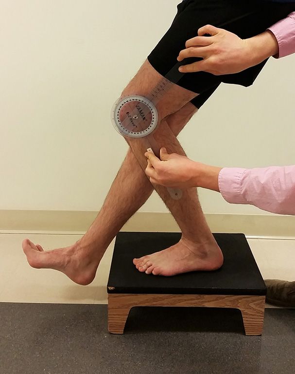
Page 2 of 14                                                                                                     Annals of Joint, 2018

   The purpose of this review is to provide an overview                movement (10). Healthcare professionals must consider
of the physical therapist’s management, including the                  not only the patient’s underlying knee pathology (e.g.,
evaluation and treatment, of the patient with PFP. We                  structural abnormalities, muscle dysfunction) but also the
begin with a brief overview of symptom onset, then discuss             patient’s psychological distress and pain neurophysiology
the importance of considering the complexities of the                  when evaluating the clinical pain experience (11). In chronic
painful experience when rehabilitating individuals with                musculoskeletal conditions, as can often become the case
PFP, particularly among those with episodic or recalcitrant            with PFP, symptoms may outlive their usefulness; although
symptoms. We then present our rehabilitation approach                  no clear definition exists, chronic pain is generally described
for a systematic physical therapy examination including                as pain that lasts “beyond the body’s usual healing time”
a thorough subjective history and objective clinical,                  and is typically three months or greater (12). Clinicians
functional, and patient-reported outcome measures. Finally,            must recognize the difference between acute (protective)
we present a comprehensive treatment approach that draws               pain and chronic pain, which may limit function and inhibit
heavily from recently published literature and clinical trials.        progress. Encouraging regular movement and exercise
                                                                       within the pain-free envelope of function (7) and, when
Symptom onset                                                          appropriate, such as in the chronic case, even beyond the
                                                                       pain-free range, may be necessary to optimize function in
PFP, or anterior knee pain, is an amalgam of conditions                patients with PFP. In such cases, graded exposure (13) may
that are typically non-traumatic in origin and result in               help maximize function even in the absence of full symptom
peripatellar and/or retropatellar knee pain. A number                  resolution.
of structures in and around the patellofemoral and                        Conscientious monitoring and progression of
tibiofemoral joints, such as the synovium or infrapatellar fat         interventions and other activities throughout rehabilitation
pad, may individually or collectively contribute to PFP (4).           is thus essential to achieving optimal outcomes. The
The patellofemoral articular cartilage itself, however, is             remainder of this review article will delineate strategies
not painful when probed directly sans anesthesia (5), likely           for conducting a thorough evaluation and creating an
due to its lack of free nerve endings (6). While a variety of          appropriate, progressive, and individualized treatment
factors may also contribute to symptom onset, disruption of            approach for PFP.
tissue homeostasis via acute injury or repetitive overloading
(i.e., high-frequency moderate loading or an isolated very
high loading event) may exceed tissue homeostasis, or the              Evaluation
envelope of function, for a given structure(s) and lead to             History
pathology and pain (7,8). Conservative management may
initially promote relative rest and avoidance of activities            A thorough history is critical for appropriately
that exacerbate the patient’s pain while attempting to limit           diagnosing (14) and optimally managing PFP (15). While
loss of muscle strength, ROM, or function. PFP, however,               one may accurately identify the relatively young, active
often persists for months or even years (3,9), requiring a             woman with atraumatic onset of anterior knee pain as
more complex rehabilitation approach.                                  the most likely candidate, men and women of all activity
                                                                       levels across a wide age range may develop PFP (16). The
                                                                       rehabilitation specialist should ask the patient to identify
The complex pain experience
                                                                       the date of symptom onset, mechanism of injury and/or
Throughout the successful management of PFP and                        antecedent events, location and quality of pain, exacerbating
especially when symptoms are chronic in nature,                        and alleviating symptoms, relevant past medical history
rehabilitation specialists must appreciate the complexity              including prior lower extremity and low back symptoms,
of the pain experience (10). In his 2016 Maley Lecture,                diagnostic imaging, occupational demands, recreational
physical therapist and pain science researcher Steven                  activities, footwear including use of orthotics, and patient
George, PT, PhD, calls for a shift in physical therapist               goals (Table 1). Pertinent past medical history may include
education, research, and clinical practice from the traditional        not only previous knee symptoms but also ankle, hip, and
direct link among pain, nociception, and injury to a more              lumbar pain, as radiculopathy from the spine to the knee
inclusive biopsychosocial model that incorporates pain with            is possible. Referred knee pain may be present due to hip

© Annals of Joint. All rights reserved.                           aoj.amegroups.com                                 Ann Joint 2018;3:40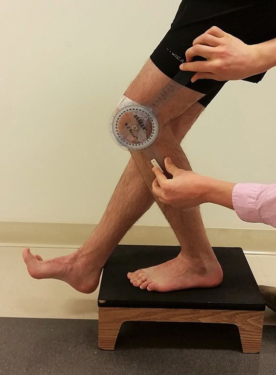
Annals of Joint, 2018                                                                                                               Page 3 of 14

 Table 1 A thorough patient history should include the following questions
 Questions                                                                           Notes

 Date of onset

 Mechanism of injury (traumatic vs. atraumatic):                                     If traumatic, consider and evaluate thoroughly for
                                                                                     alternative diagnoses including ligament sprain, meniscus
   If atraumatic, sudden or gradual onset? What factors led to symptoms (i.e.,
                                                                                     tear, fracture, etc.
   any changes in activity level, exercise, footwear, stress levels, sleep habits,
   diet or body mass)?

   If traumatic, describe event in detail including presence of swelling and time
   to swelling onset

 Chief complaint (location and quality of pain):

   Exacerbating factors (e.g., stair descent, squatting)?

   Alleviating factors (e.g., ice, heat, rest, stretching)?

 Are other symptom(s) present?                                                       If true giving way episodes are present, consider
                                                                                     ligament exam; if locking is present, consider meniscus
   If yes, any giving way/buckling, locking/clicking/popping/crepitus, stiffness?
                                                                                     involvement

 Diagnostic tests and imaging

 Relevant past medical history (e.g., previous lower extremity injury, previous      If history of back pain or unable to elicit symptoms during
 back pain with or without radiculopathy)                                            targeted knee evaluation, perform lumbar and spinal
                                                                                     radiculopathy examination

                                                                                     Consider also the hip joint as a source of knee pain,
                                                                                     particularly in the child (17,18) or older adult

 Has the patient received any prior treatment? If so, describe in detail

 What are the patient’s occupational demands?

 What recreational activities does the patient typically engage in? Are these
 activities limited? If so, how?

 Describe footwear and orthotic use                                                  Examine footwear and orthotics for wear and irregularities

 Goals for rehabilitation

pathology, such as osteoarthritis or predominantly pediatric                 limp, and limited hip ROM possibly indicative of Perthes
conditions like slipped capital femoral epiphysis (17,18),                   disease or a slipped capital femoral epiphysis) (16). Physician
thus subjective questioning and physical examination                         referral is also warranted in the case of unremitting or
should consider the hip, particularly when the practitioner                  worsening symptoms despite appropriate physical therapy
is unable to provoke the patient’s symptoms during a                         and activity modification.
thorough, targeted knee evaluation. Gradual and even
insidious onset of anterior knee pain are common in PFP
                                                                             Clinical examination
whereas acute onset of knee pain secondary to a traumatic
event merits further evaluation of the integrity of the knee                 Physical examination should incorporate a variety of
ligaments, tendons, menisci, and bone. Clinicians should                     measures including ROM, muscle length, effusion,
refer their patients to an appropriate specialist if they                    resisted isometrics, strength, balance and postural control,
suspect serious pathology (e.g., fracture or osteomyelitis) or               movement quality assessments, special tests, palpation,
non-musculoskeletal origin (e.g., cancer or infection) due to                functional evaluation, and patient reported outcome
the presence of red flags (i.e., fever, unremitting night pain,              measures. Objective assessments should guide treatment,
or increased temperature and swelling around the knee; or,                   progression, and clinical decision-making. An individualized
among adolescents or children, a leg length discrepancy,                     rehabilitation program that addresses the patient’s specific

© Annals of Joint. All rights reserved.                                aoj.amegroups.com                                      Ann Joint 2018;3:40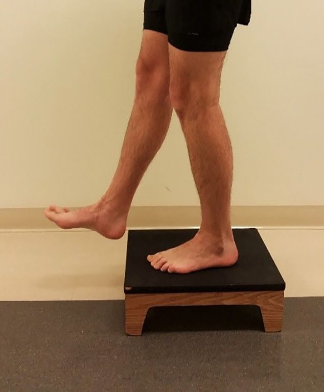
Page 4 of 14                                                                                                                Annals of Joint, 2018

 Table 2 Clinicians should monitor knee effusion throughout rehabilitation using the reliable stroke test (20) (Effusion Grading Scale of the Knee
 Joint Based on the Stroke Test)
 Grade            Test result

 Zero             No wave produced on down stroke

 Trace            Small wave on medial side with down stroke

 1+               Larger bulge on medial side with down stroke

 2+               Effusion spontaneously returns to medial side after upstroke (no down stroke necessary)

 3+               So much fluid that it is not possible to move the effusion out of the medial aspect of the knee
 Reprinted with permission from Sturgill LP, Snyder-Mackler L, Manal TJ, et al. Interrater reliability of a clinical scale to assess knee joint
 effusion. J Orthop Sports Phys Ther 2009;39:845-9. https://doi.org/10.2519/jospt.2009.3143. ©Journal of Orthopaedic & Sports Physical
         ®
 Therapy .

impairments and functional limitations is regarded as best                  exercises or home exercise program contributed to an
practice (9).                                                               exacerbation of effusion and/or other symptoms or whether
                                                                            other factors are more likely culpable. For example, asking
ROM and muscle length testing                                               a student about activities such as walking around school or
ROM of the knee as well as the ankle and hip should                         campus or attending a party may be pertinent. The use of
be assessed. The physical therapist should evaluate at a                    activity trackers to monitor movement outside of therapy is
minimum both active and passive ROM measurements of                         becoming increasingly possible and should be considered as a
tibiofemoral flexion and extension, talocrural dorsiflexion,                more accurate way to quantify activity and joint loading (24).
and femoroacetabular extension, internal and external
rotation, and flexion; other motions (e.g., hip abduction and               Resisted isometrics
adduction) or joints (e.g., subtalar eversion and inversion                 Resisted isometrics at various angles of knee flexion may be
and lumbar flexion and extension) may also be considered.                   used during the early portions of the clinical examination to
   Muscle length testing is also an important consideration                 determine what type of structure(s) is most likely involved.
as soft tissue tightness (i.e., limited flexibility) is prevalent in        A finding of “strong and painful” with resisted isometric
individuals with PFP and may contribute to symptoms (19).                   knee extension is most likely to support the diagnosis of
Evaluation of the rectus femoris, hip flexors (1- and 2-joint               PFP, although weakness is also possible, particularly in the
muscles), tensor fascia lata and iliotibial band, hamstrings,               acute phase (pain-mediated) or in long-standing, chronic
gastrocnemius, and soleus should be performed.                              cases. The clinician should evaluate resisted isometrics at
                                                                            multiple angles of knee flexion to see if there is a range
Effusion                                                                    that is more or less painful for the individual patient. The
Knee joint effusion can easily be evaluated using the stroke                clinician may use these findings to inform subsequent
test (Table 2). The stroke test is a reliable grading scale                 strength evaluations as well as treatment, selecting ranges of
that assesses the presence of intracapsular swelling (20).                  motion that are least provocative to the patient to improve
While effusion is not often present, mild effusion can occur                muscle strength and activation while avoiding exacerbation
among individuals with PFP; significant effusion is likely                  of symptoms.
indicative of more serious pathology (e.g., ligament rupture,
meniscus tear, fracture) and merits further evaluation.                     Strength
Effusion monitoring may help determine appropriate                          Strength assessments should evaluate not only the muscles
clinical progression (21,22). Increased effusion can indicate               crossing the knee joint but also the surrounding hip
when rehabilitation has exceeded the patient’s current                      and ankle musculature. Knee extensor and hip extensor,
envelope of function (7,23) and thus rehabilitation exercises               abductor, and external rotator muscle strength and
or activity should be reduced or not progressed further.                    activation are of utmost importance given their roles in
Tracking or asking the patient about outside activities                     dynamically controlling hip and knee motion and the
is critical in determining whether or not the prescribed                    association of PFP with weakness of these muscles (25-29),

© Annals of Joint. All rights reserved.                                aoj.amegroups.com                                       Ann Joint 2018;3:40
Annals of Joint, 2018 Page 5 of 14
(most) involved limb first to determine the angle of knee
flexion that is pain-free or least provocative; the clinician
can subsequently evaluate the contralateral limb in the same
position. Clinicians may also use patellar taping (see below)
to facilitate strength evaluation, enabling some patients to
complete testing with less or no pain. While we most often
use a limb symmetry index [i.e., involved limb strength/
uninvolved limb strength × 100 (%)] for comparison, PFP
is often a bilateral condition thus clinicians should interpret
limb symmetry indexes with caution. Additional evaluation
using manual muscle testing of the hip and knee muscles
may provide additional insight, especially in the case of
bilateral weakness.
Balance and postural control
Balance and postural control may be impaired in patients
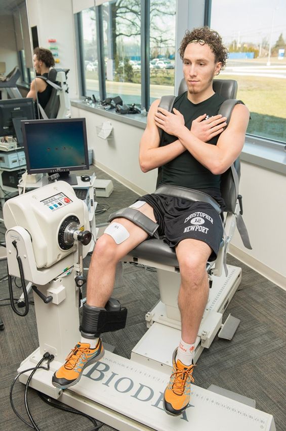
Figure 1 Quadriceps strength may be evaluated isometrically with PFP compared to healthy controls (33-35) during a
using an electromechanical dynamometer during with an electrical variety of tasks including dynamic standing balance (33),
burst superimposition technique (32) to assess muscle activation. postural stability during a stepping up and down task (34),
Clinicians may evaluate the (most) involved limb first to determine and stair climbing (35). Static balance during single leg
the angle of knee flexion that is pain-free or least provocative and stance is also impaired on the involved compared to
subsequently evaluate the contralateral limb at the same angle uninvolved limb among women with PFP (36). Fatigue of
of knee flexion for comparison. Patellar taping may be used to the hip abductors and to a lesser degree the knee extensors
alleviate pain. is associated with greater balance instability during dynamic
standing balance (33). Patients with PFP may also exhibit
especially poor postural control with their eyes closed (37).
although cause and effect are unknown (28). Interestingly, In light of these findings, it is important to assess both static
Kindel and Challis found that patients with PFP have balance with eyes opened and closed as well as dynamic
weaker hip extensors and poorer neuromuscular control balance on both the (most) involved and contralateral limb.
with the knee flexed but not extended compared to healthy To assess static balance, we evaluate single leg stance, which
controls (30), suggesting knee position may be important can be progressed in difficulty by having the patient stand
when evaluating hip musculature. A thorough evaluation on an unstable surface such as a foam pad; document the
should also strength of the core muscles, knee flexors, ankle time to error and/or number of errors in a given time (e.g.,
plantarflexors and dorsiflexors, and hip flexors, internal 30 seconds). Dynamic balance may be assessed using the
rotators, and adductors. reliable Star Excursion Balance Test (38,39).
Given the strength of the lower extremity muscles,
clinicians should evaluate lower extremity muscle, Movement assessments
particularly quadriceps, strength using an electromechanical Clinicians should consider a variety of movement quality
dynamometer when possible. When an electromechanical assessments concordant with the patient’s complaints
dynamometer is not available, one-rep max testing on and activity limitations given that aberrant mechanics
knee extension machine for quadriceps strength or hand- and neuromuscular activation patterns are often present
held dynamometer secured with a strap are acceptable in individuals with PFP (23,26,40-44). The position of
alternatives, although they overestimate strength of the dynamic knee valgus, characterized by hip adduction and
involved quadriceps (31). Electrical burst superimposition internal rotation, may be associated with PFP (23,40,44,45),
may be used to evaluate quadriceps muscle activation (i.e., thus clinicians should pay particular attention for these
inhibition) (32), but requires relatively expensive equipment aberrant mechanics. Clinicians should consider evaluating
that is unavailable to many clinicians (Figure 1). In contrast multi-joint lower extremity movements including but
to the usual order, we recommend that clinicians test the not limited to double and single leg squatting, drop jump
© Annals of Joint. All rights reserved. aoj.amegroups.com Ann Joint 2018;3:40Page 6 of 14 Annals of Joint, 2018
A B C
Figure 2 The patients stand on the involved limb on a 15-cm box (A) to begin the modified step test. We document the angle at which the
patient experiences pain and the patient’s numeric pain rating both before (B) and after (C) applying patellar taping.
landing, hopping, walking, stair ascent and descent, and quadriceps tendons, to rule out other sources of anterior knee
running. Identification of movement impairments may pain. For example, reproduction of pain with palpation of
guide not only targeted strengthening but also and perhaps the patellar tendon may indicate patellar tendinopathy; pain
more importantly neuromuscular activation exercises and at the distal pole of the patella in adolescents may indicate
movement retraining (23,40,46,47). Sinding-Larsen-Johansson Syndrome (50); and swelling and
point tenderness around the tibial tuberosity in adolescents
Step test may indicate Osgood-Schlatter Disease (16,50).
We recommend using a modification of the previously
described step test (Figure 2). The step test involves standing Functional testing
on a 15 centimeter block with hands on hips and using the Functional testing may evaluate tasks that are important to
involved limb to “slowly” and “smoothly” eccentrically lower the patient and are currently limited. Examples of functional
the body until the contralateral heel touches the floor (48). testing include the stair climb test, sit to stand test, and
A positive result is reproduction of the patient’s PFP; a 6-minute walk test. Performance as well as symptoms
positive finding is prevalent in 74% (57 of 77) of individuals should be documented.
with PFP (49) and has a modest positive likelihood ratio of
2.34 (48). In the authors’ clinical experience, we modify the Objective measures for evaluation, treatment
test by recording the angle at which pain first occurs and progression, and clinical decision-making
asking the patient to rate the pain on an 11-point numeric Evaluation, treatment progression, and clinical decision-
pain rating scale. If the test is positive, we often evaluate making like discharge and return-to-sport clearance should
the patient again on the modified step test after applying be based as much as possible on objective measures while
patellar taping (described below) to determine whether or simultaneously considering the patient’s needs and goals.
not patellar taping provides immediate relief of symptoms As mentioned above, an increase in or the presence of
and may therefore be beneficial in facilitating increased new effusion indicates that the activity has exceeded the
function in the short-term. current envelope of function and should not be progressed
further. Clinicians may also use the soreness rules (Table 3),
Palpation initially developed by Fees et al. (51) and later adapted
Individuals with PFP often have pain in or around the patella to the lower extremity by Adams et al. (21), to monitor
that may be reproduced with palpation. Clinicians should appropriate progression of activities. (While avoiding
also palpate other nearby structures, such as the patellar and pain and symptom exacerbation is critical during the early
© Annals of Joint. All rights reserved. aoj.amegroups.com Ann Joint 2018;3:40Annals of Joint, 2018 Page 7 of 14
Table 3 The soreness rules provide clinicians with a guideline to monitor symptoms and evaluate progression throughout rehabilitation (21,51)
(Soreness rules)
Criterion Action
Soreness during warm-up that continues 2 days off, drop down 1 level
Soreness during warm-up that goes away Stay at level that led to soreness
Soreness during warm-up that goes away but redevelops during session 2 days off, drop down 1 level
Soreness the day after lifting (not muscle soreness) 1 day off, do not advance program to the next level
No soreness Advance 1 level per week or as instructed by healthcare
professional
Reproduced with permission from Michael J. Axe, MD and Sage Publications, Inc. (51), available online: http://journals.sagepub.com/doi/
10.1177/03635465980260052301.
Table 4 A running progression may facilitate gradual resumption of loading; progression should occur only in the absence of increased effusion
or pain and on nonconsecutive days (Running progression*)
Level Treadmill Track
Level 1 0.1-mi walk/0.1-mi jog, repeat 10 times Jog straights/walk curves (2 mi)
Level 2 Alternate 0.1-mi walk/0.2-mi jog (2 mi) Jog straights/jog 1 curve every other lap (2 mi)
Level 3 Alternate 0.1-mi walk/0.3-mi jog (2 mi) Jog straights/jog 1 curve every lap (2 mi)
Level 4 Alternate 0.1-mi walk/0.4-mi jog (2 mi) Jog 1.75 laps/walk curve (2 mi)
Level 5 Jog full 2 mi Jog all laps (2 mi)
Level 6 Increase workout to 2.5 mi Increase workout to 2.5 mi
Level 7 Increase workout to 3 mi Increase workout to 3 mi
Level 8 Alternate between running/jogging every 0.25 mi Increase speed on straights/jog curves
*, progress to the next level when the patient is able to perform activity for 2 mi without increased effusion or pain. Perform no more than 4
times in 1 week and no more frequently than every other day. Do not progress more than 2 levels in a 7-day period. Conversion: 1 mi =1.6
km. Reproduced with permission from Tara Manal, PT, DPT, FAPTA, University of Delaware Physical Therapy Clinic.
management of acute PFP, clinicians may set a threshold Throughout the rehabilitation process, the clinicians
of acceptable symptoms (e.g., 5/10 on numeric pain must appreciate the impact of psychological factors (e.g.,
rating scale) for individuals with chronic PFP, focusing kinesiophobia) (55) and other factors (e.g., stress, sleep)
on increasing function rather than complete avoidance of on pain, particularly when a patient reports a transient
symptoms). Successful completion of a running progression increase in symptoms. Anxiety, depression, catastrophizing,
(Table 4) (21) should be pre-requisite to initiating higher and kinesiophobia may be present in individuals with
level activities. PFP and correlate with higher pain ratings and reduced
Valid and reliable patient reported outcome measures physical function (56); appropriate referral or consultation
should be completed at initial evaluation and periodically may be beneficial. Stress levels (57) and sleep duration (58)
throughout rehabilitation to monitor progress and inform also influence pain; for example, too much (>9 hours) or
rehabilitation. The Visual Analog Scale for usual pain or too little (Page 8 of 14 Annals of Joint, 2018
Treatment benefit from performing open kinetic chain exercises in
deeper ranges of knee flexion (e.g., 50°–90°) and closed
Patients with PFP present with a wide variety of underlying
kinetic chain exercises in shallower ranges (e.g., 0°–45°) (66).
pathophysiology and associated impairments (25,47). It is
Throughout the rehabilitation process, clinicians
thus imperative to individually assess each patient to identify
should design appropriate exercises that maximize muscle
and subsequently address his or her impairments, functional
strength while minimizing symptom exacerbation, using
limitations, and activity restrictions. Management of PFP
the soreness rules (Table 3) to guide progression. A recent
should consist of an individualized (47), multi-modal
study by van Rossom and colleagues provides peak and
approach with exercise therapy as the hallmark of the
mean patellofemoral joint contact forces during gait plus
plan (9,16,26,59-61).
nine functional exercises and may serve as a guide for
appropriately and gradually progressing loading during
Exercise therapy: strengthening, stretching, and aerobic rehabilitation (67). While initially during the acute
exercise stage of rehabilitation a clinician may strive to perform
only exercises that are pain-free, the goal of completely
According to the 2016 consensus statement from the eliminating movement-related pain in the chronic condition
International Patellofemoral Pain Research Committee, may be not only unrealistic but also a disservice to the
exercise therapy is the “treatment of choice” for individuals patient’s recovery (10). In such cases, setting an acceptable
with PFP (9). High-quality evidence supports exercise threshold of symptoms based on the patient’s presentation
therapy to improve pain and function in the short-, may be appropriate.
medium-, and long-term; exercise was the only intervention Stretching is another important component of
that received such a high recommendation (9). Exercise rehabilitation, as individuals with PFP often have limited
therapy should include both hip and knee strengthening ROM, particularly around the hip (19) and knee and
(9,27,62,63) using both open (non-weight-bearing) and perhaps also the ankle (25). Treatments should address
closed (weight-bearing) kinetic chain exercises (9,62). the specific ROM and muscle length restrictions identified
Open kinetic chain exercises include straight leg raises during the evaluation and may include the quadriceps,
(progress by adding ankle weights), short arc quadriceps hip flexors, hamstrings, tensor fascia lata/iliotibial band,
strengthening, knee extensions, side-lying hip abduction gastrocnemius, and/or soleus.
straight leg raise, and clamshells. Closed kinetic chain
exercises include wall sits, double- and single-leg squats,
lateral step-downs, and leg press. Strengthening of the core Joint mobilizations
(47,64) and ankle musculature should be included if the Joint mobilizations may be effective in improving pain
patient exhibits deficits or imbalances in these areas. and function among individuals with PFP when joint
Appropriate selection of open and closed chain mobilizations are directed at the knee (i.e., patellofemoral
strengthening exercises should consider the patellofemoral and tibiofemoral joint) and combined with a comprehensive
joint contact forces in each mode. Steinkamp et al. found treatment approach including exercise (59). A case study by
that comparison of patellofemoral joint contact forces Lantz et al. highlights the potential benefit of tibiofemoral
during closed (i.e., body weight squat) and open (i.e., 9 kg mobilizations in an individual with chronic PFP (68).
weighted boot) kinetic chain exercises resulted in relatively
less patellofemoral contact force in the closed kinetic chain
Patellofemoral taping
condition in less than 48° knee flexion and relatively less
patellofemoral contact force in the open kinetic chain Conflicting evidence exists regarding the efficacy of
condition in more than 48° knee flexion (65). Similar patellofemoral taping (60,69-72). We recommend using
findings have been more recently produced by Powers et al., taping in conjunction with a multi-modal, comprehensive
who added that patellofemoral joint contact force was less treatment plan if taping alleviates pain during exercises in
during quadriceps strengthening using a constant resistance rehabilitation and/or functional activities. Clinicians should
knee extension machine compared to squatting at angles evaluate the immediate effectiveness of patellofemoral
greater than approximately 45° (66). Therefore, particularly taping within an individual by assessing a functional
during the early stages of rehabilitation, patients may task pre- and post-taping that is specific to that patient’s
© Annals of Joint. All rights reserved. aoj.amegroups.com Ann Joint 2018;3:40Annals of Joint, 2018 Page 9 of 14
symptoms; if pain is alleviated then taping may help the Neuromuscular training
patient complete functional activities and exercises which
Neuromuscular activation deficits are common in
may in turn facilitate recovery. While we recommend first
individuals with PFP, especially in the hip abductors and
evaluating medial patellar glide therapeutic taping (73),
external rotators, knee extensors, and core musculature
placebo taping plus exercise may be similarly beneficial
(23,26,40,44,45). Evaluating movements during functional
to therapeutic tension taping plus exercise (60). The
tasks (described above) is essential to identifying and treating
use of patellar taping in isolation is not recommended
neuromuscular activation deficits. Strengthening alone
(9,16,60,61,69,70,73).
seldom changes mechanics (78), thus task-specific movement
retraining is likely necessary (23,40,79,80). Use of resistance
Neuromuscular electrical stimulation (NMES) tubing bands may promote activity of specific muscle groups;
for example, using resistance tubing bands around the
A 2017 Cochrane Review by Martimbianco et al. found
knees during a squat may facility hip abduction and external
limited, low-quality regarding the effect of NMES for the
treatment of PFP (74). The review concluded that very rotation. NMES may facilitate neuromuscular training, as
low-quality evidence suggests NMES reduces pain at the improvements in kinematics and muscle activity have been
end of treatment (3 to 12 weeks) but the improvement observed in a small group (N=15) of women with PFP (46).
may not be clinically relevant given the small magnitude Running mechanics and gait retraining in patients with
of change (1.63 out of 10 on the visual analog scale). The patellofemoral pain have received significant attention likely
authors found even less support for NMES on strength or due in part to the high incidence of PFP among runners (1).
function, concluding that “insufficient and inconclusive Running mechanics are often altered in individuals with
evidence” exists for the effect of NMES on treating PFP and young women may be especially prone to altered
individuals with PFP (74). While one pilot study has found mechanics such as excessive hip adduction and internal
no statistically significant differences between 38 athletes (19 rotation leading to dynamic knee valgus (23,41-43,81).
per group) who completed physiotherapy or physiotherapy Gait retraining may be considered in individuals with
plus electrical stimulation, limitations including study PFP who have aberrant running mechanics and should
design, follow-up, and stimulation parameters limit its address the specific deficits in the individual (43). Sagittal
applicability (75). Given the dose response relationship plane trunk mechanics (82) and footwear (as described by
between electrical stimulation intensity and quadriceps the Minimalist Index) (83) are related to patellofemoral
femoris muscle torque (76), we recommend using higher joint stress during running, thus should also be considered
NMES intensity levels to facilitate muscular strength during gait analysis and running retraining; forward trunk
and activation development. A 2010 systematic review lean (82) and more minimalist shoes (83) are associated with
on NMES on quadriceps strength in individuals after reduced patellofemoral joint stress. A systematic review by
anterior cruciate ligament reconstruction found that NMES Agresta and Brown found the use of real-time auditory and
combined with exercise is more effective than exercise visual feedback in conjunction with therapeutic exercise
alone at improving quadriceps muscle strength (77). We to be effective in improving lower extremity kinematics in
therefore recommend using NMES in conjunction with runners with patellofemoral, although no single method of
a comprehensive rehabilitation program in individuals feedback was deemed superior (84).
who have PFP and deficits in quadriceps strength and/
or activation. We recommend the following parameters:
Activity modification and gradual loading
10.2 cm × 12.7 cm pads on the vastus medialis and proximal
vastus lateralis muscles; 15 electrically elicited, isometric During the acute phase, activity modification characterized
contractions of the quadriceps at about 65° knee flexion (or by relative rest is likely appropriate to allow healing
the most comfortable position for the patient), 75 bursts to occur. Reintegration of loading, however, must be
per second; 10” on, 50” off, 2” ramp; and the maximum implemented and should be done in a systematic way to
tolerated intensity that elicits at least 50% maximum gradually increase and restore the envelope of function.
volitional isometric contraction (21,76). Chen et al. evaluated patellofemoral joint reaction forces
© Annals of Joint. All rights reserved. aoj.amegroups.com Ann Joint 2018;3:40Page 10 of 14 Annals of Joint, 2018
using an MRI-informed subject-specific three-dimensional criteria in patients with PFP is lacking, we recommend
model, finding that, among the four tasks evaluated, athletes achieve limb symmetry index scores of 90% of
patellofemoral joint reaction forces were highest during greater for quadriceps strength and all four hop tests (single,
running [58.2 N/kg-body weight (bwt)], followed by stair crossover, triple, and 6 meter timed) (86) prior to resuming
ascent (33.9 N/kg-bwt), stair descent (27.9 N/kg-bwt), and full participation; limb symmetry indexes, however, have
walking (10.1 N/kg-bwt) (2). In light of these findings, it limitations (87) particularly in individuals with bilateral
may be inappropriate for an individual with acute PFP to involvement thus should be interpreted with caution.
run if stair descent is painful, although individual evaluation
and clinical judgment should be considered. Recently, van
Conclusions
Rossom et al. added to Chen’s findings by evaluating peak
and mean patellofemoral joint contact forces during ten Early, appropriate rehabilitation may be critical to
functional tasks; peak patellofemoral joint contact forces preventing poor outcomes (88) and optimizing function
were lowest during gait and progressively higher in sit for individuals with PFP. We strongly recommend
down, stand up, squat, forward lunge, stair ascent, stair exercise therapy, including hip and knee strengthening and
descent, single leg hop weight acceptance phase, sideward stretching, to improve short-, medium-, and long-term
lunge, and single leg hop push-off phase (67). outcomes in individuals with PFP (9,16,26,27). A multi-
modal, individually tailored rehabilitation program should
be designed to target the patient’s specific impairments and
Other interventions
functional limitations identified during the evaluation (47).
Numerous other interventions have been proposed as Treatments may include open- and closed-chain exercises,
adjuvants or stand-alone treatments for individuals with strengthening, stretching, aerobic exercise, patellofemoral
PFP and may be considered as part of a comprehensive and tibiofemoral mobilizations, patellar taping, high-
plan of care if impairments warrant or symptoms have been intensity NMES, neuromuscular training, and gait
intractable to the more evidence-based approaches outlined retraining. Although short-term changes or reductions in
above. Foot orthotics may be beneficial in reducing pain movement often are necessary in a protective capacity, the
and improving function (16). Dry needling does not appear persistence of altered movement is a key characteristic of
to provide any additional benefit when added to a multi- chronic pain. PFP etiology is largely movement related and
modal treatment approach including manual therapy and a comprehensive conservative treatment using movement
strengthening exercise compared to manual therapy and can be successful.
strengthening exercise alone (85).
Acknowledgements
Appropriate progression and discharge
Funding: JJ Capin receives funding from the Foundation
Rehabilitation should be progressive and rooted in objective for Physical Therapy (Promotion of Doctoral Studies Level
clinical findings. Monitoring effusion and soreness should I Scholarship) and the University of Delaware (Doctoral
occur throughout rehabilitation and guide progression. Fellowship Award). L Snyder-Mackler receives funding
Use of gradual, return-to-activity training protocols, such from the National Institutes of Health: NICHD (R44-
as the running progression (Table 4) (21), may facilitate HD068054, R37-HD037985, and T32-HD007490),
appropriate progression and aid clinical decision-making. NIAMS (R01-AR048212), and NIGMS (U54-GM104941).
Discharge from physical therapy should occur when the
patient has achieved his or her goals and is equipped to
Footnote
transition to self-management or management by an athletic
trainer, strength and conditioning coach, or personal Conflicts of Interest: The authors have no conflicts of interest
trainer if available. Patient education is thus critical at this to declare.
time-point and throughout the rehabilitation process; the
patient should know what exercises to perform and how to
References
progress activity while adhering to basic principles such as
the soreness rules. Although research on return-to-sport 1. Smith BE, Selfe J, Thacker D, et al. Incidence and
© Annals of Joint. All rights reserved. aoj.amegroups.com Ann Joint 2018;3:40Annals of Joint, 2018 Page 11 of 14
prevalence of patellofemoral pain: a systematic review and Rehabil 2018;99:607-614.e1.
meta-analysis. PLoS One 2018;13:e0190892. 15. Werner S. Anterior knee pain: an update of physical
2. Chen YJ, Scher I, Powers CM. Quantification of therapy. Knee Surg Sports Traumatol Arthrosc
Patellofemoral Joint Reaction Force During Functional 2014;22:2286-94.
Activities Using a Subject-Specific Three-Dimensional 16. Crossley KM, Callaghan MJ, Van Linschoten R.
Model. J Appl Biomech 2010;26:415-23. Patellofemoral pain. Br J Sports Med 2016;50:247-50.
3. Lankhorst NE, van Middelkoop M, Crossley KM, et 17. Herngren B, Stenmarker M, Vavruch L, et al. Slipped
al. Factors that predict a poor outcome 5-8 years after capital femoral epiphysis: A population-based study. BMC
the diagnosis of patellofemoral pain: a multicentre Musculoskelet Disord 2017;18:304.
observational analysis. Br J Sports Med 2016;50:881-6. 18. Hatfield SJ, Baxter RE. Slipped Capital Femoral Epiphysis
4. Biedert RM, Sanchis-Alfonso V. Sources of anterior knee in a Patient With Knee Pain. J Orthop Sports Phys Ther
pain. Clin Sports Med 2002;21:335-47. 2012;42:482.
5. Dye SF, Vaupel GL, Dye CC. Conscious Neurosensory 19. Hamstra-Wright KL, Earl-Boehm J, Bolgla L, et al.
Mapping of the Internal Structures of the Human Knee Individuals with patellofemoral pain have less hip flexibility
Without Intraarticular Anesthesia. Am J Sports Med than controls regardless of treatment outcome. Clin J
1998;26:773-7. Sport Med 2017;27:97-103.
6. Witoński D, Wagrowska-Danielewicz M. Distribution of 20. Sturgill LP, Snyder-Mackler L, Manal TJ, et al. Interrater
substance-P nerve fibers in the knee joint in patients with reliability of a clinical scale to assess knee joint effusion. J
anterior knee pain syndrome. A preliminary report. Knee Orthop Sports Phys Ther 2009;39:845-9.
Surg Sports Traumatol Arthrosc 1999;7:177-83. 21. Adams D, Logerstedt D, Hunter-Giordano A, et
7. Dye SF. The knee as a biologic transmission with an al. Current concepts for anterior cruciate ligament
envelope of function: a theory. Clin Orthop Relat Res reconstruction: a criterion-based rehabilitation
1996;(325):10-8. progression. J Orthop Sports Phys Ther 2012;42:601-14.
8. Post WR, Dye SF. Patellofemoral Pain: An Enigma 22. Capin JJ, Behrns W, Thatcher K, et al. On-Ice Return-
Explained by Homeostasis and Common Sense. Am J to-Hockey Progression After Anterior Cruciate
Orthop (Belle Mead NJ) 2017;46:92-100. Ligament Reconstruction. J Orthop Sports Phys Ther
9. Crossley KM, Middelkoop M Van, Callaghan MJ, et al. 2017;47:324-33.
2016 Patellofemoral pain consensus statement from the 23. Willy RW, Meira EP. Current Concepts in Biomechanical
4th International Patellofemoral Pain Research Retreat, Interventions for Patellofemoral Pain. Int J Sports Phys
Manchester. Part 2: Recommended physical interventions Ther 2016;11:877-90.
(exercise, taping, bracing, foot orthoses and combined 24. Shull PB, Jirattigalachote W, Hunt MA, et al. Quantified
interventions). Br J Sports Med 2016;50:844-52. self and human movement: A review on the clinical impact
10. George SZ. Pain Management: Road Map to Revolution. of wearable sensing and feedback for gait analysis and
Phys Ther 2017;97:217-26. intervention. Gait Posture 2014;40:11-9.
11. Kittelson AJ, George SZ, Maluf KS, et al. Future 25. Witvrouw E, Werner S, Mikkelsen C, et al. Clinical
Directions in Painful Knee Osteoarthritis: Harnessing classification of patellofemoral pain syndrome: Guidelines
Complexity in a Heterogeneous Population. Phys Ther for non-operative treatment. Knee Surg Sports Traumatol
2014;94:422-32. Arthrosc 2005;13:122-30.
12. Geneen LJ, Moore RA, Clarke C, et al. Physical activity 26. Saltychev M, Dutton R, Laimi K, et al. Effectiveness of
and exercise for chronic pain in adults: an overview conservative treatment for patellofemoral pain syndrome:
of Cochrane reviews. Cochrane Database Syst Rev A systematic review and meta-analysis. J Rehabil Med
2017;4:CD011279. 2018. [Epub ahead of print].
13. Nijs J, Lluch Girbés E, Lundberg M, et al. Exercise 27. Nascimento LR, Teixeira-Salmela LF, Souza RB, et al.
therapy for chronic musculoskeletal pain: Innovation by Hip and Knee Strengthening is More Effective Than Knee
altering pain memories. Man Ther 2015;20:216-20. Strengthening Alone for Reducing Pain and Improving
14. Décary S, Frémont P, Pelletier B, et al. Validity of Activity in Individuals With Patellofemoral Pain: A
Combining History Elements and Physical Examination Systematic Review With Meta-Analysis. J Orthop Sports
Tests to Diagnose Patellofemoral Pain. Arch Phys Med Phys Ther 2018;48:19-31.
© Annals of Joint. All rights reserved. aoj.amegroups.com Ann Joint 2018;3:40Page 12 of 14 Annals of Joint, 2018
28. Rathleff MS, Rathleff CR, Crossley KM, et al. Is hip patellofemoral pain have altered biomechanics which
strength a risk factor for patellofemoral pain? A systematic targeted interventions can modify: A systematic review and
review and meta-analysis. Br J Sports Med 2014;48:1088. meta-analysis. Gait Posture 2016;45:69-82.
29. Giles LS, Webster KE, McClelland JA, et al. Does 41. Noehren B, Pohl MB, Sanchez Z, et al. Proximal and
Quadriceps Atrophy Exist in Individuals With distal kinematics in female runners with patellofemoral
Patellofemoral Pain? A Systematic Literature Review pain. Clin Biomech (Bristol, Avon) 2012;27:366-71.
With Meta-analysis. J Orthop Sports Phys Ther 42. Noehren B, Sanchez Z, Cunningham T, et al. The effect
2013;43:766-76. of pain on hip and knee kinematics during running in
30. Kindel C, Challis J. Joint Moment-Angle Properties females with chronic patellofemoral pain. Gait Posture
of the Hip Extensors in Subjects With and Without 2012;36:596-9.
Patellofemoral Pain. J Appl Biomech 2018;34:159-66. 43. Willy RW, Manal KT, Witvrouw EE, et al. Are
31. Sinacore JA, Evans AM, Lynch BN, et al. Diagnostic mechanics different between male and female runners
Accuracy of Handheld Dynamometry and 1-Repetition- with patellofemoral pain? Med Sci Sports Exerc
Maximum Tests for Identifying Meaningful Quadriceps 2012;44:2165-71.
Strength Asymmetries. J Orthop Sports Phys Ther 44. Willson JD, Kernozek TW, Arndt RL, et al. Gluteal
2017;47:97-107. muscle activation during running in females with and
32. Snyder-Mackler L, De Luca PF, Williams PR, et al. Reflex without patellofemoral pain syndrome. Clin Biomech
inhibition of the quadriceps femoris muscle after injury (Bristol, Avon) 2011;26:735-40.
of reconstuction of the anterior cruciate ligament. J Bone 45. Cowan SM, Crossley KM, Bennell KL. Altered hip and
Joint Surg Am 1994;76:555-60. trunk muscle function in individuals with patellofemoral
33. Negahban H, Etemadi M, Naghibi S, et al. The effects of pain. Br J Sports Med 2009;43:584-8.
muscle fatigue on dynamic standing balance in people with 46. Glaviano NR, Huntsman S, Dembeck A, et al.
and without patellofemoral pain syndrome. Gait Posture Improvements in kinematics, muscle activity and pain
2013;37:336-9. during functional tasks in females with patellofemoral
34. de Moura Campos Carvalho-E-Silva AP, Peixoto Leão pain following a single patterned electrical stimulation
Almeida G, Oliveira Magalhães M, et al. Dynamic postural treatment. Clin Biomech (Bristol, Avon) 2016;32:20-7.
stability and muscle strength in patellofemoral pain: Is 47. Glaviano NR, Saliba S. Impairment based rehabilitation
there a correlation? Knee 2016;23:616-21. for patellofemoral pain patients. Phys Sportsmed
35. de Oliveira Silva D, Magalhães FH, Pazzinatto MF, 2016;44:311-23.
et al. Contribution of altered hip, knee and foot 48. Nijs J, Van Geel C, Van Der Auwera C, et al. Diagnostic
kinematics to dynamic postural impairments in females value of five clinical tests in patellofemoral pain syndrome.
with patellofemoral pain during stair ascent. Knee Man Ther 2006;11:69-77.
2016;23:376-81. 49. Selfe J, Harper L, Pedersen I, et al. Four Outcome
36. Citaker S, Kaya D, Yuksel I, et al. Static balance in Measures for Patellofemoral Joint Problems.
patients with patellofemoral pain syndrome. Sports Health Physiotherapy 2001;87:507-15.
2011;3:524-7. 50. Peace KAL, Lee JC, Healy J. Imaging the infrapatellar
37. Zeinalzadeh A, Talebian S, Naghdi S, et al. Effects of tendon in the elite athlete. Clin Radiol 2006;61:570-8.
vision and cognitive load on static postural control in 51. Fees M, Decker T, Snyder-Mackler L, et al. Upper
subjects with and without patellofemoral pain syndrome. extremity weight-training modifications for the injured
Physiother Theory Pract 2018;34:276-85. athlete: A clinical perspective. Am J Sports Med
38. Gribble PA, Hertel J, Plisky P. Using the star excursion 1998;26:732-42.
balance test to assess dynamic postural-control deficits 52. Kujala UM, Jaakkola LH, Koskinen SK, et al. Scoring of
and outcomes in lower extremity injury: A literature and patellofemoral disorders. Arthroscopy 1993;9:159-63.
systematic review. J Athl Train 2012;47:339-57. 53. Crossley KM, Bennell KL, Cowan SM, et al. Analysis of
39. Kinzey SJ, Armstrong CW. The Reliability of the Star- outcome measures for persons with patellofemoral pain:
Excursion Test in Assessing Dynamic Balance. J Orthop Which are reliable and valid? Arch Phys Med Rehabil
Sports Phys Ther 1998;27:356-60. 2004;85:815-22.
40. Neal BS, Barton CJ, Gallie R, et al. Runners with 54. Ittenbach RF, Huang G, Barber Foss KD, et al. Reliability
© Annals of Joint. All rights reserved. aoj.amegroups.com Ann Joint 2018;3:40Annals of Joint, 2018 Page 13 of 14
and Validity of the Anterior Knee Pain Scale: Applications 67. van Rossom S, Smith CR, Thelen DG, et al. Knee Joint
for Use as an Epidemiologic Screener. PLoS One Loading in Healthy Adults During Functional Exercises:
2016;11:e0159204. Implications for Rehabilitation Guidelines. J Orthop
55. Sanchis-Alfonso V. Holistic approach to understanding Sports Phys Ther 2018;48:162-73.
anterior knee pain. Clinical implications. Knee Surg Sports 68. Lantz JM, Emerson-Kavchak AJ, Mischke JJ, et al.
Traumatol Arthrosc 2014;22:2275-85. Tibiofemoral Joint Mobilization in the Successful
56. Maclachlan LR, Collins NJ, Matthews MLG, et al. The Management of Patellofemoral Pain Syndrome: a Case
psychological features of patellofemoral pain: A systematic Report. Int J Sports Phys Ther 2016;11:450-61.
review. Br J Sports Med 2017;51:732-42. 69. Crossley K, Cowan SM, Bennell KL, et al. Patellar taping:
57. Østerås B, Sigmundsson H, Haga M. Perceived stress Is clinical success supported by scientific evidence? Man
and musculoskeletal pain are prevalent and significantly Ther 2000;5:142-50.
associated in adolescents: An epidemiological cross- 70. Aminaka N, Gribble PA. Patellar Taping, patellofemoral
sectional study Chronic Disease epidemiology. BMC pain syndrome, lower extremity kinematics, and dynamic
Public Health 2015;15:1-10. postural control. J Athl Train 2008;43:21-8.
58. Edwards RR, Almeida DM, Klick B, et al. Duration of 71. Ho K-Y, Epstein R, Garcia R, et al. Effects of
sleep contributes to next-day pain report in the general Patellofemoral Taping on Patellofemoral Joint Alignment
population. Pain 2008;137:202-7. and Contact Area During Weight Bearing. J Orthop
59. Jayaseelan DJ, Scalzitti DA, Palmer G, et al. The effects of Sports Phys Ther 2017;47:115-23.
joint mobilization on individuals with patellofemoral pain: 72. Edmonds DW, McConnell J, Ebert JR, et al.
a systematic review. Clin Rehabil 2018:269215517753971. Biomechanical, neuromuscular and knee pain effects
[Epub ahead of print]. following therapeutic knee taping among patients with
60. Logan CA, Bhashyam AR, Tisosky AJ, et al. Systematic knee osteoarthritis during walking gait. Clin Biomech
Review of the Effect of Taping Techniques on (Bristol, Avon) 2016;39:38-43.
Patellofemoral Pain Syndrome. Sports Health 73. Crossley KM, Marino GP, Macilquham MD, et al. Can
2017;9:456-61. patellar tape reduce the patellar malalignment and pain
61. Collins NJ, Bisset LM, Crossley KM, et al. Efficacy of associated with patellofemoral osteoarthritis? Arthritis
Nonsurgical Interventions for Anterior Knee Pain. Sports Rheum 2009;61:1719-25.
Med 2012;42:31-49. 74. Martimbianco ALC, Torloni MR, Andriolo BN, et
62. Lack S, Barton C, Sohan O, et al. Proximal muscle al. Neuromuscular electrical stimulation (NMES) for
rehabilitation is effective for patellofemoral pain: A patellofemoral pain syndrome. Cochrane Database Syst
systematic review with metaanalysis. Br J Sports Med Rev 2017;12:CD011289.
2015;49:1365-76. 75. Bily W, Trimmel L, Mödlin M, et al. Training
63. Kooiker L, Van De Port IG, Weir A, et al. Effects of Program and Additional Electric Muscle Stimulation for
Physical Therapist-Guided Quadriceps-Strengthening Patellofemoral Pain Syndrome: A Pilot Study. Arch Phys
Exercises for the Treatment of Patellofemoral Pain Med Rehabil 2008;89:1230-6.
Syndrome: A Systematic Review. J Orthop Sports Phys 76. Snyder-Mackler L, Delitto A, Stralka SW, et al. Use of
Ther 2014;44:391-B1. electrical stimulation to enhance recovery of quadriceps
64. Ferber R, Bolgla L, Earl-Boehm JE, et al. Strengthening of femoris muscle force production in patients following anterior
the hip and core versus knee muscles for the treatment of cruciate ligament reconstruction. Phys Ther 1994;74:901-7.
patellofemoral pain: A multicenter randomized controlled 77. Kim KM, Croy T, Hertel J, et al. Effects of neuromuscular
trial. J Athl Train 2015;50:366-77. electrical stimulation after anterior cruciate ligament
65. Steinkamp LA, Dillinghan MF, Markel MD, et al. reconstruction on quadriceps strength, function, and
Biomechanical considerations in patellofemoral joint patient-oriented outcomes: a systematic review. J Orthop
rehabilitation. Am J Sports Med 1993;21:438-44. Sports Phys Ther 2010;40:383-91.
66. Powers CM, Ho K-Y, Chen Y-J, et al. Patellofemoral 78. Snyder KR, Earl JE, O’Connor KM, et al. Resistance
Joint Stress During Weight-Bearing and Non—Weight- training is accompanied by increases in hip strength and
Bearing Quadriceps Exercises. J Orthop Sports Phys Ther changes in lower extremity biomechanics during running.
2014;44:320-7. Clin Biomech (Bristol, Avon) 2009;24:26-34.
© Annals of Joint. All rights reserved. aoj.amegroups.com Ann Joint 2018;3:40Page 14 of 14 Annals of Joint, 2018
79. Willy RW, Halsey L, Hayek A, et al. Patellofemoral joint Systematic Literature Review. J Orthop Sports Phys Ther
and achilles tendon loads during overground and treadmill 2015;45:576-84.
running. J Orthop Sports Phys Ther 2016;46:664-72. 85. Espí-López GV, Serra-Añó P, Vicent-Ferrando J, et
80. Bowersock CD, Willy RW, DeVita P, et al. Reduced step al. Effectiveness of Inclusion of Dry Needling in a
length reduces knee joint contact forces during running Multimodal Therapy Program for Patellofemoral Pain: A
following anterior cruciate ligament reconstruction but Randomized Parallel-Group Trial. J Orthop Sports Phys
does not alter inter-limb asymmetry. Clin Biomech Ther 2017;47:392-401.
(Bristol, Avon) 2017;43:79-85. 86. Grindem H, Snyder-Mackler L, Moksnes H, et al. Simple
81. Noehren B, Scholz J, Davis I. The effect of real-time gait decision rules can reduce reinjury risk by 84% after ACL
retraining on hip kinematics, pain and function in subjects reconstruction: the Delaware-Oslo ACL cohort study. Br J
with patellofemoral pain syndrome. Br J Sports Med Sports Med 2016;50:804-8.
2011;45:691-6. 87. Wellsandt E, Failla MJ, Snyder-Mackler L. Limb
82. Teng H-L, Powers CM. Sagittal Plane Trunk Posture Symmetry Indexes Can Overestimate Knee Function After
Influences Patellofemoral Joint Stress During Running. J Anterior Cruciate Ligament Injury. J Orthop Sports Phys
Orthop Sports Phys Ther 2014;44:785-92. Ther 2017;47:334-8.
83. Esculier JF, Dubois B, Bouyer LJ, et al. Footwear 88. Matthews M, Rathleff MS, Claus A, et al. Can we predict
characteristics are related to running mechanics in runners the outcome for people with patellofemoral pain? A
with patellofemoral pain. Gait Posture 2017;54:144-7. systematic review on prognostic factors and treatment
84. Agresta C, Brown A. Gait Retraining for Injured and effect modifiers. Br J Sports Med 2017;51:1650-60.
Healthy Runners Using Augmented Feedback: A
doi: 10.21037/aoj.2018.04.11
Cite this article as: Capin JJ, Snyder-Mackler L. The current
management of patients with patellofemoral pain from the
physical therapist’s perspective. Ann Joint 2018;3:40.
© Annals of Joint. All rights reserved. aoj.amegroups.com Ann Joint 2018;3:40You can also read