Palaeontological evidence reveals convergent evolution of intervertebral joint types in amniotes - Dinodata.de
←
→
Page content transcription
If your browser does not render page correctly, please read the page content below
www.nature.com/scientificreports
OPEN Palaeontological evidence
reveals convergent evolution
of intervertebral joint types
in amniotes
Tanja Wintrich1,2*, Martin Scaal3, Christine Böhmer4, Rico Schellhorn1, Ilja Kogan5,6,
Aaron van der Reest7 & P. Martin Sander1,8
The intervertebral disc (IVD) has long been considered unique to mammals. Palaeohistological
sampling of 17 mostly extinct clades across the amniote tree revealed preservation of different
intervertebral soft tissue types (cartilage, probable notochord) seen in extant reptiles. The distribution
of the fossilised tissues allowed us to infer the soft part anatomy of the joint. Surprisingly, we also
found evidence for an IVD in fossil reptiles, including non-avian dinosaurs, ichthyosaurs, plesiosaurs,
and marine crocodiles. Based on the fossil dataset, we traced the evolution of the amniote
intervertebral joint through ancestral character state reconstruction. The IVD evolved at least twice,
in mammals and in extinct diapsid reptiles. From this reptilian IVD, extant reptile groups and some
non-avian dinosaurs independently evolved a synovial ball-and-socket joint. The unique birds dorsal
intervertebral joint evolved from this dinosaur joint. The tuatara and some geckos reverted to the
ancestral persisting notochord.
Morphology of the vertebral column has provided zoologists with key anatomical characters for groups of
amniotes1. This includes the intervertebral disc (IVD), a soft tissue feature, which has been considered to be
unique to m ammals2–4. However, although the importance of soft tissue analysis in fossils is clearly recognised
today5–7, investigations on soft tissue in the vertebral column, namely of joints and intervertebral spaces, have not
been done5. Neither has the evolution of the intervertebral joint been reconstructed using phylogenetic methods
such as ancestral state reconstruction (ASR)8.
The intervertebral disc (IVD) is a fibrocartilaginous synarthrotic joint connecting the vertebral centra of
mammals. It provides intervertebral flexibility, allowing for a wide range of movements and is involved in shock
absorbance and transmission of mechanical forces9,10. The IVD is thus of eminent biomechanical and clinical
importance in the human vertebral column and has been studied from multiple perspectives in biological, medi-
cal and veterinary science11–13. The crucial function of the IVD is to withstand forces acting on the axial skeleton,
and gradual failure of IVD function from degenerative processes often leads to pathological symptoms14.
The mammalian IVD is composed of two distinct parts, the nucleus pulposus (NP) and the annulus fibrosus
(AF). The NP is the hydrophilic proteoglycan-rich gelatinous core of the IVD. Surrounding the NP is the AF, a
lamellate ring of spirally arranged layers of collagen I and fibrocartilage. The joint formed by the IVD is further
stabilized by intervertebral ligaments that insert in the periosteal cortex of the adjacent vertebral centra.
Due to the lack of soft tissue preservation in fossils, however, the evolutionary origins of the IVD among
Amniota (true land animals) have been poorly u nderstood10. As increasing numbers of fossil amniote specimens
1
Section Paleontology, Institute of Geosciences, University of Bonn, Nussallee 8, 53115 Bonn, Germany. 2Institute
of Anatomy, University of Bonn, Nussallee 10, 53115 Bonn, Germany. 3Institute of Anatomy II, University of
Cologne, Joseph‑Stelzmann‑Str. 9, 50937 Cologne, Germany. 4UMR 7179 CNRS, Département Adaptations
du Vivant, Muséum National D’Histoire Naturelle, case postale 55, 57 rue Cuvier, 75231 Paris Cedex 05,
France. 5Department of Palaeontology and Stratigraphy, Geological Institute, TU Bergakademie Freiberg,
Bernhard‑von‑Cotta‑Str. 2, 09596 Freiberg, Germany. 6Institute of Geology and Petroleum Technologies, Kazan
Federal University, Kremlyovskaya Str. 4/5, 420008 Kazan, Russia. 7Department of Biological Sciences, University
of Alberta, Edmonton, AB T6G 2E9, Canada. 8Dinosaur Institute, Natural History Museum Los Angeles County, 900
Exposition Boulevard, Los Angeles, CA 90007, USA. *email: tanja.wintrich@uni‑bonn.de
Scientific Reports | (2020) 10:14106 | https://doi.org/10.1038/s41598-020-70751-2 1
Vol.:(0123456789)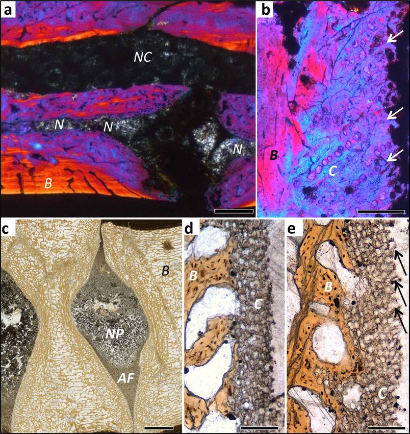
www.nature.com/scientificreports/
with preserved soft tissues are discovered, analyses of these fossils have the potential of modifying our view of
the evolution of the IVD. Since a joint is an integrated system of bone and other connective tissues, the bone
histology of the amniote centrum is important as well. Thus, the AF is anchored to the peripheral part of the two
centra via fibrocartilage and direct insertion of collagen fibres into the bone15. The NP, on the other hand, has no
direct contact with the bony surface of the centrum, being separated by a layer of hyaline cartilage (cartilaginous
endplate, see SI for details). The NP is developmentally derived from notochordal cells9–11.
In comparative anatomy, the notion prevails that reptiles lack an IVD because of their procoelous and
opisthocoelous vertebral centrum m orphology1,3,4,16, forming a synovial ball-and-socket joint. Such a joint has
a thin layer of hyaline cartilage covering the bony articular surface, with a synovial fluid-filled space separat-
ing the two articular surfaces that are peripherally connected by the joint capsule of connective tissue. Among
extant reptiles, the only exceptions to this type of joint are the tuatara Sphenodon punctatus17,18 and some gek-
konid lizards19. In the fossil record, however, we see that generalised centrum morphology does not indicate
ball-and-socket joints1 with hyaline cartilage. This agrees with higher-level amniote phylogeny that reveals the
basal condition of vertebral morphology to be an amphicoelous vertebral centrum with a notochordal canal1,10,
as already seen in the amniote sistergroup, Diadectomorpha1 (Fig. 2). The colonisation of diverse terrestrial envi-
ronments, including the conquest of the arboreal and aerial habitats, were accompanied by substantial changes
in the morphology of the vertebrae, but also in the articulations between these segments where motion o ccurs1.
Romer1 described a general evolutionary progression from amphicoelous notochordal via amphicoelous non-
notochordal to platycoelous during the transition from aquatic to terrestrial environments and hypothesized that
the ‘conjoined hollows created by amphicoelous vertebral centra were filled by modified notochordal material
or fibrous tissue’, without raising the issue of the IVD and its evolution.
Except for the well-defined IVD and synovial joints, inconsistencies in terminology describing inferred
intervertebral tissues (e.g., intervertebral cartilage, intervertebral tissue, and intervertebral pad) have caused
some confusion when referring to tissues and joint anatomy of fossils. Furthermore, if we turn to developmental
biology, mammals and birds (or archosauromorphs) are in focus of the research, not reptiles. However, regard-
less of vertebral centrum shape of extinct Amniota, there must have been connective soft tissues which allowed
three-dimensional movement of the vertebral column. In the palaeontological literature, there are few mentions
about IVDs in fossils at all such as the studies by Witzmann et al.20 and H opley21. The latter describes a spinal
pathology in a plesiosaur vertebra suggesting a disc prolapse and consisting of a Schmorl´s node, which is a
pathologic intrusion of the NP into the bony endplate. However, H opley21 does not discuss the presence of the
Schmorl´s node as evidence for IVDs in plesiosaurs but simply assumes their presence. Rothschild & Berman22
describe the preservation of the intervertebral space in the caudal vertebrae of a sauropod dinosaur. Finally,
Pérez García and Gascó23 interpret a disk-shaped fossil from the Jurassic of Spain as an ossified IVD of a marine
reptile. However, the very brief description is insufficient for evaluating this interpretation.
Here, we identify the different tissue types present in the intervertebral space of fossil amniotes based on
palaeohistological investigations of bone and preserved soft tissues (Table 1). Based on the rationale that soft
tissues associated with the skeleton might also be preservable, especially in articulated specimens, we histologi-
cally sampled diverse fossil taxa across the amniote tree for preserved intervertebral soft tissues (Table S1). If
possible, we sampled one or several dorsal vertebrae in articulation. We restricted the analysis to the dorsal
vertebral column because it shows the least morphological and functional variation (turtles being the exception)
compared to the neck and the tail columns, thus improving comparability across taxa.
For the reader to be able to understand our results without delving too deeply into the “Methods” section,
we here offer a brief review of the linkage between the fossil hard and soft tissues and the reconstruction of
intervertebral joint anatomy, including the five types of intervertebral joints encountered in this study (Fig. 1,
Table 1). In general, any vertebral centrum and any long bone in the amniote skeleton, fossil or extant, ossifies
from two domains, the endochondral domain and the periosteal domain. In former, the bone tissue has a cartilage
precursor, hence the term “replacement bone”. In the latter, bone tissue is deposited directly by an osteoblast
epithelium, the periost.
The anterior and posterior articular faces of any vertebral centrum are largely formed by endochondral
bone, whereas the vertebral surface in between is formed by periosteal bone (Fig. 1). Any cartilage pertains to
the endochondral domain (hence the name), and any ligaments and tendons inserts into the periosteal domain
via Sharpey’s fibres (Fig. 1). Different kinds of vertebral centrum shapes originate from the interplay of the rates
and directions of tissue formation in the two domains.
Beginning with the plesiomorphic amniote joint type, still seen in Sphenodon today (Fig. 1), the persistence
of the notochord in fossil samples is indicated by the presence of a notochord foramen and sometimes by
fossilised notochordal tissue. This joint already possessed a joint capsule (Fig. 1; like all other joints with the
possible exception of the fibrous bird-type joint). The presence and location of the joint capsule is indicated by
Sharpey’s fibres inserting into the periosteal bone around the margin of the articular surface. The plesiomorphic
amniote joint also possessed an AF. This is indicated by serial cartilage consisting of files of fossil chondrocytes
in the peripheral area of the articular surface, often with interspersed bone spicules with inserting fibres of the
fibrocartilage of the AF (Fig. 1b). A thin layer of fossil chondrocytes with an irregular arrangement in the center
of the articular surface as well as possible fossil notochordal cells indicate the former presence of an expanded
notochord or an incipient NP (Fig. 1e).
The former presence of an IVD is indicated by the lack of a notochordal foramen, the former presence of an
AF (see above) and sometimes the preservation of possible NP tissue. The size and shape of the former AF and
NP are indicated by the distribution of irregular cartilage vs. serial cartilage on the articular surface as well as by
the peripheral inclination of the chondrocyte files in the serial cartilage (Fig. 1). In amphicoelous vertebrae, the
NP probably was spherical in shape, and in platycoelous vertebrae, it was lens-shaped, as in humans today. The
amphicoelous joint with an IVD is extinct.
Scientific Reports | (2020) 10:14106 | https://doi.org/10.1038/s41598-020-70751-2 2
Vol:.(1234567890)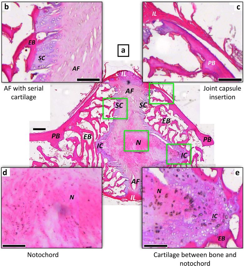
www.nature.com/scientificreports/
Fossil articular surface
morphology and fossil hard Anatomical structures and
Char. State Centrum shape and soft tissues tissues in living animal Joint type Occurrence in this study
I. Persisting notochord,
I. Notochord foramen
expanded in intervertebral space
(Figs. 3a, S3, S4, S7)
(Fig. 1a)
II. Area of expanded notochord,
II. Central concave area covered
possibly incipient nucleus pulpo-
by thin chondrocyte layer or
sus, separated by cartilage from
bone (Figs. 3a, S2, S4d)
bone (Fig. 1e)
III. Irregularly shaped and
loosely packed bodies the
III. Possible notochordal or
size of large chondrocytes or
incipient nucleus pulposus cells Stem amniotes, basal synapsids,
0 Amphicoelous, notochordal translucent, coarsely crystalline Plesiomorphic amniote
(Fig. 1d) basal reptiles, Sphenodon
matter in the intervertebral space
(Figs. 3a, S7)
IV. Peripheral convex area
with chondrocyte files (serial IV. Fibrocartilage of annulus
cartilage) and intervening bone fibrosus inserting into serial car-
spicules (Figs. S1b–d, S3b–d, tilage and bony spicules (Fig. 1b)
S4b–d, S7c)
IV. Sharpey’s fibres peripheral
to articular surface in periosteal IV. Joint capsule (Fig. 1c)
bone (Fig. S4b)
I. Notochord foramen absent
I. Notochord reduced or absent
(Figs. S8a, S10a, S11a)
II. Central concave area of
II. Globular nucleus pulposus
articular surface with thin
separated by cartilage from bone
layer of irregular chondrocytes
(Fig. 1e)
(Figs. 3d, S10d, S12c)
III. Irregularly shaped and
loosely packed bodies or grains
III. Possible nucleus pulposus
larger than chondrocytes in the
cells or notochord cells (Fig. 1d)
intervertebral space (Figs. 3c, hupehsuchians,
1 Amphicoelous, non-notochordal Intervertebral disc
S9b, S11, S12) ichthyosaurs, placodonts
IV. Peripheral convex area of
IV. Fibrocartilage of annulus
articular surface with chon-
fibrosus inserting into serial
drocyte files (serial cartilage)
cartilage and bony spicules
and intervening bone spicules
in articular surface periphery
(Figs. 3e, S8b, S9b, c, S10b,
(Fig. 1b)
S12a, b)
V. Sharpey’s fibres peripheral to
articular surface in periosteal V. Joint capsule
bone
I. Notochord foramen absent
I. Notochord absent (Fig. S5a)
(Figs. S19a, S20a, S22a, S23a)
II. Flat central area of articular II. Lens-shaped nucleus pulposus
surface with irregular chon- separated by cartilage from
drocyte cover, usually thin underlying bone (Figs. 2a, b,
(Figs. S19, S20c, S22c, S24a) S5b)
III. Irregularly shaped and
loosely packed bodies the size III. Possible nucleus pulposus
of large chondrocytes in the cells Mammals, eosauropterygians,
2 Platycoelous intervertebral space (Fig. S22b) Intervertebral disc most archosauromorphs, includ-
ing most dinosaurs
IV. Peripheral area of articular
IV. Fibrocartilage of annulus
surface with peripherally
fibrosus inserting into serial
inclined chondrocyte files (serial
cartilage and bony spicules
cartilage) and bone spicules
in articular surface periphery
(Figs. 3b, 5i–l, S6b,c, S20b, S22d,
(Figs. 2c, 5e–h, S5c)
S24b,c, S25b–e)
V. Sharpey’s fibres peripheral to
articular surface in periosteal V. Joint capsule
bone
I. Notochord foramen absent I. Notochord absent (Figs. 5a,
(Figs. S15a, S17a) S16a)
II. Convex/concave joint surface
II. Convex/concave joint surface
with unorganized chondro-
covered by hyaline cartilage
cytes or serial cartilage with
bordering on fluid-filled joint
3 Procoelous /opistho-coelous files normal to joint surface Synovial ball-and-socket Squamates, eusuchians
space encased by joint capsule
overlying bone, no bony spicules
(Figs. 2d, 5a–d, S16b,c)
(Figs. 2e,f, 5b–d), S17b,c, S17b,c)
III. Sharpey’s fibres peripheral
to articular surface in periosteal III. Joint capsule (Figs. 5a, S16b)
bone
Continued
Scientific Reports | (2020) 10:14106 | https://doi.org/10.1038/s41598-020-70751-2 3
Vol.:(0123456789)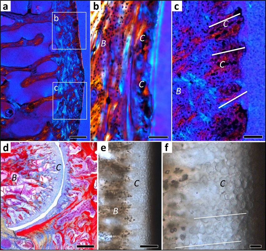
www.nature.com/scientificreports/
Fossil articular surface
morphology and fossil hard Anatomical structures and
Char. State Centrum shape and soft tissues tissues in living animal Joint type Occurrence in this study
I. Notochord foramen absent
I. Notochord absent
(Fig. S28a)
II. Convex/concave joint
II. Convex/concave surface with
surface covered by irregular,
irregular, scattered chondrocytes
thin cartilage, connective tissue
4 Procoelous in bone with fibre insertions Fibrous, bird-type Dromaeosaurs, birds
fibers inserting into bone of joint
(Figs. S26b–d, S27b,c, S28b,c)
surface
III. Sharpey’s fibres peripheral
to articular surface in periosteal III. Joint capsule
bone
Table 1. Summary of results and description of character states for ancestral state reconstruction. Relationship
between vertebral centrum shape, observed fossilised tissues (bony tissues, altered cartilaginous tissues, and
altered soft tissues), inferred anatomical structures, tissue and cell types, inferred joint type, and systematic
occurrence in this study. This table is complimentary to Fig. 1. Other pertinent figures are referenced as well.
The lack of a figure reference does not mean that the feature is not present, it simply means that it is not
illustrated. Char. state character state.
Beyond the tight fit of the articular surfaces in a socket and ball, the former presence of a synovial ball-
and-socket joint lined with articular cartilage is indicated by the even layer of fossil serial cartilage on the bony
articular surface. The fossil chondrocyte files in this layer are oriented normal to the articular surface and do
not diverge peripherally (Fig. 2d−f). In well preserved specimens, the entire layer of articular cartilage and even
the joint cavity formerly filled by synovial fluid can be observed (Fig. S15a,b).
Finally, the former presence of a fibrous bird-type joint is indicated by the close fit of the articular surfaces in
a socket and ball or in two saddles, but the fossil articular surface is mainly bony with fine fibre insertions and
only a thin layer of irregular and widely spaced fossil chondrocytes (Figs. S26–S28).
Results
Using polarised light microscopy, we observed cartilage formed in the endochondral domain in all vertebral
samples but in various altered states, of different kinds, and different distributions (Figs. 2, 3, 5; see “Methods”
section). We also observed the fibrous insertion into the periosteal bone of the connective tissue of the joint
capsule (of synovial joints) and the insertions of intervertebral ligaments. The distribution and arrangement
of bone tissue and preserved hypertrophied cartilage, hyaline cartilage and fibrocartilage allows constraining
the type of intervertebral joint and, in the case of the IVD, the size and location of the AF and NP. These were
inferred from the distribution of outwardly inclined files of hypertrophied cartilage cells (Figs. 2a–c, 3c–e, see
“Methods” section).
Especially in marine reptiles from black shales, we found soft tissue preservation across the intervertebral
space (see “Methods” section), allowing further inferences about the nature of the joint connecting adjacent
centra (Figs. 3c, S11, S19, S20). We also sampled extant taxa, in particular Sphenodon, for comparison with fossil
soft tissues and to clarify contradictory interpretations in the literature on the nature of the intervertebral joints
in this taxon (see Fig. S14a,b and SI). Turtles were excluded from the histological analysis because of the highly
modified dorsals in the crown turtles, lacking intervertebral joints, and the lack of samples from stem turtles.
Based on the detailed descriptions of the histological samples (Supplementary Text), we are now able to
review the relationships between vertebral centrum shape, observed fossilised and fresh tissues, inferred joint
tissue types, and inferred joint type (Table 1, Fig. S29). The observed fossil tissues in the histological sections
are bony tissues, altered cartilaginous tissues, and preserved soft tissues. We recognised five basic joint types
in our sample (Table 1). (1) Amphicoelous, notochordal centra show a concave area with thin cartilage of
irregularly arranged chondrocytes or smooth bone and a convex area with generally short chondrocyte files
arranged in longitudinal direction with irregular bone spicules in between (Figs. S1–S4, S7, S14). We infer that
these vertebral centra had an intervertebral joint consisting of an outer ring of fibrocartilage, best homologised
with the AF, and an expanded notochord in the central region, as is observed in Sphenodon (Figs. 1, S14). This
appears to be the plesiomorphic condition for amniotes which gave rise to all other joint t ypes10. (2) Amph-
icoelous, non-notochordal centra show a concave area with thin or thick (several cells deep) irregular cartilage
(but never bone), an outer area with chondrocyte files arranged in longitudinal direction with irregular bone
spicules in between or long diverging files with an increasing angle of divergence (hupehsuchians, ichthyosaurs,
placodonts) (Figs. 3c–e, S8–S13, S18). We infer that the taxa that show these features had a proper IVD with a
NP of notochordal origin, supported by the preservation of an altered NP in some ichthyosaurs. Importantly,
this type of morphology and joint is extinct. (3) Platycoelous centra, including those of extant mammals, show a
central area with irregular cartilage and an outer area with diverging cartilage files and bone spicules (Figs. 2a–c,
3b, 5e–l, S5, S6, S19–S26). These two areas demarcate the AF and NP of the mammalian IVD, and we infer that
fossil taxa with these features also had an IVD. (4) Procoelous and opisthocoelous vertebral centra that show
a distinct layer of hyaline cartilage arranged in files normal to the bony surface (Figs. 2d–f, 5a–d, S15–S17),
suggest the presence of a synovial joint with narrow joint cavity filled with synovial fluid, as observed in most
extant squamates. (5) Procoelous and opisthocoelous vertebral centra of fossil taxa that show only irregular, thin
(one or a few cells deep) cartilage and bone with fibre insertions (Figs. S27 and S28) suggest the presence of a
Scientific Reports | (2020) 10:14106 | https://doi.org/10.1038/s41598-020-70751-2 4
Vol:.(1234567890)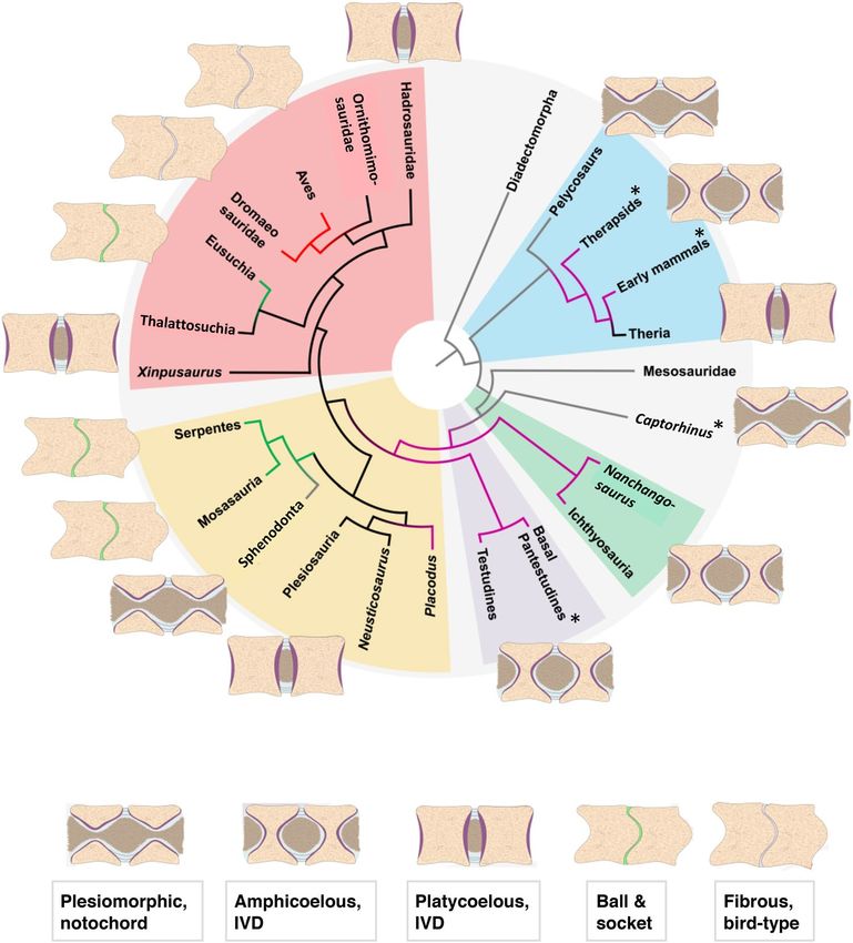
www.nature.com/scientificreports/
Figure 1. Histology of the plesiomorphic amniote joint with persisting notochord of Sphenodon punctatus
NHMW 8108. The intervertebral tissues and the two articulating mid-dorsal centra illustrate the relationship
between mineralised (fossilisable) tissues and soft tissues. (a) Oblique sagittal microtome section stained with
hematoxylin. Note that the notochord appears discontinuous in the image because of the suboptimal plane of
section. (b) Enlargement of annulus fibrosus insertion into endochondral bone of articular surface via serial
cartilage. (c) Enlargement of interspinal ligament of the joint capsule inserting into periosteal bone of the
centrum peripheral surface. (d) Enlargement of notochordal tissue in intervertebral space. (e) Enlargement of
contact between notochordal tissue and endochondral bone of articular surface of centrum with intervening
irregular cartilage. Scale bar in (a) represents 100 µm, and scale bars in (b–e) represent 40 µm. AF annulus
fibrosus, EB endochondral bone, IC irregular cartilage, IL intervertebral ligament, N notochordal tissue, PB
periosteal bone, SC serial cartilage.
fibrocartilage joint as in extant birds. We found evidence for intervertebral ligaments inserting in the periosteal
bone in all types of joints.
We then performed ancestral character state reconstructions (ASR)8 for a consensus amniote phylogeny,
allowing us to trace the evolution of the amniote intervertebral joint (Fig. 4). Both parsimony (Fig. 4) and
maximum likelihood-based ASR agree (Fig. S29). Looking at the tissue level, we explain the pathway of amniote
vertebral joint evolution envisaged by Romer1 by combining fossil connective tissue evidence with principles
of bone and cartilage formation and growth. The basal amniote joint, as exemplified by the iconic sail-backed
synapsid Dimetrodon and the stem amniote Diadectes, must have contained a notochord constricted by the ver-
tebral centrum and expanded in the space between the centra (Fig. 4). The vertebral centrum was a ring of bone
of largely periosteal origin with a thin endochondral layer, modified from the condition in basal t etrapods24. In
the joint, two such rings of bone were connected by a ring of fibrocartilage forming an AF, as seen in Spheno-
don (Figs. 1, S14) and modern geckos19. The fibrocartilage inserted in the thin endochondral domain of each
Scientific Reports | (2020) 10:14106 | https://doi.org/10.1038/s41598-020-70751-2 5
Vol.:(0123456789)www.nature.com/scientificreports/
Figure 2. Histology of mammalian and squamate intervertebral spaces. (a) Extant Phoca vitulina IGPB M 60,
sagittal ground section of dorsal vertebral centrum showing part of the bony endplate in cross-polarised light
with lambda filter. Note that the colors in this and the following two images do not result from histological
staining but from polarised light. (b) Enlargement of area b in (a) showing a thin layer of cartilage. Note the
irregular arrangement of the chondrocyte lacunae. This tissue is overlain by the nucleus pulposus (NP) of
the IVD in life. (c) Enlargement of area c in (a) showing cartilage chondrocytes in inclined files (white lines)
embedded in fibrous bony tissue. This represents the insertion of the annulus fibrosus (AF) of the IVD. (d)
Extant Python sp. IGPB R 662, transverse microtome section of the synovial joint connecting the dorsal
vertebral centrum. This joint is formed by hyaline cartilage and a thin intervertebral space filled with synovial
fluid in life. (e) Fossil Mosasaurus missouriensis IGPB Goldfuß 1230, close up of the joint surface in sagittal
section of a dorsal vertebral centrum. Note the globular structures arranged in files representing fossilised
hyaline cartilage. (f) Enlargement of (e), white lines highlight some files. Scale bars in (a, d) represent 500 µm,
scale bars in (b, c, e) represent 100 µm, and scale bar in (f) represents 20 µm. B bone tissue, C cartilage.
centrum and was covered on the outside by a ligamentous joint capsule consisting of intervertebral ligaments
and inserting into the periosteal bone, as is seen in Sphenodon today (Figs. 1, S14). The AF and the intervertebral
ligaments are the plesiomorphic condition for the holospondylous centra of amniotes. Next, the ring of bone
closed up in its centre, resulting in a round aggregation of notochordal cells, the incipient NP. Because this stage
is extinct, we cannot be sure of the exact nature of this incipient NP, i.e. whether it was the same structure as
the NP in extant taxa or consisted of the remaining isolated pieces of the notochord. To a certain extent, this is
a semantic issue. However, we found a probable altered NP preserved in ichthyosaur fossils from the Middle
Triassic and Early Jurassic (Figs. 3c, S9, S11). The NP was separated from the endochondral bone surface by a
layer of hyaline cartilage, the cartilaginous endplate (Figs. 3c–e, S11). At this stage, there still was little growth
in the bony endplate, as indicated by the retention of the amphicoelous centrum. Closure of the notochordal
canal happened convergently twice, once in therapsids and once in the reptile lineage, in early diapsids such as
ichthyosaurs25 (Fig. 4).
From this stage evolved a platycoelous or slightly amphicoelous centrum with an IVD by greatly increased
cartilage cell proliferation in the endochondral domain and subsequent endochondral ossification, filling in
the space formerly occupied by notochordal tissue and fully developing it into an NP. The periphery of the
articular surfaces remained connected by fibrocartilage of the AF. This evolutionary transition happened at
least twice, in therian mammals and in higher diapsids (Fig. 4). Alternatively, the platycoelous centrum with
an IVD evolved independently in eosauropterygians (a clade of marine reptiles to which plesiosaurs belong),
and in archosauromorphs (Fig. 4), in addition to mammals. The platycoelous centrum with an IVD was then
retained in most archosauromorphs, including basal crocodiliforms and most dinosaurs. Modern crocodiles
evolved a synovial ball-and-socket joint convergently with lepidosaurs (Fig. 4). The evolutionary transition to
the ball-and-socket joint of extant reptiles involved the reduction of the NP and the AF, i.e., the cartilage and
Scientific Reports | (2020) 10:14106 | https://doi.org/10.1038/s41598-020-70751-2 6
Vol:.(1234567890)www.nature.com/scientificreports/
Figure 3. Histology of mesosaur, ichthyosaur and dinosaur intervertebral spaces, all fossil ground sections.
(a) Stereosternum tumidum IGPB R 622, sagittal section of two articulated dorsal vertebral centra with
intervertebral space, showing the notochordal amphicoelous shape and the persisting notochord. Image is
in cross-polarised light with lambda filter. (b) Hadrosauridae indet. UALVP 59650. Close up of the articular
surface showing obliquely arranged mineralised fibres in between poorly defined files of chondrocyte lacunae
(arrows). Image is in cross-polarised light with lambda filter. (c) Stenopterygius sp. IGPB R 661 sagittal section
of two articulated centra showing the amphicoelous shape. Note the differentiation of the content of the
intervertebral space into a coarse into a fine fraction, probably representing the nucleus pulposus (NP) and
the annulus fibrosus (AF). (d) Enlargement of the concave part of the articular surface, showing a thin layer of
irregularly arranged chondrocyte lacunae, underlying the nucleus pulposus (NP). (e) Enlargement of the convex
part of the articular surface, showing obliquely arranged files of chondrocyte lacunae, representing the insertion
of the annulus fibrosus (AF) (arrows). Scale bar in (a) represents 500 µm, scale bar in (b) represents 100 µm,
scale bar in (c) represents 2 mm, scale bars in (d, e) represent 100 µm. AF annulus fibrosus, B bone tissue, C
cartilage, N notochord, NC neural canal, NP nucleus pulposus.
notochordal components of the joint, and the development of a synovial space. The transition also involved
strong anteroposterior asymmetry, with the formation of endochondral bone suppressed in the socket part of
the joint and hypertrophied in the ball part, as seen in mosasaurs and snakes in this study (Figs. S15–S17, see SI).
The dinosaurs included in our study also retained the platycoelous centrum with the IVD except for the most
derived non-avian dinosaurs, i.e., dromaeosaurs. In these, cartilage is greatly reduced, suggesting that the dorsal
vertebral centra of these dinosaurs had the same fibrous connection as in extant birds.
Discussion
Histological investigations of fossil hard tissues have become a powerful tool in p alaeontology26, but in general
they have been limited to bone and dental tissues. It is known that different soft tissue components of bone,
such as osteons, bone collagen fibres, osteoblasts, and even blood vessels may preserve in deep time27,28. Apart
from the bone, integumentary soft tissues may be p reserved5,29 and, very rarely, internal o
rgans30,31. It has long
been understood that the anoxic conditions leading to the deposition of black shales permits the preservation of
such soft parts, primarily of the integument, meaning that the potential exists that non-mineralised connective
tissues will preserve. Hence, ideally, the articulated segments of vertebral column that we studied were still at
Scientific Reports | (2020) 10:14106 | https://doi.org/10.1038/s41598-020-70751-2 7
Vol.:(0123456789)www.nature.com/scientificreports/
Figure 4. Ancestral state reconstruction using parsimony of the different types of dorsal intervertebral joints
in the phylogeny of the higher clades of amniotes (see “Methods” and SM for details). The schematic drawings
of the intervertebral joints are explained in the text. Grey branches indicate persisting notochord. Purple
branches indicate an intervertebral disc between amphicoelous centra. Black branches indicate an intervertebral
disc between platycoelous centra. Green branches indicate a synovial joint with hyaline cartilage in between
procoelous or opisthocoelous centra. Red branches indicate a fibrous cartilage joint. Key to tissue colors: beige,
bone; purple, cartilaginous endplate; light blue, AF; brown, NP; green, articular cartilage of synovial joint;
grey, fibrous joint cartilage. Key to clade colors: light blue, Synapsida; light green, Ichthyosauria; light purple,
Testudines; light yellow, Lepidosauromorpha; light red, Archosauromorpha. Asterisks indicate fossil taxa for
which intervertebral soft tissue anatomy was inferred based on morphological and histological descriptions in
the literature.
least partially embedded in the matrix. This is important to be able to compare potentially preserved soft parts
in the intervertebral spaces with the host sediment.
Patterns of cartilage fossilisation. Cartilage fossilises well if it is mineralised, i.e., if the living chon-
drocytes mineralise in spherulitic arrangements of apatite crystallites, as in the case of calcified cartilage of
chondrichthyans. However, fossilisation of cartilage has been little studied, and any evidence of cartilage in a
fossil traditionally has been subsumed by palaeontologist unter the term "calcified cartilage”, even if the tissue in
question was not mineralised in life (e.g., Ref.32). There is true calcified cartilage in extinct amniotes, i.e., serial
cartilage that was not replaced by bone but was fully mineralised in the living animal. An example is provided
by some pachypleurosaurs, small Triassic sauropterygians, in which these tissues served to increase skeletal
mass33,34. The term calcified cartilage, however, is also used for the zone of hypertrophy of chondrocytes in the
growth plate of amniote long b ones35. This cartilage is replaced completely by endochondral bone during the
Scientific Reports | (2020) 10:14106 | https://doi.org/10.1038/s41598-020-70751-2 8
Vol:.(1234567890)www.nature.com/scientificreports/
growth phase of the animal, and its presence suggests immaturity. Whereas the hypertrophy zone may show
increased mineralisation35, favoring fossilisation, the chondrocytes do not show the characteristic spherulitic
radial crystallite arrangement of true calcified cartilage. We note that true calcified cartilage was not encountered
in this study.
Patterns in intervertebral joint evolution. The convergent evolution of the IVD in synapsids and rep-
tiles by closure of the notochordal canal and evolution of an NP from the notochord had remained unrecognised
for two reasons: First, the general belief was that amphicoelous vertebrae did not house an IVD based on the
only extant amphicoelous amniotes, i.e., Sphenodon and some geckos. We now recognise the presence of an IVD
in extinct amphicoelous reptiles, e.g., in ichthyosaurs, hupehsuchians, probably non-mammalian therapsids,
and possibly stem tutles (Fig. 4). The condition in these taxa is different from the plesiomorphic condition of a
continuous notochord. The second reason is that reptile clades that had evolved an IVD went extinct at the end
of the Cretaceous, after giving rise to lineages with synovial ball-and-socket joints and bird-type intervertebral
joints (not proper IVDs with a NP). However, we consider it unlikely that these types of joints were the decisive
factor in survival of this mass extinction. Synovial ball-and-socket joints also evolved in Jurassic sauropodo-
morphs dinosaurs36 and in some non-avian theropod d inosaurs37, all of which went extinct at the end of the Cre-
taceous. Note that in some recent s tudies3,38, the term ‘avian IVD’ or similar is used. This has created confusion
because in these same studies, it is stated or illustrated that the avian ‘IVD’ lacks a NP and is not homologous
to the IVD proper. Given the complex distribution of the different types of intervertebral joints on the amniote
tree, further studies on well-preserved fossils in combination with soft tissue histology of extant taxa and devel-
opmental studies hold great p otential10. The evolutionary scenario presented here results in hypotheses that are
testable using classical embryology and developmental genetics.
Conclusions
Preserved soft tissues, bone histology and articular surface morphology inform on the nature of the intervertebral
articulation in fossil amniotes. Currently, this understanding is restricted to dorsal vertebrae, and the approach
should be extended to cervical vertebrae in particular. Remains of articular cartilage are regularly present in
fossils and can be observed in palaeohistological thin sections. The shape and organisation of the chondrocytes
can be used to infer presence and size of an AF and NP. Occasionally, remains of the notochord and the NP also
are encountered in such sections. The preserved tissues lead us to infer that amphicoelous, notochordal vertebrae
had an intervertebral joint consisting of a continuous notochord surrounded by a peripheral AF. This is the ple-
siomorphic amniote type and must have been present in stem amniotes, basal synapsids, and basal reptiles. It is
also seen Sphenodon and some geckos today. We also infer that amphicoelous, non-notochordal vertebrae were
connected by an intervertebral disc with a NP surrounded by an AF. This was the situation in hupehsuchians,
ichthyosaurs, placodonts, probably non-mammalian synapsids, and possibly stem turtles. Fossil platycoelous
vertebrae possessed a proper IVD resembling that of extant mammals. Among reptiles, a proper IVD must have
been present in eosauropterygians and most archosauromorphs, including most non-avian dinosaurs. How-
ever, this reptilian IVD went extinct at the end of the Cretaceous with the extinction of non-avian dinosaurs
and plesiosaurs Finally, ancestral state reconstruction on a consensus phylogeny of amniotes using maximum
parsimony and maximum likelihood algorithms in the software Mesquite indicates that the intervertebral disc
evolved convergently at least twice, once on the mammalian stem line and once in early diapsids. Similarly, a
ball-and-socket joint evolved at least twice convergently.
Methods
Acquisition of histological and morphological data. For this study, we histologically sampled 22
specimens representing 19 taxa of different amniote clades which include mammalians, non-mammalian synap-
sids, and reptiles (including non-avian dinosaurs and birds) (see Table S1). We used histological ground sections
(for fossil taxa), histological demineralised sections (for extant taxa) and morphological data, i.e., the shape of
the bony articular surface of the vertebral centra (see Table 1). For the stem reptile Captorhinus, we used the his-
tological images in Ref.32. For the morphological part, we checked the general applicability of our observations
with information from the rich literature on amniote vertebral centrum morphology, including another three
taxa for which morphological (non-mammalian therapsids, basal mammals) and also histological information
(Alligator) is available. Extant turtles are a special case because they lack mobile intervertebral joints between
the dorsal vertebrae which are integrated into the carapace. The stem turtles Pappochelys, Odontochelys, and
Eorhynchochelys lack a carapace and have non-notochordal amphicoelous v ertebrae39–41. No histological data are
available for these taxa, but we included the published morphological information in some analyses.
Sampling and histology of fossil and extant non‑decalcified material. Two types of fossil mate-
rial were sampled for this study. First, segments of the dorsal vertebral column of articulated skeletons as well
as isolated but articulated segments of at least two centra. In the latter, we focused on specimen in which the
vertebrae are preserved in close articulation. These specimens originally also pertained to articulated skeletons
that were collected in an incomplete state, however. The incomplete state presumably is due to loss to weathering
before discovery. The second type is isolated vertebral centra that were sampled if articulated column segments
were not available.
Articulated column segments are primarily found in conservation deposits such as black shales (e.g., the Posi-
donienschiefer Formation of southern Germany), but isolated vertebrae derive from a host of different sediment
types. The segments of vertebral column and isolated vertebrae were sectioned as exactly in the sagittal plane as
possible. In the case of longer segments (e.g., Mesosaurus, Stenopterygius, Neusticosaurus, etc.), there was some
Scientific Reports | (2020) 10:14106 | https://doi.org/10.1038/s41598-020-70751-2 9
Vol.:(0123456789)www.nature.com/scientificreports/
degree of curvature of the vertebral column, but we attempted to intersect at least two adjacent centra in the exact
sagittal plane. The half of the segment that represented the closest approximation to the sagittal plane was then
processed into a petrographic thin section following standard m ethods26. Thin sections were ground to a thick-
ness of 50–80 µm, depending on the degree of dark staining. Thin sections were then examined with a standard
polarising microscope, either a Leica DLMP or a Zeiss Axio Imager und normal and cross-polarised light with
and without a lambda filter. Images were taken with a Leica digital camera and processed with ImageAccess
EasyLab 7 software.
Dental tissues and bone fossilises very well at the histological level, and observations using light microscopy
are directly transferrable from fossil to extant comparative m aterial26,35. Histological descriptions of fossil hard
tissues accordingly use the same terminology as for extant amniotes, and this terminology is discussed in detail by
Francillon-Vieillot et al.35. In normal light, all types of bone tissue (periosteal, endochondral, secondary) show a
brownish stain of the matrix developed during f ossilisation28. Osteocyte lacunae are generally visible in this bone
matrix. Remains of cartilaginous tissues, on the other hand, generally lack the brownish stain, and are whitish
or light greyish translucent (Fig. 5). In polarised light, bone is easily distinguished from any kind of cartilage
as well because of the birefringence of the bone apatite crystallites. Cartilage and other soft tissue remains lack
birefringence or show the birefringence of the templating mineral, e.g., calcite (Fig. 5).
Although extant material is typically studied in decalcified microtome sections, ground sections (petro-
graphic thin sectioning) of fresh bone can be studied as well. The advantage is a better identification of the bone
tissue types because decalcification destroys the birefringence of the bone tissue. In addition, the observational
relationship between bone, cartilage, and connective tissue is the same as in fossils, offering an intermediate in
the comparison between fossil ground sections and microtome sections. We studied some extant material using
this technique (Table S1).
Microtome histology of extant material. Two taxa (Sphenodon and Python) were successfully sampled
by decalcified microtome section in the sagittal plane of the dorsal and caudal region of the vertebral column
from consecutive vertebrae (Table S1). The Sphenodon specimen used for sectioning is from the original collec-
tion of Reischek made in 1890 for the NHMW. Because it has been stored in ethanol for more than a century, the
material was extremely desiccated and tough and therefore difficult to section with the microtome. After several
washes in ethanol, rehydration and refixation with 4% paraformaldehyde, the samples were demineralised for
50 days in 8% EDTA at pH 8. After re-transfer into ethanol, the samples were embedded in paraffin following
standard procedures. For sectioning on a standard microtome at 10 µm, special blades for hard tissue were used
(N35HR microblades, pfm medical). After mounting on silane-coated glass slides, the sections were treated by
HE staining and Heidenhain Azan staining according to standard procedures.
The Python samples were generated from fresh frozen material from a historical collection at the IGPB. The
muscular tissue was partially removed from the vertebral column without damaging the vertebral ligaments.
After fixation for 7 days in 4% PFA, the samples were washed and demineralised for 33 days in 8% EDTA at pH
8. Whole-mount preparations were cut with a razor blade in the median plane. Samples for histological sec-
tions were embedded in paraffin, sectioned at 10 µm with a standard microtome and treated by HE staining and
Heidenhain Azan staining according to standard procedures. Note that ground sections in polarised light may
appear in a similar color scheme to stained microtome sections (e.g., Figs. 2, 5), but the meaning and origin of
these colors is radically different.
Preservation of cartilage and inferences on intervertebral tissues in fossils. Our ground histo-
logical sections of fossils allow different observations and inferences, depending on the quality of preservation,
about the nature of the tissues occupying the space between the bony end plates of successive vertebrae (Fig. 1,
Table 1). These inferences are based on the principles of vertebral centrum and joint differentiation and growth
(see “Introduction” and SI). All of the isolated vertebral centra in our study preserve histological evidence for the
nature of the cartilage covering the bony end plate, including cartilage preserved in different states of alteration
by fossilisation and of lacunae that once housed the chondrocytes. Although frequently noted in palaeohisto-
logical descriptions (e.g., Ref.32), fossil cartilage has been little studied. The identification as fossil cartilage is
based on the direct comparison with cartilage in extant animals. Just as fossilised bone tissue can be directly
compared with living bone tissue in histological ground sections (e.g., Ref.35), so can fossilised cartilage (Figs. 2,
3, 5). Cartilage cells and extracellular matrix may either have become mineralised during fossilisation or leave
empty spaces surrounded by bone matrix and later filled in by diagenetic minerals. Both, cell size and cell shape
are readily comparable with those in fresh cartilage, and the different types of cartilage produced by the chond-
roblasts can be observed (Fig. 5).
Bone tissue and fossilised hypertrophied cartilage of the endplate also offer histological correlates of other
intervertebral soft tissues (Fig. 1, Table 1). Thus, the direction and presence/absence of files of cartilage cells
(chondrocytes) and anchoring fibres in the bone allow mapping of the AF and NP or notochordal tissue (Fig. 1).
The arrangement of the chondrocytes in files results from the directed growth of the cartilage in rows by chon-
drocyte division, hence the term ‘serial cartilage’ (Table 1). This mapping approach is based on the numerous
descriptions and illustrations in the literature and our own observations (e.g., Fig. 1). Human IVDs (e.g., Ref.2)
and other mammalian IVDs (e.g., Ref.42, Fig. 2, and Ref.43, Fig. 7) are abundantly figured in the medical literature
(although not always correctly labelled). Our own observations cover the histology of humans and other extant
mammals (e.g., Phoca vitulina, see Figs. 5, S5, S6, SI) and reptiles (e.g., Sphenodon punctatus, see Fig. 1 and
Fig. S14, and Phython sp.; see Figs. 2, 5, and Figs. S16). Sphenodon is particularly important because it represents
the only extant example of the ancestral condition of the amniote intervertebral joint (Figs. 1, 4).
Scientific Reports | (2020) 10:14106 | https://doi.org/10.1038/s41598-020-70751-2 10
Vol:.(1234567890)www.nature.com/scientificreports/
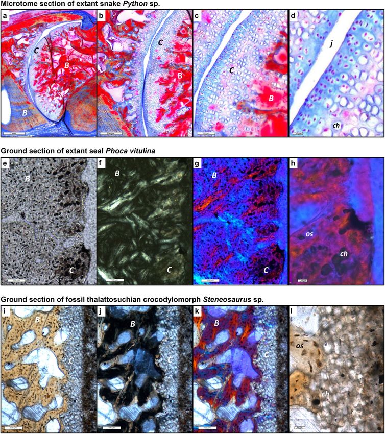
Figure 5. Intervertebral articular cartilage in extant and fossil amniotes and different types of sectioning and
light microscopy. (a–d) Microtome section stained with Azan of extant joint with cartilage of extant snake
Python sp. IGPB R 662, synovial ball-and-socket joint at increasing magnifications in normal transmitted
light. Blue is cartilage and other connective tissues, red is bone. Note the serial cartilage, i.e., the arrangement
of the cartilage cells in files, especially in (c). (d) Cartilage layer with differentiating (left) and hypertrophied
(right) cartilage cells, note the size increase from left to right and the globular shape of the hypertrophied
chondrocytes, as in (h) and (l). (e–h) Ground section of extant seal Phoca vitulina IGPB M 60, joint with
IVD, peripheral area of articular surface. (e) Section in normal transmitted light. (f) Section in cross-
polarised light. (g) Cross-polarised light and lambda filter. (h) Close-up of g. (i–l) Ground section of fossil
thalattosuchian crocodilomorph Steneosaurus IGPB R 663, joint with IVD, peripheral area of articular surface.
(i) Section in normal light. (j) Section in cross-polarised light. (k) Cross polarised light and lambda filter. (l)
Close-up of (i). Note that the fossil cartilage does not show the same birefringence as the bone. B bone tissue, C
cartilage, ch chondrocyte, j joint space, os osteocyte.
Scientific Reports | (2020) 10:14106 | https://doi.org/10.1038/s41598-020-70751-2 11
Vol.:(0123456789)www.nature.com/scientificreports/
Our observations and literature search indicate that the hypertrophied cartilage cells are organized into files
(serial cartilage) in the region of attachment of the annulus fibrosus (Fig. 1b, Table 1). The inclination of the files
relative to the bony endplate (or articular surface) increases outwards, reflecting the orientation of the collagen
fibres in the fibrocartilage of the AF (Fig. 1b). On the other hand, the central region of the joint, the location of
the NP or notochordal tissue, is only underlain by loosely organized hypertrophied cartilage (Fig. 1e). Two types
of anchoring fibres are also preserved in the fossils. In fact, they are commonly better visible in fossils than in
ground and particularly microtome sections of extant vertebrae. The optical birefringence is not as well devel-
oped in fresh bone as in fossil bone, and not all stains of microtome sections pick up collagen fibres (e.g., they
are not seen in the HE-stained Sphenodon sections (Fig. 1c). One fibre type is the classical Sharpey’s fibres that
insert in the periosteal bone near the anterior and posterior end of the centra (Fig. 5a,b), indicating the location
of the joint capsule formed by intervertebral ligaments (Figs. 1c, S16b). Note that The other types are fibres that
insert in the endochondral bone and hypertrophied cartilage, anchoring the AF to the end plate (Figs. 1b, 5).
Their distribution and angle of insertion thus also can be used to infer the size and structure of the AF (Fig. 1b,
Table 1). For details, see the description of the individual specimens in the SI and Figs. S1–S28.
Preservation of non‑mineralised skeletal and connective tissues. It has long been understood that
the anoxic conditions leading to the deposition of black shales permit the preservation of soft parts, primarily
of the integument, meaning that the potential exists that non-mineralised connective tissues will preserve (e.g.,
Ref.44). We sectioned articulated segments of vertebral column from black shale settings which appear to pre-
serve altered or templated joint connective tissues other than hyaline cartilage, i.e., the fibrocartilage of the AF
and the cells of the NP. Hence, ideally, the articulated segments of vertebral column that we studied were still at
least partially embedded in the matrix. This is important to be able to compare potentially preserved soft parts
in the intervertebral spaces with the host sediment. For details, see the description of the individual specimens
in the SI and Figs. S1–S28.
Optimization of intervertebral articulation on the amniote tree. For the reconstruction of the
evolution of the intervertebral articulation in amniotes, we used ancestral character state reconstruction (ASR)8.
We built a consensus tree of the amniotes sampled based on the literature, including 24 tip taxa. The phylog-
eny is based in most parts on Müller et al.45 and Chen et al.46 and is consistent with a new analysis of meso-
saur relationships47. The phylogenetic position of thalattosaurs, represented in our study by Xinpusaurus, varies
between basal neodiapsid affinities45, basal lepidosauromorph affinities48, as part of a clade including hupeh-
suchians and ichthyosaurs46, and the traditional view of basal archosauromorph affinities which we expressed
in Fig. 4 and Fig. S29a. We implemented this uncertainty in different ASRs but did not find any influence on the
outcome to the overall analysis (Fig. S29).
Commonly turtles are considered archosauromorphs, if not archosaurs, a view based on molecular evidence,
which does indeed point towards archosauromorph a ffinities45. However, current morphological phylogenetic
analysis including transitional fossil overwhelmingly recover basal diapsid or lepidosauromorph a ffinities39,41,50.
Note that these morphological results were obtained independently by three different teams, without any overlap
in authorship in the last four years, postdating molecular analyses. This is why we place turtles as basal Diapsida
(Fig. 4 and Fig. S29)39,50. Because the phylogenetic position of turtles remains controversial beyond their diapsid
affinities and because no histological data are available for stem turtle vertebral centra, we ran the ASR both
with and without turtles to test whether their inclusion nevertheless might have an effect on the outcome of the
ASR. In the analysis including turtles, we placed them in the least nested position on the diapsid stem (Fig. 4),
but more derived than Ichthyosauria, and we coded their vertebrae as non-notochordal a mphicoelous37–39. We
recognised five character states describing the different types of intervertebral joints based on our morphological
and histological observations and inferences. These character states are as follows and are described in Table 1:
0, amphicoelous centrum with notochordal canal, continuous notochord; 1, non-notochordal amphicoelous
centrum with IVD; 2, platycoelous centrum with IVD; 3, synovial ball-and-socket joint; 4, fibrous ball-and-
socket joint. Data for non-mammalian Therapsida, early mammals, and Eusuchia were taken from the literature
(Table S1). We then entered the consensus phylogeny and the states for the tip taxa into a NEXUS file (Sup-
plementary NEXUS file 1) and analyzed it in Mesquite v. 3.024351 using both maximum parsimony (MP) and
maximum likelihood (ML). For the ML reconstruction, we used the one-parameter Markov k-state probability
model. The inclusion of turtles had no effect on the analysis, and we excluded them from further consideration.
Received: 20 March 2020; Accepted: 23 July 2020
References
1. Romer, A. S. Osteology of the Reptiles 772 (The University of Chicago Press, Chicago, 1956).
2. Hall, B. K. Bones and Cartilage. Developmental and Evolutionary Skeletal Biology, 2nd edn, 902 (Academic Press, San Diego, 2015).
3. Bruggeman, B. J. et al. Avian intervertebral disc arises from rostral sclerotome and lacks a nucleus pulposus: implications for
evolution of the vertebrate disc. Dev. Dyn. 241, 675–683 (2012).
4. Witzmann, F., Schwarz-Wings, D., Hampe, O., Fritsch, G. & Asbach, P. Evidence of spondyloarthropathy in the spine of a phytosaur
(Reptilia: Archosauriformes) from the Late Triassic of Halberstadt Germany. PLoS ONE 9, e85511 (2014).
5. Schweitzer, M. H. Soft tissue preservation in terrestrial Mesozoic vertebrates. Annu. Rev. Earth Planet. Sci. 39, 187–216 (2011).
6. Briggs, D. E. G. & Summons, R. E. Ancient biomolecules: Their origins, fossilization, and role in revealing the history of life.
BioEssays 36, 482–490 (2014).
Scientific Reports | (2020) 10:14106 | https://doi.org/10.1038/s41598-020-70751-2 12
Vol:.(1234567890)www.nature.com/scientificreports/
7. Service, R. F. Researchers close in on ancient dinosaur proteins. “Milestone” paper opens door to molecular approach. Science 355,
441–442 (2017).
8. Pontarotti, P. & Hue, I. in Evolutionary Biology. Convergent Evolution, Evolution of Complex Traits, Concepts and Methods (ed.
Pontarotti, P.) 3–21 (Springer Nature, Heidelberg, 2016).
9. Pattappa, G. et al. Diversity of intervertebral disc cells: phenotype and function. J. Anat. 221, 480–496 (2012).
10. Cox, M. K. & Serra, R. The Intervertebral Disc 33–51 (Springer, Vienna, 2014).
11. Scaal, M. Early development of the vertebral column. Semin. Cell Dev. Biol. 49, 83–91 (2016).
12. D. Peterson, J.D. Bronzino, eds, Biomechanics. Principles and Applications. (CRC Press, New York, 2008).
13. I. M. Shapiro, M. V. Risbud, eds, The Intervertebral Disc. Molecular and Structural Studies of the Disc in Health and Disease.
(Springer, Wien, 2014).
14. Chan, W. C., Au, T. Y., Tam, V., Cheah, K. S. & Chan, D. Coming together is a beginning: the making of an intervertebral disc.
Birth Defects Res. Part C Embryo Today 102, 83–100 (2014).
15. Nosikova, Y. S., Santerre, J. P., Grynpas, M., Gibson, G. & Kandel, R. A. Characterization of the annulus fibrosus-vertebral body
interface: identification of new structural features. J. Anat. 221, 577–589 (2012).
16. Zug, G. R., Vitt, L. J. & Caldwell, J. P. Herpetology: An Introductory Biology of Amphibians and Reptiles 630 (Academic Press, San
Diego, San Francisco, New York, Boston, London, Sydney, Tokio, 2001).
17. Gadow, H. F. On the evolution of the vertebral column of Amphibia and Amniota. Philos. Trans. R. Soc. Lond. B 187, 1–57 (1896).
18. Gadow, H. F. The Evolution of the Vertebral Column. A Contribution to the Study of Vertebrate Phylogeny 356 (Cambridge University
Press, London, 1933).
19. Holder, L. A. The comparative morphology of the axial skeleton in the Australian Gekkonidae. Zool. J. Lin. Soc. 44, 300–335 (1960).
20. Witzmann, F. et al. Vertebral pathology in an ornithopod dinosaur: a hemivertebra in Dysalotosaurus lettowvorbecki from the
Jurassic of Tanzania. Anat. Rec. 291, 1149–1155 (2008).
21. Hopley, P. J. Plesiosaur spinal pathology: the first fossil occurrence of Schmorl’s nodes. J. Vertebr. Paleontol. 21, 253–260 (2001).
22. Rothschild, B. M. & Berman, D. S. Fusion of caudal vertebrae in Late Jurassic sauropods. J. Vertebr. Paleontol. 11, 29–36 (1991).
23. Pérez García, A. & Gascó, F. Preservación excepcional de un disco intervertebral attribuido a un reptil marino, descubierto en la
Sierra de Albarracín (Cordillera Ibérica, España) en el siglo XIX. Geogaceta 48, 75–78 (2010).
24. Danto, M., Witzmann, F. & Fröbisch, N. B. Vertebral development in Paleozoic and Mesozoic tetrapods revealed by paleohistologi-
cal data. PLoS ONE 11, e0152586 (2016).
25. Houssaye, A., Nakajima, Y. & Sander, P. M. Structural, functional, and physiological signals in ichthyosaur vertebral centrum
microanatomy and histology. Geodiversitas 40, 161–170 (2018).
26. Padian, K., & Lamm, E.-T. (eds) Bone Histology of Fossil Tetrapods. Advancing Methods, Analysis, and Interpretation 285 (University
of California Press, Berkeley, 2013).
27. Schweitzer, M. H., Wittmeyer, J. L., Horner, J. R. & Toporski, J. K. Soft-tissue vessels and cellular preservation in Tyrannosaurus
rex. Science 307, 1952–1955 (2005).
28. Wiemann, J. et al. Fossilization transforms vertebrate hard tissue proteins into N-heterocyclic polymers. Nat.Commun. 9, 4741
(2018).
29. McNamara, M. E. et al. Reconstructing carotenoid-based and structural coloration in fossil skin. Curr. Biol. 26, 1075–1082 (2016).
30. Maldanis, L. et al. Heart fossilization is possible and informs the evolution of cardiac outflow tract in vertebrates. eLife 5, 14691–
14612 (2016).
31. Wang, X. et al. Archaeorhynchus preserving significant soft tissue including probable fossilized lungs. Proc. Natl. Acad. Sci. USA
115, 11555–11560 (2018).
32. LeBlanc, A. R. H., MacDougall, M. J., Haridiy, Y., Scott, D. & Reisz, R. R. Caudal autotomy as anti-predatory behaviour in Palaeozoic
reptiles. Sci. Rep. 8, 3328 (2018).
33. de Ricqlès, A., de Buffrénil, V. in Secondary Adaptation of Tetrapods to Life in Water (eds Mazin, J.-M., de Buffrénil, V.) 289–310
(Verlag Dr. Friedrich Pfeil, Munich, 2001).
34. Houssaye, A. Bone histology of aquatic reptiles: what does it tell us about secondary adaptation to an aquatic life? Biol. J. Lin. Soc.
108, 3–21 (2013).
35. Francillon-Vieillot, H. et al. in Skeletal Biomineralization: Patterns, Processes and Evolutionary Trends (ed. Carter, J. G.) Vol. 1,
471–530 (Van Nostrand Reinhold, New York, 1990).
36. Fronimos, J. A. & Wilson, J. A. Concavo-convex intervertebral joints stabilize the vertebral column in sauropod dinosaurs and
crocodylians. Ameghiniana 54, 151–176 (2017).
37. Brett-Surman, M. K., Holtz, T. R. & Farlow, J. O. The Complete Dinosaur 2nd edn, 1128 (Indiana University Press, Bloomington,
2012).
38. Rashid, D. J. et al. Avian tail ontogeny, pygostyle formation, and interpretation of juvenile Mesozoic specimens. Sci. Rep. 8, 9014
(2018).
39. Schoch, R. R. & Sues, H. D. A Middle Triassic stem-turtle and the evolution of the turtle body plan. Nature 523, 584–587 (2015).
40. Li, C., Wu, X.-C., Rieppel, O., Wang, L.-T. & Zhao, L.-J. An ancestral turtle from the Late Triassic of southwestern China. Nature
456, 497–501 (2008).
41. Li, C., Fraser, N. C., Rieppel, O. & Wu, X.-C. A Triassic stem turtle with an edentulous beak. Nature 560, 476–497 (2018).
42. Shu, C. C. et al. A histopathological scheme for the quantitative scoring of intervertebral disc degeneration and the therapeutic
utility of ault mesenchymal stem cells for intervertebral disc regeneration. Int. J. Mol. Sci. 18, 1–31 (2016).
43. Ohnishi, T. et al. In vivo mouse intervertebral disc degeneration model based on a new histological classification. PLoS ONE 11,
e0160486 (2016).
44. Lindgren, J. et al. Soft-tissue evidence for homeothermy and crypsis in a Jurassic ichthyosaur. Nature 564, 359–365 (2018).
45. Müller, J. et al. Homeotic effects, somitogenesis and the evolution of vertebral numbers in recent and fossil amniotes. Proc. Natl.
Acad. Sci. USA 107, 2118–2123 (2010).
46. Chen, X.-H., Motani, R., Cheng, L., Jiang, D.-Y. & Rieppel, O. The enigmatic marine reptile Nanchangosaurus from the Lower
Triassic of Hubei, China and the phylogenetic affinities of Hupehsuchia. PLoS ONE 9, e102361 (2014).
47. Laurin, M. & Piñeiro, G. H. A reassessment of the taxonomic position of mesosaurs, and a surprising phylogeny of early amniotes.
Front. Earth Sci. 5, 1–13 (2017).
48. Neenan, J. M., Li, C., Rieppel, O. & Scheyer, T. M. The cranial anatomy of Chinese placodonts and the phylogeny of Placodontia
(Diapsida: Sauropterygia). Zool. J. Lin. Soc. 175, 415–428 (2015).
49. Lee, M. S. Y. Turtle origins: insights from phylogenetic retrofitting and molecular scaffolds. J. Evol. Biol. 26, 2729–2738 (2013).
50. Bever, G. S., Lyson, T. R., Field, D. J. & Bhullar, B.-A.S. Evolutionary origin of the turtle skull. Nature 525, 239–242 (2015).
51. Maddison, W. P., Maddison, D. R. Mesquite: a Modular System for Evolutionary Analysis. Version 3.02. http://www.mesquitepr
oject.org (2015).
Acknowledgements
Samples were provided by Liu Jun (Hefei University of Technology, Hefei, China); Thorsten Wappler (Hessisches
Landesmuseum Darmstadt, Germany); Silke Schweiger (Naturhistorisches Museum Wien, Vienna, Austria);
Scientific Reports | (2020) 10:14106 | https://doi.org/10.1038/s41598-020-70751-2 13
Vol.:(0123456789)You can also read