The Impact of Lidocaine on Adipose-Derived Stem Cells in Human Adipose Tissue Harvested by Liposuction and Used for Lipotransfer - MDPI
←
→
Page content transcription
If your browser does not render page correctly, please read the page content below
International Journal of
            Molecular Sciences

Article
The Impact of Lidocaine on Adipose-Derived Stem
Cells in Human Adipose Tissue Harvested by
Liposuction and Used for Lipotransfer
Felix Grambow 1, * , Rico Rutkowski 2,† , Fred Podmelle 1,† , Katrin Schmoeckel 3 ,
Florian Siegerist 4 , Grzegorz Domanski 5 , Matthias W. Schuster 1 and Grazyna Domanska 3
 1    Department of Oral and Maxillofacial Surgery/Plastic Surgery, University Medicine Greifswald,
      17489 Greifswald, Germany
 2    Department of Oral and Maxillofacial Surgery, University Medical Center Hamburg Eppendorf,
      20251 Hamburg, Germany
 3    Department of Immunology, University Medicine Greifswald, 17489 Greifswald, Germany
 4    Department of Anatomy and Cell Biology, University Medicine Greifswald, 17489 Greifswald, Germany
 5    Department of Neonatology and Pediatric Intensive Care, University Medicine Greifswald,
      17489 Greifswald, Germany
 *    Correspondence: felix.grambow@med.uni-greifswald.de; Tel.: +49-(0)-3834-867160
 †    These authors contributed equally to this work.

 Received: 24 March 2020; Accepted: 17 April 2020; Published: 20 April 2020

 Abstract: The local anesthetic lidocaine, which has been used extensively during liposuction, has been
 reported to have cytotoxic effects and therefore would be unsuitable for use in autologous lipotransfer.
 We evaluated the effect of lidocaine on the distribution, number, and viability of adipose-derived stem
 cells (ASCs), preadipocytes, mature adipocytes, and leukocytes in the fatty and fluid portion of the
 lipoaspirate using antibody staining and flow cytometry analyses. Adipose tissue was harvested from
 11 female patients who underwent liposuction. Abdominal subcutaneous fat tissue was infiltrated
 with tumescent local anesthesia, containing lidocaine on the left and lacking lidocaine on the right
 side of the abdomen, and harvested subsequently. Lidocaine had no influence on the relative
 distribution, cell number, or viability of ASCs, preadipocytes, mature adipocytes, or leukocytes in the
 stromal-vascular fraction. Assessing the fatty and fluid portions of the lipoaspirate, the fatty portions
 contained significantly more ASCs (p < 0.05), stem cells expressing the preadipocyte marker Pref-1
 (p < 0.01 w/lidocaine, p < 0.05 w/o lidocaine), and mature adipocytes (p < 0.05 w/lidocaine, p < 0.01
 w/o lidocaine) than the fluid portions. Only the fatty portion should be used for transplantation. This
 study found no evidence that would contraindicate the use of lidocaine in lipotransfer. Limitations of
 the study include the small sample size and the inclusion of only female patients.

 Keywords: lipofilling; autologous lipotransfer; adipose-derived stem cells; lidocaine

1. Introduction
       Autologous lipotransfer has become a standard procedure in plastic and reconstructive surgery.
It has been successfully used in many areas ranging from breast reconstruction, through augmentation
in facial lipoatrophy and scar correction to the adjustment of body proportions [1–5]. Some
challenges remain to be addressed with respect to its long-term unpredictability of volume stability,
e.g., in high-volume grafts and lipofilling of irradiated tissue [6–8]. There is still no final consent on the
optimal surgical techniques to be used for lipotransfer [9–14].
       Adipose tissue harvested by liposuction contains mainly mature adipocytes, erythrocytes,
leukocytes, and, most importantly for high and predictable graft take, cells of the stromal-vascular

Int. J. Mol. Sci. 2020, 21, 2869; doi:10.3390/ijms21082869                          www.mdpi.com/journal/ijms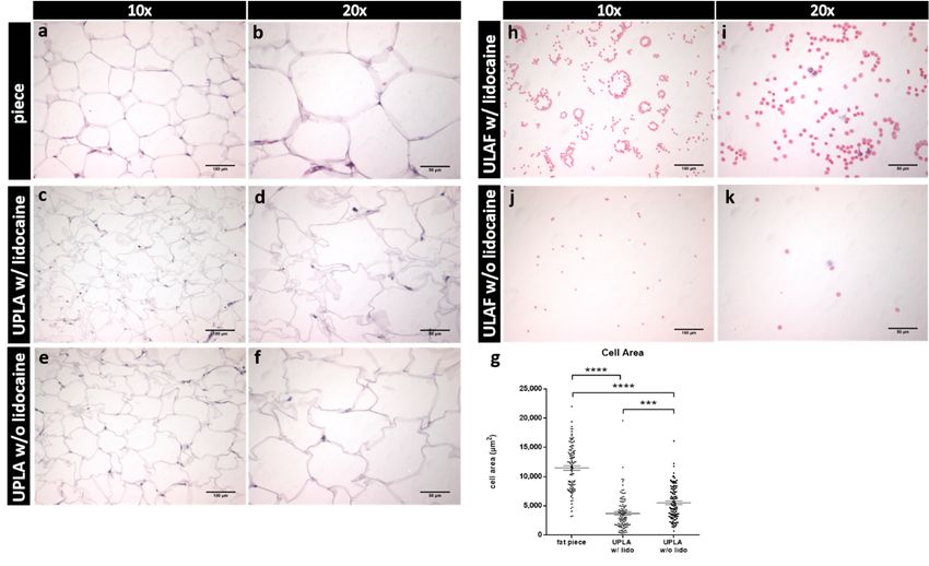
Int. J. Mol. Sci. 2020, 21, 2869                                                                        2 of 19

fraction (SVF), including preadipocytes and adipose-derived stem cells (ASCs) [15–17].
ASCs’ phenotype resembles that of bone marrow-derived mesenchymal stem cells (MSCs) and can
differentiate into several cell types, such as adipocytes [18,19], osteoblasts [18,20], chondrocytes [20],
myocytes [18], and tenocytes [21]. These features make ASCs of high interest for use in regenerative
medicine [22]. In contrast, preadipocytes are already restricted to evolve exclusively into adipocytes [23].
      Lidocaine, the most commonly used and best understood local anesthetic, has been shown
to have minimal systemic toxicity when used in tumescent local anesthesia (TLA) and is therefore
recommended as the anesthetic agent for liposuction. A survey from the American Society for Aesthetic
Plastic Surgery showed that the majority of American physicians use a tumescent solution containing
0.5 mg/mL of lidocaine [24–26].
      However, in vitro trials have shown that lidocaine exerts some potentially damaging effects on
ASCs, which are crucial for transplant survival [27,28]. Consequently, a potentially cytotoxic effect of
lidocaine would prevent its use as local anesthetic for lipotransfer and would result in a procedure
conducted under general anesthesia [29].
      In this study, we focused on the effect of lidocaine, applied during TLA prior to liposuction, on
cells of the SVF. Adipose-derived stem cells have been found to be crucial for transplant survival
by promoting angiogenesis and decreasing cell death and therefore, were the main focus of this
research [30,31].

2. Results

2.1. Basic Demographics and Clinical Data of the Patients
     In total, 11 female patients, who presented randomly at the clinic for Maxillofacial Surgery/Plastic
Surgery, Greifswald, Germany, for abdominoplasty, were included in this study. Patients’ basic
demographics and clinical history are summarized in Table 1. On average, they were 42 years old
(range from 19 to 60 years, standard error of the mean- SEM = 3.5), had a body-mass-index (BMI)
of 30.2 kg/m2 (SEM = 2.06), and waist-to-hip ratio of 0.86 (SEM = 0.03). None of the patients were
reported to have diabetes mellitus type 1 or 2.

      Table 1. In Table 1, the demographics (age, body-mass-index (BMI), waist/hip ration) and clinical
      history (weight gain/loss, diabetes) are shown for each patient.

       Patient       Age           BMI    Weight Gain/Loss in Last 6 Months     W:H Ratio    Diabetes
       1           43          26        no                                   0.854         no
       2           44          35        yes, −5 kg                           1.026         no
       3           35          48        no                                   0.91          no
       4           30          30        yes, −5 kg                           0.63          no
       5           41          26        yes, −5 kg                           0.78          no
       6           27          26        yes, −5 kg                           0.85          no
       7           48          29        no                                   0.85          no
       8           37          25        no                                   0.97          no
       9           52          34        no                                   0.96          no
       10          60          25        yes, −5 kg                           0.83          no
       11          19          32        no                                   0.8           no
       Mean        42          30.2                                           0.86
       SEM         3.54        2.06                                           0.03

2.2. Hematoxylin-Eosin (HE) Staining of Unprocessed Lipoaspirate and Surgically Resected Fat Tissue
     The two fractions resulting from sedimentation after liposuction are termed unprocessed
lipoaspirate (UPLA), which is the fatty supernatant portion, and unprocessed liposuction aspirate fluid
(ULAF), which is the fluid portion of the lipoaspirate. Fat tissue was harvested by liposuction with
Int. J. Mol. Sci. 2020, 21, x FOR PEER REVIEW 4 of 19
2.2. Hematoxylin-Eosin (HE) Staining of Unprocessed Lipoaspirate and Surgically Resected Fat Tissue
The two fractions resulting from sedimentation after liposuction are termed unprocessed
lipoaspirate (UPLA), which is the fatty supernatant portion, and unprocessed liposuction aspirate
Int. J. Mol. Sci. 2020, 21, 2869 3 of 19
fluid (ULAF), which is the fluid portion of the lipoaspirate. Fat tissue was harvested by liposuction
with (w/) and without (w/o) the use of lidocaine. Furthermore, in one patient, a piece of fat tissue
containing lidocaine was dissected.
(w/) and without (w/o) the use of lidocaine. Furthermore, in one patient, a piece of fat tissue containing
A significant mechanical impact of the liposuction procedure on the integrity of mature
lidocaine was dissected.
adipocytes was observed. Cell areas of adipocytes obtained by surgical resection were significantly
A significant
larger (p < 0.001)mechanical
than UPLA impact of the
samples liposuction
harvested w/andprocedure on the(Figure
w/o lidocaine integrity
1g).ofFurthermore,
mature adipocytes
wasUPLAobserved. Cell areas of adipocytes obtained by surgical resection were significantly
cells of the sample w/o lidocaine were significantly larger (p < 0.001) than cells larger the< 0.001)
of (p
than lidocaine-containing sample (Figure 1c–f). As shown in Figure 1a–g, liposuction performed with a of the
UPLA samples harvested w/and w/o lidocaine (Figure 1g). Furthermore, UPLA cells
pressure
sample w/o of 600 mmHg
lidocaine were(=0.8 bar) appears
significantly (p < 0.001)
to damage
larger maturethan adipocytes,
cells of leading to cell shrinking. sample
the lidocaine-containing
Subsequently,
(Figure 1c–f). As shown thein ULAF
Figure was
1a–g,assessed
liposuctionhistologically
performed(Figure 1h–k). We
with a pressure of 600observed
mmHgthat (=0.8 bar)
erythrocytes were the predominant cell population
appears to damage mature adipocytes, leading to cell shrinking. in the ULAF. Additionally, some leukocytes
were found in this fraction.
Figure
Figure1. 1.The
Thefatfatpiece
piece and
and the unprocessed
unprocessedfatty fatty portion
portion (UPLA)
(UPLA) afterafter paraffin
paraffin embedding
embedding and and
Hematoxylin-Eosin
Hematoxylin-Eosin(HE) (HE)staining, aswell
staining, as wellasasthe
theunprocessed
unprocessed fluidfluid portion
portion (ULAF) (ULAF) after
after HE HE staining
staining
are are shown.
shown. Slides
Slides wereobserved
were observedin inaa light
light microscope.
microscope. InInpanels
panels (a)(a,b),
and (b),
a fata fat piece
piece harvested
harvested bybysurgical
surgical w/lidocaine
extraction extraction w/lidocaine
is seen in is seen
10× in 10×20×
and andmagnification.
20× magnification. InInpanels
panels (c,d),
(c) andthe
(d),UPLA
the UPLA
harvested
harvested by w/lidocaine
by liposuction liposuction w/lidocaine
and in panelsand in panels
(e,f) w/o(e) and (f) w/o
lidocaine lidocaine
is shown inis10×
shown
andin20×
10× magnification.
and 20×
magnification. In panel (g), the
In panel (g), the cell area (in µm2 )cell area (in µm2) of the cross-sections from the fat tissue piece and
of the cross-sections from the fat tissue piece and the UPLA w/and w/o
the UPLA w/and w/o lidocaine of 100 random mature adipocytes (20 cells in 5 fields of views = 100
lidocaine of 100 random mature adipocytes (20 cells in 5 fields of views = 100 cells, 10× magnification)
cells, 10× magnification) was compared. The mean and standard error of the mean are shown. *** p <
was compared. The mean and standard error of the mean are shown. *** p < 0.001; **** p < 0.0001.
0.001; **** p < 0.0001. In (h) and (i), the ULAF of the lipoaspirate w/lidocaine, and in (j) and (k), w/o
In (h,i), the isULAF
lidocaine shownofinthe
10× lipoaspirate w/lidocaine, and in (j,k), w/o lidocaine is shown in 10× and
and 20× magnification.
20× magnification.
2.3. Hematoxylin-Eosin (HE) Staining, Immunostaining and Flow Cytometry of Processed Lipoaspirate
Subsequently, the ULAF was assessed histologically (Figure 1h–k). We observed that erythrocytes
The main purpose of the research was to quantify the potential effects of lidocaine on ASCs,
were the predominant cell population in the ULAF. Additionally, some leukocytes were found in
preadipocytes, mature adipocytes, and leukocytes number and live vs. dead status, found, after an
this fraction.
isolation process, inside the SVF of the processed lipoaspirate. The two fractions resulting from the
2.3. Hematoxylin-Eosin (HE) Staining, Immunostaining and Flow Cytometry of Processed Lipoaspirate
The main purpose of the research was to quantify the potential effects of lidocaine on ASCs,
preadipocytes, mature adipocytes, and leukocytes number and live vs. dead status, found, after an
isolation process, inside the SVF of the processed lipoaspirate. The two fractions resulting from the
isolation process are termed processed lipoaspirate (PLA), which is the fatty supernatant portion,
and liposuction aspirate fluid (LAF), which is the fluid portion of the lipoaspirate.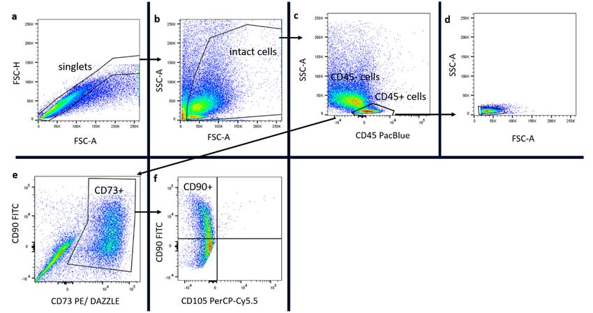
Int.J.J.Mol.
Int.     Mol. Sci.
              Sci. 2020,
                   2020, 21,
                         21,x2869
                              FOR PEER REVIEW                                                                           5 of 1919
                                                                                                                          4 of

 isolation process are termed processed lipoaspirate (PLA), which is the fatty supernatant portion,
 and Figure
       liposuction  aspirate
               2 gives       fluid (LAF),
                       a histological      which is the
                                      illustration       fluid
                                                    of the     portion
                                                            cells  of theofSVF.
                                                                            the lipoaspirate.
                                                                                Only nucleated cells were visible,
        Figure  2 gives  a histological illustration  of  the  cells of
indicating the complete lysis of erythrocytes. Further differentiation   the  SVF.and
                                                                                    Only  nucleated cells
                                                                                       quantification     were
                                                                                                      of cell types
 visible, indicating the complete lysis of erythrocytes. Further differentiation and quantification of
was conducted by flow cytometry.
 cell types was conducted by flow cytometry.

       Figure 2. This figure presents the lysed SVF of the lipoaspirate of the fluid (LAF) and fatty portion
      Figure 2. This figure presents the lysed SVF of the lipoaspirate of the fluid (LAF) and fatty portion
       (PLA), which was later used for flow cytometry. Slides were observed in a light microscope. In (a)
      (PLA), which was later used for flow cytometry. Slides were observed in a light microscope. In (a,e),
       and (e), the LAF w/lidocaine, and in (b) and (f), the LAF w/o lidocaine is seen in 10× and 20×
      the LAF w/lidocaine, and in (b,f), the LAF w/o lidocaine is seen in 10× and 20× magnification. In panel
       magnification. In panel (c) and (g), PLA w/lidocaine, and in (d) and (h), PLA w/o lidocaine is shown
      (c,g), PLA w/lidocaine, and in (d,h), PLA w/o lidocaine is shown in 10× and 20× magnification.
       in 10× and 20× magnification.

      The cytotoxic effect of lidocaine was quantified by determining the relative distribution and the
      The cytotoxic effect of lidocaine was quantified by determining the relative distribution and
absolute number of nucleated cell populations of the SVF, harvested w/or w/o lidocaine. In addition,
 the absolute number of nucleated cell populations of the SVF, harvested w/or w/o lidocaine. In
the ratio ofthe
 addition,     living
                   ratioto
                         ofdead
                             livingcells  wascells
                                     to dead    evaluated    using phenotypic
                                                     was evaluated                  markers.
                                                                       using phenotypic      markers.
      A  significantly      higher    percentage      of nucleated    cells
      A significantly higher percentage of nucleated cells were found insidewere    found    inside
                                                                                                  the the
                                                                                                       PLAPLA
                                                                                                            w/ow/o     lidocaine
                                                                                                                  lidocaine
compared      to   the  LAF     w/o  lidocaine    (p  <  0.01) in proportion     to  all events    (cells
 compared to the LAF w/o lidocaine (p < 0.01) in proportion to all events (cells and cell fragments)      and  cell  fragments)
counted
 counted byby flow
                 flow cytometry.
                       cytometry. The   ThePLAPLAw/lidocaine
                                                    w/lidocainealsoalso  contained
                                                                       contained       significantly
                                                                                    significantly       more
                                                                                                     more      nucleated
                                                                                                           nucleated    cellscells
than  the  LAF     w/lidocaine      (p <  0.05).  The  absolute   number     of nucleated     cells
 than the LAF w/lidocaine (p < 0.05). The absolute number of nucleated cells was significantly higherwas  significantly   higher
inside  the  PLA     w/o   lidocaine    compared      to the LAF   w/o  lidocaine    (p
 inside the PLA w/o lidocaine compared to the LAF w/o lidocaine (p < 0.05). There were no< 0.05).  There   were   no  significant
differences    in the relative
 significant differences            distribution
                                in the              and absolute
                                        relative distribution    andnumber
                                                                      absoluteofnumber
                                                                                   nucleated     cells between
                                                                                             of nucleated          the samples
                                                                                                             cells between
w/or  w/o lidocaine
 the samples     w/or w/o from    the same
                               lidocaine      type
                                           from  theof  isolates.
                                                      same  type of isolates.
      The influence
      The   influence of  of lidocaine
                              lidocaineon  ondistinct
                                               distinctsubpopulations
                                                          subpopulations  of of
                                                                              thethe
                                                                                  SVF,   such
                                                                                      SVF,      as ASCs
                                                                                             such         (CD45-,
                                                                                                    as ASCs         CD73+,
                                                                                                              (CD45-,    CD73+,
 CD90+, and CD105-),
CD90+,             CD105-), preadipocytes
                                 preadipocytes(Pref-1+
                                                    (Pref-1+FABP4-),
                                                               FABP4-), mature
                                                                            mature adipocytes
                                                                                      adipocytes  (Pref-1-  FABP4+),
                                                                                                      (Pref-1-  FABP4+), and and
 leukocytes (CD45+),
leukocytes      (CD45+), was  wasassessed
                                     assessedasasdescribed
                                                     describedininthethemethods
                                                                          methods   section.
                                                                                      section. TheTheimplemented
                                                                                                        implemented   gating
                                                                                                                          gating
 strategy  is shown     in  Figures    3 and  4. In Figure  5a,b  results of nucleated
strategy is shown in Figures 3 and 4. In Figure 5a,b results of nucleated cells are shown. cells are  shown.
      The results showed a significantly higher percentage (p < 0.01) and absolute number (p < 0.05)
of stem cells inside the PLA compared to the LAF, which contained almost no ASCs. No differences
between the samples w/and w/o lidocaine were found (Figure 5c,d). With regard to cell viability,
no differences between the two PLA treatments were detected (n = 5, x: mean, SEM: standard error
of the mean). The alive rate w/lidocaine was x = 25.5% SEM = 15.6%, and w/o lidocaine x = 28.9%
SEM = 18.1%; the absolute cell number of alive cells w/lidocaine was x = 25,109/mL SEM = 15,828/mL,
and w/o lidocaine x = 32,113/mL SEM = 23,670/mL (Table 2).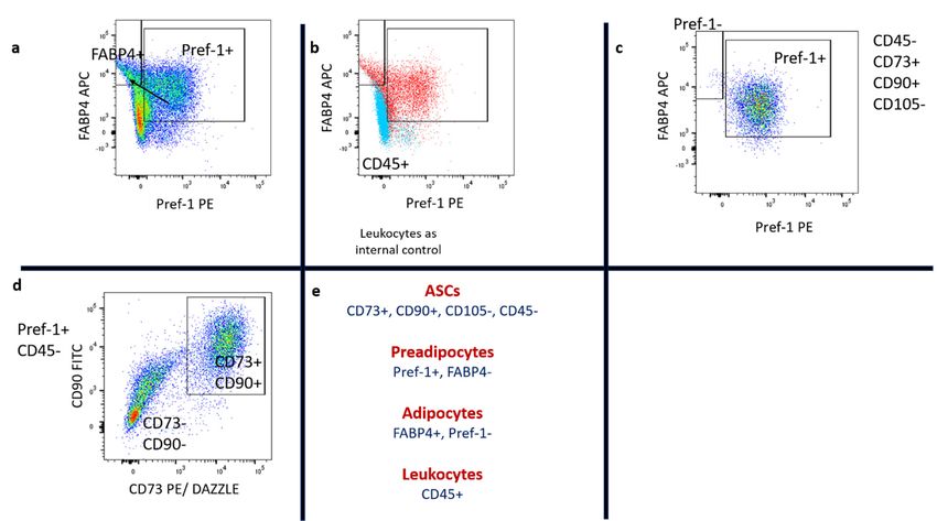
Int. J. Mol. Sci. 2020, 21, 2869 5 of 19
Int. J. Mol. Sci. 2020, 21, x FOR PEER REVIEW 6 of 19
Int. J. Mol. Sci. 2020, 21, x FOR PEER REVIEW 6 of 19
Figure 3. Part I of the gating strategy applied in the study, shown here with PLA cells of one patient.
Figure 3. Part
Figure 3. Part
In (a),
I ofIwere
(a), singlets
of
thethe
singlets were
gatingstrategy
gating
gated. In
strategyapplied
In (b)
(b) of
applied in
of all
in the
all singlets,
the study,
singlets, intact
study,
intact cells
shown
shown
cells were
here
herewith
were gated,
withPLA
gated, leaving
PLA cells
leaving cell
of one
cells patient.
of one
cell fragments
patient. In
fragments aside.
aside.
In gated.
(a), singlets
In (c)
(c) of were
of all gated.
all intact
intact cells, In (b)
cells, CD45 of
+ all singlets,
cells were
CD45+ cells were gated.intact
gated. In cells
In (d), were
(d), CD45+
CD45+ cellsgated,
cells are leaving
are shown
shown in cell fragments
in SSC/FSC.
SSC/FSC. In aside.
In (e), the In (c)
(e), the
In + cells were
of allCD45-
intactcell
cells, CD45
population was was assessed gated.
assessed for In
for CD73(d), CD45+
CD73 expression. cells
expression. In are
In (f), shown
(f), these in SSC/FSC.
these CD45-,
CD45-, CD73+ In
CD73+ cells (e),wereCD45-
the
cells were
CD45- cell population
cell population
assessed for was
for CD105 assessed
CD105 and for
and CD90 CD73
CD90 expression. expression.
expression. Stem In
Stem cells (f), these
cells appear
appear to CD45-,
to be CD73+
be CD45-,
CD45-, CD73+,cells were
CD73+, CD90+, assessed
CD90+, and and for
assessed
CD105 and CD90 expression. Stem cells appear to be CD45-, CD73+, CD90+, and CD105-.
CD105-.
CD105-.
Figure 4. Part II of the gating strategy. In (a), intact cells expressing Pref-1 and FABP4 were gated.
Figure 4. Part
Figure 4. Part II II
ofofthe
thegating
gating strategy.
strategy. In
In(a),
(a),intact
intactcells expressing
cells Pref-1
expressing and FABP4
Pref-1 were gated.
and FABP4 were gated.
Preadipocytes lose
Preadipocytes lose Pref-1
Pref-1 expression
expression and
and enrich
enrich FABP4
FABP4 during
during further
further differentiation
differentiation toto mature
mature
Preadipocytes lose Pref-1 expression and enrich FABP4 during further differentiation to mature
adipocytes. (b)
adipocytes. (b) Preadipocytes
Preadipocytes andand adipocytes
adipocytes were
were gated
gated by
by using
using leukocytes
leukocytes asas an
an internal
internal control.
control.
adipocytes. (b)
(c) CD45-, Preadipocytes
CD45-, CD73+,
CD73+, andand CD90+ and
CD90+ cellsadipocytes
cells were were
were assessed gated
assessed for by
for Pref-1 using leukocytes
Pref-1 expression.
expression. In
In the as
the PLAan internal
PLA sample, control.
sample, most
most (c)
(c)
CD45-, CD73+,
stem cells and
cells expressedCD90+
expressed Pref-1. cells
Pref-1. In were
In (d), assessed
(d), Pref-1+
Pref-1+ and for Pref-1
and CD45-
CD45- cells expression.
cells were In
were assessed the
assessed forPLA sample,
for CD73
CD73 and most
and CD90
CD90 stem
stem
cellsexpression
expressedandPref-1.
foundInto(d),
be Pref-1+
partly and CD45-
CD73+ and cells were
CD90+. (e) assessed
Specific for CD73
marker and
profiles forCD90
stem expression
cells,
expression and found to be partly CD73+ and CD90+. (e) Specific marker profiles for stem cells,
andpreadipocytes,
found to be partly
preadipocytes, mature CD73+ and
adipocytes, CD90+.
and (e)
leukocytesSpecific
are
mature adipocytes, and leukocytes are shown.
marker
shown. profiles for stem cells, preadipocytes,
mature adipocytes, and leukocytes are shown.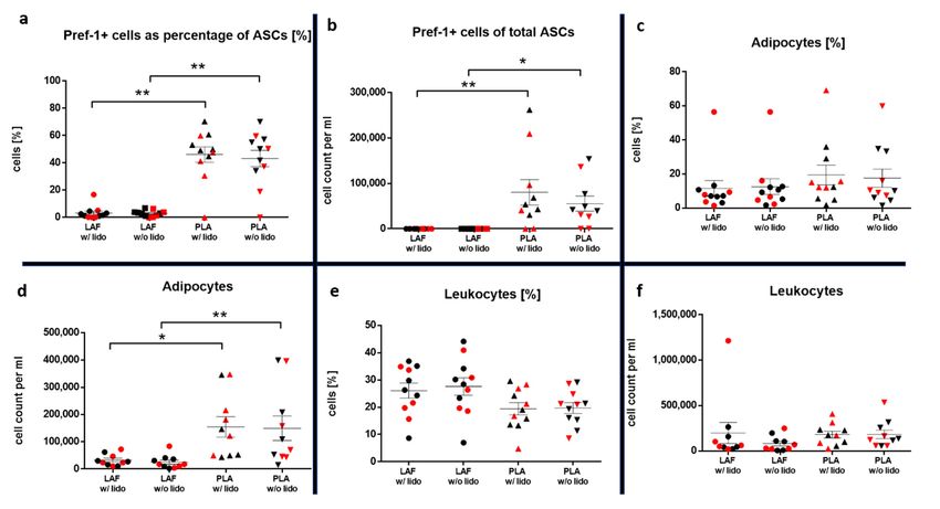
between the samples w/and w/o lidocaine were found (Figure 5c,d). With regard to cell viability, no
        differences between the two PLA treatments were detected (n= 5, x̅: mean, SEM: standard error of
        the mean). The alive rate w/lidocaine was x̅ = 25.5% SEM = 15.6%, and w/o lidocaine x̅ = 28.9% SEM
        = 18.1%; the absolute cell number of alive cells w/lidocaine was x̅ = 25,109/mL SEM = 15,828/mL,
        and w/o lidocaine x̅ = 32,113/mL SEM = 23,670/mL (Table 2).
Int. J. Mol. Sci.
             The2020, 21, 2869
                  preadipocyte  cell yield in the PLA was not significantly higher compared to the LAF, as 6 of 19
        shown in Figure 5e,f.

         Figure
     Figure      5. graphs
            5. The  The graphs
                           showshow      the statistical
                                   the statistical       comparison
                                                     comparison        of relative
                                                                   of the the relative distribution
                                                                                   distribution   (n =(n11)
                                                                                                         = and
                                                                                                            11) and
                                                                                                                 absolute
         absolute number  (n = 10)  of all cells (a,b), ASCs (c,d), and preadipocytes   (e,f) inside the
     number (n = 10) of all cells (a,b), ASCs (c,d), and preadipocytes (e,f) inside the SVF of the fluid SVF  of the(LAF)
         fluid (LAF) and fatty (PLA) portion w/ and w/o lidocaine. Samples of each patient are marked in
     and fatty (PLA) portion w/ and w/o lidocaine. Samples of each patient are marked in every group.
         every group. The mean and standard error of the mean in each group is shown. For multiple
     The mean and standard error of the mean in each group is shown. For multiple comparisons, the
         comparisons, the Kruskal–Wallis test was used. The α-value was at 0.05. Patients with a BMI ≥ 30
     Kruskal–Wallis test was used. The α-value was at 0.05. Patients with a BMI ≥ 30 are shown in red.
          are shown in red.

          No influence on the number or viability of preadipocytes inside the SVF of PLA and LAF was
     Table 2. Mean (x) and standard error of the mean (SEM) of alive ASCs and alive preadipocytes (Zombie-)
     observed (n= 5, x̅: mean, SEM: standard error of the mean). The PLA alive rate w/lidocaine was x̅ =
     in the absolute number per mL and as a percentage of all counted stem cells and preadipocytes
     70% SEM = 15%, and w/o lidocaine x̅ = 77.9% SEM = 8.5%; the PLA absolute cell number of alive
     (n = 5 patients).
     cells w/lidocaine was x̅ = 88,756/mL SEM = 44,481/mL, w/o lidocaine x̅ = 87,814/mL SEM =
     58,406/mL; LAF alive rate: w/lidocaine x̅ = 93% SEM = 3.4%, w/o lidocaine      x̅ = 93% SEM
                                                                                 w/Lido       w/o =Lido
                                                                                                    2.59%;
     and LAF absolute cell number of alive cells: w/lidocaine x̅ = 38,380/mL SEM = 21,419/mL, w/o
                                                                        X          25.5          28.9
     lidocaine x̅ = 15,848/mL SEM = 7851/mL (Table 2). %
                                                                       SEM         15.6          18.1
            ASCs (viable)            PLA
                                                                        X         25,109       32,113
                                                       Cells/mL
                                                                       SEM        15,828       23,670
                                                                                X            93              93
                                                                %
                                                                               SEM           3.4            2.59
                                         LAF
                                                                                X          38,380          15,848
                                                            Cells/mL
        Preadipocyte (viable)                                                  SEM         21,419           7851
                                                                                X            70             77.9
                                                                %
                                                                               SEM           15              8.5
                                         PLA
                                                                                X          88,756          87,814
                                                            Cells/mL
                                                                               SEM         44,411          58,406

      The preadipocyte cell yield in the PLA was not significantly higher compared to the LAF, as
shown in Figure 5e,f.
      No influence on the number or viability of preadipocytes inside the SVF of PLA and LAF was
observed (n = 5, x: mean, SEM: standard error of the mean). The PLA alive rate w/lidocaine was
x = 70% SEM = 15%, and w/o lidocaine x = 77.9% SEM = 8.5%; the PLA absolute cell number of alive
cells w/lidocaine was x = 88,756/mL SEM = 44,481/mL, w/o lidocaine x = 87,814/mL SEM = 58,406/mL;
LAF alive rate: w/lidocaine x = 93% SEM = 3.4%, w/o lidocaine x = 93% SEM = 2.59%; and LAF absolute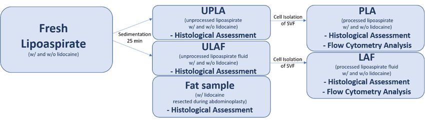
compared to the LAF (Figure 6a,b).
         Mature adipocytes were found in a low number, compared to preadipocytes, in the SVF. A
 significantly higher absolute number of cells was found inside the PLA compared to the LAF (p <
 0.05 w/lidocaine, p < 0.01 w/o lidocaine, Figure 6c,d). No influence of lidocaine on the count or
 viability    of 2020,
Int. J. Mol. Sci. this type   of cell was detected (n = 5, x̅: mean, SEM: standard error of the mean). The7 PLA
                        21, 2869                                                                                of 19
 alive rate w/lidocaine was x̅ = 71.2% SEM = 10.7%, w/o lidocaine x̅ = 71.5% SEM = 11.4%; PLA
 absolute cell number of alive cells: w/lidocaine x̅ = 119,035/mL SEM = 46,475/mL, w/o lidocaine x̅ =
cell  number of
 148,586/mL           alive
                    SEM      cells: w/lidocaine
                          = 51,609/mL;           x = 38,380/mL
                                           LAF alive             SEM = 21,419/mL,
                                                     rate: w/lidocaine                w/o lidocaine
                                                                         x̅ = 74.8% SEM               = 15,848/mL
                                                                                           = 8%, w/ox lidocaine   x̅ =
SEM     =  7851/mL      (Table   2).
 72.5% SEM = 8.7%; and LAF absolute cell number of alive cells: w/lidocaine x̅ = 24,944/mL SEM =
        Stem w/o
 7587/mL,      cellslidocaine
                      expressing  x̅ =the preadipocyte
                                       28,163/mL SEM =marker     Pref-1 occurred more frequently in the relative
                                                         7081/mL).
distribution       (p <  0.01)  and   absolute number  (p
         Leukocytes numbers inside the LAF were, compared < 0.01 w/lidocaine,
                                                                          to thep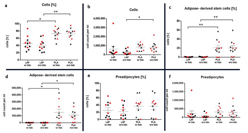
124,613/mL SEM = 42,567/mL, w/o lidocaine x̅ = 143,169/mL SEM = 55,985/mL; LAF alive rate:
  w/lidocaine x̅ = 79.1% SEM = 15.4%, w/o lidocaine x̅ = 77.8% SEM = 15.6%; and LAF absolute cell
  number of alive cells: w/lidocaine x̅ = 78,334/mL SEM = 46,606/mL, w/o lidocaine x̅ = 46,139/mL
  SEM = 21,961/mL).
Int. J. Mol. Sci. 2020, 21,statistical
         Additional         2869                                                                           8 of 19
                                        analysis (one-sample t-test) of the relative distribution and absolute
  numbers of particular cell types (ASCs, preadipocytes, ASCs expressing preadipocyte marker Pref-
  1, mature and
adipocytes,       adipocytes,    andwas
                       leukocytes)     leukocytes)
                                          conductedwas
                                                     to conducted
                                                        address theto   address
                                                                      sample    the sample heterogeneity.
                                                                             heterogeneity.               We did
                                                                                            We did not observe   a
  not   observe     a significant   inter-patient
significant inter-patient variability (p > 0.05). variability (p > 0.05).

  2.4.Evaluation
2.4.   EvaluationofofLidocaine
                       LidocaineDistribution
                                 DistributionUsing
                                              UsingGC-MS
                                                    GC-MS

      AsAsdescribed
             describedininthethe  method
                               method        section,
                                          section,    lidocaine-free
                                                   lidocaine-free    and
                                                                   and     lidocaine-containingTLA
                                                                        lidocaine-containing        TLAwaswas  injected
                                                                                                             injected
  inside
inside  thethe  right
             right andand
                        leftleft
                             sideside  of the
                                   of the      lower
                                           lower      abdomen,
                                                  abdomen,       respectively.
                                                             respectively.      Analysis
                                                                            Analysis of theoffat
                                                                                              the  fat tissue
                                                                                                 tissue       by using
                                                                                                         by using gas
  gas  chromatography        showed     that  lidocaine  was only  found   inside the left abdomen.
chromatography showed that lidocaine was only found inside the left abdomen. The right side of the      The  right side
  of the abdomen,
abdomen,               whichas
             which served       served   as a control,
                                  a control,   was not was  not contaminated
                                                        contaminated            by lidocaine
                                                                      by lidocaine             (Figure 7).
                                                                                     (Figure 7).

     Figure
       Figure   The
             7. 7.   results
                   The       of the
                        results  of semiquantitative   analysis
                                    the semiquantitative        with with
                                                            analysis  a GC-MS    method
                                                                           a GC-MS       of the unprocessed
                                                                                      method                    fluid
                                                                                                 of the unprocessed
     portion  (ULAF)   of the right and  left lower abdomen    are shown.  To  evaluate the method,
       fluid portion (ULAF) of the right and left lower abdomen are shown. To evaluate the method, thethe  lidocaine
     concentration   was determined
       lidocaine concentration          in 2 patients.
                                  was determined     in The right abdomen,
                                                        2 patients. The right which served
                                                                              abdomen,       as aserved
                                                                                         which    control,aswas  free
                                                                                                             a control,
     ofwas
        lidocaine.
            free of lidocaine.
3. Discussion
  3. Discussion
      Several studies have been carried out on the influence of lidocaine on fat tissue, delivering an
         Several studies have been carried out on the influence of lidocaine on fat tissue, delivering an
inconsistent picture. Some studies described cytotoxic effects [28,32], while others did not see signs of
  inconsistent picture. Some studies described cytotoxic effects [28,32], while others did not see signs
cell damage [33,34]. The use of different methods makes it difficult to compare these results.
  of cell damage [33,34]. The use of different methods makes it difficult to compare these results.
      Research on the cytotoxicity of lidocaine was first published in 1995 by Moore et al. [35]. They
         Research on the cytotoxicity of lidocaine was first published in 1995 by Moore et al. [35]. They
postulated that lidocaine potently inhibits glucose transport and lipolysis in mature adipocytes as
  postulated that lidocaine potently inhibits glucose transport and lipolysis in mature adipocytes as
well as their growth in culture using in vitro experiments. Since then, the topic has been the subject of
  well as their growth in culture using in vitro experiments. Since then, the topic has been the subject
controversy. Shoshani et al. [33] and Livaoglu et al. [34] did not find effects of lidocaine on the graft take
  of controversy. Shoshani et al. [33] and Livaoglu et al. [34] did not find effects of lidocaine on the
of fat tissue in animal models. In contrast, Keck et al. [28] showed a negative effect of lidocaine on ASCs’
  graft take of fat tissue in animal models. In contrast, Keck et al. [28] showed a negative effect of
viability after 30 min of incubation in vitro, while using a much higher concentration of lidocaine than
  lidocaine on ASCs’ viability after 30 min of incubation in vitro, while using a much higher
usual for tumescence local anesthesia (2% vs. 0.062% in the therapeutic concentration). Girard et al. [32]
  concentration of lidocaine than usual for tumescence local anesthesia (2% vs. 0.062% in the
demonstrated significant cytotoxicity of lidocaine on ASCs in vitro, using a therapeutic concentration
of 0.8 mg/mL. This effect only occurred after 24 h of incubation. However, this might not reflect human
in vivo conditions, because the metabolic half-life of lidocaine is about 90 min in humans [36]. In their
study from 2016, Goldman et al. [37] used a method that resembles ours. They also realized liposuction
w/and w/o lidocaine on symmetrical body parts, performed by the same surgeon. In contrast, they
used a lower lidocaine concentration (0.3 mg/mL) and did not evaluate the presence or absence of
lidocaine on each of the harvested sides. Goldman et al. [37] concluded that removing lidocaine from
tumescent significantly reduced ASCs apoptosis in the lipoaspirate. Even using a concentration of 0.6
mg/mL, these findings could not be confirmed in this study.
      TLA is not only used for analgesia but also for tissue inflation and vasoconstriction to reduce
trauma and bleeding during liposuction. As stated, the Goldman study [37] described apoptotic effects
of lidocaine on ASCs and therefore recommended the use of tumescent liposuction without lidocaine
and to carry out lipotransfer under general anesthesia. They suggested infiltrating lidocaine at a
Int. J. Mol. Sci. 2020, 21, 2869 9 of 19
later step for post-operative pain reduction. The results of our study contradict that view. Lidocaine
influenced neither the relative distribution, nor absolute numbers, nor the life vs. death status of
ASCs. Additionally, general anesthesia has a much higher risk in cardiorespiratory events than TLA,
which has been shown to be safe up to a 35 mg/mL concentration of lidocaine [25,38]. Complications
in liposuction are more common when performed under general anesthesia [39]. Furthermore, an
additional TLA injection with lidocaine after liposuction would be a rather impractical approach.
It would complicate the surgical procedure, not only in a logistical, but also in a time-sensitive manner.
The protocol used in our study is frequently used in the host clinic to harvest, process, and
transplant/analyze harvested fat tissue and was adopted as a standard operating procedure (SOP).
Potential variability in cell yields and variability in a heterogenous population (age, body mass index)
was excluded through intra-individual comparison using symmetrical areas of the lower abdomen
w/ and w/o lidocaine [40–42]. The intra-individual approach helped to eliminate fluctuations of cell
yields that may occur during isolation and cell staining. Results showed no influence of a BMI ≥ 30 on
cell count, which supports the validity of the results obtained by this approach. All liposuctions were
performed by a single surgeon.
In addition, it was shown that when this protocol is used, lidocaine was detected only inside the
LAF of the right abdomen and only cell populations from the right lower abdomen were in contact
with lidocaine. This improves the reliability of the data collected.
One may argue that washing or centrifugation could, in comparison to sedimentation, decrease
the contact time with lidocaine, leading to improved ASCs viability. However, several studies
concluded that no processing technique is superior to another, and that centrifugation force greater
than 1200× g causes cell damage [9]. This study used sedimentation for 25 min as an easy and accessible
standard method.
To characterize cell status right before transplantation, viability has to be assessed. Cell death can
be caused by either apoptosis or necrosis. Apoptosis is characterized by cell shrinking, fragmentation,
and phagocytosis without inflammation. During apoptosis, phosphatidylserine is translocated from the
inner to the outer leaflet of the membrane, where it can be detected by annexin V staining. In contrast,
necrosis involves cell swelling, membrane perforation, and phagocytosis with inflammation [43]. Most
studies that reported a cytotoxic effect of lidocaine found necrosis [32,44], whereas others claimed
apoptosis [37] to be the involved mechanism. Zombie NIR™ staining, which does not differentiate
between apoptosis and necrosis, was used in this study. Classic necrosis markers like propidium
iodide (PI) have the disadvantage that they may leak out of cells within a short period of time [45].
Using a lidocaine concentration of 0.6 mg/mL in TLA did not show a negative influence of lidocaine on
cell viability.
In contrast to this study, Goldman et al. [37] did not use a phenotypical marker profile to distinguish
between different SVF cell populations. Different research groups have struggled to find a single
definition of markers expressed by ASCs. Especially, the expression of the hematopoietic stem cell
marker CD34 has been debated intensively [46,47]. The aim was therefore to use minimal criteria of
markers to define ASCs that most research groups could relate to. The International Society of Cell
Therapy (ISCT) released a statement paper in 2005 that established minimal criteria, defining bone
marrow-derived MSCs as positive for CD73, CD90, and CD105 and lacking the presence of the leukocyte
marker CD45 [48]. ASCs have been found to be of perivascular origin [46,49–51]. Zimmerlin et al. [52]
evaluated the presence of MSC markers on perivascular cells and found them to express MSC markers
up to 95.5%. Crisan et al. [53] also observed the expression of these surface proteins on perivascular
cells. Other works questioned the expression of CD105 in freshly isolated fat tissue and suggested that
it might be only expressed under culture conditions [54]. Our results show that freshly isolated ASCs
do not express CD105, which is consistent with the findings of Yoshimura et al. [54]. We therefore
consider CD105 a stem-cell marker in cell culture but not in freshly isolated ASCs.
Ultimately, this study defines ASCs as positive for CD73 and CD90, and negative for CD45 and
CD105. Preadipocytes are already restricted to evolve into adipocytes and have been shown to expressInt. J. Mol. Sci. 2020, 21, 2869 10 of 19
Pref-1, which acts as an inhibitor of adipogenesis and is downregulated in mature adipocytes [23,55–57].
This study’s findings showed that Pref-1-positive ASCs were almost entirely located inside the PLA.
This leads to the conclusion that the PLA is the source of stem cells that are restricted towards an
adipogenic differentiation and therefore only the fatty portion should be used for transplantation.
Distinct cell populations were isolated using specific surface markers to keep the time between
liposuction and data acquisition as short as possible. This approach gave the opportunity to obtain data
after a short processing time, without possible variability in cell yields or viability due to cultivation of
adherent cells for 24 h. The time from liposuction to transplantation usually takes from 30 min up to
2 h, depending on the surgeon and the type of surgery. Therefore, in this study, data were obtained
in a similar processing time compared to a lipotransfer procedure. During further development,
preadipocytes enrich FABP4, which has been used extensively as a marker for mature adipocytes [58].
Adipocytes are mitotic inactive cells that can be renewed by ASCs. In lipotransfer, they act as a
filler for volume augmentation. Histological evaluation of the unprocessed lipoaspirate has shown that
vibration-assisted liposuction has a negative mechanical impact on the integrity of mature adipocytes
in an intra-individual approach. Cellular area was used as a surrogate parameter for cellular lysis upon
liposuction. In mature adipocytes, cellular damage will cause rapid lysis, leakage of fat vacuoles, and
therefore a decrease of the cell size [59]. ASCs are thought to be located inside the adipose connective
tissue [46,49–51]. Whether the mechanical process of liposuction has a negative influence on ASC
viability or a positive impact on graft survival by releasing ASCs from their original location in between
adipocytes needs to be evaluated in further research.
In lipotransfer, adipocytes act as fillers for volume augmentation. It is thus important to know
where to find mature adipocytes and to evaluate whether lidocaine has an impact on these cells.
Adipocytes are buoyant cells. However, this study found a low number of mature adipocytes present
inside the SVF even after an isolation process. Nevertheless, the main source of mature adipocytes is
the upper layer of the unprocessed lipoaspirate. Transplantation of SVF cells only would therefore not
fulfill the purpose of volume augmentation in lipofilling.
The influence of lidocaine on the SVF of the LAF was investigated in our study. In a previous
study, Yashimuro et al. [54] found that a comparable number of cells can be harvested from the
erythrocyte-lysed SVFs of PLA and LAF, with fewer LAF cells being adherent. In their study, they
used plating of CD34 and CD45 expression to identify potential stem cells. Since the expression of
CD34 in human adipose tissue is still debated, mesenchymal stem cell markers were used that have
been shown to be expressed on perivascular stem cells in adipose tissue [46,47,50]. ASCs were located
almost entirely inside the PLA.
Being a major component of the human immune system, leukocytes play a key role as a regulator
in inflammation and anti-inflammation processes inside human fat tissue [60]. The effect of lidocaine
on the leukocyte cell count and viability inside the SVF of PLA and LAF was therefore evaluated.
Yashimuro et al. [54] showed a higher cell yield of blood-derived CD45+ cells inside the SVF of the
LAF compared to the PLA. According to our study’s findings, the LAF and PLA are made up of a
comparable percentage and absolute cell count of leukocytes. No impact of lidocaine was found on the
viability of CD45+ cells.
It is important to emphasize that this study did not use long-term cultivation of ASCs to evaluate the
effect of the potential damage done by liposuction with lidocaine on long-term survival. The conditions
in cell culture do not resemble those found at the recipient site in lipotransfer due to the lack of
structural, biochemical, and signaling interactions with surrounding tissues and cells [61]. Cell culture
was therefore not used for this study. In vivo murine models as used by Shoshani et al. [33] are not
comparable to the conditions found in humans.
To address the question of cell viability after liposuction, cell life vs. death status was assessed using
Zombie NIR™ staining. Zombie NIR™ is an amine-reactive dye, which crosses the cell membranes of
dead cells, thus producing information about the viability status.evaluate the effect of the potential damage done by liposuction with lidocaine on long-term
survival.
The conditions in cell culture do not resemble those found at the recipient site in lipotransfer due to
the lack of structural, biochemical, and signaling interactions with surrounding tissues and cells
[61]. Cell culture was therefore not used for this study. In vivo murine models as used by Shoshani
Int. J. Mol. Sci. 2020, 21, 2869 11 of 19
et al. [33] are not comparable to the conditions found in humans.
To address the question of cell viability after liposuction, cell life vs. death status was assessed
using Zombie NIR™ staining. Zombie NIR™ is an amine-reactive dye, which crosses the cell
The results obtained in this study give information about the cell numbers and life vs. dead status
membranes of dead cells, thus producing information about the viability status.
right before transplantation. The chosen study design can offer no conclusions about the long- term
The results obtained in this study give information about the cell numbers and life vs. dead
survival
statusofright
cellsbefore
in celltransplantation.
culture. The chosen study design can offer no conclusions about the
long- term survival of cells in include
The two main limitations the gender and small sample size. Only adult female patients
cell culture.
were evaluated, which limits the generalizability
The two main limitations include the gender and ofsmall
the results. There
sample size. Onlywere nofemale
adult malepatients
patients and
no children included
were evaluated, in this
which study,
limits so no conclusions
the generalizability can be There
of the results. drawnwere
about thesepatients
no male population
and nogroups.
Thechildren included
small sample in prevented
size this study, so no conclusions
reasonable can
use of be drawn about
regression these
analysis to population
control forgroups. The
intra-individual
small sample
variability. size prevented
Intra-individual reasonableand
assessments use one-sample
of regressiont-tests
analysis to applied
were control for intra-individual
instead, as described in
variability. Intra-individual assessments and one-sample t-tests were applied
the statistics section. One factor that has been described to influence the SVF cell yield instead, as described
is the BMI of a
in the statistics section. One factor that has been described to influence the SVF cell yield is the BMI
patient [40]. To further control for inter-patient variability, obese patients (BMI ≥ 30) were highlighted
of a patient [40]. To further control for inter-patient variability, obese patients (BMI ≥ 30) were
in red. Our results showed no significant inter-individual differences in cell numbers between obese
highlighted in red. Our results showed no significant inter-individual differences in cell numbers
andbetween
non-obese patients.
obese and non-obese patients.
4. Methods
4. Methods
Figure 8 gives
Figure ananoverview
8 gives overviewof
of the
the processing steps.
processing steps.
Figure
Figure 8. The
8. The studystudy workflow
workflow showing
showing the processing
the processing and evaluation
and evaluation of the of theportion,
fatty fatty portion,
unprocessed
unprocessed (UPLA) and processed (PLA); and the fluid portion, unprocessed
(UPLA) and processed (PLA); and the fluid portion, unprocessed (ULAF), and processed (ULAF), and
(LAF). In
processed (LAF). In n = 1 patient, a fat piece was resected during abdominoplasty and used for
n = 1 patient, a fat piece was resected during abdominoplasty and used for histological evaluation.
histological evaluation.
4.1. Written Informed Consent and Ethical Approval
4.1. Written Informed Consent and Ethical Approval
Written informed consent was obtained from each patient. The study was carried out following
Written informed consent was obtained from each patient. The study was carried out
the rules of the Declaration of Helsinki and was reviewed and approved by the ethics committee
following the rules of the Declaration of Helsinki and was reviewed and approved by the ethics
of the University
committee of theMedicine Greifswald
University Medicine (Nr. BB 050/16,
Greifswald 05/16).
(Nr. BB All05/16).
050/16, experiments were performed
All experiments were in
accordance with local guidelines overseen by the University Medicine Greifswald and the University
performed in accordance with local guidelines overseen by the University Medicine Greifswald and
of Greifswald, Greifswald,
the University Mecklenburg-Western
of Greifswald, Pomerania. Pomerania.
Greifswald, Mecklenburg-Western
4.2. 4.2.
Tissue Harvesting
Tissue bybyLiposuction
Harvesting Liposuction and Abdominoplasty
and Abdominoplasty
The right and left lower abdomen were prepared with different compositions of TLA. The right side
was infiltrated with TLA containing 1 l saline (0.9%) and 0.25 mL epinephrine (1 mg/mL). The left side
was prepared with 1 l saline (0.9%) containing 30 mL lidocaine 2% (600 mg) and 0.25 mL epinephrine
(1 mg/mL). TLA was infiltrated under constant pressure with an 18G cannula at 20 ◦ C temperature
(Figure 9a,b).
Liposuction was performed with the vibration-assisted method, using VibraSat with 5000 cycles
per minute. Rapid Extraction 9-hole cannula (diameter = 4 mm; length = 350 mm) was used under
a constant negative pressure of 600 mmHg (0.8 bar). Liposuction was performed first on the right
abdomen to avoid contamination of the VibraSat system with lidocaine. Lipoaspirate was collected
in a falcon tube and put in an upright position for 25 min, during which the fat tissue separated by
sedimentation into a fatty portion designated as unprocessed lipoaspirate (UPLA) and a fluid portion
designated as unprocessed liposuction aspirate fluid (ULAF) (Figure 9c).°C temperature (Figure 9a,b).
Liposuction was performed with the vibration-assisted method, using VibraSat with 5000
cycles per minute. Rapid Extraction 9-hole cannula (diameter = 4 mm; length = 350 mm) was used
under a constant negative pressure of 600 mmHg (0.8 bar). Liposuction was performed first on the
right abdomen to avoid contamination of the VibraSat system with lidocaine. Lipoaspirate was
Int. J. Mol. Sci. 2020,
collected 21, 2869tube and put in an upright position for 25 min, during which the fat tissue 12 of 19
in a falcon
separated by sedimentation into a fatty portion designated as unprocessed lipoaspirate (UPLA) and
a fluid portion designated as unprocessed liposuction aspirate fluid (ULAF) (Figure 9c).
In one
In one patient,
patient,lidocaine
lidocaine containing fattissue
containing fat tissue was
was dissected
dissected as aas a piece
piece by surgical
by surgical extraction
extraction
during abdominoplasty (Figure
during abdominoplasty (Figure 9d). 9d).
Figure 9. This figure shows the important steps of the harvesting method done by liposuction and
Figure 9. This figure shows the important steps of the harvesting method done by liposuction and
abdominoplasty. In (a), 1l TLA with w/o lidocaine and 1l w/lidocaine (0.6 mg/mL; 0.062%) were
abdominoplasty. In (a), 1l TLA with w/o lidocaine and 1l w/lidocaine (0.6 mg/mL; 0.062%) were
prepared. Both contained 0.25mg of adrenalin. Panel (b) shows the marked abdomen just before
prepared. Both contained
TLA infiltration. 0.25mg
The right lowerof adrenalin.
abdomen wasPanel (b) shows
infiltrated the marked
w/o lidocaine abdomen
and the just before TLA
left w/lidocaine.
infiltration.
After liposuction, the lipoaspirate of the right (w/o) and left (w/) lower abdomen underwent 25 min After
The right lower abdomen was infiltrated w/o lidocaine and the left w/lidocaine.
liposuction, the lipoaspirate
of sedimentation. of the right
The unprocessed fatty (w/o)
portionand left (w/)
(UPLA) was lower
floatingabdomen underwentfluid
on the unprocessed 25 min of
sedimentation.
portion (ULAF) (c). Panel (d) shows the fat piece of the left (w/lido) abdomen, harvested by surgicalportion
The unprocessed fatty portion (UPLA) was floating on the unprocessed fluid
extraction
(ULAF) during
(c). Panel abdominoplasty.
(d) shows the fat piece of the left (w/lido) abdomen, harvested by surgical extraction
during abdominoplasty.
4.3. Hematoxylin-Eosin (HE) Staining of Unprocessed Lipoaspirate and Fat Piece
4.3. Hematoxylin-Eosin (HE) Staining of Unprocessed Lipoaspirate and Fat Piece
In order to characterize cell damage to the fat tissue during liposuction as entirely as possible,
the
In mechanical impact of liposuction
order to characterize cell damageontomature
the fatadipocytes of the
tissue during UPLA was
liposuction as first evaluated
entirely as possible,
the histologically by using
mechanical impact HE staining.on mature adipocytes of the UPLA was first evaluated histologically
of liposuction
by using HE staining.
The surgically resected fat piece and the UPLA with (w/) and without (w/o) lidocaine (3 samples)
were embedded in paraffin and HE stained according to the procedure described below.
Half a gram of each fat tissue sample was put in 4% paraformaldehyde over night, after which they
were put through an ascending alcohol series (50%, 70%, 80%, 90%, 96%, 99%) for 20 and two times xylol
for 100 . Samples were immersed into liquid paraffin (T = 60 ◦ C) overnight and afterwards cooled down
to 20 ◦ C. Three histological slides were cut from each sample to be stained with hematoxylin-eosin
(HE). For this, the slides were put twice into xylol for 50 , a descending alcohol series (100%, 96%, 90%,
80%, 70%, 50% each 20 ), and stained by putting the slides through distilled water for 20 , hematoxylin
for 20 , tap water for 50 , eosin for 50 , distilled water for 10 , an ascending alcohol series (70% 10 , 80%
10 , 90% 10 , 96% 10 , 96% 10 100% 30 ), and twice in xylol (30 each). To quantify the damage caused by
liposuction, the cell area of 100 random adipocytes from each slide (20 cells in five fields = 100 cells,
10× magnification) were measured by using ImageJ software (Research Services Branch, National
Institute of Mental Health, Bethesda, MD, USA). Damaged cells appeared smaller in their surface area.Int. J. Mol. Sci. 2020, 21, 2869 13 of 19
For further evaluation of the lipoaspirate, glass slides of ULAF were prepared using a cytospin
technique (200× g, 50 ). Slides were incubated in 4% paraformaldehyde for 100 and HE stained as
described above. Cells were assessed morphologically.
4.4. Processing of Lipoaspirate
4.4.1. Cell Isolation from ULAF and UPLA
In order to isolate cells from the ULAF and the UPLA, the samples had to be processed differently,
as described below. The two fractions resulting from the isolation process are termed processed
lipoaspirate (PLA) and liposuction aspirate fluid (LAF).
Samples w/and w/o lidocaine, 25 mL each, were allowed to sediment for 25 min, resulting in 4
samples (ULAF w/and w/o lidocaine; UPLA w/and w/o lidocaine).
Samples of 5 mL ULAF w/and w/o lidocaine were pipetted onto a 100-µm nylon mesh of a cell
strainer and centrifugated at 300× g for 5’. The pellets were resuspended in 500 µL phosphate-buffered
saline (PBS) and centrifugated again at 500× g for 6´. Material remaining on the mesh and supernatants
were removed after every step. Pellets containing the stroma-vascular fraction (SVF) were resuspended
in 500 µL of PBS.
UPLA samples were treated with Collagenase I and Collagenase II to maximize the viability and
yield of isolated cells. Then, 10 mL of Liberase TL working solution (Roche, Germany) was prepared
by mixing 1 mL of Liberase TL (20 U/mL in PBS) and 9 mL of HBSS containing Ca2+ and Mg2+ (Gibco,
USA). Then, 5 mL of Liberase TL working solution were added to 5 mL of UPLA w/or w/o lidocaine
and incubated for 1 h at 37 ◦ C. Samples were pipetted onto a 100-µm nylon mesh of a cell strainer,
washed with 10 mL of wash buffer (HBSS with 10% FCS but without Ca2+ and Mg2+ , Gibco, USA)
containing DNase (20,000 IU/mL, 0.13 mL, Roche, Germany), and 5 mL of wash buffer without DNase.
Samples were centrifugated at 300× g for 50 . Material remaining on the mesh and supernatants were
removed. Pellets were resuspended in 25% Percoll (Sigma, St. Louis, MO, USA) and a density gradient
centrifugation with 500× g, 20 min, T = 18 ◦ C was performed. The superior liquid was removed gently,
with 5 mL remaining inside the falcon tube. Cells, located at the bottom of the tube, were diluted in
20 mL of wash buffer and centrifugated at 300× g for 50 . Samples were than resuspended in 500 µL of
PBS and centrifugated again at 500× g for 60 . Supernatants were removed after centrifugation. Pellets
containing the SVF were finally resuspended in 500 µL of PBS.
4.4.2. HE Staining of a Single Cell Suspension
Erythrocytes in the SVF suspension were selectively lysed with lysis buffer and histological
slides were prepared using a cytospin technique (200× g, 50 ). Cells were HE stained and assessed
morphologically. Methods were used as described above.
4.4.3. Immunostaining and Measurement by Flow Cytometry
Immunostaining was implemented to identify distinct cell populations. Surface molecular markers
CD73, CD90, CD105, and CD45 were used for distinction between leukocytes and adipose-derived
stem cells (ASCs). Pref-1 and FABP4 were used to identify preadipocytes and mature adipocytes.
The presence of Pref-1 within the stem cell population was also evaluated. In 5 patients, Zombie NIR™
staining was included to distinguish between living and dead cells.
First, cells were washed with PBS and centrifuged for 60 at 1200 rpm (250× g) at room temperature
(RT). Then, 1 µL of NIR (Zombie NIR™ Fixable Viability Kit, #423106, Biolegend, San Diego, CA, USA)
was added. Cells were incubated for 300 at RT in darkness and then washed with FACS buffer (BD
FACS-Flow, #342003, 2% FCS, 2mM EDTA, 0.02% NaN3, Fisher Scientific GmbH, Berlin, Germany),
centrifuged for 60 at 1200 rpm (250× g) at RT. Then, 1 µL of Fc-Block (#130-092-575, Miltenyi Biotec
GmbH, Germany) was added and the mixture was incubated for 50 at 4 ◦ C. Afterwards, 50 µL of
primary antibody mix: Pref-1 (PE, rat IgG1 clone 24-11, #D187-5, MBL International, Woburn, MA,Int. J. Mol. Sci. 2020, 21, 2869 14 of 19
USA), CD73 (PE/Dazzle, Mouse IgG1, κ Clone AD2, #344019, Biolegend, USA), CD90 (FITC, Mouse
IgG1, κ Clone 5E10, #328107, Biolegend, USA), CD105 (PerCP-Cy5.5, Mouse IgG1, κ Clone 43A3,
#323215, Biolegend, USA), CD45 (PacBlue, Mouse IgG1, κ Clone HI30, #304021, Biolegend, USA) was
added and cells were incubated for 300 at 4 ◦ C. In CD73, CD90, and CD105, FMO was used. For Pref-1
staining, isotype control (PE, rat IgG1, clone 1H5, #M080-5, MBL International, USA) was used. To lyse
erythrocytes, 4 mL of lysis buffer (#555899, BD Pharm Lyse, BD, USA) was added and incubated for
70 at RT in darkness. The mixture was centrifuged for 60 at 1200 rpm (250× g), washed again with
FACS buffer, and centrifuged for 60 at 1200 rpm (250× g). Then, 100 µL of solution A Fix & Perm
(#GAS-002, ADG, Vienna, Austria) were added for 150 at RT in darkness and cells were washed with
Saponin-FACS-buffer (FACS buffer with 0.1 % Saponin, Merck, Germany), and centrifuged again for
60 at 1200 rpm (250× g) at RT. Then, 100 µL of solution B with intracellular antibody FABP4 (#13063r,
polyclonal rabbit IgG, Bioss Antibodies, Woburn, MA, USA) were added for 150 at RT in darkness. Cells
were washed with Saponin-FACS-buffer and centrifuged for 60 at 1200 rpm (250× g). Subsequently,
100 µL of solution B with secondary antibody (APC, goat F(ab’)2, #31984, Life Technologies, Carlsbad,
CA, USA) were added for 150 at RT in darkness. Finally, cells were washed with Saponin FACS buffer,
centrifuged for 60 at 1200 rpm (250× g), resuspended in 100 µL of FACS buffer, and measured.
In the gating strategy, first, singlets were gated and intact cells detected, leaving cell fragments
aside. Intact cells were assessed for CD45 expression, and the CD45- population was evaluated for
the expression of CD73, CD90, and CD105. ASCs were found to express the pattern CD45-, CD73+,
CD90+, and CD105-. ASCs were stained with Zombie NIR™ to assess their viability. Zombie+ cells
were counted as non-viable.
Preadipocytes and mature adipocytes were identified by using Pref-1 and FABP4 staining.
Preadipocytes showed high Pref-1 expression and enriched FABP4 during further differentiation to
mature adipocytes. Preadipocytes (Pref-1+ FABP4-) and adipocytes (Pref-1- FABP4+) were gated by
using leukocytes (CD45+) as an internal control.
Subsequently, the co-expression of stem cell markers (CD45, CD73, CD90, and CD105) with
preadipocyte and adipocyte markers (Pref-1 and FABP4) was evaluated.
For this purpose, CD45-, CD73+, CD90+, and CD105- cells were assessed for Pref-1 and FABP4
co-expression. Preadipocytes, leukocytes, and adipocytes were assessed for viability by Zombie NIR™
staining. Zombie+ cells were counted as non-viable. Compensation for individual molecular markers
was done with single stained fixed cells (NIR and FABP4) and beads (Pref-1, CD45, CD73, CD90,
CD105, BD, USA). The implemented gating strategy is shown in Figures 3 and 4.
The absolute number of SVF cells was counted using BD Trucount™ Tubes (BD Biosciences,
USA). For this, a known number of beads was added to the samples. Beads were counted by flow
cytometry and the absolute cell numbers were therefore calculated. Erythrocytes were than selectively
lysed, using lysis buffer, and the absolute number of remaining cells per mL was determined again
using BD Trucount™ Tubes. The method differentiated between cell fragments and intact cells using
flow cytometry.
Cell populations of PLA and LAF samples w/and w/o lidocaine were expressed either on a
percentage basis (n = 11) or in absolute numbers (n = 10). The number of measured patients differed
due to technical issues using the BD Trucount™ tubes. Therefore, the absolute cell number could not
be determined in the first patient. Viability was only determined in the last 5 patients. The mean (x)
and standard error of the mean (SEM) of alive ASCs, preadipocytes, mature adipocytes, and leukocytes
were calculated in absolute cell numbers/mL and as a percentage of all counted cells of each cell type
found inside the PLA and LAF w/and w/o lidocaine.
4.5. GC-MS Evaluation of Method
During the surgery, lidocaine could have diffused into the lidocaine-free right side of the lower
abdomen. Lidocaine concentration was determined inside the LAF of the right and left lower abdomen
to prove the method’s reliability.You can also read