A Single Cysteine Residue in the Translocation Pathway of the Mitosomal ADP/ATP Carrier from Cryptosporidium parvum Confers a Broad Nucleotide ...
←
→
Page content transcription
If your browser does not render page correctly, please read the page content below
International Journal of
            Molecular Sciences

Article
A Single Cysteine Residue in the Translocation
Pathway of the Mitosomal ADP/ATP Carrier from
Cryptosporidium parvum Confers a Broad
Nucleotide Specificity
Martin S. King † , Sotiria Tavoulari † , Vasiliki Mavridou          , Alannah C. King     ,
John Mifsud and Edmund R. S. Kunji *
 Medical Research Council Mitochondrial Biology Unit, The Keith Peters Building,
 Cambridge Biomedical Campus, Hills Road, Cambridge CB2 0XY, UK; msk@mrc-mbu.cam.ac.uk (M.S.K.);
 st632@mrc-mbu.cam.ac.uk (S.T.); vm363@mrc-mbu.cam.ac.uk (V.M.); ak2022@mrc-mbu.cam.ac.uk (A.C.K.);
 John.Mifsud@cantab.net (J.M.)
 * Correspondence: ek@mrc-mbu.cam.ac.uk; Tel.: +44-1223-252-850
 † These authors contributed equally to this work.

 Received: 21 October 2020; Accepted: 23 November 2020; Published: 26 November 2020

 Abstract: Cryptosporidium parvum is a clinically important eukaryotic parasite that causes the disease
 cryptosporidiosis, which manifests with gastroenteritis-like symptoms. The protist has mitosomes,
 which are organelles of mitochondrial origin that have only been partially characterized. The genome
 encodes a highly reduced set of transport proteins of the SLC25 mitochondrial carrier family of
 unknown function. Here, we have studied the transport properties of one member of the C. parvum
 carrier family, demonstrating that it resembles the mitochondrial ADP/ATP carrier of eukaryotes.
 However, this carrier has a broader substrate specificity for nucleotides, transporting adenosine,
 thymidine, and uridine di- and triphosphates in contrast to its mitochondrial orthologues, which have
 a strict substrate specificity for ADP and ATP. Inspection of the putative translocation pathway
 highlights a cysteine residue, which is a serine in mitochondrial ADP/ATP carriers. When the serine
 residue is replaced by cysteine or larger hydrophobic residues in the yeast mitochondrial ADP/ATP
 carrier, the substrate specificity becomes broad, showing that this residue is important for nucleotide
 base selectivity in ADP/ATP carriers.

 Keywords: substrate specificity and selectivity; substrate binding site; adenine nucleotide translocase;
 adenine nucleotide translocator; SLC25 mitochondrial carrier family

1. Introduction
     Cryptosporidium parasites are clinically important parasitic protists that cause cryptosporidiosis,
a disease that presents with a gastroenteritis-like syndrome [1]. Among immuno-compromised
individuals, cryptosporidiosis is associated with severe, life-threatening illnesses for which there is no
effective therapy [2]. Cryptosporidium subspecies have been responsible for major outbreaks in the
developed world [3–5]. Notably, in the developing world, 10–30% of individuals are asymptomatic
cyst excretors [6]. In 2004, cryptosporidiosis was included in the World Health Organization (WHO)
Neglected Diseases Initiative [7]. Despite its significance for public health, the biology and metabolism
of Cryptosporidium parasites remain poorly understood. Cryptosporidium parvum and hominis are the
two species responsible for the majority of human infections.

Int. J. Mol. Sci. 2020, 21, 8971; doi:10.3390/ijms21238971                       www.mdpi.com/journal/ijms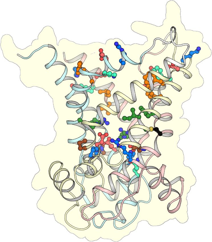
Int. J. Mol. Sci. 2020, 21, 8971                                                                    2 of 18

      Cryptosporidium parvum, in common with a range of anaerobic/microaerophilic eukaryotic lineages,
contains a mitochondrion-related organelle, the mitosome [8]. Unlike mitochondria, mitosomes do
not contain genetic information, meaning that all of the proteins are encoded by the nuclear
genome and imported into the organelle. The metabolic pathways present in mitosomes differ
from those in mitochondria. In C. parvum, the mitosome does not produce ATP through oxidative
phosphorylation, but instead, the organism relies on glycolysis or substrate-level phosphorylation
within the mitosome [8,9]. It lacks pyruvate dehydrogenase, most TCA cycle enzymes, and most of the
subunits of ATP synthase, except the α and β subunits [8,9].
      Despite major metabolic differences, parasitic protozoa are known to contain mitochondrial protein
homologues, indicative of a mitochondrial origin of the mitosome. The genome of C. parvum has been
sequenced [10] and contains eight homologues of the mitochondrial carrier family (SLC25), including a
putative orthologue of the mitochondrial ADP/ATP carrier. Mitochondrial carriers are responsible for
the transport of metabolites across the mitochondrial inner membrane, linking biochemical pathways in
the cytosol with those in the mitochondrial matrix [11–13]. In eukaryotic mitochondria, the ADP/ATP
carrier replenishes the cell with metabolic energy by importing ADP into the mitochondrion for
conversion to ATP and by exporting the synthesized ATP to the cytosol [14].
      The ADP/ATP carrier is the best-characterized member of the mitochondrial carrier family (SLC25),
and currently the only member for which structural information is available [15–17]. Structural and
functional analyses have highlighted several important features. The ADP/ATP carrier functions as a
monomer [18–22]. It has the tripartite sequence repeats typical of SLC25 members [23] and a three-fold
pseudo-symmetrical structure consisting of six transmembrane helices with a translocation pathway
through the center of the protein [24]. Each of the three domains consists of two transmembrane helices
and a short amphipathic helix in the matrix loop [15]. The odd-numbered helices H1, H3, and H5
have a sharp kink, called the Pro-kink, where a proline residue [15] or serine residue, which mimics
proline [16], is located. The proline/serine is the first residue of the signature motif Px[DE]xx[RK].
      Two salt bridge networks have been identified, one on the matrix side and one on the cytoplasmic
side of the carrier. The matrix salt bridge network, also part of the signature motif Px[DE]xx[RK],
forms in the cytoplasmic state, when the substrate binding site is open to the intermembrane
space [15,16,25]. The cytoplasmic network forms in the matrix state, when the substrate binding site
is open to the mitochondrial matrix [16,26–28]. Disruption and formation of these two networks, in
an alternating way, leads to changes in accessibility of the substrate binding site to one or the other
side of the membrane [26,28]. Glutamine braces stabilize interactions of the matrix network [16],
whereas tyrosine braces stabilize interactions of the cytoplasmic network [28]. The tyrosine brace
and cytoplasmic network residues are part of another consensus sequence [YF][DE]xx[RK] on the
even-numbered helices H2, H4, and H6 [26,28]. Beneath the networks are sets of residues that provide
a 15 Å insulation layer [13,28].
      The human and bovine isoforms of mitochondrial ADP/ATP carriers are known to have a strict
substrate specificity for ADP and ATP, and the deoxy variants [29–32]. No transport is observed
for AMP, purine nucleotides GDP or GTP, or the pyrimidine nucleotides TDP, TTP, CDP, CTP, UDP,
and UTP. A single substrate binding site has been located in the central cavity by in silico analyses,
by using chemical and distance constraints [33,34], symmetry analysis [26], and molecular dynamics
simulations [32,35,36]. The consensus site has three contact points; contact points I and III consist
of positively charged residues that bind the phosphate moieties of the nucleotide, whereas contact
point II is part of a hydrophobic pocket that binds the adenine moiety [17,33,34]. The residues of this
binding site are accessible in the cavity of the matrix and cytoplasmic state [28,37]. However, their role
in substrate binding has not been demonstrated directly by experimental or structural work.
      Here, we present a detailed functional analysis of the mitosomal ADP/ATP carrier from
Cryptosporidium parvum (CpAAC). Our results demonstrate that the parasitic carrier can carry out
ADP/ATP hetero-exchange and is inhibited by the canonical inhibitors carboxyatractyloside (CATR)
and bongkrekic acid (BKA), similar to mitochondrial ADP/ATP carriers. However, it exhibits a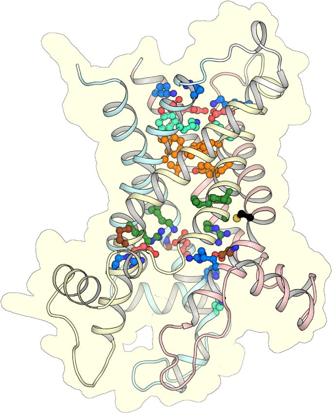
Int. J. Mol. Sci. 2020, 21, 8971                                                                      3 of 18

unique substrate selectivity profile, which includes thymidine and uridine di- and trinucleotides,
in addition to adenine nucleotides. By comparing the sequence of the C. parvum homologue
with those of mitochondrial ADP/ATP carriers, a cysteine residue is identified, which is in the
proximity of contact point II in the proposed binding site and is conserved as serine in mitochondrial
orthologues. By mutating the equivalent serine in yeast mitochondrial ADP/ATP carrier Aac2p to
cysteine, we have converted a carrier with stringent substrate specificity for ADP and ATP to one
that can transport additional nucleotides, matching the substrate specificity profile of the C. parvum
orthologue. These results advance our understanding of the metabolic processes in Cryptosporidium
and provide a molecular explanation for the differences in its substrate specificity as compared with
mitochondrial ADP/ATP carriers.

2. Results

2.1. Mitochondrial Carriers of Cryptosporidium parvum
      To identify members of the mitochondrial carrier family in Cryptosporidium parvum, BLAST searches
were carried out using the yeast mitochondrial carriers as templates. The search identified eight
putative members on the genome, which all contained the signature motifs Px[DE]xx[RK] and
tripartite sequence repeats typical of members of the mitochondrial carrier family (SLC25) [12,13,23,25].
Subsequently, we used two approaches to assign a putative function to these carriers. First, we aligned
the identified sequences to the yeast carriers to find the closest homologues. Second, we identified
the three contact points [33,34] and asymmetric residues [26] of the proposed substrate binding
site to obtain clues about their potential substrates. These analyses identified close homologues of
yeast in C. parvum, such as a putative mitosomal ADP/ATP carrier (Cgd8_1210, Q5CW91), NAD+ or
FAD carrier (Cgd1_2370, Q5CSK1), oxoglutarate/malate carrier (partial gene) (Cgd1_600, A3FQC0),
phosphate carrier (Cgd2_520, Q5CU30), Mtm1p-like carrier (cgd6_3880, Q5CWS9), Mrs3p-like carrier
(Cgd2_1030, A3FQ60), Sam5p or Mme1p-like carrier (cgd6_2350, Q5CX62), and an unknown carrier
(cgd6_2360, Q5CX61). Here, we will study, in detail, the putative mitosomal ADP/ATP carrier,
called CpAAC, and compare it with mitochondrial ADP/ATP carriers. This comparison is only possible
for this member of the mitosomal carrier complement because AAC is the best characterized member
of the SLC25 family and the only one for which atomic structures are available [15,16,37].
      The importance of ADP/ATP carriers in ATP generation means that most eukaryotes with
mitochondria possess at least one, enabling a phylogenetic analysis of a wide range of species.
C. parvum belongs to the Apicomplexa and the phylogenetic tree based on the sequences reflects
this relationship (Figure 1). The mitosomal ADP/ATP carriers of C. parvum and Cryptosporidum
ubiquitum belong to the early branch of the clade of Apicomplexa, which also contains the Hematozoa
(e.g., Plasmodium falciparum) and Coccidia (Trypanosoma gondii).

2.2. The Mitosomal ADP/ATP Carrier Retains Structural and Functional Characteristics of the
Mitochondrial AAC
     The mitosomal carrier has all of the key structural and functional features required for transport,
such as the Pro-kink [15,16], matrix salt bridge network [15,16,25], glutamine braces [16], cytoplasmic salt
bridge network [16,26–28], tyrosine braces [28] (Figures 2 and 3), as well as gate and small residues,
required for the conformational state interconversions [12,13,28].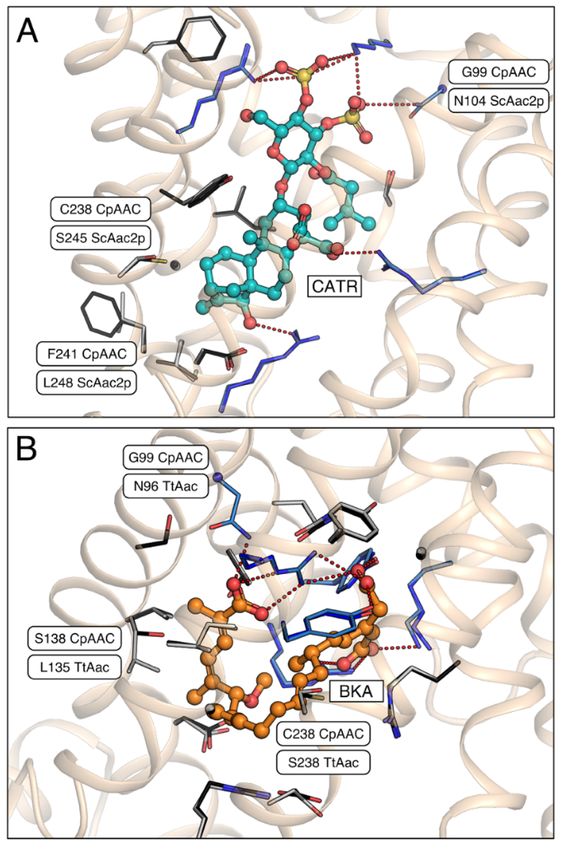
Int. J. Mol. Sci. 2020, 21, 8971                                                                                                                      4 of 18

                               Int. J. Mol. Sci. 2020, 21, x                                                                         4 of 18

                                      Figure 1. Phylogenetic relationship of mitosomal and mitochondrial ADP/ATP carriers. An unrooted
      Figure 1. Phylogenetic      relationship
                             phylogenetic              of mitosomal
                                           tree of homologous   mitochondrialand     mitochondrial
                                                                                and mitosomal               ADP/ATP
                                                                                               ADP/ATP carrier   sequences forcarriers.
                                                                                                                               a        An unrooted
                             selection of eukaryotic organisms based on alignments. The tree was calculated using RAxML. The
      phylogenetic tree ofmitosomal
                              homologous             mitochondrial and mitosomal ADP/ATP carrier sequences for a
                                         ADP/ATP carrier of the parasite Cryptosporidium parvum is in an early branch of the
      selection of eukaryotic       organisms
                             Apicomplexa,   as expectedbased       on
                                                        on the basis     alignments.
                                                                     of species phylogeny.     The tree was calculated using RAxML.
      The mitosomal ADP/ATP            carrier
                       2.2. The Mitosomal   ADP/ATP of Carrier
                                                        the parasite          Cryptosporidium
                                                                Retains Structural                     parvumof is
                                                                                    and Functional Characteristics  the in an early branch of the
                       Mitochondrial AAC
      Apicomplexa, as expected          on the basis of species phylogeny.
                                      The mitosomal carrier has all of the key structural and functional features required for transport,
                     Int. J. Mol.such
                                  Sci. 2020, 21,Pro-kink
                                        as the   x       [15,16], matrix salt bridge network [15,16,25], glutamine braces [16], cytoplasmic 5 of 18
                               salt bridge network [16,26–28], tyrosine braces [28] (Figures 2 and 3), as well as gate and small
                               residues, required for the conformational state interconversions [12,13,28].

                           Figure 2. The putative ADP/ATP carrier of C. parvum is homologous to mitochondrial ADP/ATP
      Figure 2. The carriers.
                       putative      ADP/ATP
                               Alignment   of CpAACcarrier       of C. parvum
                                                      with the mitochondrial          is homologous
                                                                                ADP/ATP     carrier of Bos taurusto   mitochondrial
                                                                                                                   (BtAAC1),   Homo       ADP/ATP
      carriers. Alignment        of CpAAC
                      sapiens (HsAAC1),            with the
                                            Saccharomyces        mitochondrial
                                                           cerevisiae                   ADP/ATPthermophila
                                                                      (ScAac2p), Thermothelomyces                       of Bosand
                                                                                                           carrier (TtAac),       taurus (BtAAC1),
                      Entamoeba histolytica (EhAAC). The transmembrane α-helices (H1-6), linker α-helices (l12, l34, l56),
      Homo sapiens (HsAAC1), Saccharomyces cerevisiae (ScAac2p), Thermothelomyces thermophila (TtAac),
                      and matrix α-helices (h12, h34, h56) are indicated. Repeats 1, 2, and 3 are colored in pastel blue, yellow,
      and Entamoeba and
                      histolytica    (EhAAC).
                           red, respectively.        The transmembrane
                                              The residues                           α-helices
                                                            of the Pro-kink (P, brown),             (H1-6),(MN,
                                                                                          matrix network         linker     α-helices
                                                                                                                      red and  blue),  (l12, l34, l56),
                      glutamine-brace   (Q, cyan), substrate binding  site (green), contact points  (black  circles),
      and matrix α-helices (h12, h34, h56) are indicated. Repeats 1, 2, and 3 are colored in pastel blue, yellow,     tyrosine-brace
                      (Y, cyan), and cytoplasmic network (CN, red and blue) are indicated.
      and red, respectively. The residues of the Pro-kink (P, brown), matrix network (MN, red and blue),
      glutamine-brace (Q, cyan), substratecytoplasmicbinding
                                                        state       site (green), contactmatrix     pointsstate(black circles), tyrosine-brace
      (Y, cyan), and cytoplasmic network (CN, red and blue) are indicated.                                               IS

                                                                                cytoplasmic network
                                                                                 tyrosine brace
                                                                                 aromatic plug

                                                                             substrate binding site                             IM
                                                                          Pro kink / matrix network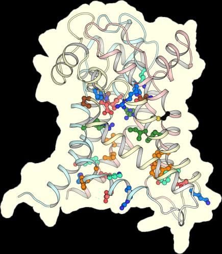
Figure 2. The putative ADP/ATP carrier of C. parvum is homologous to mitochondrial ADP/ATP
                      carriers. Alignment of CpAAC with the mitochondrial ADP/ATP carrier of Bos taurus (BtAAC1), Homo
                      sapiens (HsAAC1), Saccharomyces cerevisiae (ScAac2p), Thermothelomyces thermophila (TtAac), and
                      Entamoeba histolytica (EhAAC). The transmembrane α-helices (H1-6), linker α-helices (l12, l34, l56),
                      and matrix α-helices (h12, h34, h56) are indicated. Repeats 1, 2, and 3 are colored in pastel blue, yellow,
                      and red, respectively. The residues of the Pro-kink (P, brown), matrix network (MN, red and blue),
Int. J. Mol. Sci. 2020, 21, 8971                                                                                                    5 of 18
                      glutamine-brace (Q, cyan), substrate binding site (green), contact points (black circles), tyrosine-brace
                      (Y, cyan), and cytoplasmic network (CN, red and blue) are indicated.

                                          cytoplasmic state                                      matrix state
                                                                                                                       IS

                                                                       cytoplasmic network
                                                                        tyrosine brace
                                                                         aromatic plug

                                                                     substrate binding site                            IM

                                                                 Pro kink / matrix network
                                                                     glutamine brace

                                                                                                                      MM

       Figure 3. CpAAC has all of the key features of a functional ADP/ATP. Structural models of CpAAC, in
       the cytoplasmic state (left) and matrix state (right), based on Protein Data Bank (PDB) entries 4c9g [16]
       and 6gci [28] respectively, calculated by SWISS-MODEL [38]. Repeats 1, 2, and 3 are colored in pastel
       blue, yellow, and red, respectively. The residues of the Pro-kink (brown), matrix network (red and blue),
       glutamine-brace (cyan), substrate binding site (green), aromatic plug (orange), tyrosine-brace (cyan),
       and cytoplasmic network (red and blue) are indicated. Residue C238 in CpAAC is indicated by a red
       arrowhead. IS, intermembrane space; IM, mitochondrial inner membrane; MM, mitochondrial matrix.

     To test its ability to transport nucleotide substrates, we expressed the CpAAC carrier in the
cytoplasmic membrane of Lactococcus lactis. We have previously shown that this organism is an
excellent host for the expression of mitochondrial carrier proteins in a functional state, including the
human mitochondrial ADP/ATP carrier HsAAC1 [32,39–45]. CpAAC was expressed in the cytoplasmic
membrane to higher levels than HsAAC1 (Figure 4C), enabling further functional characterization by
transport assays (Figure 4).
     To determine the transport activity of CpAAC, lactococcal membranes expressing the carrier were
fused with liposomes to form fused vesicles. In eukaryotic cells, transport of adenine nucleotides by
the mitochondrial ADP/ATP carriers is driven by the chemical gradients of the substrates and the
membrane potential, which also determines the directionality [14]. However, the transport activity of
these carriers is fully reversible, and they can catalyze both hetero-exchange and homo-exchange of
adenine nucleotides in vitro [14,17]. Therefore, to assess transport activity, we extruded the vesicles in
the presence of 5 mM unlabeled ADP to incorporate ADP inside the fused vesicles, removed external
nucleotide by gel filtration, and initiated homo-exchange with the addition of 1.5 µM [14 C]-ADP
on the outside. CpAAC exhibited high rates of ADP homo-exchange, which could be inhibited by
the canonical inhibitors carboxyatractyloside (CATR) and bongkrekic acid (BKA) of mitochondrial
ADP/ATP carriers (Figure 4A,B).
     The structures of both the bovine and yeast isoforms in the cytoplasmic state reveal the basis
of CATR inhibition [15,16]. Of the thirteen residues involved in binding of CATR, ten are strictly
conserved in CpAAC (Figure 5A). Residue N104 in ScAac2p, which is replaced by a glycine residue in
CpAAC (G99), might form a hydrogen bond with the sulphate moiety of CATR, but it is not consistently
observed in the available structures (see [16] for an analysis) (Figure 5A). Two other residues, which are
involved in weak van der Waals interactions, have been replaced in CpAAC, but they are unlikely to
interfere with binding (Figure 5A). In agreement with these observations, CATR inhibition of ADP
homo-exchange by CpAAC is nearly complete (Figure 4B).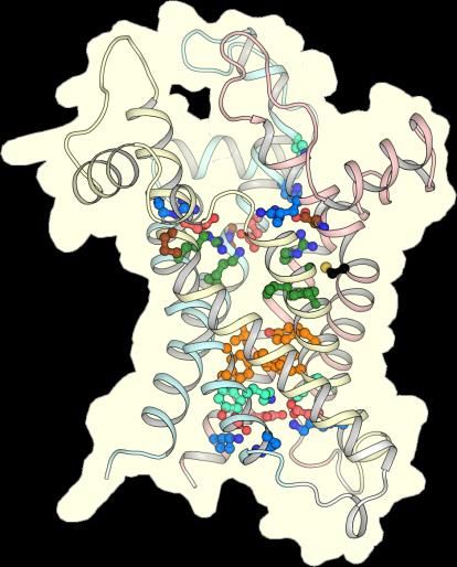
To test its ability to transport nucleotide substrates, we expressed the CpAAC carrier in the
cytoplasmic membrane of Lactococcus lactis. We have previously shown that this organism is an
excellent host for the expression of mitochondrial carrier proteins in a functional state, including the
human mitochondrial ADP/ATP carrier HsAAC1 [32,39–45]. CpAAC was expressed in the
cytoplasmic membrane to higher levels than HsAAC1 (Figure 4C), enabling further functional
Int. J. Mol. Sci. 2020, 21, 8971 6 of 18
characterization by transport assays (Figure 4).
Figure 4. CpAAC
CpAAC expressed
expressed in in Lactococcus
Lactococcus lactis
lactis retains
retains functional
functional characteristics
characteristics of the
the mitochondrial
mitochondrial
ADP/ATP carriers.(A)
ADP/ATP carriers. (A)ADP
ADPtransport
transportbybyCpAAC
CpAAC can
can be be inhibited
inhibited by by carboxyatractyloside
carboxyatractyloside (CATR)
(CATR) and
bongkrekic
and acid (BKA),
bongkrekic the canonical
acid (BKA), inhibitors
the canonical of mitochondrial
inhibitors AACs. Fused
of mitochondrial AACs. membrane vesicles of
Fused membrane
lactococcal
vesicles membranes
of lactococcal expressingexpressing
membranes CpAAC were CpAAC preloaded with 5 mMwith
were preloaded ADP in the
5 mM ADPabsence
in the(squares)
absence
or presence
(squares) or of 20 µM of
presence CATR20 μM (triangles) or BKA (circles).
CATR (triangles) or BKAThe emptyThe
(circles). vector
empty controls
vectorare shownare
controls as
invertedastriangles. 14
shown inverted Transport
triangles. was initiated
Transport by initiated
was the addition of 1.5
by the µM [ C]-ADP;
addition of 1.5 μM (B)[ The
14 specific
C]-ADP; (B)initial
The
transportinitial
specific rates, when the applied
transport chemical
rates, when thegradients
applied of radio-labeled
chemical and cold
gradients substrates areand
of radio-labeled maximal,
cold
are derivedare
substrates from the linear
maximal, are parts
derived of the uptake
from curves,
the linear typically
parts of the in the first
uptake minute,
curves, corrected
typically forfirst
in the the
amount corrected
minute, of protein.for Thethedata are represented
amount of protein. byThe thedata
meanareand standard by
represented deviation
the mean of two
andbiological
standard
repeats, each
deviation performed
of two biologicalinrepeats,
quadruplicate; (C) Western
each performed blot of cytoplasmic
in quadruplicate; (C) Westernmembranes of L. lactis
blot of cytoplasmic
strains expressing CpAAC or human AAC1 (HsAAC1), which was characterized previously by [32],
or transformed with empty vector. The band of CpAAC (approximately 33 kDa) was detected with
a chicken antibody raised against the antigen YPLDTVRRRMMMT and anti-chicken-horseradish
peroxidase conjugate. It is marked with an arrow. 10 µg of total protein was loaded per lane.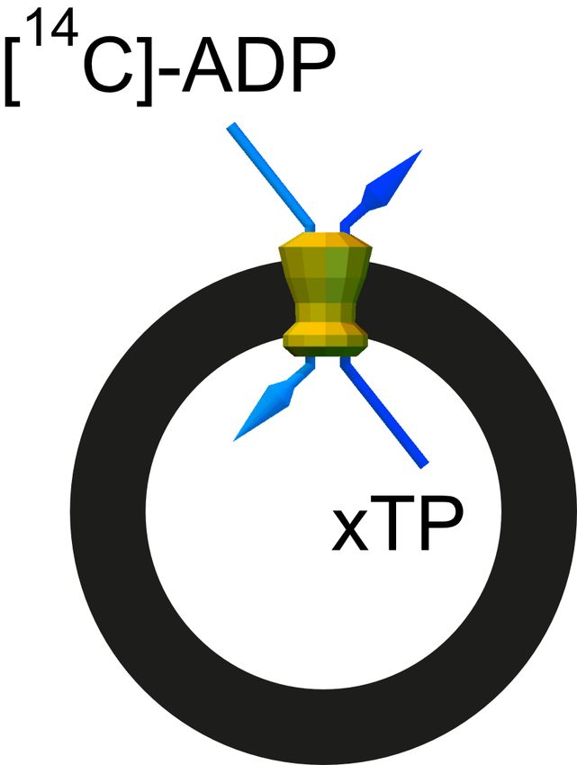
CATR inhibition [15,16]. Of the thirteen residues involved in binding of CATR, ten are strictly
          conserved in CpAAC (Figure 5A). Residue N104 in ScAac2p, which is replaced by a glycine residue
          in CpAAC (G99), might form a hydrogen bond with the sulphate moiety of CATR, but it is not
          consistently observed in the available structures (see [16] for an analysis) (Figure 5A). Two other
          residues, which are involved in weak van der Waals interactions, have been replaced in CpAAC, but
Int. J. Mol. Sci.are
          they    2020, 21, 8971to interfere with binding (Figure 5A). In agreement with these observations, CATR
                     unlikely                                                                                       7 of 18
          inhibition of ADP homo-exchange by CpAAC is nearly complete (Figure 4B).

     Figure Figure  5. Carboxyatractyloside and bongkrekic acid binding to CpAAC. (A) Bound inhibitor
              5. Carboxyatractyloside      and bongkrekic acid binding to CpAAC. (A) Bound inhibitor
             carboxyatractyloside (CATR, teal) in the structure of ScAac2p (PDB entry 4c9g) [16] with the
     carboxyatractyloside (CATR, teal) in the structure of ScAac2p (PDB entry 4c9g) [16] with the
     superimposed structural model of CpAAC, which was determined by SWISS-MODEL [38] using the
     ScAac2p structure (PDB entry 4c9g) as a template, following the alignment in Figure 2; (B) Bound
     inhibitor bongkrekic acid (BKA, orange) in the structure of TtAac (PDB entry 6gci) [28] with
     superimposed structural model of CpAAC, based on TtAac (PDB entry 6gci). All carriers are
     shown as a wheat cartoon with the inhibitors in ball-and-stick representations. Amino acid residues
     that form salt bridges or hydrogen bonds, or van der Waals interactions with the inhibitors, are shown
     in blue or grey carbon atoms stick representations, respectively. Interacting residues that are different
     between the structures and the CpAAC models are labeled.

     Comparison of the model of CpAAC with the structure of the ADP/ATP carrier from
Thermothelomyces thermophila (TtAac) in complex with bongkrekic acid (BKA) [28,37] shows that
all polar interactions are fully conserved, except for N96 in TtAac, which is G99 in CpAAC (Figure 5B).
Two residues L135 and S238 in TtAac are involved in van der Waals interactions with BKA, but they
are different in CpAAC, being S138 and C238, respectively. Transport by CpAAC is inhibited to 83%
by bongkrekic acid, which could be explained by these substitutions interfering to a small extend with
the binding of BKA (Figure 4B).

2.3. The Mitosomal ADP/ATP Carrier from Cryptosporidium parvum Has a Broad Nucleotide Specificity Profile
      Human and bovine ADP/ATP carriers have a strict substrate specificity for ADP, ATP and their
deoxy variants [29–32]. Here, we tested whether the substrate specificity of CpAAC was similar to
that of mitochondrial ADP/ATP carriers. We determined the substrate specificity using two methods.
First, competition experiments were carried out, in which radiolabeled ADP uptake into vesicles
was monitored in the presence of a 1667-fold excess of external non-labeled nucleotides (Figure 6).
There was almost complete inhibition of ADP homo-exchange by CpAAC by a broad range of di-and
trinucleotides, but not mononucleotides, except for AMP (Figure 6). However, these experiments show
that these nucleotides can compete with ADP for binding to some extent, when present in large excess,
but they do not demonstrate that these nucleotides are transported by CpAAC.
First, competition experiments were carried out, in which radiolabeled ADP uptake into vesicles was
monitored in the presence of a 1667-fold excess of external non-labeled nucleotides (Figure 6). There
was almost complete inhibition of ADP homo-exchange by CpAAC by a broad range of di-and
trinucleotides, but not mononucleotides, except for AMP (Figure 6). However, these experiments
show thatSci.
Int. J. Mol. these
2020,nucleotides
21, 8971 can compete with ADP for binding to some extent, when present in large
8 of 18
excess, but they do not demonstrate that these nucleotides are transported by CpAAC.
Figure
Figure6.6.Nucleotide
Nucleotidecompetition
competition experiments
experiments of ADP/ADP homo-exchange
of ADP/ADP homo-exchangeby byCpAAC.
CpAAC.Percentage
Percentageof
ofinhibition
inhibition of
14 [ 14C]-ADP/ADP homo-exchange in the presence of 2.5 mM of non-radiolabeled
of [ C]-ADP/ADP homo-exchange in the presence of 2.5 mM of non-radiolabeled nucleotides
nucleotides (1667-fold
(1667-fold excess). excess). Transport
Transport wasby
was initiated initiated by the of
the addition addition
1.5 µMof[14
1.5 μM [14C]-ADP
C]-ADP to fusedtovesicles,
fused
vesicles,
preloaded preloaded
with 5 mM withnon-radiolabeled
5 mM non-radiolabeled
ADP. The ADP.
dataThe
are data are represented
represented by the
by the mean andmean and
standard
standard
deviationdeviation of two biological
of two biological repeats,
repeats, each each performed
performed in quadruplicate.
in quadruplicate.
Second, we performed hetero-exchange experiments with all compounds that effectively competed
for ADP uptake to verify whether they were transported by CpAAC (Figure 7A). For this purpose,
non-labeled compounds were loaded into fused membrane vesicles, and hetero-exchange was initiated
by the addition of radio-labeled ADP and the transport rate was monitored (see inset Figure 7).
Only when the internal compound is transported out can uptake of the radiolabeled ADP into the fused
vesicles occur. The initial rates of ADP and ATP uptake by CpAAC were 1.71 ± 0.49 and 2.25 ± 1.45 nmol
[14 C]-ADP mg−1 total protein min−1 , respectively, as compared with the rates of TDP, TTP, UDP, and
UTP uptake by CpAAC which were 3.15 ± 1.90, 3.62 ± 1.58, 2.52 ± 0.28, and 4.12 ± 2.00 nmol [14 C]-ADP
mg−1 total protein min−1 , respectively. Hetero-exchange was also observed for inosine and cytosine di-
and trinucleotides, but at much lower rates (0.76 ± 0.15, 1.21 ± 0.38, 1.04 ± 0.32, and 1.09 ± 0.29 nmol
[14 C]-ADP mg−1 total protein min−1 for IDP, ITP, CDP, and CTP, respectively). The rate of transport for
guanosine nucleotides was low, despite near complete inhibition in the competition assays, suggesting
these nucleotides can compete for substrate binding when present in large excess, but they are
not transported.
For comparison, we tested the substrate specificity of ScAac2p, for which a structure is available [16],
in the same way. Indeed, ScAac2p shares the substrate specificity profile of the human and bovine
mitochondrial ADP/ATP carriers, showing the highest rates of transport for ADP, ATP, dADP, and dATP,
but no transport for TDP, TTP, UDP, and UTP (Figure 7B). These data show that CpAAC has a broader
substrate specificity as compared with ScAac2p and previously characterized orthologues [31,32].and 2.25 ± 1.45 nmol [14C]-ADP mg−1 total protein min−1, respectively, as compared with the rates of
TDP, TTP, UDP, and UTP uptake by CpAAC which were 3.15 ± 1.90, 3.62 ± 1.58, 2.52 ± 0.28, and 4.12
± 2.00 nmol [14C]-ADP mg−1 total protein min−1, respectively. Hetero-exchange was also observed for
inosine and cytosine di- and trinucleotides, but at much lower rates (0.76 ± 0.15, 1.21 ± 0.38, 1.04 ±
0.32, and 1.09 ± 0.29 nmol [14C]-ADP mg−1 total protein min−1 for IDP, ITP, CDP, and CTP,
respectively). The rate of transport for guanosine nucleotides was low, despite near complete
Int. J. Mol. Sci. 2020, 21, 8971 9 of 18
inhibition in the competition assays, suggesting these nucleotides can compete for substrate binding
when present in large excess, but they are not transported.
CpAAC
6.0 A
Initial uptake rate (nmol [14C]-ADP mg-1 of total protein min-1)
5.0
4.0
3.0
2.0
1.0
0.0
ATP
dATP
CTP
UTP
ADP
AMP
dADP
CDP
GTP
GDP
ITP
IDP
TTP
TDP
UDP
ScAac2p
6.0 B
5.0
4.0
3.0
2.0
1.0
0.0
ATP
ADP
AMP
dATP
dADP
CTP
CDP
GTP
GDP
ITP
IDP
TTP
TDP
UTP
UDP
Figure 7. CpAAC
Figure 7. CpAAC has ahas a broadersubstrate
broader substrate specificity than thethan
specificity yeast ADP/ATP
the yeastcarrier ScAac2p. Fused
ADP/ATP carrier ScAac2p.
lactococcal membranes expressing (A) CpAAC or (B) ScAac2p were loaded with 2.5 mM of the
Fused lactococcal membranes expressing (A) CpAAC or (B) ScAac2p were loaded with 2.5 mM of the
indicated substrate (X), and hetero-exchange was initiated by the addition of 1.5 μM [14C]-ADP on the
indicated substrate
outside (see(X), andThe
insets). hetero-exchange wasrates
initial specific transport initiated by the addition
were determined of 1.5
over the linear partµM [14 C]-ADP on
of the
the outside (see insets). The initial specific transport rates were determined over the linear part of the
uptake curve, typically in the first 60 s. Data are represented by the mean and standard deviation of
initial hetero-exchange rates of two biological repeats with six technical repeats in total.
uptake curve, typically in the first 60 s. Data are represented by the mean and standard deviation of
initial hetero-exchange rates of two biological repeats with six technical repeats in total.
2.4. A Single Residue in the Substrate Translocation Pathway Can Broaden the Substrate Selectivity of the Yeast
ADP/ATP Carrier
To investigate the molecular basis of the observed substrate specificity of CpAAC, we compared
the sequences of ADP/ATP carriers that had a strict specificity for ADP and ATP with the one of
C. parvum (Figure 2). The only difference in the residues predicted to be near the putative adenine
binding site is C238 in C. parvum, which is a serine residue in the yeast (S245), bovine (S227) and human
(S228) mitochondrial orthologs. In fact, a cysteine residue in that position is only observed in C. parvum
and other cryptosporidia, and in the mitosomal ADP/ATP carrier of Entamoeba histolytica.
Next, we mutated serine 245 of ScAac2p to cysteine, as found in C. parvum, to analyze its effect
on the substrate specificity of the carrier. To get a more complete picture, we also introduced other
substitutions, such as the hydrophobic amino acid residues alanine, isoleucine, leucine, and valine and
the polar amino acid residue threonine, which has similar properties as serine. To test the substrate
specificity of each mutant variant in hetero-exchange we loaded, in each case, fused membrane vesicles
with ATP, CTP, GTP, ITP, TTP, or UTP and initiated transport by the addition of radio-labeled ADP
(Figure 8). All mutants were capable of ADP/ATP hetero-exchange; the rates in µmol [14 C]-ADP mg−1
of AAC min−1 for S245A (0.47 ± 0.06) and S245C (0.53 ± 0.03) were the same as wild-type (0.53 ± 0.02),
the rates for S245I (0.72 ± 0.07) and S245L (0.67 ± 0.13) were slightly higher, and the rates for S245T
(0.29 ± 0.02) and S245V (0.37 ± 0.11) were slightly lower than the wild-type. For all mutants, the rates
for CTP, GTP, and ITP hetero-exchange were either similar, or slightly lower than the wild-type but
overall, very low. However, there was a significant increase in the rate of ADP/TTP and ADP/UTPInt. J. Mol. Sci. 2020, 21, 8971 10 of 18
hetero-exchange for the S245C, S245I, and S245L variants as compared with the wild-type. The rate
of ADP/TTP hetero-exchange in µmol [14 C]-ADP mg−1 of AAC min−1 for ScAac2p was 0.08 ± 0.02,
whereas for S245L, the transport rates were seven-times higher than the wild-type (0.56 ± 0.22),
the highest observed. Similarly, the rate of ADP/UTP hetero-exchange in µmol [14 C]-ADP mg−1 of
AAC min−1 for S245L was more than four-times higher than that of the wild-type (0.45 ± 0.17 for
S245L versus 0.10 ± 0.06). Interestingly, the rates of ADP/CTP hetero-exchange for S245C (0.06 ± 0.02),
S245I (0.08 ± 0.03) and S245L (0.14 ± 0.05) were not markedly different from the wild-type (0.11 ± 0.02),
despite the structural similarity between these pyrimidine nucleotides. These data indicate that the
amino acid residue in position 238 in CpAAC and 245 in ScAac2p is critical for determining the
substrate specificity of the carrier, suggesting it might have a role in nucleotide base selectivity.
Initial uptake rate (µmol [14C]-ADP mg-1 of AAC min-1)
1.0 ScAac2p ScAac2p
ScAac2p S245A ScAac2p S245C
0.8
0.6
0.4
0.2
0.0
1.0 ScAac2p ScAac2p
ScAac2p S245L ScAac2p S245T
0.8
0.6
0.4
0.2
0.0
ATP CTP GTP ITP TTP UTP ATP CTP GTP ITP TTP UTP
Figure 8. Nucleotide hetero-exchange by wild-type and S245 mutants of ScAac2p. Vesicles of lactococcal
membranes expressing wild-type ScAac2p (black bars) or mutant carriers at position S245 (white bars)
were loaded with 2.5 mM ATP, CTP, GTP, ITP, TTP, or UTP, and transport was initiated with the external
addition of 1.5 µM [14 C]-ADP. The specific initial transport rates were determined over the linear part
of the uptake curve, typically in the first 60 s. Data are represented by the mean and standard deviation
of two biological repeats with six technical repeats in total.
3. Discussion
We have used bioinformatics to identify putative mitochondrial carriers encoded by the genome
of the parasitic protist Cryptosporidium parvum. Whereas most eukaryotes have between 30 and
60 mitochondrial carriers [46], C. parvum has only eight, in agreement with the highly reduced
functions of the mitosome. A similar reduction in the mitochondrial carrier complement has beenInt. J. Mol. Sci. 2020, 21, 8971 11 of 18
observed previously for other eukaryotic parasites, which occurred in adaptation to their host cell
exploitation [43,47]. One potential reason is that parasites can acquire nutrients from the host
cells, meaning that many synthetic pathways and accompanying transport steps become redundant,
leading to gene loss and a more reduced genome.
In this study, we show that the genome of C. parvum encodes a mitosomal ADP/ATP carrier protein
(CpAAC), phylogenetically related to mitochondrial ADP/ATP carriers, but with a broad substrate
specificity. We have previously characterized the substrate specificity of the human mitochondrial
ADP/ATP carrier [32], the mitosomal ADP/ATP carrier of the microaerophilic human parasite Entamoeba
histolytica (EhAAC) [43], and here, the yeast mitochondrial ADP/ATP carrier ScAac2p (Figure 7B).
These analyses are consistent with previous studies on ADP/ATP carriers from other species, showing
that the substrate specificity is confined to ADP and ATP, and their deoxy variants [29–31]. In contrast,
CpAAC has gained the ability to transport a broader range of di- and tri-nucleotides, i.e., ADP, ATP,
dADP, and dATP, but also TDP, TTP, UDP, and UTP (Figure 7A), which is unprecedented. CpAAC is
much more closely related to mitochondrial ADP/ATP carriers with respect to the conserved structural
and functional features than the mitosomal carrier of Entamoeba histolytica EhAAC [43], which is much
more divergent (Figure 2). Although EhAAC also has a cysteine residue in a similar position as C238
of CpAAC, the rest of the putative binding site is not conserved, making it difficult to rationalize the
differences in specificity (Figure 2). EhAAC is much smaller in size, has unusually short loops, and has
substantial differences in other conserved features of mitochondrial ADP/ATP carriers, such as the
networks and braces (Figure 2).
The differences between CpAAC and other characterized mitochondrial ADP/ATP carriers provide
new insights into the molecular determinants of substrate binding and transport. One way of explaining
these differences is by carrying out docking studies or molecular dynamics simulations, but there is a
fundamental problem. We have analyzed the energetics of the transport cycle by treating the ADP/ATP
carrier as a nanomachine that moves stochastically and continuously from the matrix to cytoplasmic
conformation under the influence of thermal energy [48]. This analysis shows that the tightest binding
of the substrate occurs in the occluded state [48], which is a defined state [49], but its structure has not
been determined.
Still, it should be possible to compare the properties of the substrate binding site with those of the
substrates, in particular, the nucleotide bases. We had previously proposed a putative binding site
using a novel binding site search procedure [33,34], which has been confirmed by other bioinformatic
studies and molecular dynamics simulations [26,32,35,36]. The proposed binding site in the yeast
ADP/ATP carrier ScAac2p consists of positively charged residues K38, R96, and R294, which may
form ionic interactions with the phosphate moieties of ADP and ATP [33,34] (Figure 9A). It contains a
tyrosine residue Y203, which may form aromatic stacking arrangements with the adenine ring [32–35].
Residue Y203 is flanked by residues G199, I200, which have been implicated in substrate binding
previously [33,34], as well as S245, and S242, which are in the vicinity (Figure 9A). Together, these five
residues form a fairly large hydrophobic pocket, which could function as an adenine binding site
(Figure 9A). What about the properties of the substrates? Electrostatic surface calculations of the
nucleotide bases have shown that cytosine, guanine, and to a lesser extent inosine are quite polar
because of strong partial negative charges at the ketone groups [50]. Adenine, thymidine, and uridine
have a more even electron distribution and are consequently less polar [50].
What can we learn about the substrate specificity by comparing the properties of the substrate
binding sites of the single replacement mutants (Figure 8) and those of the nucleotide bases? First,
the nucleotide hetero-exchange experiments clearly show that S245 is not essential for binding of
adenine nucleotides, as the replacement mutants can still transport adenine nucleotides with similar
rates. This result indicates that the hydroxyl group of S245 is not directly involved in adenine binding,
but this residue must still be involved in substrate selectivity, given the specificity broadening observed
in CpAAC (Figure 7A).Int. J. Mol. Sci. 2020, 21, 8971 12 of 18
Second, substitutions introducing a larger and more hydrophobic group than S245 into the putative
adenine binding pocket of ScAac2p, such as S245C, S245I, and S245L (Figure 9C–E), enable transport of
uridine and thymidine nucleotides (Figure 8). The pyrimidine bases, thymidine and uridine, are smaller
than adenosine and, consequently, might fit loosely in the adenine binding pocket of the wild-type
Int. J. Mol. Sci. 2020, 21, x 12 of 18
yeast protein. The substitutions may make the binding of uridine and thymidine bases possible by a
matrix
better fit, which to cytoplasmic
still allows adenine conformation under because
to bind, the influence of thermal
they are allenergy [48]. This analysis
hydrophobic shows Cytosine is
in nature.
that the tightest binding of the substrate occurs in the occluded state [48], which is a defined state
also smaller than adenine, but more polar than the other pyrimidine bases, and will not be tolerated
[49], but its structure has not been determined.
well in this hydrophobic
Still, it shouldsite. The purine
be possible bases,
to compare the guanine and
properties of the inosine, are bigger
substrate binding and
site with more
those of polar than
adenine and the
aresubstrates,
not tolerated in particular,
in thethe nucleotide
largely bases. We hadbinding
hydrophobic previously proposedThe
pocket. a putative
S245Tbinding
mutation leads to
site using a novel binding site search procedure [33,34], which has been confirmed by other
the introduction of a larger
bioinformatic studiespolar group indynamics
and molecular the hydrophobic adenine binding
simulations [26,32,35,36]. pocket
The proposed (Figure
binding site 9F), and to
a reduction in ADP/ATP
in the yeast ADP/ATP hetero-exchange
carrier ScAac2p (Figure
consists of8), potentially
positively chargedbecause of less
residues K38, effective
R96, and R294, binding of
which mayAs
the adenine moiety. form ionic interactions
expected, with the phosphate
this substitution moieties
does of ADP andefficient
not promote ATP [33,34] (Figure 9A).
binding of uridine and
It contains a tyrosine residue Y203, which may form aromatic stacking arrangements with the adenine
thymidine nucleotides.
ring [32–35]. Residue Y203 is flanked by residues G199, I200, which have been implicated in substrate a significant
The smaller substitutions, S245V and S245A, do not introduce
change in properties of the binding
binding previously pocket
[33,34], as well (Figure
as S245, 9B,G)
and S242, whichand their
are in substrate
the vicinity (Figurespecificities
9A). Together,are similar to
these five
that of wild-type residues
(Figure 8).form
Thus,a fairly
thelarge hydrophobic pocket,
hydrophobicity andwhich
size ofcould
thefunction
adenine as an adenine site are two
binding
binding site (Figure 9A). What about the properties of the substrates? Electrostatic surface
key factors that work oftogether
calculations to determine
the nucleotide bases have shownthe that
specificity of nucleotide
cytosine, guanine, base
and to a lesser selection
extent inosine rather than
specific interactions.
are quite polar because of strong partial negative charges at the ketone groups [50]. Adenine,
thymidine, and uridine have a more even electron distribution and are consequently less polar [50].
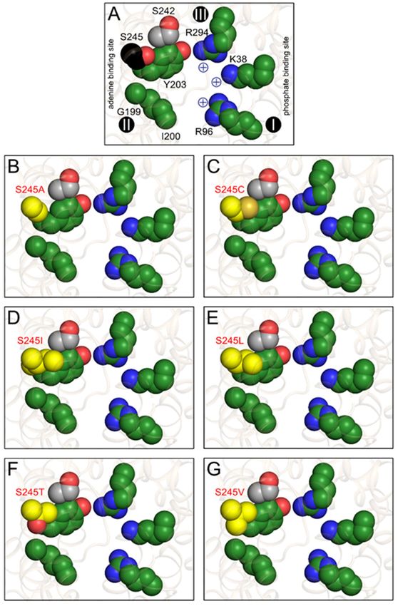
Figure 9. S245 substitutions change the properties of the putative substrate binding pocket. (A)
Figure 9. S245 substitutions change the properties of the putative substrate binding pocket.
Consensus substrate binding site of the yeast ADP/ATP carrier ScAac2p (PDB entry 4c9g) based on
(A) Consensus substrate analysis
computational binding site of the
[16,17,28,37]. yeast
The ADP/ATP
putative carriersite
adenine binding ScAac2p (PDB
(G199, I200, and entry
Y203) and4c9g) based on
computational positively
analysischarged residues (K38, R96,
[16,17,28,37]. Theand R294) involved
putative in binding
adenine of the phosphate
binding site (G199, moieties
I200,of the
and Y203) and
nucleotides are shown in green, whereas S242 and S245, which are close to this site, are shown in grey
positively charged residues (K38, R96, and R294) involved in binding of the phosphate moieties of
the nucleotides are shown in green, whereas S242 and S245, which are close to this site, are shown in
grey and black, respectively. The contact points I, II, an III on transmembrane helices H2, H4, and H6,
respectively, are also indicated as black spheres with roman numerals [33,34]. Putative substrate
binding site in (A) wild-type; (B) S245A; (C) S245C; (D) S245I; (E) S245L; (F) S245T; and (G) S245V with
the substitutions, shown in yellow.Int. J. Mol. Sci. 2020, 21, 8971 13 of 18
Overall, our findings provide new insights into the substrate specificity of the ADP/ATP carriers,
as well as potential metabolic functions of Cryptosporidium parvum. Interestingly, the serine at position
245 is strictly conserved among mitochondrial ADP/ATP carriers. These results may explain why
there is a strong selection against mutations at this position. If other nucleotides are transported as
well, key processes, such as equimolar exchange of ADP/ATP, as well as nucleotide transport for DNA
and RNA synthesis could become deregulated in mitochondria. These processes do not occur in the
mitosome of C. parvum, and thus there is no selective pressure against broadening of the substrate
specificity. Given the absence of oxidative phosphorylation in C. parvum, it is likely that CpAAC is
responsible for importing ATP into the mitosome for conserved, energy-requiring processes, such as
iron-sulphur cluster synthesis [51–53]. The broader substrate specificity of the C. parvum carrier is
intriguing, as the parasite does not have mitochondrial DNA requiring nucleotide import for synthesis
and transcription. However, mitosomal function may also require the import of a broad range of
nucleotides, given that C. parvum relies on salvage pathways for supply of purine and pyrimidine
nucleotides [54]. In any case, iron-sulphur cluster synthesis is an essential process in eukaryotes, which
requires the import of ATP into the mitosome, and thus CpAAC might be an interesting drug target for
treatment of cryptosporidiosis.
4. Materials and Methods
4.1. In Silico Analysis
To identify the sequences of mitochondrial carriers from Cryptosporidium parvum, BLAST searches
of the genome sequence stored at the National Centre for Biotechnology Information databases
were performed. Protein sequences for the yeast mitochondrial carriers and the ADP/ATP carrier
of Entamoeba histolytica were used as search templates, using an expected threshold of 1. Identified
sequences were aligned with CLUSTALW [55], redundancy was removed by CD-HIT [56], and the
sequences were curated manually. BLAST searches of the putative carrier proteins to the yeast genome
allowed us to predict their most likely function. ADP/ATP carrier sequences for a range of different
eukaryotic species were gathered using NCBI BLAST and Uniprot. Jalview was used to align the
sequences. The loop regions of the carriers were removed by using the location of the loops in the known
structures (PDB entries 4c9g [16] and 6gci [28]). Once the helical regions were isolated, the alignment
was stored as a Phylip file. The phylogenetic tree was generated with RAxML, using a maximum
likelihood method [57]. The LG matrix was used with a gamma model of heterogeneity. The tree was
visualized using the Interactive Tree of Life browser (https://itol.embl.de). Structural models of CpAAC
in the cytoplasmic state and matrix state, based on PDB entries 4c9g [16] and 6gci [28], were calculated
by SWISS-MODEL, following the alignment, as shown in Figure 2 [38].
4.2. DNA Constructs and Mutagenesis
The Cryptosporidium parvum CpAAC gene was codon-optimized, synthesized (GenScript,
Piscataway, NJ, USA), and cloned into the Lactococcus lactis expression vector pNZ8048 under the
control of a nisin A-inducible promoter. The expression strains for the human ADP/ATP carrier
HsAAC1 [32] and yeast ADP/ATP carrier [39,41] have been described previously. L. lactis strain NZ9000
was transformed with the resulting plasmid by electroporation [45]. Mutagenesis was performed
using standard PCR procedures. Plasmids were isolated by miniprep (Qiagen, Hilden, Germany),
according to the instructions with one alteration; 10 mg mL−1 lysozyme was added to the lysis buffer
and the resuspended cells were incubated at 55 ◦ C for 20 min prior to lysis. Gene insertions were
confirmed by sequencing.
4.3. Growth of Lactococcus lactis and Membrane Isolation
Precultures of L. lactis were obtained by inoculating M17 medium supplemented with 1% (w/v)
glucose and 5 µg mL−1 chloramphenicol from glycerol stocks and incubating the cultures overnightInt. J. Mol. Sci. 2020, 21, 8971 14 of 18
at 30 ◦ C with no aeration. Cells were diluted to a starting OD600 of 0.1 in fresh medium and grown
at 30 ◦ C with no aeration until the OD600 reached 0.5. Expression of the recombinant proteins was
induced by addition of nisin A with a dilution of 1:10,000 of spent M17 medium from nisin A-excreting
L. lactis strain NZ9700. Cells were grown for a further 2 h at 30 ◦ C, harvested by centrifugation
(6,000× g, 10 min, 4 ◦ C), resuspended in 10 mM PIPES pH 7.0, 50 mM NaCl (pH 7) (PIPES buffer),
and collected by centrifugation as before. Cells were subsequently resuspended in 50 mL PIPES buffer
and disrupted mechanically with a cell disruptor (Constant Cell Disruption Systems, Daventry, UK)
at 30 kpsi. Whole cells and debris were removed by centrifugation (10,800× g, 15 min, 4 ◦ C) and the
membranes were collected by ultracentrifugation (138,000× g, 1 h, 4 ◦ C). Pellets were resuspended in
PIPES buffer and stored in liquid nitrogen.
4.4. SDS-PAGE and Western Blotting
The membrane proteins were separated by SDS-PAGE (4–20% tris-glycine gel, Novex,
ThermoFisher, Waltham, MA, USA) and stained using Instant Blue (Expedeon, Cambridge, UK).
For Western blotting, the proteins were transferred to PVDF membranes using semi-dry transfer
apparatus, and then blocked in buffer containing 5% milk powder and 0.4% Tween 20, for one hour, at
room temperature. The membranes were subsequently probed with an anti-ADP/ATP carrier antibody
raised in chicken (antigen: YPLDTVRRRMMMT) at 1:20,000 dilution for one hour, followed by an
anti-chicken-HRP conjugate at 1:20,000 dilution for another hour. The membrane was developed using
Amersham ECL Western blotting detection system (Little Chalfont, Buckinghamshire, UK) for 30 min.
4.5. Membrane Vesicle Fusions
Escherichia coli polar lipid extract and egg yolk phosphatidylcholine (25 mg mL−1 in chloroform
from Avanti Polar Lipids, Alabaster, AL, USA) were mixed in a mass ratio of 3:1. The chloroform was
evaporated under a stream of nitrogen and the lipids were washed with an equal volume of diethyl
ether. When dry, the lipid mixture was resuspended in PIPES buffer with a homogenizer to a final
concentration of 20 mg mL−1 and frozen in liquid nitrogen. To make membrane fusions, 1 mg L. lactis
membranes was mixed with 5 mg liposomes (extruded 11 times through a 1 µm polycarbonate filter),
diluted to a final volume of 1 mL with PIPES buffer, and fused by seven cycles of freezing in liquid
nitrogen and thawing at room temperature, before storage in liquid nitrogen.
4.6. Transport Assays
On the day of the experiment, 1 mL of membrane vesicle fusions were thawed, and 100
µL PIPES with 55 mM substrate was added. Vesicles were extruded 11 times through a 1 µm
polycarbonate filter, passed through a pre-equilibrated PD10 column to remove external substrate,
and collected in PIPES buffer. Transport assays were carried out using a Hamilton MicroLab Star
robot (Hamilton Robotics Ltd., Birmingham, UK). Transport of [14 C]-labeled ADP was initiated by
the addition of 100 µL PIPES buffer with 1.5 µM [14 C]-ADP (2.22 GBq mmol−1 ) to 5 µg vesicles in a
MultiScreenHTS-HA 96-well filter plate (pore size 0.45 µm, Millipore, Merck, Darmstadt, Germany),
and stopped at 0 s, 10 s, 20 s, 30 s, 45 s, 60 s, 150 s, 5 min, 7.5 min, 10 min, and 15 min by the addition
of 200 µL ice-cold PIPES buffer and filtration, followed by two additional wash steps with 200 µL
ice-cold PIPES buffer. Levels of radioactivity in the vesicles were measured by the addition of 200 µL
MicroScint-20 (Perkin Elmer, Waltham, MA, USA) and by quantifying the amount of radioactivity with
a TopCount scintillation counter (Perkin Elmer). Initial rates were determined from the linear part of
the uptake curves, typically the first 60 s.
Author Contributions: Conceptualization, M.S.K., S.T., J.M. and E.R.S.K.; methodology, M.S.K., S.T., V.M., A.C.K.
and J.M.; validation, M.S.K., S.T., V.M., A.C.K., J.M. and E.R.S.K.; formal analysis, M.S.K., S.T., V.M., A.C.K., J.M.
and E.R.S.K.; investigation, M.S.K., S.T., V.M., A.C.K., J.M. and E.R.S.K.; resources, M.S.K., S.T. and E.R.S.K.;
data curation, M.S.K., S.T., V.M., A.C.K., J.M. and E.R.S.K.; writing—original draft preparation, M.S.K., S.T. and
E.R.S.K.; writing—review and editing, M.S.K., S.T., V.M., A.C.K., J.M. and E.R.S.K.; visualization, M.S.K., S.T.,Int. J. Mol. Sci. 2020, 21, 8971 15 of 18
A.C.K. and E.R.S.K.; supervision, S.T and E.R.S.K.; project administration, E.R.S.K.; funding acquisition, E.R.S.K.
All authors have read and agreed to the published version of the manuscript.
Funding: This research was funded by the Medical Research Council UK, programme grant MC_UU_00015/1.
Acknowledgments: We wish to dedicate this paper to the memory of H. Ronald Kaback (1936–2019), who was a
pioneer in the study of transport processes. Kaback was the scientific grandfather of E.R.S.K. via Wil N. Konings
(1937–2014) and of S.T. via Stathis Frillingos and Gary Rudnick.
Conflicts of Interest: The authors declare no conflict of interest.
Abbreviations
BKA Bongkrekic acid
CATR Carboxyatractyloside
BtAAC1 ADP/ATP carrier isoform 1 from Bos taurus
CpAAC ADP/ATP carrier from Cryptosporidium parvum
HsAAC1 ADP/ATP carrier isoform 1 from Homo sapiens
EhAAC ADP/ATP carrier from Entamoeba histolytica
PDB Protein Data Bank
ScAac2p ADP/ATP carrier isoform 2 from Saccharomyces cerevisiae
TtAac ADP/ATP carrier from Thermothelomyces thermophila
References
1. Chalmers, R.M.; Davies, A.P. Minireview: Clinical cryptosporidiosis. Exp. Parasitol. 2010,
124, 138–146. [CrossRef]
2. Abubakar, I.; Aliyu, S.H.; Arumugam, C.; Usman, N.K.; Hunter, P.R. Treatment of cryptosporidiosis in
immunocompromised individuals: Systematic review and meta-analysis. Br. J. Clin. Pharmacol. 2007,
63, 387–393. [CrossRef]
3. Cicirello, H.G.; Kehl, K.S.; Addiss, D.G.; Chusid, M.J.; Glass, R.I.; Davis, J.P.; Havens, P.L. Cryptosporidiosis
in children during a massive waterborne outbreak in Milwaukee, Wisconsin: Clinical, laboratory and
epidemiologic findings. Epidemiol. Infect. 1997, 119, 53–60. [CrossRef]
4. Mac Kenzie, W.R.; Hoxie, N.J.; Proctor, M.E.; Gradus, M.S.; Blair, K.A.; Peterson, D.E.; Kazmierczak, J.J.;
Addiss, D.G.; Fox, K.R.; Rose, J.B.; et al. A massive outbreak in Milwaukee of Cryptosporidium infection
transmitted through the public water supply. N. Engl. J. Med. 1994, 331, 161–167. [CrossRef]
5. Chalmers, R.M. Waterborne outbreaks of cryptosporidiosis. Annali dell’Istituto Superiore di Sanita 2012,
48, 429–446. [CrossRef]
6. Current, W.L.; Garcia, L.S. Cryptosporidiosis. Clinics in Laboratory Medicine. Clin. Lab. Med. 2020,
40, 873–897. [CrossRef]
7. Savioli, L.; Smith, H.; Thompson, A. Giardia and Cryptosporidium join the ‘Neglected Diseases Initiative’.
Trends. Parasitol. 2006, 22, 203–208. [CrossRef]
8. Makiuchi, T.; Nozaki, T. Highly divergent mitochondrion-related organelles in anaerobic parasitic protozoa.
Biochimie 2014, 100, 3–17. [CrossRef] [PubMed]
9. Mogi, T.; Kita, K. Diversity in mitochondrial metabolic pathways in parasitic protists Plasmodium
and Cryptosporidium. Parasitol. Int. 2010, 59, 305–312. [CrossRef]
10. Abrahamsen, M.S.; Templeton, T.J.; Enomoto, S.; Abrahante, J.E.; Zhu, G.; Lancto, C.A.; Deng, M.; Liu, C.;
Widmer, G.; Tzipori, S.; et al. Complete genome sequence of the apicomplexan, Cryptosporidium parvum.
Science 2004, 304, 441–445. [CrossRef]
11. Palmieri, F.; Scarcia, P.; Monné, M. Diseases Caused by Mutations in Mitochondrial Carrier Genes SLC25:
A Review. Biomolecules 2020, 10, 655. [CrossRef]
12. Kunji, E.R.S.; King, M.S.; Ruprecht, J.J.; Thangaratnarajah, C. The SLC25 Carrier Family: Important Transport
Proteins in Mitochondrial Physiology and Pathology. Physiology 2020, 35, 302–327. [CrossRef]
13. Ruprecht, J.J.; Kunji, E.R.S. The SLC25 Mitochondrial Carrier Family: Structure and Mechanism.
Trends Biochem. Sci. 2020, 45, 244–258. [CrossRef]
14. Klingenberg, M. The ADP and ATP transport in mitochondria and its carrier. Biochim. Biophys. Acta 2008,
1778, 1978–2021. [CrossRef]Int. J. Mol. Sci. 2020, 21, 8971 16 of 18
15. Pebay-Peyroula, E.; Dahout-Gonzalez, C.; Kahn, R.; Trezeguet, V.; Lauquin, G.J.; Brandolin, G. Structure of
mitochondrial ADP/ATP carrier in complex with carboxyatractyloside. Nature 2003, 426, 39–44. [CrossRef]
16. Ruprecht, J.J.; Hellawell, A.M.; Harding, M.; Crichton, P.G.; McCoy, A.J.; Kunji, E.R.S. Structures of yeast
mitochondrial ADP/ATP carriers support a domain-based alternating-access transport mechanism. Proc.
Natl. Acad. Sci. USA 2014, 111, E426–E434. [CrossRef]
17. Kunji, E.R.S.; Aleksandrova, A.; King, M.S.; Majd, H.; Ashton, V.L.; Cerson, E.; Springett, R.; Kibalchenko, M.;
Tavoulari, S.; Crichton, P.G.; et al. The transport mechanism of the mitochondrial ADP/ATP carrier.
Biochim. Biophys. Acta 2016, 1863, 2379–2393. [CrossRef]
18. Kunji, E.R.S.; Ruprecht, J.J. The mitochondrial ADP/ATP carrier exists and functions as a monomer.
Biochem. Soc. Trans. 2020, 48, 1419–1432. [CrossRef]
19. Kunji, E.R.S.; Crichton, P.G. Mitochondrial carriers function as monomers. Biochim. Biophys. Acta 2010,
1797, 817–831. [CrossRef]
20. Bamber, L.; Slotboom, D.-J.; Kunji, E.R.S. Yeast mitochondrial ADP/ATP carriers are monomeric in detergents
as demonstrated by differential affinity purification. J. Mol. Biol. 2007, 371, 388–395. [CrossRef]
21. Bamber, L.; Harding, M.; Monné, M.; Slotboom, D.-J.; Kunji, E.R.S. The yeast mitochondrial ADP/ATP carrier
functions as a monomer in mitochondrial membranes. Proc. Natl. Acad. Sci. USA 2007, 104, 10830–10834.
[CrossRef] [PubMed]
22. Bamber, L.; Harding, M.; Butler, P.J.G.; Kunji, E.R.S. Yeast mitochondrial ADP/ATP carriers are monomeric in
detergents. Proc. Natl. Acad. Sci. USA 2006, 103, 16224–16229. [CrossRef] [PubMed]
23. Saraste, M.; Walker, J.E. Internal sequence repeats and the path of polypeptide in mitochondrial ADP/ATP
translocase. FEBS Lett. 1982, 144, 250–254. [CrossRef]
24. Kunji, E.R.S.; Harding, M. Projection structure of the atractyloside-inhibited mitochondrial ADP/ATP carrier
of Saccharomyces cerevisiae. J. Biol. Chem. 2003, 278, 36985–36988. [CrossRef]
25. Nelson, D.R.; Felix, C.M.; Swanson, J.M. Highly conserved charge-pair networks in the mitochondrial carrier
family. J. Mol. Biol. 1998, 277, 285–308. [CrossRef] [PubMed]
26. Robinson, A.J.; Overy, C.; Kunji, E.R.S. The mechanism of transport by mitochondrial carriers based on
analysis of symmetry. Proc. Natl. Acad. Sci. USA 2008, 105, 17766–17771. [CrossRef] [PubMed]
27. King, M.S.; Kerr, M.; Crichton, P.G.; Springett, R.; Kunji, E.R.S. Formation of a cytoplasmic salt bridge
network in the matrix state is a fundamental step in the transport mechanism of the mitochondrial ADP/ATP
carrier. Biochim. Biophys. Acta 2016, 1857, 14–22. [CrossRef]
28. Ruprecht, J.J.; King, M.S.; Zogg, T.; Aleksandrova, A.A.; Pardon, E.; Crichton, P.G.; Steyaert, J.; Kunji, E.R.S. The
molecular mechanism of transport by the mitochondrial ADP/ATP carrier. Cell 2019, 176, 435–447. [CrossRef]
29. De Marcos Lousa, C.; Trézéguet, V.; Dianoux, A.-C.; Brandolin, G.; Lauquin, G.J.-M. The human mitochondrial
ADP/ATP carriers: Kinetic properties and biogenesis of wild-type and mutant proteins in the yeast S. cerevisiae.
Biochemistry 2002, 41, 14412–14420. [CrossRef]
30. Dolce, V.; Scarcia, P.; Iacopetta, D.; Palmieri, F. A fourth ADP/ATP carrier isoform in man:
Identification, bacterial expression, functional characterization and tissue distribution. FEBS Lett. 2004,
579, 633–637. [CrossRef]
31. Pfaff, E.; Klingenberg, M. Adenine nucleotide translocation of mitochondria. 1. Specificity and control.
Eur. J. Biochem. 1968, 6, 66–79. [CrossRef] [PubMed]
32. Mifsud, J.; Ravaud, S.; Krammer, E.-M.; Chipot, C.; Kunji, E.R.S.; Pebay-Peyroula, E.; Dehez, F. The substrate
specificity of the human ADP/ATP carrier AAC1. Mol. Membr. Biol. 2013, 30, 160–168. [CrossRef] [PubMed]
33. Kunji, E.R.S.; Robinson, A.J. The conserved substrate binding site of mitochondrial carriers.
Biochim. Biophys. Acta 1757, 1237–1248. [CrossRef]
34. Robinson, A.J.; Kunji, E.R.S. Mitochondrial carriers in the cytoplasmic state have a common substrate binding
site. Proc. Natl. Acad. Sci. USA 2006, 103, 2617–2622. [CrossRef]
35. Dehez, F.; Pebay-Peyroula, E.; Chipot, C. Binding of ADP in the mitochondrial ADP/ATP carrier is driven by
an electrostatic funnel. J. Am. Chem. Soc. 2008, 130, 12725–12733. [CrossRef]
36. Wang, Y.; Tajkhorshid, E. Electrostatic funneling of substrate in mitochondrial inner membrane carriers. Proc.
Natl. Acad. Sci. USA 2008, 105, 9598–9603. [CrossRef]
37. Ruprecht, J.J.; Kunji, E.R.S. Structural changes in the transport cycle of the mitochondrial ADP/ATP carrier.
Curr. Opin. Struct. Biol. 2019, 57, 135–144. [CrossRef]You can also read