Performance of the Polydopamine-Graphene Oxide Composite Substrate in the Osteogenic Differentiation of Mouse Embryonic Stem Cells - MDPI
←
→
Page content transcription
If your browser does not render page correctly, please read the page content below
International Journal of
               Molecular Sciences

Article
Performance of the Polydopamine-Graphene Oxide Composite
Substrate in the Osteogenic Differentiation of Mouse
Embryonic Stem Cells
Na Young Shim and Jung Sun Heo *

                                          Department of Maxillofacial Biomedical Engineering and Institute of Oral Biology, School of Dentistry,
                                          Kyung Hee University, 26 Kyunghee-daero, Dongdaemun-gu, Seoul 02447, Korea; sny4845@hanmail.net
                                          * Correspondence: heojs@khu.ac.kr; Tel.: +82-2-961-0470

                                          Abstract: Graphene oxide (GO) is a biocompatible material considered a favorable stem cell culture
                                          substrate. In this study, GO was modified with polydopamine (PDA) to facilitate depositing GO
                                          onto a tissue culture polystyrene (PT) surface, and the osteogenic performance of the PDA/GO
                                          composite in pluripotent embryonic stem cells (ESCs) was investigated. The surface chemistry of
                                          the PDA/GO-coated PT surface was analyzed by scanning electron microscopy (SEM) and X-ray
                                          photoelectron spectroscopy (XPS). A high cell viability of ESCs cultured on the PDA/GO composite-
                                          coated surface was initially ensured. Then, the osteogenic differentiation of the ESCs in response to
                                          the PDA/GO substrate was assessed by alkaline phosphatase (ALP) activity, intracellular calcium
                                          levels, matrix mineralization assay, and evaluation of the mRNA and protein levels of osteogenic
                                          factors. The culture of ESCs on the PDA/GO substrate presented higher osteogenic potency than that

                                   on the uncoated control surface. ESCs cultured on the PDA/GO substrate expressed significantly
                                          higher levels of integrin α5 and β1, as well as bone morphogenetic protein receptor (BMPR) types
Citation: Shim, N.Y.; Heo, J.S.
                                          I and II, compared with the control groups. The phosphorylation of extracellular signal-regulated
Performance of the
Polydopamine-Graphene Oxide
                                          kinase (ERK)1/2, p38, and c-Jun-N-terminal kinase (JNK) mitogen-activated protein kinases (MAPKs)
Composite Substrate in the                was observed in ESCs culture on the PDA/GO substrate. Moreover, BMP signal transduction by
Osteogenic Differentiation of Mouse       SMAD1/5/8 phosphorylation was increased more in cells on PDA/GO than in the control. The
Embryonic Stem Cells. Int. J. Mol. Sci.   nuclear translocation of SMAD1/5/8 in cells was also processed in response to the PDA/GO substrate.
2021, 22, 7323. https://doi.org/          Blocking activation of the integrin α5/β1, MAPK, or SMAD signaling pathways downregulated
10.3390/ijms22147323                      the PDA/GO-induced osteogenic differentiation of ESCs. These results suggest that the PDA/GO
                                          composite stimulates the osteogenic differentiation of ESCs via the integrin α5/β1, MAPK, and
Academic Editor: Francesca Paino          BMPR/SMAD signaling pathways.

Received: 25 May 2021
                                          Keywords: mouse embryonic stem cells; graphene oxide; polydopamine; osteogenic differentiation;
Accepted: 5 July 2021
                                          integrins; bone morphogenetic receptors (BMPRs)
Published: 7 July 2021

Publisher’s Note: MDPI stays neutral
with regard to jurisdictional claims in
                                          1. Introduction
published maps and institutional affil-
iations.                                        Macro-, micro-, and nanoscale extracellular matrix (ECM) organization provides a
                                          dynamic microenvironment to facilitate pivotal cell functions, including cell survival,
                                          migration, proliferation, and differentiation [1,2]. The biophysical roles of the ECM include
                                          being a critical regulator of stem cell behavior and function. The developmental fate of
Copyright: © 2021 by the authors.
                                          embryonic stem cells (ESCs) is also determined not only by soluble signaling molecules but
Licensee MDPI, Basel, Switzerland.
                                          also by the ECM contents, comprising the stem cell niche [3]. Since many biomaterials for
This article is an open access article    tissue engineering and regenerative medicine aim to modulate the ECM, which governs
distributed under the terms and           stem cell differentiation and tissue construction, identifying biological ECM substitutes
conditions of the Creative Commons        and how the engineered ECM governs stem cell performance is critical.
Attribution (CC BY) license (https://           Recently, carbon-based materials have received increased attention in various biomed-
creativecommons.org/licenses/by/          ical engineering applications, such as osteogenesis [4], bone replacement [5], and drug
4.0/).

Int. J. Mol. Sci. 2021, 22, 7323. https://doi.org/10.3390/ijms22147323                                          https://www.mdpi.com/journal/ijms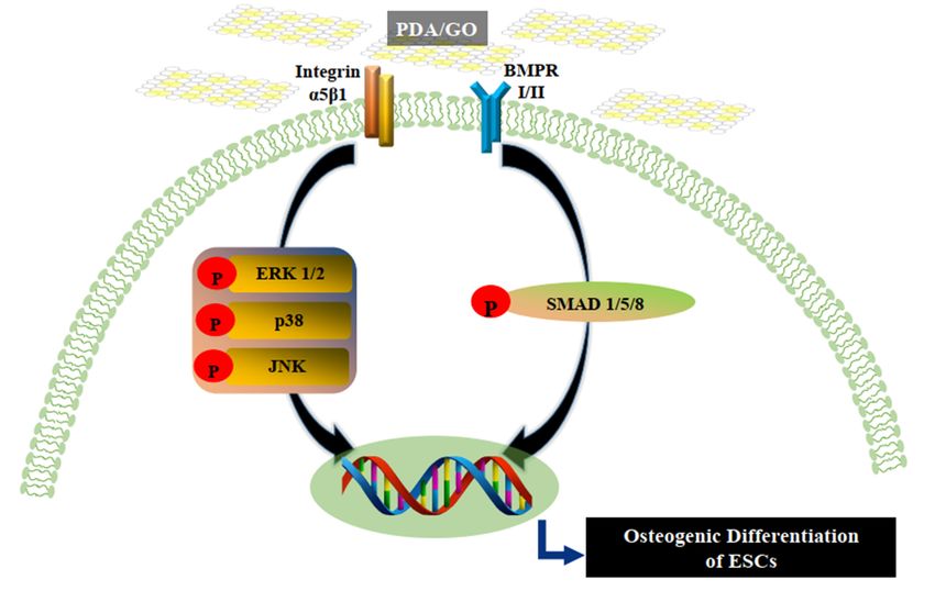
Int. J. Mol. Sci. 2021, 22, 7323                                                                                             2 of 15

                                   delivery systems [6]. Graphene, a carbon-based nanomaterial with versatile physico-
                                   chemical properties, has attracted increased research attention in various bio-engineering
                                   fields [7]. Graphene is a flat monolayer of carbon atoms patterned into a honeycomb,
                                   two-dimensional (2D) lattice, and it serves as a biocompatible and implantable platform for
                                   stem cell culture and artificial microenvironments [8–10]. Moreover, the biofunctionalized
                                   capability of graphene and its derivative, graphene oxide (GO), has highlighted these
                                   carbon nanomaterials in regenerative medicine and biotechnology studies [11,12]. GO has
                                   been shown to strengthen induced pluripotent stem cell (iPSC) growth and to promote
                                   spontaneous differentiation [10]. Graphene and GO encouraged the cardiomyogenic or
                                   neuronal differentiation of ESCs [13,14]. GO-filmed substrates also intensified the differen-
                                   tiation of mouse ESCs to the hematopoietic lineage [15]. Growing evidence has indicated
                                   that a graphene surface provides a biocompatible nanoscale to accelerate the differenti-
                                   ation of human mesenchymal stem cells (hMSCs) into a bone cell specification without
                                   cellular toxicity [8,16,17]. GO-incorporated poly (lactic-co-glycolic acid) (PLGA) nanofiber
                                   structures also enhance the proliferation and osteogenic differentiation of hMSCs [18].
                                   Collectively, these results indicate the potential of graphene and GO-based biomaterials for
                                   stem cell-related biomedical applications.
                                         Graphene and its derivatives significantly promote ECM–integrin clustering signaling
                                   and promote the long-term adhesion of human neuronal stem cells [19,20]. GO also
                                   strengthens the adherence of mouse ESCs in controlling the ESC differentiation potency [21].
                                   Moreover, the integrin signaling pathway is involved in the biocompatibility of GO for the
                                   self-renewal of ESCs [22]. Many integrin-connected signaling networks influence not only
                                   cell adhesion to ECM substrates but also specific cell lineage commitments [23–25]. Thus,
                                   cell-graphene/GO substrate interactions can contribute to considerable cellular processes
                                   from the onset of adhesion operation.
                                         Graphene and GO can be further functionalized by other biomaterials through various
                                   chemical bonds, such as covalent bonds, ionic bonds, and Van der Waals forces [26,27].
                                         To establish effective strategies to fabricate functional matrixes or films, 3,4-dihydroxy-
                                   L-phenylalanine (DOPA) derived from mussel adhesive threads has been used because of
                                   its extraordinarily adhesion properties [28]. Dopamine (DA), a catecholamine that contains
                                   catechol and amine functional groups, can self-polymerize to polydopamine (PDA) at an
                                   alkaline pH level, coating any virtual surface and presenting mussel-derived adhesive
                                   properties [28]. Based on these properties, PDA can be applied as an intermediate layer to
                                   deposit GO onto various substrates. Thus, this study employed PDA to securely attach GO
                                   nanosheets to the surface and manufactured carbon-based nanoscale ECM with GO and
                                   PDA hybrid materials (PDA/GO).
                                         Although several studies have reported the involvement of GO in ESC self-renewal
                                   and differentiation into the hematopoietic or neuronal lineage, to date, few studies have
                                   investigated the influences of GOs on the osteogenic differentiation of ESCs. The present
                                   study investigated how the PDA/GO composite substrate influences the osteogenic differ-
                                   entiation of ESCs. Moreover, the interaction of ESCs and this engineered microenvironment
                                   was demonstrated by certain signaling mechanisms to promote and control the fate of ESCs.

                                   2. Results
                                   2.1. PDA/GO Substrate Promotes the Osteogenic Differentiation of ESCs
                                        The morphology of the patterned PDA/GO substrate was first observed by SEM.
                                   The SEM images demonstrated a relative PDA coating or multilayered PDA/GO layer on
                                   the substrate (Figure 1A). The atomic constitution by XPS indicated that C1s is the most
                                   predominant element on the PT surface. The PDA-coated surface demonstrated increased
                                   concentrations of O1s and N1s compared with that on the PT surface. When PDA/GO was
                                   deposited on the surfaces, the concentrations of C1s and O1s were increased compared
                                   with those on the PDA surface (Figure 1B). These results suggest that the PDA or PDA/GO
                                   substrate was successfully immobilized on the PT culture surface.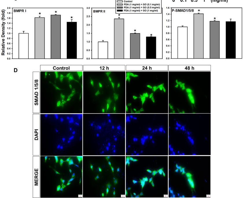
Int. J. Mol. Sci. 2021, 22, x FOR PEER REVIEW 3 of 15
concentrations of O1s and N1s compared with that on the PT surface. When PDA/GO was
deposited on the surfaces, the concentrations of C1s and O1s were increased compared
Int. J. Mol. Sci. 2021, 22, 7323 3 of 15
with those on the PDA surface (Figure 1B). These results suggest that the PDA or PDA/GO
substrate was successfully immobilized on the PT culture surface.
Figure1.1.Surface
Figure Surfacecharacterization
characterization of
of PDA/GO
PDA/GOsubstrates. (A)(A)
substrates. Topographical features
Topographical of the
features ofPT
thesurface, PDA PDA
PT surface, (1 mg/mL)-
(1 mg/mL)-
coated, and PDA/GO (1 mg/mL + 1 mg/mL)-coated surfaces were assessed by scanning electron
coated, and PDA/GO (1 mg/mL + 1 mg/mL)-coated surfaces were assessed by scanning electron microscopymicroscopy at ×10 k mag-
at ×10 k
nification. (B) Relative atomic composition of each sample.
magnification. (B) Relative atomic composition of each sample.
Before observing the osteogenic response of ESCs, cell viability on PDA/GO was first
Before observing the osteogenic response of ESCs, cell viability on PDA/GO was first
assessed after cells were cultured on PDA/GO-coated (0.1, 0.5, 1 mg/mL) or uncoated (con-
assessed after cells were cultured on PDA/GO-coated (0.1, 0.5, 1 mg/mL) or uncoated
trol) surfaces for 1, 5, 10, and 14 days. No difference was found in the cell viability between
(control) surfaces for 1, 5, 10, and 14 days. No difference was found in the cell viability
ESCs cultured on the PDA/GO and control surface, indicating no cytotoxicity of the pre-
between
sent ESCs cultured
combination of PDA/GOon the PDA/GO
substrates and 2A).
(Figure control surface, indicating
Subsequently, no cytotoxicity
the osteogenic differ- of
the present combination of PDA/GO substrates (Figure 2A). Subsequently,
entiation of ESCs was explored by analyzing the ALP activity and intracellular calcium the osteogenic
differentiation
levels of ESCs
([Ca2+]i). The ALP was
activityexplored
in cellsby onanalyzing
the PDA/GO the substrate
ALP activity
was and intracellular
increased com- cal-
cium levels ([Caof2+ ] ). The ALP activity in cells on the PDA/GO substrate was increased
pared with those the
i control group on day 4 and was further increased on day 7 (Figure
compared
2B). Similar with
to ALPthose of the
activity, thecontrol group
[Ca2+]i was onincreased
also day 4 and was on
in cells further
PDA/GOincreased on day 7
in a dose-
(Figure 2B). Similar to ALP 2+ ] was also increased in cells on PDA/GO
dependent manner (Figure 2C).activity, the [Ca
Extracellular calciumi deposits were also analyzed. Figure
in aindicates
2D dose-dependent
that calcium manner
deposits (Figure 2C). Extracellular
were increased in ESCs on calcium
the PDA/GO deposits were
substrate also ana-
com-
lyzed.with
pared Figure 2Dofindicates
those the control that calcium
groups deposits
(Figure wereosteogenic
2D). The increasedeffect
in ESCs onPDA/GO
of the the PDA/GO
substrate
substratewas assessed
compared by following
with those of the thecontrol
gene and protein
groups expression
(Figure 2D). Theof the osteogenic
osteogenic effect of
markers.
the PDA/GO The mRNAsubstrateexpression of osteogenic
was assessed target genes
by following (ALP,
the gene and osterix (OSX),
protein runt-re- of the
expression
lated transcription
osteogenic markers. factor
The2 mRNA
(RUNX2), osteocalcin
expression of (OCN), and target
osteogenic osteopontin
genes(OPN)) was in-(OSX),
(ALP, osterix
creased in cells on a PDA/GO coating compared with that of
runt-related transcription factor 2 (RUNX2), osteocalcin (OCN), and osteopontin the control group and(OPN))
showed the highest level in cells on 1 mg/mL of PDA/GO (Figure 3A–E).
was increased in cells on a PDA/GO coating compared with that of the control group and Western blot
analysis
showedalso the showed
highest that
levelOCN andon
in cells OSX protein levels
1 mg/mL were increased
of PDA/GO (Figurein3A–E).
cells cultured
Western blot
on PDA/GO substrate on day 7 of osteogenic induction (Figure 3F,G).
analysis also showed that OCN and OSX protein levels were increased in cells cultured on
PDA/GO substrate on day 7 of osteogenic induction (Figure 3F,G).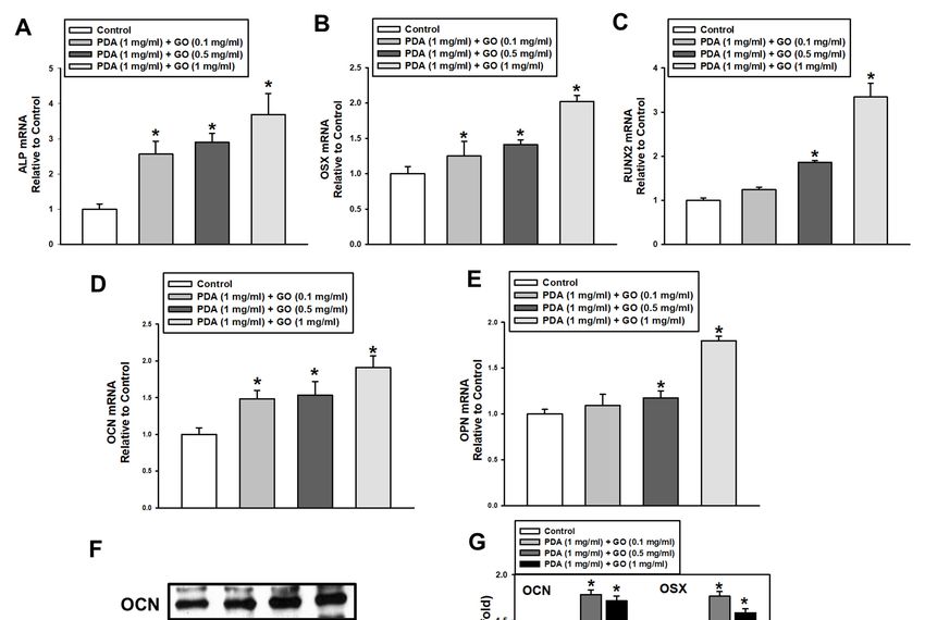
Int. J. Mol. Sci. 2021, 22, 7323                                                                                                                           4 of 15
Int. J. Mol. Sci. 2021, 22, x FOR PEER REVIEW                                                                                                  4 of 15

      Figure       2. Osteogenic differentiation of ESCs cultured on PDA/GO-modified surfaces. (A) Cells were cultured on the
      Figure 2. Osteogenic differentiation of ESCs cultured on PDA/GO-modified surfaces. (A) Cells were cultured on the
      PDA/GO
      PDA/GO substratesubstrate  (composite
                               (composite      of PDA
                                           of PDA    withwith  1 mg/mL
                                                           1 mg/mL         and variable
                                                                     and variable           GO concentration
                                                                                     GO concentration    with 0.1,with
                                                                                                                    0.5, 0.1,
                                                                                                                         or 1 0.5, or 1 mg/mL)
                                                                                                                              mg/mL)    for 1, 5, for 1, 5,
      10,
      10, and
          and 14    14days,
                      days,and
                            andthen,
                                  then,
                                     thethe
                                          cellcell viability
                                               viability waswas   assessed
                                                              assessed       as described
                                                                       as described    in the in the Materials
                                                                                              Materials         and Methods.
                                                                                                         and Methods.               (B) ALP(C)
                                                                                                                          (B) ALP activity,    activity, (C)
      [Ca 2+]i], and
      [Ca2+       , and(D)
                        (D)Alizarin
                            AlizarinRed
                                      RedS staining
                                            S         were
                                                staining    evaluated
                                                          were         after
                                                                evaluated    4, 7,
                                                                             after 14,
                                                                                    4, or
                                                                                       7,  21
                                                                                          14, days
                                                                                              or  21of osteogenic
                                                                                                     days  of      induction.
                                                                                                              osteogenic        The  values
                                                                                                                             induction.   Thearevalues are
                i
      presented as means ± SD (n = 3). * p < 0.05 vs. the control value at each time point.
      presented       as means ± SD (n = 3). * p < 0.05 vs. the control value at each time point.

                                        2.2. Integrin α5/β1 and BMPR I/II Signaling Pathways in ESCs on the PDA/GO Substrate
                                             To understand the molecular mechanisms underlying the link between ESCs and
                                        the PDA/GO substrate, we explored whether integrins, as an adhesion receptor, and
                                        bone morphogenetic protein receptors (BMPRs), as representative osteogenic-functioning
                                        receptors, are associated with PDA/GO substrate-derived ESC-osteolineage commitment.
                                        The protein levels of integrins were analyzed in cells cultured on the PDA/GO substrate
                                        after 7 days of culture (Figure 4A). The α5 and β1 subunits of integrins showed increased
                                        protein levels in cells on the PDA/GO substrate compared with those in the control groups
                                        (Figure 4A). These increases were dose dependent according to the GO concentration.
                                        Western blot analysis showed that the type I and II BMPR levels were significantly increased
                                        when cells were cultured on the PDA/GO substrate (Figure 4B). SMAD 1/5/8 are signal
                                        transducers, which are activated by BMP receptors and mediate BMP signaling pathways.
                                        SMAD1/5/8 phosphorylation was elevated in cells on the PDA/GO substrate than in the
                                        control (Figure 4B,C). The nuclear translocation of SMAD1/5/8 was also confirmed using
                                        immunofluorescence staining (Figure 4D). Thus, activation of the BMPR-SMAD1/5/8
                                        signaling pathway may provide an additional osteoinductive signal for ESC-osteogenic
                                        differentiation on the PDA/GO substrate.
Int. J. Mol. Sci. 2021, 22, 7323 5 of 15
Int. J. Mol. Sci. 2021, 22, x FOR PEER REVIEW 5 of 15
Figure 3. Effect
Figure of of
3. Effect PDA/GO
PDA/GO on osteogenic-related
osteogenic-related gene
gene andand protein
protein expression.
expression. (A–E)
(A–E) The Theexpression
mRNA mRNA expression
of ALP, OSX, of ALP, OSX,
RUNX2,
RUNX2, OCN,OCN,andand
OPNOPN was
was analyzedafter
analyzed after7-day
7-day osteogenic
osteogenic induction
inductionbyby
real-time RT-PCR.
real-time (F) The
RT-PCR. protein
(F) The levelslevels
protein of of OCN
OCN (5.5 kDa) and OSX (45 kDa) were determined by Western blot. (G) The bars denote the density relative to β-actin.
(5.5 kDa) and OSX (45 kDa) were determined by Western blot. (G) The bars denote the density relative to β-actin. The
The values are expressed as means ± SD (n = 3). * p < 0.05 vs. control value.
values are expressed as means ± SD (n = 3). * p < 0.05 vs. control value.
2.2. Integrin α5/β1 and BMPR I/II Signaling Pathways in ESCs on the PDA/GO Substrate
This study also examined whether ESC osteogenic differentiation on the PDA/GO
To understand the molecular mechanisms underlying the link between ESCs and the
substrate requires the
PDA/GO substrate, we activation of mitogen-activated
explored whether protein kinases
integrins, as an adhesion receptor,(MAPKs),
and bone which are
responsible
morphogenetic forprotein
osteogenic differentiation
receptors and bone formation
(BMPRs), as representative [29,30]. The re-
osteogenic-functioning activation of
MAPKs, such
ceptors, are as extracellular
associated signal-regulated
with PDA/GO kinase
substrate-derived (ERK)1/2, p38,commitment.
ESC-osteolineage and c-Jun-N-terminal
kinase (JNK),
The protein wasofassessed
levels by measuring
integrins were analyzed inthe phosphorylated
cells form of substrate
cultured on the PDA/GO each MAPK using
after 7 days
Western of analysis
blot culture (Figure 4A).5A).
(Figure The α5
Theand β1 subunitsof
expression of P-ERK1/2,
integrins showed increased
P-p38, and P-JNK was
protein levels in
upregulated incells
cellsonon
thethe
PDA/GO
PDA/GOsubstrate compared
substrate with those with
compared in the that
control
ingroups
the control. No
(Figure 4A). These increases were dose dependent according to the GO concentration.
significant differences were found in the expression of total ERK1/2, p38, and JNK among
Western blot analysis showed that the type I and II BMPR levels were significantly in-
all the experimental groups. To further understand this extracellular-leading intracellular
creased when cells were cultured on the PDA/GO substrate (Figure 4B). SMAD 1/5/8 are
signaling pathway,
signal transducers, the influence
which of by
are activated integrin α5/β1 in
BMP receptors andMAPK
mediateactivation was evaluated
BMP signaling
using integrin α5/β1 siRNA. The knockdown efficiency of integrin α5/β1 siRNA was first
confirmed when the transfection of integrin α5/β1 siRNA downregulated the protein levels
of each integrin in cells on the PDA/GO substrate (Figure 5B). The phosphorylation of
ERK1/2, p38, and JNK was decreased by the knockdown of integrin α5/β1, indicating that
integrin α5/β1 mediates MAPK signaling pathways during ESC osteogenic differentiation
in response to the PDA/GO substrate.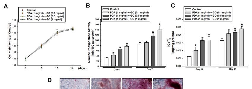
pathways. SMAD1/5/8 phosphorylation was elevated in cells on the PDA/GO substrate
                                         than in the control (Figure 4B,C). The nuclear translocation of SMAD1/5/8 was also con-
Int. J. Mol. Sci. 2021, 22, 7323         firmed using immunofluorescence staining (Figure 4D). Thus, activation of the BMPR- 6 of 15
                                         SMAD1/5/8 signaling pathway may provide an additional osteoinductive signal for ESC-
                                         osteogenic differentiation on the PDA/GO substrate.

      FigureFigure  4. Effect of PDA/GO on integrin α5/β1, MAPKs, BMPR I/II, and SMAD 1/5/8 signaling pathways. The cells were
              4. Effect of PDA/GO on integrin α5/β1, MAPKs, BMPR I/II, and SMAD 1/5/8 signaling pathways. The cells were
            cultured on the PDA/GO substrate for 7 days, and the protein levels of (A) integrin α5 (138 kDa), integrin β1 (150 kDa),
      cultured
            and on
                 (B)the
                     BMPRPDA/GO
                            I (50–55 substrate
                                      kDa), BMPRforII7(115
                                                       days,  and
                                                           kDa), andthephosphorylation
                                                                        protein levels levels
                                                                                        of (A)ofintegrin  α5 (138
                                                                                                  SMAD 1/5/8       kDa),
                                                                                                               (52–56   kDa)integrin    β1 (150 kDa),
                                                                                                                               were analyzed.
      and (B)
            (C)BMPR    I (50–55
                The bars   denotekDa),  BMPRrelative
                                   the density  II (115tokDa),  andThe
                                                           β-actin.   phosphorylation    levelsasofmeans
                                                                         values are expressed       SMAD    1/5/8
                                                                                                         ± SD       (52–56
                                                                                                               (n = 3). * p < kDa)   were
                                                                                                                               0.05 vs.     analyzed.
                                                                                                                                        control
            value.
      (C) The  bars(D)  Nuclear
                     denote   thetranslocation of SMAD
                                   density relative       1/5/8 was
                                                      to β-actin.    assessed
                                                                   The  valuesbyare
                                                                                  immunofluorescence
                                                                                     expressed as means staining
                                                                                                            ± SD(scale
                                                                                                                   (n =bar,
                                                                                                                          3). *20pμm).
                                                                                                                                  < 0.05 vs. control
      value. (D) Nuclear translocation of SMAD 1/5/8 was assessed by immunofluorescence staining (scale bar, 20 µm).
confirmed when the transfection of integrin α5/β1 siRNA downregulated the protein lev-
els of each integrin in cells on the PDA/GO substrate (Figure 5B). The phosphorylation of
ERK1/2, p38, and JNK was decreased by the knockdown of integrin α5/β1, indicating that
integrin
Int. J. Mol. α5/β1
Sci. 2021, 22, 7323 mediates MAPK signaling pathways during ESC osteogenic differentiation
7 of 15
in response to the PDA/GO substrate.
Figure 5. Effect of PDA/GO on MAPKs signaling pathways. The cells were cultured on the PDA/GO substrate for
7 Figure
days and5. Effect
(A) of PDA/GO
Phosphorylation on MAPKs
of ERK1/2 (42–44signaling pathways.
kDa), p38 (38 The(46–54
kDa), and JNK cells kDa)
werewascultured on(B)the
assessed. Cells were
PDA/GOwith
transfected substrate
integrin for 7 days and
α5/β1-specific (A) Phosphorylation
siRNA, ofofERK1/2
and the protein levels (42–44
integrin α5, kDa),
β1, and p38 (38p38,
the ERK1/2, kDa),andand
JNK
JNK (46–54 kDa)
phosphorylation levelswas
were assessed. (B) 4Cells
examined after were
days of transfected
osteogenic with integrin α5/β1-specific siRNA, and
induction.
the protein levels of integrin α5, β1, and the ERK1/2, p38, and JNK phosphorylation levels were
examined after 4 days2.3.
of Integrin α5/β1,
osteogenic MAPKs, and BMPRs/SMAD Mediate ESC Osteogenic Differentiation on the
induction.
PDA/GO Substrate
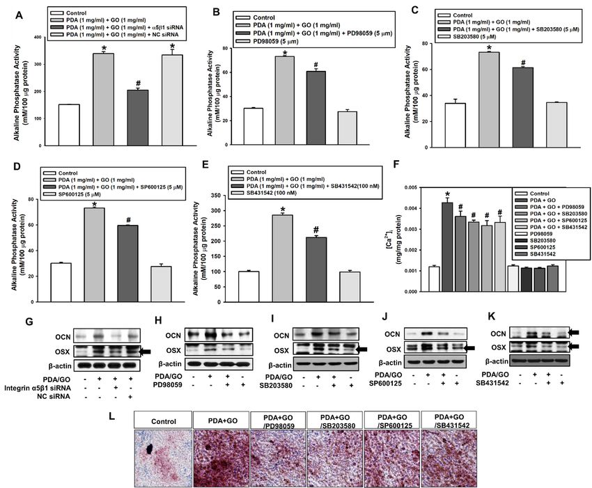
Considering that the activation of integrin α5/β1, MAPK, and BMPR/SMAD sig-
2.3. Integrin α5/β1, MAPKs, and BMPRs/SMAD
naling pathways Mediate ESC Osteogenic
is related to PDA/GO-derived Differentiation
ESC osteogenesis, on the
we investigated
the PDA/GO Substrate influence of individual pathways in the osteogenic differentiation of ESCs on the PDA/GO
substrate. Figure 6A demonstrates that the knockdown of integrin α5/β1 reduced the
Considering that the activation of integrin α5/β1, MAPK, and BMPR/SMAD signal-
PDA/GO-induced ESC ALP activity. Western blot analysis confirmed the downregulation
ing pathways is related
of OCNto and
PDA/GO-derived
OSX protein levelsESC after osteogenesis, we investigated
integrin α5/β1-knockdown (Figure the
6G). influ-
Next, the
ence of individual connection
pathwaysbetween
in theMAPKosteogenic differentiation
activation of ESCs
and PDA/GO-derived ESConosteogenesis
the PDA/GO was ex-
amined. When cells were treated with ERK inhibitor (PD98059),
substrate. Figure 6A demonstrates that the knockdown of integrin α5/β1 reduced the p38 inhibitor (SB203580),
JNK inhibitor (SP600125), or SMAD inhibitor (SB431542), the ALP activity and [Ca2+ ]i of
PDA/GO-induced ESC ALP activity. Western blot analysis confirmed the downregulation
ESCs cultured on the PDA/GO were downregulated (Figure 6B–F), as well as OCN and
of OCN and OSX protein levels
OSX protein after
levels integrin
(Figure 6H–K). α5/β1-knockdown
Calcium deposits were (Figure 6G). in
also decreased Next,
ESCs the
on the
connection betweenPDA/GO
MAPK substrate
activation withand
eachPDA/GO-derived
inhibitor (Figure 6L).ESC osteogenesis was exam-
ined. When cells were treated with ERK inhibitor (PD98059), p38 inhibitor (SB203580),
JNK inhibitor (SP600125), or SMAD inhibitor (SB431542), the ALP activity and [Ca2+]i ofInt. J. Mol. Sci. 2021, 22, x FOR PEER REVIEW 8 of 15
Int. J. Mol. Sci. 2021, 22, 7323 ESCs cultured on the PDA/GO were downregulated (Figure 6B–F), as well as OCN and 8 of 15
OSX protein levels (Figure 6H–K). Calcium deposits were also decreased in ESCs on the
PDA/GO substrate with each inhibitor (Figure 6L).
FigureFigure
6. Role6. Role
of theofintegrin
the integrin α5/β1,
α5/β1, MAPK,and
MAPK, andBMPR/SMAD
BMPR/SMAD signaling pathways
signaling pathwaysin the PDA/GO-mediated
in the PDA/GO-mediated osteogenic
osteogenic
differentiation of ESCs. Cells were transfected with integrin α5/β1-specific siRNA, or treated with PD98059 (5 μM),
differentiation of ESCs. Cells were transfected with integrin α5/β1-specific siRNA, or treated2+with PD98059 (5 µM),
SB203580 (5 μM), SP600125 (5 μM), or SB431542 (100 nM) for 48 h before (A–E) ALP activity, (F) [Ca ]i, (G–K) Western
SB203580 2+ , (G–K) Western blot
blot (5 µM), SP600125
analysis, (5 µM),Red
and (L) Alizarin or SB431542
S staining. (100 nM) for
The values are48expressed
h before as
(A–E) ALP
means activity,
± SD (n = 4).(F)
* p[Ca
< 0.05]ivs. each control
analysis, and
value, (L)# pAlizarin
and < 0.05 vs.Red value. The values are expressed as means ± SD (n = 4). * p < 0.05 vs. each control value,
S staining.
PDA/GO
and # p < 0.05 vs. PDA/GO value.
3. Discussion
The present study showed that a PDA/GO composite-coated cell culture substrate
3. Discussion
can effectively contribute to ESC osteogenic differentiation through the integrin α5/β1,
The present study showed that a PDA/GO composite-coated cell culture substrate can
MAPK, and BMPR/SMAD signaling pathways. There is an increased demand for opti-
effectively contribute to ESCsystems
mized microenvironmental osteogenic differentiation
to maintain through
self-renewal the integrin
or facilitate α5/β1, MAPK,
the differentia-
and BMPR/SMAD signaling pathways. There is an increased demand
tion of adult and embryonic stem cells [31,32]. Previous studies have shown that for optimized
novel mi-
croenvironmental
biomaterials that systems toextracellular
imitate the maintain self-renewal or facilitate
microenvironment and inthe vivodifferentiation
construction en- of adult
and embryonic
courage stem cells
the efficient [31,32]. Previous
differentiation of stem studies have
cells to the shown
desired cellthat novel
lineage biomaterials
[33,34]. Thus, that
the present
imitate study demonstrated
the extracellular that the culture
microenvironment and inof ESCs on the PDA/GO-coated
vivo construction encouragesurface
the efficient
promotes osteogenic
differentiation of stem differentiation
cells to theofdesired
ESCs. Few
cellstudies
lineagehave investigated
[33,34]. Thus,thetheosteogenic
present study
effect of the PDA/GO
demonstrated composite
that the culture on ESCs
of ESCs andPDA/GO-coated
on the even on MSCs. One studypromotes
surface showed that a
osteogenic
differentiation of ESCs. Few studies have investigated the osteogenic effect of the PDA/GO
composite on ESCs and even on MSCs. One study showed that a PDA-inspired GO and
titanium scaffold promoted bone marrow-derived MSC adhesion and proliferation, as
well as development of nanostructured environments for bone regeneration [35]. Our
PDA/GO-functionalized culture substrate is a potential strategy to produce large numbers
of osteogenic cells from ESCs.
The interplay between stem cells and the engineered extracellular microenvironment
for a practical stem cell differentiation system must be investigated further. The presentInt. J. Mol. Sci. 2021, 22, 7323 9 of 15
study provides the underlying molecular processes for ESC osteogenic differentiation on
a physicochemical PDA/GO substrate. The first cue mediating the mechanical signal of
PDA/GO to ESCs was the cell surface receptors integrin α5/β1 in the present study. Previ-
ous studies have shown that the gene and protein expression levels of integrin α5/β1 were
increased during human MSC differentiation to osteoblasts [36–38]. Osteoblast adhesion
on certain ECM proteins was achieved through binding to αvβ1 integrin [39]. Various
studies have demonstrated that dynamic expression of different integrins is required for
the osteogenic differentiation of MSCs [40,41]. The use of graphene material suggests the
pivotal role of integrin β1 in ECM roughness recognition, which is involved in osteoblast
maturation and MSC differentiation on graphitic carbon-coated surfaces [42]. Other reports
have shown that the protein expression of integrin β1 is increased on graphene-coated
Si/SiO2 substrates by significantly promoting the differentiation of MSCs into bone cells [8].
Integrin β1 binds 12 different α subunits, including the α5 subunit, in osteoblasts and
osteoprogenitor cells. Moreover, integrin β1 mediates cell adhesion to bone matrix and
promotes osteogenic cell proliferation and differentiation, indicating that integrin β1 sig-
naling plays a major function in bone formation [43,44]. To date, numerous studies have
shown that integrins α5/β1 participate in the osteogenic differentiation of osteoprogenitors
and MSCs as previously mentioned. We also suggest that integrin α5/β1 can introduce
the differentiation of ESCs into the osteogenic lineage when they are cultured on the
PDA/GO substrate.
In the present study, integrin α5/β1 led to the activation of ERK1/2, p38, and JNK
MAPKs as outside-relayed intracellular pathways during ESC osteogenic differentiation in
response to PDA/GO. These MAPKs are the best-characterized downstream signaling path-
ways of the matrix microenvironment–integrin interactions in osteogenic cell types [44–46].
Consistent with the current results, MAPKs have been frequently reported as a key player
for the osteogenic differentiation of various types of stem cells [47–49]. Regarding previous
studies and our findings, these integrin–MAPK stepwise processes trigger the osteogenic
induction of ESCs in response to the PDA/GO culture substrate.
Interestingly, the BMP receptors, members of the transforming growth factor-β (TGF-
β) superfamily, were suggested as the other cell-receiving signals from the PDA/GO
substrate. GO mechanistically interacts with multiple cell surface receptors [50–52]. How-
ever, the PDA or GO material can activate BMP receptors in ESCs or other stem cell models.
One previous study reported that GO activated TGF-β receptor/SMAD2/3 signaling
to trigger new metastases of human cancer cells [53]. Insufficient data exist to identify
PDA/GO-related osteogenic signaling pathways; however, our findings showed that the
PDA/GO substrate facilitates the presentation of BMP receptors in ESCs and enhances
ESC osteogenic activity. BMPRs and canonical SMAD signaling are widely studied in the
bone biology field [54–56]. Several studies have verified that SMAD-dependent BMP and
TGF-β signaling pathways manage both osteoblast and osteoclast function; thus, they play
potential roles in skeletal development, bone formation, and bone homeostasis [57–60].
The present study demonstrated increased BMPR type I and II protein levels and activa-
tion of SMAD 1/5/8, receptor-regulated SMADs (R-SMADs), which are responsible for
PDA/GO-derived ESC differentiation into osteolineage cells. A previous study reported
that BMPR recognition and the phosphorylation of SMAD 1/5/8 signaling promoted the
in vitro osteogenic differentiation of C2C12 cells in a magnesium-modified calcium phos-
phate matrix model [61]. Thus, the designed ECM substrates sensing the BMPR/SMAD
signaling axis enable biomaterials to attend osteoinductive performance.
Although the present study suggests a model for ESC osteogenic differentiation on
PDA/GO composite by the initiation of both integrin α5/β1 and BMPRs, the reciprocal
interactions between these receptors is still unclear. Further study of this ambiguous issue
is warranted.
This study systematically investigated the osteogenic bioactivity of PDA/GO compos-
ite as a substrate material with ESCs. When ESCs were cultured on PDA/GO substrates,
cells significantly exhibited the osteogenic differentiation through integrin α5/β1, MAPK,Although the present study suggests a model for ESC osteogenic differentiation on
PDA/GO composite by the initiation of both integrin α5/β1 and BMPRs, the reciprocal
interactions between these receptors is still unclear. Further study of this ambiguous issue
Int. J. Mol. Sci. 2021, 22, 7323 is warranted. 10 of 15
This study systematically investigated the osteogenic bioactivity of PDA/GO compo-
site as a substrate material with ESCs. When ESCs were cultured on PDA/GO substrates,
cells significantly exhibited the osteogenic differentiation through integrin α5/β1, MAPK,
and
and BMPR
BMPRI/II-SMAD
I/II-SMAD 1/5/8 signaling pathways
1/5/8 signaling pathways (Figure
(Figure 7).
7). Finally,
Finally, the
thePDA/GO
PDA/GO culture
culture
system may provide a stem cell niche–mimetic environment to control
system may provide a stem cell niche–mimetic environment to control stem stem cell fate
cell and
fate anda
facile and promising strategy for bone tissue engineering and regenerative medicine.
a facile and promising strategy for bone tissue engineering and regenerative medicine.
Figure 7. Hypothesized model of the PDA/GO-mediated osteogenic differentiation of ESCs. When ESCs are cultured on
Figure 7. Hypothesized model of the PDA/GO-mediated osteogenic differentiation of ESCs. When ESCs are cultured on
PDA/GO-modified surfaces, integrin α5/β1 and BMPR I/II are recognized, leading to MAPK or SMAD signaling pathway
PDA/GO-modified surfaces, integrin α5/β1 and BMPR I/II are recognized, leading to MAPK or SMAD signaling pathway
activation and eventually promoting the osteogenic differentiation of ESCs.
activation and eventually promoting the osteogenic differentiation of ESCs.
4.
4. Materials
Materials and
and Methods
Methods
4.1. Materials
4.1. Materials
Fetal bovine serum (FBS) was supplied by Gibco-BRL (Gaithersburg, MD, USA).
Fetal bovine serum (FBS) was supplied by Gibco-BRL (Gaithersburg, MD, USA). An-
Antibodies used for Western blot analysis and immunofluorescence staining were obtained
tibodies used for Western blot analysis and immunofluorescence staining were obtained
from Santa Cruz Biotechnology (Santa Cruz, CA, USA). The chemicals, including L-3,4-
from Santa Cruz Biotechnology (Santa Cruz, CA, USA). The chemicals, including L-3,4-
dihydroxyphenylalanine (L-DOPA) and graphene oxide dispersion, were purchased from
dihydroxyphenylalanine (L-DOPA) and graphene oxide dispersion, were purchased from
Sigma Chemical Company (St. Louis, MO, USA). Other laboratory materials were acquired
Sigma
from Chemical
SPL LifescienceCompany (St.Korea).
(Pocheon, Louis, MO, USA). Other laboratory materials were ac-
quired from SPL Lifescience (Pocheon, Korea).
4.2. Mouse ESC Culture and Embryoid Body Formation
4.2. Mouse ESC Culture and Embryoid Body Formation
Mouse ESCs (ES-E14TG2a (ATCC® CRL-1821™)) were supplied from the Ameri-
Mouse
can Type ESCs Collection
Culture (ES-E14TG2a (ATCC® VA,
(Manassas, CRL-1821™))
USA). ESCwere supplied
culture from the American
was performed as in our
Type Culture
previous reportCollection
[62]. To (Manassas, VA, USA).
develop embryoid ESC culture
bodies was performed
(EBs), dissociated cellsaswere
in our pre-
aggre-
vious report
gated [62]. To
by hanging develop
drop with embryoid
2000 cellsbodies (EBs),
in 20 µL dissociated
of DMEM. Wecells
usedwere aggregated
5-day-old by
EBs in
every experiment.
4.3. Preparation of the PDA/GO Composite Substrate
The PDA solution was prepared by dissolving 1 mg of L-DOPA in 1 mL of 10 mM
Tris buffer base (pH 8.5; Sigma-Aldrich, St. Louis, MI, USA). Then, GO dispersion was
added to the PDA solution (1 mg/mL) under magnetic stirring at room temperature for
24 h. The final concentration of GO in the PDA solution was 0.1, 0.5, or 1 mg/mL forInt. J. Mol. Sci. 2021, 22, 7323 11 of 15
individual experiments. The polystyrene (PT) culture surface was coated with PDA/GO
composite solution overnight at room temperature and washed three times with sterile
phosphate-buffered saline (PBS). Then, the PDA/GO-modified surfaces were dried in a
vacuum oven.
4.4. Characterization of the PDA/GO-Coated Surface
Images of PDA/GO composite-coated surfaces were identified with scanning electron
microscopy (SEM; S-4700, Hitachi, Tokyo, Japan). The specimens were rinsed with PBS
and then freeze-dried overnight before SEM operation. The atomic composition of the
PDA/GO-modified surfaces was assessed by X-ray photoelectron spectroscopy (XPS) as
in our previous report [63]. The surface composition amount was obtained from the XPS
survey spectra.
4.5. Cell Viability Assay
Cell viability was performed using the cell counting kit-8 (CCK-8) assay as in our pre-
vious report [62]. Briefly, cells were cultured on PDA/GO substrate (0, 0.1, 0.5, 1 mg/mL)
for 1, 5, 10, and 14 days, and the CCK-8 assay was performed according to the manufac-
turer’s instructions. The optical density was observed at a wavelength of 450 nm using an
ELISA reader system (Triad; DYNEX, Chantilly, VA, USA). The proportion of cell viability
was designated relative to the control.
4.6. Alkaline Phosphatase Activity Assay
Alkaline phosphatase activity was assessed as in our previous report [62]. Briefly, cells
were plated onto PDA/GO composite-coated 60 mm dishes (10–15 EBs per dish). ALP ac-
tivity was evaluated after 4 and 7 days of osteogenic induction using the p-nitrophenylphos-
phate (pNPP) procedure. The ALP enzyme activity was denoted as mM/100 µg of protein.
4.7. Intracellular Calcium Quantification Assay
Quantification of the intracellular calcium level was evaluated based on our previ-
ous report [62]. Briefly, cells were plated onto PDA/GO composite-coated 60 mm dishes
(10–15 EBs per dish). Fourteen and twenty-one days after osteogenic induction, the intra-
cellular calcium concentration was measured using a calcium assay kit (BioAssay Systems,
Hayward, CA, USA) according to the manufacturer’s information. The optical density was
read at 612 nm. The calcium level was expressed as mg/mg of protein.
4.8. Alizarin Red Staining
Alizarin Red staining was achieved as described in our previous report [64]. The cells
were fixed with 4% paraformaldehyde for 15 min and rinsed three times with PBS. Then,
the cells were stained with 2% Alizarin Red S solution (pH 4.2) for 5 min, and unbound
dye residue was washed with PBS. The stained images of different surfaces were observed
using a light microscope.
4.9. Osteogenic-Related Gene Expression Analysis
Real-time reverse transcription-polymerase chain reaction (RT-PCR) was conducted
as in our previous report [62] to measure the mRNA expression of osteogenic genes. The
primers used were as follows: 50 -GAC TGG TAC TCG GAT AAC GA-30 (forward) and 50 -
TGC GGT TCC AGA CAT AGT GG-30 (reverse) for ALP; 50 -CCAACTTCCTGTGCTCCGTG-
30 (forward) and 50 -TCTTGCCTCGTCCGCTCC-30 (reverse) for Runx2; 50 -TGA AAC GAG
TCA GCT CTG GAT G-30 (forward) and 50 -TGA AAT TCA TGG CTG TGG AA-30 (reverse)
for OPN; 50 -TGA GGA GGA AGT TCA CTA TGG-30 (forward) and 50 -TTC TTT GTG CCT
GCT TTG C-30 (reverse) for OSX; 50 -ATG AGA GCC CTC ACA CTC CTC-30 (forward) and
50 -GCC GTA GAA GCG CCG ATA GGC-30 (reverse) for OCN; and 50 -GCT CTC CAG AAC
ATC ATC C-30 (forward) and 50 -TGC TTC ACC ACC TTC TTG-30 (reverse) for GAPDH.Int. J. Mol. Sci. 2021, 22, 7323 12 of 15
4.10. Western Blot Analysis
Western blot analysis was performed as in our previous report [63]. Briefly, the
primary (anti-OCN, anti-OSX, anti-integrin α5, anti-integrin β1, anti-BMPR I, anti-BMPR
II, anti-SMAD1/5/8, anti-P-SMAD1/5/8, anti-ERK1/2, anti-P-ERK1/2, anti-p38, anti-
P-p38, anti-JNK, anti-P-JNK, or anti-β-actin; Santa Cruz Biotechnology) and secondary
antibodies (goat anti-rabbit immunoglobulin G (IgG) or goat anti-mouse IgG conjugated to
horseradish peroxidase) were employed with the dilutions recommended by the supplier.
The blots were developed using enhanced chemiluminescence (Santa Cruz Biotechnology)
and developed using X-ray film (Eastman-Kodak, Rochester, NY, USA).
4.11. Immunofluorescence Staining
Nuclear translocation of SMAD1/5/8 was detected by time-dependent immunofluo-
rescence staining. The cells were incubated with SMAD1/5/8 antibodies at 4 ◦ C overnight
and then with Alexa Fluor 488 goat anti-rabbit IgG for 2 h. Fluorescence images were
developed using a fluorescence microscope (Fluoview 300; Olympus, Tokyo, Japan).
4.12. SiRNA Transfection
Small interfering RNA (siRNA) transfection was conducted as in our previous re-
port [64]. Briefly, the cells were transfected with either an integrin α5/β1 siRNA (25 nM)
or a negative control siRNA (scrambled) for 48 h using a transfection reagent (RNAiMAX,
Invitrogen, Waltham, MA, USA) according to the manufacturer’s manual before being
subjected to ALP activity and Western blot analysis.
4.13. Statistical Analysis
All the data were expressed as means ± standard deviation. One-way analysis of
variance was used for multiple comparisons (Duncan’s multiple range test). Analyses were
obtained using SPSS software (ver. 10.0; SPSS Inc., Chicago, IL, USA). A p-value < 0.05
were considered statistically significant.
Author Contributions: Conceptualization, J.S.H.; Methodology and Investigation, N.Y.S.; Data Cura-
tion, J.S.H. and N.Y.S.; Writing, original draft preparation, J.S.H.; Visualization, N.Y.S.; Supervision,
J.S.H.; Project Administration, J.S.H.; Funding Acquisition, J.S.H. All authors have read and agreed
to the published version of the manuscript.
Funding: This work was supported by a National Research Foundation of Korea (NRF) grant funded
by the Korea government (MSIP) (No. 2018R1D1A1B07047538).
Institutional Review Board Statement: Not applicable.
Informed Consent Statement: Not applicable.
Data Availability Statement: Not applicable.
Conflicts of Interest: The authors declare no conflict of interest.
References
1. Daley, W.P.; Peters, S.B.; Larsen, M. Extracellular matrix dynamics in development and regenerative medicine. J. Cell Sci. 2008,
121, 255–264. [CrossRef] [PubMed]
2. Kerativitayanan, P.; Carrow, J.K.; Gaharwar, A.K. Nanomaterials for engineering stem cell responses. Adv. Healthc. Mater. 2015,
411, 1600–1627. [CrossRef] [PubMed]
3. Czyz, J.; Wobus, A. Embryonic stem cell differentiation: The role of extracellular factors. Differentiation 2001, 68, 167–174.
[CrossRef] [PubMed]
4. Xu, A.; Liu, X.; Gao, X.; Deng, F.; Deng, Y.; Wei, S. Enhancement of osteogenesis on micro/nano-topographical carbon fiber-
reinforced polyetheretherketone-nanohydroxyapatite biocomposite. Mater. Sci. Eng. C Mater. Biol. Appl. 2015, 48, 592–598.
[CrossRef]
5. Bennett, D.B.; Hill, J.C.; Dennison, J.; O’Brien, S.; Mantel, J.L.; Isaac, G.H.; Beverland, D.E. Metal-carbon fiber composite femoral
stems in hip replacements: A randomized controlled parallel-group study with mean ten-year follow-up. J. Bone Jt. Surg. Am.
2014, 96, 2062–2069. [CrossRef] [PubMed]Int. J. Mol. Sci. 2021, 22, 7323 13 of 15
6. Chen, J.; Chen, S.; Zhao, X.; Kuznetsova, L.V.; Wong, S.S.; Ojima, I. Functionalized single-walled carbon nanotubes as rationally
designed vehicles for tumor targeted drug delivery. J. Am. Chem. Soc. 2008, 130, 16778–16785. [CrossRef]
7. Zhu, Y.; Murali, S.; Cai, W.; Li, X.; Suk, J.W.; Potts, J.R.; Ruoff, R.S. Graphene and graphene oxide: Synthesis, properties, and
applications. Adv. Mater. 2010, 22, 3906–3924. [CrossRef]
8. Nayak, T.R.; Andersen, H.; Makam, V.S.; Khaw, C.; Bae, S.; Xu, X.; Ee, P.L.; Ahn, J.H.; Hong, B.H.; Pastorin, G.; et al. Graphene for
controlled and accelerated osteogenic differentiation of human mesenchymal stem cells. ACS Nano 2011, 5, 4670–4678. [CrossRef]
9. Lee, W.C.; Lim, C.H.; Shi, H.; Tang, L.A.; Wang, Y.; Lim, C.T.; Loh, K.P. Origin of enhanced stem cell growth and differentiation on
graphene and graphene oxide. ACS Nano 2011, 5, 7334–7341. [CrossRef]
10. Chen, G.Y.; Pang, D.W.; Hwang, S.M.; Tuan, H.Y.; Hu, Y.C. A graphene based platform for induced pluripotent stem cells culture
and differentiation. Biomaterials 2012, 33, 418–427. [CrossRef]
11. Cai, Y.; Li, H.; Du, B.; Yang, M.; Li, Y.; Wu, D.; Zhao, Y.; Dai, Y.; Wei, Q. Ultrasensitive electrochemical immunoassay for BRCA1
using BMIM·BF4-coated SBA-15 as labels and functionalized graphene as enhancer. Biomaterials 2011, 32, 2117–2123. [CrossRef]
[PubMed]
12. Feng, L.; Chen, Y.; Ren, J.; Qu, X. A graphene functionalized electrochemical aptasensor for selective label-free detection of cancer
cells. Biomaterials 2011, 32, 2930–2937. [CrossRef] [PubMed]
13. Lee, T.J.; Park, S.; Bhang, S.H.; Yoon, J.K.; Jo, I.; Jeong, G.J.; Hong, B.H.; Kim, B.S. Graphene enhances the cardiomyogenic
differentiation of human embryonic stem cells. Biochem. Biophys. Res. Commun. 2014, 452, 174–180. [CrossRef]
14. Lv, M.; Zhang, Y.; Liang, L.; Wei, M.; Hu, W.; Li, X.; Huang, Q. Effect of graphene oxide on undifferentiated and retinoic
acid-differentiated SH-SY5Y cells line. Nanoscale 2012, 4, 3861–3866. [CrossRef] [PubMed]
15. Eva, G.; Maria, I.; Monika, S.; Claudio, S.; Sebastian, H.; Susan, J.K.; Valerie, K.; Georges, L.; Aravind, V.; Kiran, B. Graphene
Oxide promotes embryonic stem cell differentiation to haematopoietic lineage. Sci. Rep. 2016, 6, 25917.
16. Newby, S.D.; Masi, T.; Griffin, C.D.; King, W.J.; Chipman, A.; Stephenson, S.; Anderson, D.E.; Biris, A.S.; Bourdo, S.E.; Dhar,
M. Functionalized Graphene Nanoparticles Induce Human Mesenchymal Stem Cells to Express Distinct Extracellular Matrix
Proteins Mediating Osteogenesis. Int. J. Nanomed. 2020, 15, 2501–2513. [CrossRef]
17. Lv, L.W.; Liu, Y.S.; Zhang, P.; Gu, M.; Bai, X.S.; Xiong, C.Y.; Zhou, Y.S. Transcriptomics and Functional Analysis of Graphene-
Guided Osteogenic Differentiation of Mesenchymal Stem Cells. Chin. J. Dent. Res. 2018, 21, 101–111.
18. Luo, Y.; Shen, H.; Fang, Y.; Cao, Y.; Huang, J.; Zhang, M.; Dai, J.; Shi, X.; Zhang, Z. Enhanced Proliferation and Osteogenic Differ-
entiation of Mesenchymal Stem Cells on Graphene Oxide-Incorporated Electrospun Poly (lactic-co-glycolic acid) Nanofibrous
Mats. ACS Appl. Mater. Interfaces 2015, 7, 6331–6339. [CrossRef]
19. Hong, S.W.; Lee, J.H.; Kang, S.H.; Hwang, E.Y.; Hwang, Y.S.; Lee, M.H.; Han, D.W.; Park, J.C. Enhanced neural cell adhesion and
neruite out-growth on graphene-based biomimetic substrates. BioMed. Res. Int. 2014, 2014, 212149. [CrossRef]
20. Yang, K.; Lee, J.; Lee, J.S.; Kim, D.; Chang, G.E.; Seo, J.; Cheong, E.; Lee, T.; Cho, S.W. Graphene oxide hierarchical patterns for the
derivation of electrophysiologically functional neuron-like cells from human neural stem cells. ACS Appl. Mater. Interfaces 2016, 8,
17763–17774. [CrossRef]
21. Yang, D.; Li, T.; Xu, M.; Gao, F.; Yang, J.; Yang, Z.; Le, W. Graphene oxide promotes the differentiation of mouse embryonic stem
cells to dopamine neurons. Nanomedicine 2014, 9, 2445–2455. [CrossRef] [PubMed]
22. Jing, G.; Wang, Z.; Zhuang, X.; He, X.; Wu, H.; Wang, Q.; Cheng, L.; Liu, Z.; Wang, S.; Zhu, R. Suspended graphene oxide
nanosheets maintain the self-renewal of mouse embryonic stem cells via down-regulating the expression of Vinculin. Biomaterials
2018, 171, 1–11. [CrossRef]
23. Hamidouche, Z.; Fromigué, O.; Ringe, J.; Häupl, T.; Marie, P.J. Crosstalks between integrin alpha 5 and IGF2/IGFBP2 signalling
trigger human bone marrow-derived mesenchymal stromal osteogenic differentiation. BMC Cell Biol. 2010, 11, 44. [CrossRef]
[PubMed]
24. Sipilä, K.; Haag, S.; Denessiouk, K.; Käpylä, J.; Peters, E.C.; Denesyuk, A.; Hansen, U.; Konttinen, Y.; Johnson, M.S.; Holmdahl,
R.; et al. Citrullination of collagen II affects integrin-mediated cell adhesion in a receptor-specific manner. FASEB J. 2014, 28,
3758–3768. [CrossRef]
25. Tang, C.H.; Yang, R.S.; Huang, T.H.; Lu, D.Y.; Chuang, W.J.; Huang, T.F.; Fu, W.M. Ultrasound stimulates cyclooxygenase-2
expression and increases bone formation through integrin, focal adhesion kinase, phosphatidylinositol 3-kinase, and Akt pathway
in osteoblasts. Mol. Pharmacol. 2006, 69, 2047–2057. [CrossRef]
26. Li, B.; Zhang, X.Y.; Yang, J.Z.; Zhang, Y.J.; Li, W.X.; Fan, C.H.; Huang, Q. Influence of polyethylene glycol coating on biodistribution
and toxicity of nanoscale graphene oxide in mice after intravenous injection. Int. J. Nanomed. 2014, 9, 4697–4707. [CrossRef]
[PubMed]
27. Kanakia, S.; Toussaint, J.D.; Mullick, C.S.; Tembulkar, T.; Lee, S.; Jiang, Y.P.; Lin, R.Z.; Shroyer, K.R.; Moore, W.; Sitharaman, B.
Dose ranging, expanded acute toxicity and safety pharmacology studies for intravenously administered functionalized graphene
nanoparticle formulations. Biomaterials 2014, 35, 7022–7031. [CrossRef]
28. Lee, H.; Dellatore, S.M.; Miller, W.M.; Messersmith, P.B. Mussel-inspired surface chemistry for multifunctional coatings. Science
2007, 318, 426–430. [CrossRef]
29. Cao, Y.; Shi, R.; Yang, H.; Zhang, J.; Ge, L.; Gao, R.; Fan, Z. Epiregulin promotes osteogenic differentiation and inhibits neurogenic
trans-differentiation of adipose-derived mesenchymal stem cells via MAPKs pathway. Cell Biol. Int. 2020, 44, 1046–1058.
[CrossRef] [PubMed]Int. J. Mol. Sci. 2021, 22, 7323 14 of 15
30. Yang, X.; Yang, Y.; Zhou, S.; Gong, X.; Dai, Q.; Zhang, P.; Jiang, L. Puerarin Stimulates Osteogenic Differentiation and Bone
Formation Through the ERK1/2 and p38-MAPK Signaling Pathways. Curr. Mol. Med. 2018, 17, 488–496. [CrossRef] [PubMed]
31. Riddell, J.; Gazit, R.; Garrison, B.S.; Guo, G.; Saadatpour, A.; Mandal, P.K.; Ebina, W.; Volchkov, P.; Yuan, G.C.; Orkin, S.H.; et al.
Reprogramming committed murine blood cells to induced hematopoietic stem cells with defined factors. Cell 2014, 157, 549–564.
[CrossRef]
32. Sandler, V.M.; Lis, R.; Liu, Y.; Kedem, A.; James, D.; Elemento, O.; Butler, J.M.; Scandura, J.M.; Rafii, S. Reprogramming human
endothelial cells to haematopoietic cells requires vascular induction. Nature 2014, 511, 312–318. [CrossRef]
33. Choi, J.S.; Mahadik, B.P.; Harley, B.A. Engineering the hematopoietic stem cell niche: Frontiers in biomaterial science. Biotechnol. J.
2015, 10, 1529–1545. [CrossRef]
34. Ireland, R.G.; Simmons, C.A. Human Pluripotent Stem Cell Mechanobiology: Manipulating the Biophysical Microenvironment
for Regenerative Medicine and Tissue Engineering Applications. Stem Cells 2015, 33, 3187–3196. [CrossRef] [PubMed]
35. Han, L.; Sun, H.; Tang, P.; Li, P.; Xie, C.; Wang, M.; Wang, K.; Weng, J.; Tan, H.; Ren, F.; et al. Mussel-inspired graphene oxide
nanosheet-enwrapped Ti scaffolds with drug-encapsulated gelatin microspheres for bone regeneration. Biomater. Sci. 2018, 6,
538–549. [CrossRef]
36. Sun, M.; Chi, G.; Xu, J.; Tan, Y.; Xu, J.; Lv, S.; Xu, Z.; Xia, Y.; Li, L.; Li, Y. Extracellular matrix stiffness controls osteogenic
differentiation of mesenchymal stem cells mediated by integrin alpha5. Stem Cell Res. Ther. 2018, 9, 52. [CrossRef] [PubMed]
37. Hamidouche, Z.; Fromigue, O.; Ringe, J.; Häupl, T.; Vaudin, P.; Pagès, J.C.; Srouji, S.; Livne, E.; Marie, P.J. Priming integrin alpha5
promotes human mesenchymal stromal cell osteoblast differentiation and osteogenesis. Proc. Natl. Acad. Sci. USA 2009, 106,
18587–18591. [CrossRef] [PubMed]
38. Saidak, Z.; Le Henaff, C.; Azzi, S.; Marty, C.; da Nascimento, S.; Sonnet, P.; Marie, P.J. Wnt/beta-catenin signaling mediates
osteoblast differentiation triggered by peptide-induced alpha5beta1 integrin priming in mesenchymal skeletal cells. J. Biol. Chem.
2015, 290, 6903–6912. [CrossRef]
39. Moussa, F.M.; Hisijara, I.A.; Sondag, G.R.; Scott, E.M.; Frara, N.; Abdelmagid, S.M.; Safadi, F.F. Osteoactivin promotes osteoblast
adhesion through HSPG and alphavbeta1 integrin. J. Cell. Biochem. 2014, 115, 1243–1253. [CrossRef]
40. Lee, H.M.; Seo, S.R.; Kim, J.; Kim, M.K.; Seo, H.; Kim, K.S.; Jang, Y.J.; Ryu, C.J. Expression dynamics of integrin alpha2, alpha3,
and alphaV upon osteogenic differentiation of human mesenchymal stem cells. Stem Cell Res. Ther. 2020, 11, 210. [CrossRef]
41. Zheng, H.; Li, X.; Chen, Y.; Zhou, R.; Zhao, H.; Qian, C. Integrin subunits alphaV and beta3 promote the osteogenic differentiation
of umbilical cord blood mesenchymal stem cells. Int. J. Clin. Exp. Pathol. 2018, 11, 2008–2016.
42. Olivares-Navarrete, R.; Rodil, S.E.; Hyzy, S.L.; Dunn, G.R.; Almaguer-Flores, A.; Schwartz, Z.; Boyan, B.D. Role of integrin
subunits in mesenchymal stem cell differentiation and osteoblast maturation on graphitic carbon-coated microstructured surfaces.
Biomaterials 2015, 51, 69–79. [CrossRef] [PubMed]
43. Zimmerman, D.; Jin, F.; Leboy, P.; Hardy, S.; Damsky, C. Impaired bone formation in transgenic mice resulting from altered
integrin function in osteoblasts. Dev. Biol. 2000, 220, 2–15. [CrossRef] [PubMed]
44. Marie, P.J. Targeting integrins to promote bone formation and repair. Nat. Rev. Endocrinol. 2013, 9, 288–295. [CrossRef]
45. Brunner, M.; Jurdic, P.; Tuckerman, J.P.; Block, M.R.; Bouvard, D. New insights into adhesion signaling in bone formation. Int.
Rev. Cell Mol. Biol. 2013, 305, 1–68. [PubMed]
46. Lu, M.; Zhuang, X.; Tang, K.; Wu, P.; Guo, X.; Yin, L.; Cao, H.; Zou, D. Intrinsic Surface Effects of Tantalum and Titanium on
Integrin alpha5beta1/ERK1/2 Pathway-Mediated Osteogenic Differentiation in Rat Bone Mesenchymal Stromal Cells. Cell.
Physiol. Biochem. 2018, 51, 589–609. [CrossRef] [PubMed]
47. Kim, H.Y.; Park, S.Y.; Choung, S.Y. Enhancing effects of myricetin on the osteogenic differentiation of human periodontal ligament
stem cells via BMP-2/Smad and ERK/JNK/p38 mitogen-activated protein kinase signaling pathway. Eur. J. Pharmacol. 2018, 834,
84–91. [CrossRef]
48. Zhu, D.; Deng, X.; Han, X.F.; Sun, X.X.; Pan, T.W.; Zheng, L.P.; Liu, Y.Q. Wedelolactone Enhances Osteoblastogenesis through
ERK- and JNK-mediated BMP2 Expression and Smad/1/5/8 Phosphorylation. Molecules 2018, 23, 561. [CrossRef]
49. Liang, L.; Zhou, W.; Yang, N.; Yu, J.; Liu, H. ET-1 Promotes Differentiation of Periodontal Ligament Stem Cells into Osteoblasts
through ETR, MAPK, and Wnt/β-Catenin Signaling Pathways under Inflammatory Microenvironment. Mediat. Inflamm. 2016,
2016, 8467849. [CrossRef]
50. Horvath, L.; Magrez, A.; Burghard, M.; Kern, K.; Forro, L.; Schwaller, B. Evaluation of the toxicity of graphene derivatives on cells
of the lung luminal surface. Carbon 2013, 64, 45–60. [CrossRef]
51. Bidram, E.; Sulistio, A.; Cho, H.J.; Amini, A.; Harris, T.; Zarrabi, A.; Qiao, G.; Stewart, A.; Dunstan, D.E. Targeted Graphene Oxide
Networks: Cytotoxicity and Synergy with Anticancer Agents. ACS Appl. Mater. Interfaces 2018, 10, 43523–43532. [CrossRef]
52. Dudek, I.; Skoda, M.; Jarosz, A.; Szukiewicz, D. The Molecular Influence of Graphene and Graphene Oxide on the Immune
System under In Vitro and In Vivo Conditions. Arch. Immunol. Ther. Exp. 2016, 64, 195–215. [CrossRef] [PubMed]
53. Zhu, J.; Li, B.; Xu, M.; Liu, R.; Xia, T.; Zhang, Z.; Xu, Y.; Liu, S. Graphene Oxide Promotes Cancer Metastasis through Associating
with Plasma Membrane to Promote TGF-β Signaling-Dependent Epithelial-Mesenchymal Transition. ACS Nano 2020, 14, 818–827.
[CrossRef]
54. Wang, M.; Li, J.; Ye, Y.; He, S.; Song, J. SHED-derived conditioned exosomes enhance the osteogenic differentiation of PDLSCs via
Wnt and BMP signaling in vitro. Differentiation 2020, 111, 1–11. [CrossRef] [PubMed]Int. J. Mol. Sci. 2021, 22, 7323 15 of 15
55. Wang, J.; Wang, M.; Chen, F.; Wei, Y.; Chen, X.; Zhou, Y.; Yang, X.; Zhu, X.; Tu, C.; Zhang, X. Nano-Hydroxyapatite Coating
Promotes Porous Calcium Phosphate Ceramic-Induced Osteogenesis Via BMP/Smad Signaling Pathway. Int. J. Nanomed. 2019,
14, 7987–8000. [CrossRef]
56. Chen, X.; Zhang, S.; Chen, X.; Hu, Y.; Wu, J.; Chen, S.; Chang, J.; Wang, G.; Gao, Y. Emodin promotes the osteogenesis of MC3T3-E1
cells via BMP-9/Smad pathway and exerts a preventive effect in ovariectomized rats. Acta Biochim. Biophys. Sin. 2017, 49, 867–878.
[CrossRef]
57. Yang, J.H.; Kim, S.C.; Kim, K.M.; Jang, C.H.; Cho, S.S.; Kim, S.J.; Ku, S.K.; Cho, I.J.; Ki, S.H. Isorhamnetin Attenuates Liver Fibrosis
by Inhibiting TGF-β/Smad Signaling and Relieving Oxidative Stress. Eur. J. Pharmacol. 2016, 783, 92–102. [CrossRef] [PubMed]
58. Yoon, B.S.; Ovchinnikov, D.A.; Yoshii, I.; Mishina, Y.; Behringer, R.R.; Lyons, K.M. Bmpr1a and Bmpr1b have overlapping
functions and are essential for chondrogenesis in vivo. Proc. Natl. Acad. Sci. USA 2005, 102, 5062–5067. [CrossRef]
59. Sánchez-Duffhues, G.; Hiepen, C.; Knaus, P.; Ten Dijke, P. Bone morphogenetic protein signaling in bone homeostasis. Bone 2015,
80, 43–59. [CrossRef]
60. Wang, H.; Chen, F.; Li, J.; Wang, Y.; Jiang, C.; Wang, Y.; Zhang, M.; Xu, J. Vaspin antagonizes high fat-induced bone loss in rats
and promotes osteoblastic differentiation in primary rat osteoblasts through Smad-Runx2 signaling pathway. Nutr. Metab. 2020,
17, 9. [CrossRef]
61. Ding, S.; Zhang, J.; Tian, Y.; Huang, B.; Yuan, Y.; Liu, C. Magnesium modification up-regulates the bioactivity of bone morpho-
genetic protein-2 upon calcium phosphate cement via enhanced BMP receptor recognition and Smad signaling pathway. Colloids
Surf. B Biointerfaces 2016, 145, 140–151. [CrossRef] [PubMed]
62. An, S.Y.; Lee, H.J.; Lee, S.C.; Heo, J.S. Supplement of nitric oxide through calcium carbonate-based nanoparticles contributes
osteogenic differentiation of mouse embryonic stem cells. Tissue Cell 2020, 66, 101390. [CrossRef] [PubMed]
63. Lee, J.S.; Lee, J.C.; Heo, J.S. Polydopamine-assisted BMP-2 immobilization on titanium surface enhances the osteogenic potential of
periodontal ligament stem cells via integrin-mediated cell-matrix adhesion. J. Cell Commun. Signal. 2018, 12, 661–672. [CrossRef]
[PubMed]
64. Lee, J.S.; Kim, E.; Han, S.; Kang, K.L.; Heo, J.S. Evaluating the oxysterol combination of 22(S)-hydroxycholesterol and 20(S)-
hydroxycholesterol in periodontal regeneration using periodontal ligament stem cells and alveolar bone healing models. Stem
Cell Res. Ther. 2017, 8, 276. [CrossRef] [PubMed]You can also read


