Wolfram syndrome 1 gene negatively regulates ER stress signaling in rodent and human cells - JCI
←
→
Page content transcription
If your browser does not render page correctly, please read the page content below
Research article

               Wolfram syndrome 1 gene negatively
              regulates ER stress signaling in rodent
                         and human cells
       Sonya G. Fonseca,1 Shinsuke Ishigaki,1 Christine M. Oslowski,1 Simin Lu,1 Kathryn L. Lipson,1,2
                 Rajarshi Ghosh,1 Emiko Hayashi,1 Hisamitsu Ishihara,3 Yoshitomo Oka,3
                                M. Alan Permutt,4 and Fumihiko Urano1,5
   1Program in Gene Function and Expression, University of Massachusetts Medical School, Worcester. 2Department of Physical and Biological Sciences,

 Western New England College, Springfield, Massachusetts. 3Division of Molecular Metabolism and Diabetes, Tohoku University Graduate School of Medicine,
         Sendai, Japan. 4Division of Endocrinology, Metabolism, and Lipid Research, Washington University School of Medicine, St. Louis, Missouri.
                                        5Program in Molecular Medicine, University of Massachusetts Medical School.

      Wolfram syndrome is an autosomal-recessive disorder characterized by insulin-dependent diabetes mellitus,
      caused by nonautoimmune loss of β cells, and neurological dysfunctions. We have previously shown that
      mutations in the Wolfram syndrome 1 (WFS1) gene cause Wolfram syndrome and that WFS1 has a protective
      function against ER stress. However, it remained to be determined how WFS1 mitigates ER stress. Here we
      have shown in rodent and human cell lines that WFS1 negatively regulates a key transcription factor involved
      in ER stress signaling, activating transcription factor 6α (ATF6α), through the ubiquitin-proteasome pathway.
      WFS1 suppressed expression of ATF6α target genes and repressed ATF6α-mediated activation of the ER stress
      response element (ERSE) promoter. Moreover, WFS1 stabilized the E3 ubiquitin ligase HRD1, brought ATF6α
      to the proteasome, and enhanced its ubiquitination and proteasome-mediated degradation, leading to sup-
      pression of ER stress signaling. Consistent with these data, β cells from WFS1-deficient mice and lymphocytes
      from patients with Wolfram syndrome exhibited dysregulated ER stress signaling through upregulation of
      ATF6α and downregulation of HRD1. These results reveal a role for WFS1 in the negative regulation of ER
      stress signaling and in the pathogenesis of diseases involving chronic, unresolvable ER stress, such as pancre-
      atic β cell death in diabetes.

Introduction                                                                             to prevent hyperactivation of the UPR (9). However, the mecha-
Productive folding of secretory proteins and degradation of misfolded                    nism underlying this phenomenon has yet to be elucidated.
proteins are essential to ensure normal cell function. Both these pro-                     WFS1, a transmembrane protein localized to the ER (10), has
cesses occur in the ER. Perturbations in ER function cause an imbal-                     previously been shown to be a UPR component that mitigates ER
ance between these processes, leading to accumulation of misfolded                       stress response in cells (11). Mutations in the gene encoding WFS1
and unfolded proteins in the organelle, a state called ER stress. Cells                  cause Wolfram syndrome, a genetic form of diabetes, optic atro-
cope with ER stress by activating an ER stress signaling network, also                   phy, neurodegeneration, and psychiatric illness (12, 13). Recent
called the unfolded protein response (UPR). Activation of the UPR                        reports also indicated that WFS1 polymorphisms are associated
not only results in the upregulation of gene expression for molecular                    with type 2 diabetes (14–16). Accumulating evidence indicates that
chaperones, but expands the size of the ER, decreases general protein                    β cell death and neuronal cell dysfunction in Wolfram syndrome
translation to reduce the ER workload, and degrades abnormal pro-                        are attributed to high levels of ER stress signaling in affected cells
teins accumulated in the ER (1, 2). As long as the UPR can mitigate                      (11, 17–19). However, the function of WFS1 in the UPR has been
ER stress, cells can produce proper amounts of proteins in response to                   unclear. Here we showed that WFS1 controls a regulatory feed-
the need for them and perform their normal functions.                                    back loop of the ER stress signaling network. Activation of the
  Activating transcription factor 6α (ATF6α) is 1 of the 3 mas-                          ER stress response element (ERSE) by ATF6α was attenuated by
ter regulators of the UPR (1). ATF6 encodes a bZIP-containing                            WFS1 expression. WFS1 recruited ATF6α to an E3 ligase, HRD1,
transcription factor localized to the ER membrane (3). Under ER                          and the proteasome, where it enhanced ATF6α degradation, thus
stress, the N-terminal DNA binding domain of ATF6α is cleaved                            suppressing the UPR. Inducible overexpression of WFS1 thereby
and released from the ER (3–5). The bZIP domain of ATF6α                                 decreased expression levels of ATF6α target genes, such as BiP and
then translocates into the nucleus and upregulates downstream                            XBP-1. These results indicate that WFS1 has an important func-
target genes, such as BiP and XBP-1, that function in protein                            tion in the negative regulation of a feedback loop of the ER stress
folding and processing (3, 4, 6). Therefore, deletion of ATF6α                           signaling network and prevents secretory cells from death caused
compromises the secretory pathway during ER stress (7, 8). It                            by dysregulation of this signaling pathway.
has been reported that the noncleaved form of ATF6α is unsta-
ble and quickly degraded by the ubiquitin-proteasome pathway                             Results
                                                                                         WFS1 forms an ER stress–mediated complex with ATF6α and suppresses
Conflict of interest: The authors have declared that no conflict of interest exists.     its activity. In order to further define the role of WFS1 in the UPR,
Citation for this article: J Clin Invest. 2010;120(3):744–755. doi:10.1172/JCI39678.     we assessed whether WFS1 expression affects the function of UPR

744                                   The Journal of Clinical Investigation    http://www.jci.org    Volume 120    Number 3    March 2010
research article

Figure 1
WFS1 interacts with ATF6α in an ER stress–dependent manner and suppresses ATF6α transcriptional activation. (A) COS7 cells were trans-
fected with a full-length ATF6α expression plasmid or ΔATF6α with a WFS1 plasmid together with the following luciferase reporter genes: ATF6α
binding site reporter gene ATF6GL3, ATF6α mutant site reporter ATF6m1GL3, and GRP78 promoter reporter gene ERSE. Relative intensity
of luciferase was then measured (n = 3). (B) Protein lysates from the luciferase assay were analyzed by IB using anti-HA (ATF6α), anti-Flag
(WFS1), and anti-actin antibodies. ATF6α and ΔATF6α are denoted by single and double asterisks, respectively. (C) COS7 cells were transfect-
ed with a full-length ATF6α expression plasmid with a BiP expression plasmid, WFS1 expression plasmid, or WFS1 and BiP expression plasmid
together with the GRP78 reporter gene (n = 3). (D) An anti-WFS1 antibody was used to IP WFS1 protein from INS1 832/13 cells untreated (UT)
or treated with the ER stress inducer DTT (1 mM) for 0.5, 1.5, or 3 hours. IPs were then subject to IB analysis using anti-ATF6α, anti-WFS1, and
anti-actin antibodies (n = 3). (E) INS1 832/13 cells were treated with DTT (1 mM) for 2 hours and then chased in normal media for 0, 1, or 2 hours.
WFS1 was subjected to IP from cell lysates, and IPs were analyzed by IB using anti-ATF6α, anti-WFS1, and anti-actin antibodies (n = 3).

components. Transcriptional activity of a transmembrane transcrip-                element (ERSE). This reporter was induced by both full-length
tion factor and master regulator of the UPR, ATF6α, is attenuated                 ATF6α and ΔATF6α; however, only full-length ATF6α activity was
by WFS1 expression. Under ER stress, the N-terminal DNA binding                   suppressed by WFS1 expression (Figure 1A). In addition, full-length
domain of ATF6α is cleaved and released from the ER to upregu-                    ATF6α protein expression decreased when it was coexpressed with
late UPR target genes in the nucleus (3–5). As expected, when full-               WFS1 (Figure 1B). BiP has previously been shown to anchor full-
length ATF6α was transfected with the ATF6α binding site reporter                 length ATF6α to the ER membrane and prevent ATF6α activation
gene ATF6GL3, this reporter was induced 12-fold by ATF6α (20),                    (6, 21). To compare the ability of WFS1 to suppress ATF6α with that
an induction reduced to 3-fold by cotransfection with WFS1 (Fig-                  of BiP, the GRP78 promoter reporter was cotransfected with full-
ure 1A). ATF6α has also been shown to strongly activate the BiP/                  length ATF6α and BiP, with full-length ATF6α and WFS1, or with
GRP78 promoter (4). To confirm that WFS1 regulates ATF6α tran-                    full-length ATF6α, BiP, and WFS1. Suppression of ATF6α activity
scriptional activity on the BiP/GRP78 promoter, full-length ATF6α                 by WFS1 was stronger than that by BiP (Figure 1C). Collectively,
or cleaved ATF6α (ΔATF6α) was cotransfected with WFS1 and a rat                   these results indicate that WFS1 suppresses ATF6α transcriptional
GRP78 promoter reporter gene containing the ER stress response                    activity before its translocation to the nucleus.

                               The Journal of Clinical Investigation    http://www.jci.org    Volume 120    Number 3    March 2010                745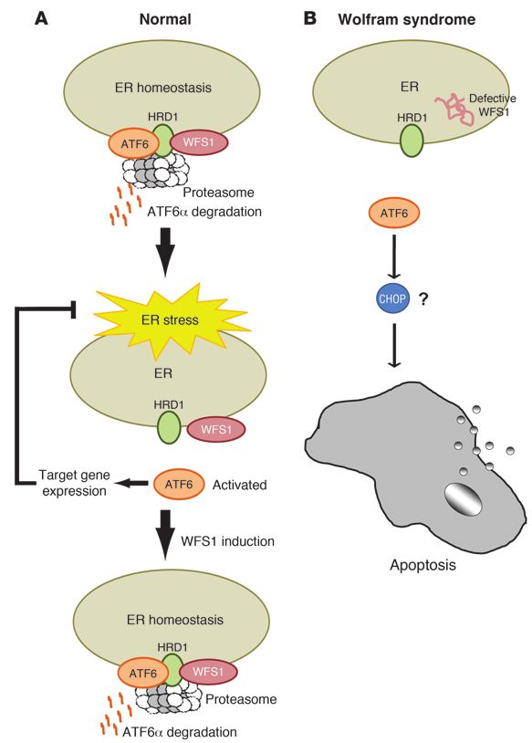
research article

746                The Journal of Clinical Investigation    http://www.jci.org    Volume 120    Number 3    March 2010
research article

Figure 2                                                                          target, BiP. Upregulation of BiP by WFS1 inhibition was cancelled
WFS1 regulates ATF6α protein levels. (A) IB analysis measured                     out by ATF6α inhibition (Supplemental Figure 4).
ATF6α and WFS1 levels in MIN6 cells expressing shGFP (control) or                   ATF6α protein levels were also measured in INS-1 832/13
shWFS1, as well as in MIN6 cells expressing shWFS1 or expressing
                                                                                  cells overexpressing WFS1. Full-length and nuclear ATF6α pro-
shWFS1 and rescued with WFS1 (n = 3). (B) IB analysis measuring
ATF6α, WFS1, IRE1α, and PERK levels in INS1 832/13 cells (treated
                                                                                  tein levels were suppressed in these cells, whereas there was no
with 2 mM DTT for 3 hours) overexpressing GFP (control) or WFS1                   significant change in protein levels of the other 2 master regu-
(n = 3). (C) Quantitative real-time PCR analysis of BiP, total Xbp-1,             lators of the UPR, IRE1 and PERK (Figure 2B). IRE1 and PERK
Chop, Ero1-α, Glut2, and Ins2 mRNA levels in INS1 832/13 cells over-              protein expression levels were not decreased even with higher
expressing GFP (control) or WFS1 (n = 3). (D) IB analysis of ATF6α                levels of WFS1 expression (Supplemental Figure 5). Suppression
and WFS1 in COS7 cells transfected with ATF6α-HA or ATF6α-HA and                  of ATF6α protein expression was also seen in a neuronal cell line
WFS1-FLAG at 2 different ratios, and in INS1 832/13 cells expressing              (Supplemental Figure 6). ATF6 target gene mRNA levels were
inducible WFS1 and treated with or without MG132. (E) IB analysis of
                                                                                  also suppressed in β cells overexpressing WFS1 (Figure 2C). The
ATF6α and WFS1 in MIN6 cells expressing shWFS1 and transfect-
ed with WT WFS1-FLAG or mutant P724L WFS1-FLAG and G695V                          relationship of WFS1 and ATF6 protein expression was found
WFS1-FLAG (n = 3). (F) IB analysis measuring ATF6α and WFS1 lev-                  to be dose-dependent: increased expression of WFS1 leads to a
els in INS1 832/13 cells expressing WT WFS1 or P724L WFS1 (n = 3).                decrease in ATF6 protein expression (Supplemental Figure 7).
(G) WFS1 was subjected to IP from COS7 cells expressing ATF6α-HA                  We asked whether this relationship was proteasome dependent.
or ATF6α-HA with WT, P724L, or G695V WFS1-Flag using an anti-                     Treatment of 2 WFS1-overexpressing cell lines with the pro-
Flag antibody. IPs and input proteins were analyzed using anti-HA and             teasome inhibitor MG132 rescued ATF6α protein levels (Figure
anti-Flag antibodies. **P < 0.01.                                                 2D). We cloned 2 missense mutants, WFS1 P724L and WFS1
                                                                                  G695V, and 1 inactivating mutant, WFS1 ins483fs/ter544, from
                                                                                  patient samples (13). Mutant variants of WFS1 did not affect
  Both WFS1 and ATF6α are transmembrane proteins localized to                     ATF6α protein levels in MIN6 cells expressing shRNA directed
the ER (3, 10), raising the possibility that the suppression of the               against WFS1 (Figure 2E and Supplemental Figure 8). This was
ATF6α reporter by WFS1 might be mediated by direct interaction                    also confirmed in INS-1 832/13 cells expressing the missense
between the WFS1 and ATF6α proteins. To confirm this, the asso-                   mutant WFS1 P724L (Figure 2F) and in neuronal cells express-
ciation of WFS1 with ATF6α was examined in the pancreatic β cell                  ing the missense mutant WFS1 G695V (Supplemental Figure 9).
line INS-1 832/13. WFS1 associated with ATF6α under nonstress                     Although ATF6α weakly interacted WFS1 P724L and WFS1
conditions (Figure 1D). To examine whether this interaction was                   G695V, there was no significant decrease in ATF6α protein levels
maintained during ER stress conditions, the cells were treated with               in these cells (Figure 2G).
the ER stress inducer dithiothreitol (DTT), which caused a disso-                   To assess the impact of WFS1 on ATF6α protein degradation,
ciation of ATF6α from WFS1 in a time-dependent manner, with                       cycloheximide experiments were performed. In MIN6 cells express-
almost complete dissociation 3 hours after treatment (Figure 1D).                 ing shRNA directed against WFS1, there was a block in ATF6α
This ER stress–dependent interaction was also observed in cells                   protein degradation, whereas in cells overexpressing WFS1, there
treated with another ER stress inducer, thapsigargin (Supplemental                was minimal ATF6α protein expression (Figure 3, A and B). WFS1
Figure 1; supplemental material available online with this article;               could not enhance the degradation of 2 other ER proteins suscep-
doi:10.1172/JCI39678DS1). To confirm that this interaction was                    tible to misfolding, TCRα and mutant alpha-1-antitrypsin NHK3
recovered after stress, cells were treated for 2 hours with DTT and               (refs. 22–24 and Supplemental Figure 10), which indicates that
then chased in normal media. As expected, the interaction of ATF6                 WFS1 specifically degrades ATF6α protein. WFS1 also enhanced
and WFS1 began to recover after a 1-hour chase (Figure 1E). This                  the ubiquitination of ATF6α. In cells expressing shRNA directed
interaction was also seen in the neuronal cell line Neuro2A (Supple-              against WFS1, there was a decrease in ATF6α ubiquitination after
mental Figure 2). Together, these data suggest that ATF6 is released              blocking proteasome activity (Figure 3C), whereas in cells over-
from WFS1 under stress in order to activate its target UPR genes.                 expressing WFS1, there was an enhancement of ATF6α ubiqui-
  WFS1 has a function in the degradation of ATF6α through the ubiquitin-          tination (Figure 3D). In Wfs1–/– mouse pancreata, ATF6α protein
proteasome pathway. Suppression of ATF6α transcriptional activity                 expression was strikingly high compared with control littermate
by WFS1 and the formation of an ATF6α-WFS1 complex led to the                     pancreata (Figure 3E), indicating that WFS1 functions in ATF6α
prediction that WFS1 regulates ATF6α function at the posttrans-                   protein expression in vivo. In samples from patients with WFS1
lational level. To test this prediction, we derived a pancreatic β cell           mutations, there was a higher expression of ATF6α protein com-
line, MIN6 cells, stably expressing a shRNA directed against WFS1.                pared with control samples (Supplemental Figure 11). Together,
Full-length as well as nuclear ATF6α protein levels increased approx-             these results indicate that WFS1 is important for regulating
imately 2-fold compared with control cells (Figure 2A). To confirm                ATF6α protein expression. When WFS1 was not present, there was
that upregulation of ATF6α protein is directly regulated by WFS1,                 increased expression of ATF6α protein and hyperactivation of its
we reintroduced a lentivirus expressing WFS1 into the cells express-              downstream effectors. This suggests that in response to ER stress,
ing shRNA directed against WFS1; ATF6 protein expression levels                   ATF6α escapes from WFS1-dependent degradation, is cleaved in
were again reduced when WFS1 was reintroduced (Figure 2A).                        the Golgi to its active form, and then translocates to the nucleus
  ATF6α mRNA was unchanged in the WFS1-knockdown INS-1                            to upregulate its UPR target genes.
832/13 cells, but ATF6α target genes, such as p58IPK and BiP (7, 8),                These data raised the possibility that WFS1 recruits ATF6α
were upregulated as predicted (Supplemental Figure 3). To further                 to the proteasome for its degradation. As we predicted, WFS1
confirm that this upregulation is directly regulated by ATF6α, we                 formed a complex with the proteasome (Figure 4A). When glyc-
suppressed ATF6α expression by siRNA in WFS1 knockdown INS-                       erol-gradient fractionation was performed on ER-isolated lysates,
1 832/13 cells and then measured expression levels of its major                   the proteasome ATF6α and WFS1 comigrated in the same high–

                               The Journal of Clinical Investigation    http://www.jci.org    Volume 120    Number 3    March 2010                747
research article Figure 3 WFS1 enhances ATF6α ubiquitination and degradation. (A) IB analysis measuring ATF6α, WFS1, and actin levels in MIN6 cells stably expressing shGFP (control) or shWFS1 treated with 40 μM cycloheximide (CX) for 0, 2, and 4 hours (n = 3). (B) IB analysis measuring ATF6α, WFS1, and actin levels in INS1 832/13 cells expressing GFP (control) or WFS1 treated with 40 μM cycloheximide for 0, 2, and 6 hours (n = 3). (C) ATF6α was subjected to IP using an anti-ATF6α antibody from an INS1 832/13 cells inducibly expressing shWFS1 (treated for 48 hours with 2 μM doxycycline) and treated with MG132 (20 μM) for 3 hours. IPs were then subjected to IB with anti-ubiquitin and anti-ATF6α antibodies, and input lysates were blotted with anti-ATF6α, anti-WFS1, and anti-actin antibodies (n = 3). (D) ATF6α was subjected to IP using an anti-ATF6α antibody, from INS1 832/13 cells overexpressing GFP (control) or WFS1, then treated with MG132 (0.1 μM) overnight. IPs were subjected to IB with anti-ubiquitin and anti-ATF6α antibodies. Input lysates were subjected to IB with anti-ATF6α, anti-WFS1, and anti-actin antibodies (n = 3). (E) Wfs1–/– and WT littermate mouse pancreata were analyzed by immunohistochemistry using anti-ATF6α and anti-insulin antibodies. Scale bars: 50 μm. 748 The Journal of Clinical Investigation http://www.jci.org Volume 120 Number 3 March 2010
research article
Figure 4
WFS1 forms a complex with the proteasome and ATF6α. (A) WFS1 was subjected to IP from INS1 832/13 cells using an anti-WFS1 specific
antibody. IPs were subjected to IB with anti–alpha 5 20S proteasome and anti-WFS1 antibodies. (B) IB analysis measuring CREB, actin, and
PDI levels using whole cell lysates or ER-isolated lysates of INS1 832/13 cells. ER-isolated lysates of INS1 832/13 cells were also subjected
to fractionation using a 10%–40% glycerol gradient. Fractions were analyzed by IB using anti–alpha 5 20s proteosome, anti-ATF6α, and anti-
WFS1 antibodies. Lanes were run on separate gels and were not contiguous. (C) WFS1 was subjected to IP from a mixture of fractions 10 and
11 using an anti-WFS1 antibody, and IP products were subjected to IB analysis using anti-alpha 5 20s proteosome, anti-ATF6α, and anti-WFS1
antibodies. ATF6 was subjected to IP from a mixture of fractions 9 and 12, and IP products were analyzed by IB with anti–alpha 5 20s proteo-
some and anti-ATF6α (n = 3).
molecular weight fractions, and a complex between them was sion of missense and inactivating WFS1 mutants did not increase
formed (Figure 4, B and C). or decrease HRD1 expression (Figure 5E). To determine whether
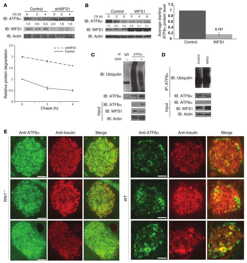
WFS1 stabilizes HRD1, which functions as an E3 ligase for ATF6α. WFS1 mutants interact with HRD1, comparable amounts of WT
Because WFS1 is localized to the ER membrane and recruits ATF6α and missense mutant WFS1 proteins were expressed together
to the proteasome, but is not itself an E3 ligase, we searched for with HRD1 in COS7 cells, and the interaction was monitored
ER-localized E3 ligases with which WFS1 could interact. A top can- by co-IP. HRD1 interacted with WT WFS1, but not with WFS1
didate was the ER-resident E3 ligase HRD1, which has a known role mutants (Figure 5F). Collectively, these results demonstrated that
in ER stress signaling (25, 26). SEL1/HRD3, which has an impor- WFS1 stabilizes and enhances the function of the E3 ligase HRD1
tant function in hydroxy-3-methylglutaryl-CoA reductase (HMG-R) through direct binding.
degradation (27), has been shown to interact with and stabilize Based on the ability of WFS1 to regulate ATF6α protein, as well
HRD1 (28), raising the possibility that WFS1 may also have a simi- as its function in stabilizing HRD1, it followed that WFS1 may be
lar function and could interact with HRD1. Indeed, WFS1 and recruiting ATF6α to HRD1 and that ATF6α is a substrate of HRD1.
HRD1 formed a complex (Figure 5A). We next asked whether WFS1 Indeed, HRD1 interacted with ATF6α (Figure 6A). In glycerol-gradi-
also plays a role in HRD1 protein expression. Inducible suppression ent fractionation experiments of ER-isolated lysates, HRD1, ATF6α,
of WFS1 in INS-1 832/13 cells expressing shRNA directed against and WFS1 were found to form a complex (Supplemental Figure 13).
WFS1 suppressed HRD1 protein expression (Figures 5B). To test We next analyzed the interaction between ATF6α and HRD1 under
the effect of WFS1 on HRD1 protein stability, we performed cyclo- ER stress conditions. ATF6α was released from HRD1 by DTT and
heximide experiments using MIN6 cells stably expressing shRNA thapsigargin treatments (Figure 6B), which indicates that the inter-
directed against WFS1. HRD1 protein expression was significantly action between these proteins is disrupted by ER stress. To study the
decreased in WFS1 knockdown cells compared with control cells, relationship between HRD1 and ATF6α protein expression levels,
and it was difficult to measure the stability of HRD1 (Figure 5C). the stability of ATF6α protein was measured in MIN6 cells stably
We further confirmed the effects of WFS1 on HRD1 protein expressing shRNA directed against HRD1 and control cells. HRD1
expression in vivo using Wfs1 –/– mice. As expected from the suppression in cells enhanced ATF6α protein stability (Figure 6C).
results using β cell lines, HRD1 expression was undetectable in In contrast, overexpression of HRD1 enhanced ATF6α protein deg-
islets of Wfs1–/– mice (Figure 5D). In addition, in samples from radation (Figure 6D). HRD1 also enhanced ATF6α ubiquitination,
patients with Wolfram syndrome, there was less HRD1 protein and lack of HRD1 decreased ATF6α ubiquitination (Figure 6, E and
expression compared with control samples (Supplemental Figure F). Collectively, these results indicate that the WFS1-HRD1 complex
12A). HRD1 expression did not affect WFS1 protein expression enhances ATF6α ubiquitination and degradation.
(Supplemental Figure 12B).
We next sought to compare the effects of WT WFS1 and WFS1 Discussion
mutants on HRD1 protein expression. Ectopic expression of WT In this study, we provide evidence that WFS1 plays a crucial role in
WFS1 increased HRD1 protein expression, whereas ectopic expres- regulating ATF6α transcriptional activity through HRD1-mediated
The Journal of Clinical Investigation http://www.jci.org Volume 120 Number 3 March 2010 749research article Figure 5 WFS1 interacts with and stabilizes the E3 ligase HRD1. (A) Hrd1 was subjected to IP from INS1 832/13 cells, and IPs were subjected to IB analysis using anti-WFS1 and anti-Hrd1 antibodies (n = 3). (B) Total lysates from INS1 832/13 cells inducibly expressing shWFS1 (treated with 2 μM doxycycline for 48 hours) were analyzed by IB using anti-WFS1, anti-Hrd1, and anti-actin antibodies (n = 3). (C) IB analysis measuring HRD1 levels in MIN6 cells stably expressing shGFP (control) or shWFS1 treated with 40 μM cycloheximide for 0, 0.5, 1, and 2 hours (n = 3). (D) Wfs1–/– and WT littermate mouse pancreata were analyzed by immunohistochemistry using anti-HRD1 and anti-insulin antibodies (n = 3). Scale bars: 100 μm. (E) COS7 cells were transfected with pcDNA3, HRD1–c-Myc, HRD1–c-Myc and WT WFS1, or HRD1–c-Myc and WFS1 mutants (P724L, G695V, and ins483fs/ter544) expression plasmids. Expression levels of HRD1–c-Myc, WFS1, and actin were measured by IB using anti–c-Myc, anti-WFS1, and anti-actin antibodies, respectively. WT and mutant WFS1 are denoted by single and double asterisks, respectively. (F) COS7 cells were transfected with pcDNA3, HRD1–c-Myc, HRD1–c-Myc and WT WFS1-Flag, HRD1–c-Myc and WFS P724L-Flag, and HRD1–c-Myc and WFS1 G695V-Flag expression plasmids. The lysates were subjected to IP with anti-Flag antibody and IB with anti–c-Myc antibody to study the interaction between HRD1 and WFS1. ubiquitination and proteasome-mediated degradation of ATF6α conditions. When stress is applied to the ER, such as through the protein. Based upon the data provided, we propose a pathway for chemical ER stress inducer DTT, ATF6α is released from WFS1. the negative-feedback regulation of the ER stress signaling network It is then released from the ER membrane and translocates to the by WFS1 (Figure 7). In healthy cells, WFS1 prevents dysregulated nucleus, where it upregulates stress signaling targets. At later time ER stress signaling by recruiting ATF6α to HRD1 and the protea- points, WFS1 is induced by ER stress, causing eventual degradation some for ubiquitin-mediated degradation under non–ER stress of ATF6α when ER homeostasis is established. In patients with 750 The Journal of Clinical Investigation http://www.jci.org Volume 120 Number 3 March 2010
research article

                                                                                                                Figure 6
                                                                                                                HRD1 is an E3 ligase for ATF6α. (A) HRD1
                                                                                                                was subjected to IP from INS1 832/13 cells
                                                                                                                treated for 3 hours with 30 μM MG132. IPs
                                                                                                                and input proteins were analyzed by IB
                                                                                                                using anti-ATF6α and anti-HRD1 antibod-
                                                                                                                ies. Lanes were run on the same gel but
                                                                                                                were noncontiguous (white line). (B) An anti-
                                                                                                                HRD1 antibody was used to IP HRD1 protein
                                                                                                                from INS1 832/13 cells untreated or treated
                                                                                                                with DTT (1 mM) and thapsigargin (Tg; 1 μM)
                                                                                                                for 3 hours. IPs were then subjected to IB
                                                                                                                analysis using anti-ATF6α, anti-HRD1, and
                                                                                                                anti-actin antibodies (n = 3). (C) IB analysis
                                                                                                                measuring ATF6α levels in MIN6 cells sta-
                                                                                                                bly expressing shGFP (control) or shRNA
                                                                                                                shHRD1 treated with 40 μM cycloheximide
                                                                                                                for 0, 4, and 6 hours (n = 3). (D) COS7 cells
                                                                                                                transfected with ATF6α-HA expression
                                                                                                                plasmid (control) or ATF6α-HA together
                                                                                                                with Hrd1-myc expression plasmids (Hrd1)
                                                                                                                were treated with 40 μM cycloheximide for
                                                                                                                0, 4, and 6 hours. Whole cell lysates were
                                                                                                                subjected to IB with an anti-HA antibody
                                                                                                                (n = 3). (E) ATF6α was subjected to IP using
                                                                                                                an anti-ATF6α antibody from INS1 832/13
                                                                                                                cells either mock transfected (control) or
                                                                                                                transfected with a Hrd1-Myc expression
                                                                                                                plasmid and treated with MG132 (20 μM) for
                                                                                                                3 hours. IPs were then subjected to IB with
                                                                                                                anti-ubiquitin and anti-ATF6α antibodies, and
                                                                                                                input lysates were blotted with anti-ATF6α,
                                                                                                                anti–c-Myc, and anti-actin antibodies (n = 3).
                                                                                                                (F) ATF6 was subjected to IP using an anti-
                                                                                                                ATF6α antibody from MIN6 cells stably
                                                                                                                expressing shGFP (control) or shHRD1 and
                                                                                                                treated with MG132 (20 μM) for 3 hours. IPs
                                                                                                                were then subjected to IB with anti-ubiquitin
                                                                                                                and anti-ATF6α antibodies, and input lysates
                                                                                                                were blotted with anti-HRD1, anti-ATF6α,
                                                                                                                and anti-actin antibodies (n = 3).

Wolfram syndrome or Wfs1–/– mice, ATF6α escapes from this pro-                   from ischemia/reperfusion-mediated apoptosis (31). This effect is
teasome-dependent degradation, leading to dysregulated ATF6α                     also mediated by BiP and GRP94 upregulation. Conversely, a recent
signaling. This ATF6α hyperactivation caused by the lack of WFS1                 report has shown that ATF6α upregulation attenuates diet-induced
is probably involved in β cell apoptosis.                                        obesity and insulin resistance (32). More importantly, it has been
   It has previously been shown that WFS1-deficient β cells are                  shown that WT mice are better protected from ER stress in vivo
susceptible to ER stress–mediated apoptosis (11, 18, 29). We con-                than are ATF6α knockout mice. ATF6α knockout hepatocytes have
firmed that knockdown of WFS1 made β cells sensitive to ER                       been shown to be more sensitive to ER stress–mediated cell death
stress–mediated cell death (Supplemental Figure 14). In addition,                compared with control hepatocytes (7). It is not surprising that pre-
we found that ectopic expression of an active form of ATF6α in β                 vious studies found induction of ATF6α to have beneficial effects
cells caused apoptosis (Supplemental Figure 15). Although this fits              on cell function and cell survival, because ATF6α is a major regu-
to our model that hyperactivation of ATF6α has a harmful effect on               lator for BiP, a central molecular chaperone in the ER (7, 8). The
β cells and leads to apoptosis, it may be contrary to previous studies           main conclusion of the present study is that chronic dysregulation
showing beneficial effects of ATF6α upregulation. For example, it                of the UPR, more specifically hyperactivation of ATF6α signaling,
has been shown that activation of the ATF6α pathway by a chemi-                  has a negative effect on β cell survival. It has been suggested that
cal compound protects neuronal cells from ER stress–mediated                     the UPR regulates both adaptive and apoptotic effectors (2, 33).
apoptosis (30). This effect is mediated by BiP upregulation. The                 The balance between these effectors depends on the nature of the
induction of ATF6α has also been shown to protect cardiomyocytes                 ER stress, whether it is tolerable or unresolvable. Thus, the UPR

                              The Journal of Clinical Investigation    http://www.jci.org    Volume 120    Number 3    March 2010                         751
research article
Figure 7
WFS1 controls steady-state levels of ATF6α protein and acti-
vation. (A) In normal cells, WFS1 recruits the ER transcription
factor ATF6α to the E3 ligase Hrd1 under non–ER stress condi-
tions. Hrd1 marks ATF6α with ubiquitin for proteasomal degra-
dation. Under ER stress, ATF6α dissociates from WFS1 and
undergoes proteolysis, and its soluble aminoportion, p60ATF6α,
translocates to the nucleus, where it upregulates ER stress tar-
get genes, such as BiP, CHOP, and XBP-1. At later time points,
WFS1 is induced by ER stress, which causes the eventual
degradation of ATF6α. (B) In patients with Wolfram syndrome
or Wfs1–/– mice, ATF6α escapes from the proteasome-depen-
dent degradation, leading to chronic hyperactivation of ATF6α
signaling. This ATF6α hyperactivation is involved in apoptosis
through apoptotic effectors of the UPR, such as CHOP.
enhancing the activity of a key ER-resident E3 ligase, HRD1
(28). Thus, a loss of functional WFS1 may affect ER stress
levels in 2 ways: (a) enhancing ATF6α signaling by increas-
ing the pool of ATF6α, and (b) destabilizing HRD1 protein
and thus its activity. The latter would independently con-
tribute to ER stress by promoting the buildup of unfolded
and misfolded proteins in the ER. In support of this is
our present finding that silencing of HRD1 in β cell lines
indeed led to mild ER stress (Supplemental Figure 16).
It has previously been shown that HRD1 is regulated by
the IRE1–XBP-1 pathway (26) and is activated at a later
time point during ER stress. The ATF6α pathway, how-
ever, is activated at an earlier phase (36). Thus, WFS1 may
also function as a switch from the ATF6α pathway to the
IRE1–XBP-1 pathway, through the stabilization of HRD1
and consequent destruction of ATF6α protein. A previous
publication has reported that WFS1 deficiency could lead
to increased HRD1 expression (17), contrary to our find-
ings. This discrepancy could be attributed to the fact that
WFS1 deficiency can cause dysregulated ER stress signaling
and can lead to hyperactivation of the IRE1–XBP-1 path-
acts as a binary switch between life and death. Our results demon- way under some circumstances.
strated that, in patients with Wolfram syndrome and Wfs1–/– mice, It has been established that WFS1 is induced under ER stress
unresolvable ER stress occurs in β cells and neurons, leading to a (11). However, WFS1 increased steadily over a 24-hour time peri-
switch toward apoptosis. od (data not shown). ATF6α upregulation, on the other hand,
ER stress is caused by both physiological and pathological stim- occurred much more rapidly. Thus, the initial pool of WFS1 protein
uli that can lead to the accumulation of unfolded and misfolded induced under stress may not have an inhibitory effect on ATF6α
proteins in the ER. Physiological ER stress can be caused by a large protein. In addition, we have shown that the ER-resident chap-
biosynthetic load placed on the ER, for example, during postpran- erone BiP also bound to ATF6α. The release of BiP from ATF6α
dial stimulation of proinsulin biosynthesis in pancreatic β cells. when unfolded/misfolded proteins accumulate in the ER may be
This stimulation leads to the activation of ER stress signaling and a key step in how ATF6α escapes WFS1-mediated proteolysis. BiP
enhancement of insulin synthesis (34). Under physiological ER binding may be essential for the interaction of ATF6α and WFS1,
stress conditions, activation of ER stress signaling must be tightly and, upon release, cause a conformational change in ATF6α, lead-
regulated because hyperactivation or chronic activation of this sig- ing to its consequent release from WFS1.
naling pathway can cause cell death. For example, when eukaryotic WFS1 is highly expressed in pancreatic β cells that are specialized
translation initiation factor 2α, a downstream component of ER for the production and regulated secretion of insulin to control
stress signaling, is hyperphosphorylated by the compound salu- blood glucose levels. In β cells, ER stress signaling needs to be tight-
brinal in pancreatic β cells, apoptosis is induced in these cells (35). ly regulated to adapt to the frequent fluctuations of blood glucose
Our results showed that WFS1 has an important function in the levels and to produce the proper amount of insulin in response to
tight regulation of ER stress signaling through its interaction with the need for it (34, 37). To achieve tight regulation, mammals may
a key transcription factor, ATF6α, thereby protecting cells from the have developed WFS1 as a regulator of HRD1 function in addition
damaging effects of hyperactivation of this signaling pathway. to SEL1. Higher expression of WFS1 in β cells, therefore, prevents
On the basis of our present results, we believe WFS1 plays a hyperactivation of ER stress signaling in these cells that are particu-
similar role in mammals as HRD3 does in yeast: stabilizing and larly sensitive to disruption of ER homeostasis and dysregulation
752 The Journal of Clinical Investigation http://www.jci.org Volume 120 Number 3 March 2010research article
of the UPR. Therefore, WFS1 has a role in protecting β cells from University, Japan). Hrd1-myc plasmid was a gift from M. Kaneko and M.
death by acting as an ER stress signaling suppressor. Nomura (Hokkaido University, Sapporo, Japan). TCRα plasmids were pro-
Mutations in the gene encoding WFS1 cause Wolfram syn- vided by R. Kopito (Stanford University, California), and NHK3 plasmids
drome, a genetic form of diabetes and neurodegeneration. It has were a gift from K. Nagata (Kyoto University). Entry vectors, destination
been proposed that a high level of ER stress causes β cell death and vectors, and viral plasmids for establishing lentiviral and retroviral cell lines
neurodegeneration in this disorder. Collectively, our results sug- were provided by E. Campeau (University of Massachusetts Medical School;
gest that a loss-of-function mutation of WFS1 causes the instabil- ref. 45). shRNA against WFS1 and GFP were purchased from the shRNA
ity of an E3 ligase, HRD1, leading to the upregulation of ATF6α Library Core Facility at the University of Massachusetts Medical School.
protein and hyperactivation of ATF6α signaling. Therefore, we IB. Cells were lysed in ice-cold TNE buffer (50 mM Tris HCl, pH 7.5; 150
predict that a loss-of-function or hypomorphic mutation of the mM NaCl; 1 mM EDTA; and 1% NP-40) containing a protease inhibitor
WFS1, HRD1, or ATF6α genes can cause ER stress–related disor- cocktail (Sigma-Aldrich) for 15 minutes on ice. Lysates were then cleared by
ders, such as diabetes, neurodegeneration, and bipolar disorder. centrifuging the cells at 12,000 g for 20 minutes at 4°C. Lysates were normal-
Indeed, it has been shown recently that common variants in WFS1 ized for total protein (30 μg/lane), separated using a 4%–20% linear gradi-
confer risk of type 2 diabetes (14–16), and there is a link between ent SDS-PAGE (BioRad), and electroblotted. Anti-WFS1 antibody was a gift
WFS1 mutations and type 1A diabetes (38, 39). It has also been from Y. Oka (Tohoku University). Anti-actin and anti-FLAG antibodies were
shown that ATF6α polymorphisms and haplotypes are associated purchased from Sigma-Aldrich. Anti-HA antibody was purchase from Stress-
with impaired glucose homeostasis and type 2 diabetes (40). Exces- gen, and anti-ATF6 and anti-GFP antibodies were purchased from Santa
sive β cell loss is a component of both type 1 and type 2 diabetes Cruz Biotechnology. Antigen retrieval was used for the anti-ATF6 antibody.
(41); therefore, WFS1 may have a key role in the protection of these Anti-ubiquitin and anti-IRE1 antibodies were purchased from Cell Signal-
cells from apoptosis through the tight regulation of ER stress sig- ing. Anti–alpha 5 20s proteasome antibody was purchased from Biomol, and
naling, thereby suppressing the diabetes phenotype. In addition, anti-PERK antibody was purchased from Rockland Inc. Anti–c-myc antibody
about 60% of patients with Wolfram syndrome have some mental was purchased from Roche. The anti–alpha-1-antitrypsin antibody was pur-
disturbance such as severe depression, psychosis, or organic brain chased from DakoCytomation. Anti-Hrd1 antibody was generated in rab-
syndrome, as well as impulsive verbal and physical aggression (42). bits using a KLH-conjugated synthetic peptide, TCRMDVLRASLPAQS. The
The heterozygotes who do not have Wolfram syndrome are 26-fold antibody specificity was tested by peptide/antigen competition.
more likely than noncarriers to have a psychiatric hospitalization DTT chase. INS-1 832/13 cells were treated with 1 mM DTT for 2 hours.
(43). The relative risk of psychiatric hospitalization for depression The DTT was washed out with normal media (RPMI 1640 supplemented
was estimated to be 7.1 in these heterozygotes (44). Therefore, it is with 10% FBS) for 0, 1, or 2 hours. Cells were lysed and subjected to IP with
possible that dysregulation of a negative feedback loop of ER stress anti-WFS1 antibodies.
signaling may have a pathological role in psychiatric illness. Fractionation. The ER was isolated from INS-1 832/13 cells using an Endo-
In this study, we focused on determining the physiological func- plasmic Reticulum Isolation kit (Sigma-Aldrich). The ER pellet was then
tion of WFS1 in ER stress signaling because of its implication in lysed in ice-cold TNE buffer containing 1% NP-40 and protease inhibitors,
diabetes, neurodegeneration, and bipolar disorder. We propose and the lysates were cleared and normalized. The ER lysates (1.0 ml) were
that WFS1 has a critical function in the regulation of ER stress sig- loaded on top of a glycerol gradient (10%–40%) prepared in PBS contain-
naling and prevents secretory cells, such as pancreatic β cells, from ing 1 mM DTT and 2 mM ATP and centrifuged at 4°C and 80,000 g for 20
dysfunction and premature death caused by hyperactivation of ER hours. We collected 32 fractions from the top of the tubes. Of each frac-
stress signaling through its interaction with the transcription fac- tion, 200 μl was precipitated with acetone, and the remaining pellet was
tor ATF6α. WFS1 could therefore be a key target for prevention lysed with 50 μl sample buffer. Precipitated proteins were then separated
and/or therapy of ER stress–mediated diseases such as diabetes, using a 4%–20% linear gradient SDS-PAGE and electroblotted.
neurodegenerative diseases, and bipolar disorder. IP. Cells were lysed in ice-cold TNE buffer with protease inhibitors for
15 minutes on ice; the lysates were then cleared by centrifuging the cells at
Methods 12,000 g for 20 minutes at 4°C. For IP of endogenous WFS1, 500 μg whole
Cell culture. Rat insulinoma cells (INS1 832/13) were a gift from C. New- cell extract from each sample was incubated with Protein G Sepharose 4
gard (Duke University Medical Center, Durham, North Carolina) and Fast Flow beads (GE Healthcare) and 4 μg anti-WFS1 antibody overnight
cultured in RPMI 1640 supplemented with 10% FBS. Mouse insulinoma at 4°C with rotation. After incubation, the beads were washed 3 times with
cells (MIN6) were maintained in DMEM with 15% FBS and 1% sodium TNE buffer followed by a final wash in 1× PBS. The IPs were resolved by
pyruvate. COS7 and Neuro2A cells were cultured in DMEM supplemented SDS-PAGE and then subjected to IB. For IP of ATF6, 6 μg anti-ATF6 anti-
with 10% FBS. For generation of cells inducibly overexpressing WFS1 and body was used; for HA, 2 μg anti-HA antibody was used; and for Hrd1, 4 μg
GFP, INS-1 832/13 cells stably expressing pTetR were transduced with a anti-Hrd1 antibody was used. As a control, lysates were subjected to IP as
lentivirus expressing human WFS1-FLAG or GFP and cultured in 2 μM described above using rabbit IgG.
doxycycline for 24 hours prior to protein/RNA isolation. For generation of Real-time PCR. Total RNA was isolated from the cells using the RNeasy
cells stably suppressing WFS1 or GFP, MIN6 cells were transduced with a Mini Kit (Qiagen) and reverse transcribed using 1 μg total RNA from
retrovirus expressing shRNA against mouse WFS1 or GFP. For overexpres- cells with Oligo-dT primer. For the thermal cycle reaction, the iQ5 sys-
sion of ATF6, Hrd1, and WFS1, COS7 cells were transfected with ATF6- tem (BioRad) was used at 95°C for 10 minutes, then 40 cycles at 95°C
HA and WFS1-FLAG expression plasmids using FuGENE 6 transfection for 10 seconds, and 55°C for 30 seconds. The relative amount for each
reagent (Roche Applied Science). As a control for coexpression, equivalent transcript was calculated by a standard curve of cycle thresholds for serial
amounts of pcDNA3 plasmid was used. DTT, cycloheximide, and MG132 dilutions of cDNA sample and normalized to the amount of actin. PCR
were purchased from Sigma-Aldrich. was performed in triplicate for each sample; all experiments were repeated
Plasmids. ATF6 plasmids were provided by R. Prywes (Columbia Univer- 3 times. The following sets of primers and Power SYBR Green PCR Master
sity, New York). GRP78 reporter plasmid was provided by K. Mori (Kyoto Mix (Applied Biosystems) were used for real-time PCR: rat actin, GCAAAT-
The Journal of Clinical Investigation http://www.jci.org Volume 120 Number 3 March 2010 753research article
GCTTCTAGGCGGAC and AAGAAAGGGTGTAAAACGCAGC; rat BiP, than 0.01 was considered statistically significant. In the figures, numbers
TGGGTACATTTGATCTGACTGGA and CTCAAAGGTGACTTCAATCT- below the lanes of blots denote relative protein amounts, as quantified by
GGG; rat Chop, AGAGTGGTCAGTGCGCAGC and CTCATTCTCCT- ImageJ software, normalized to the respective control. All graphical data are
GCTCCTTCTCC; rat total XBP-1, TGGCCGGGTCTGCTGAGTCCG shown as mean ± SD.
and ATCCATGGGAAGATGTTCTGG; rat ERO1-α, GAGAAGCTGTA-
ATAGCCACGAGG and GAGCCTTTCAATAAGCGGACTG; rat GLUT2, Acknowledgments
GTGTGAGGATGAGCTGCCTAAA and TTCGAGTTAAGAGGGAGCGC; We thank Keiji Tanaka, Hideki Yashiroda, Reid Gilmore, Sara
rat INS2, ATCCTCTGGGAGCCCCGC and AGAGAGCTTCCACCAAG. Evans, and Michael R. Green for comments on the manuscript;
Luciferase assay. COS7 cells were mock transfected, transfected with full- Julie Zhu for statistical analysis; Karen Sargent, Jessica Crisci, Yuan
length ATF6 or ΔATF6 with pcDNA3.0, or transfected with ATF6 with Lee, Linda Leehy, and Cris Welling for technical assistance; and
WFS1 expression plasmids along with rat GRP78 (ERSE) promoter lucif- Carlos Fonseca for support and advice. This work was supported
erase reporter gene, WT ATF6 binding site luciferase reporter gene (ATF- by grants from NIDDK, NIH (R01 DK067493), the Diabetes and
6GL3), or mutant ATF6 binding site luciferase reporter gene (ATF6m1GL3) Endocrinology Research Center at the University of Massachusetts
using Lipofectamine 2000 (Invitrogen). At 48 hours after transfection, Medical School (5 P30 DK32520), the Juvenile Diabetes Research
lysates were prepared using a Luciferase Assay System kit (Promega). The Foundation International, the Worcester Foundation for Biomedi-
light produced from the samples was read by a standard plate reading lumi- cal Research, and the Iacocca Foundation to F. Urano.
nometer. Each sample was read in triplicate and normalized against the sig-
nal produced from mock wells. All experiments were repeated 3 times. Received for publication April 27, 2009, and accepted in revised
Wfs1–/– animals. Wfs1–/– mice were provided by M.A. Permutt (Washing- form January 6, 2010.
ton University, St. Louis, Missouri). All procedures were reviewed and
approved by the University of Massachusetts Medical School IACUC Address correspondence to: Fumihiko Urano, Program in Gene
(assurance no. A-3306-01). Function and Expression, University of Massachusetts Medical
Statistics. To determine whether the treatment was significantly different School, Worcester, MA 01605-2324. Phone: 508.856.6012; Fax:
from the control, 2-tailed paired Student’s t test was used. A P value less 508.856.4650; E-mail: Fumihiko.Urano@umassmed.edu.
1. Ron D, Walter P. Signal integration in the endo- coding for a predicted transmembrane protein. EMBO Rep. 2001;2(5):415–422.
plasmic reticulum unfolded protein response. Nat Hum Mol Genet. 1998;7(13):2021–2028. 25. Kaneko M, et al. A different pathway in the endo-
Rev Mol Cell Biol. 2007;8(7):519–529. 13. Inoue H, et al. A gene encoding a transmembrane plasmic reticulum stress-induced expression of
2. Rutkowski DT, Kaufman RJ. That which does not protein is mutated in patients with diabetes mel- human HRD1 and SEL1 genes. FEBS Lett. 2007;
kill me makes me stronger: adapting to chronic ER litus and optic atrophy (Wolfram syndrome). Nat 581(28):5355–5360.
stress. Trends Biochem Sci. 2007;32(10):469–476. Genet. 1998;20(2):143–148. 26. Yamamoto K, et al. Human HRD1 promoter carries
3. Haze K, Yoshida H, Yanagi H, Yura T, Mori K. 14. Lyssenko V, et al. Clinical risk factors, DNA vari- a functional unfolded protein response element to
Mammalian transcription factor ATF6 is synthe- ants, and the development of type 2 diabetes. which XBP1 but not ATF6 directly binds. J Biochem.
sized as a transmembrane protein and activated by N Engl J Med. 2008;359(21):2220–2232. 2008;144(4):477–486.
proteolysis in response to endoplasmic reticulum 15. Franks PW, et al. Replication of the association 27. Hampton RY, Gardner RG, Rine J. Role of 26S pro-
stress. Mol Biol Cell. 1999;10(11):3787–3799. between variants in WFS1 and risk of type 2 dia- teasome and HRD genes in the degradation of 3-
4. Yoshida H, et al. ATF6 activated by proteolysis betes in European populations. Diabetologia. 2008; hydroxy-3-methylglutaryl-CoA reductase, an inte-
binds in the presence of NF-Y (CBF) directly to the 51(3):458–463. gral endoplasmic reticulum membrane protein.
cis-acting element responsible for the mamma- 16. Sandhu MS, et al. Common variants in WFS1 confer Mol Biol Cell. 1996;7(12):2029–2044.
lian unfolded protein response. Mol Cell Biol. 2000; risk of type 2 diabetes. Nat Genet. 2007;39(8):951–953. 28. Gardner RG, et al. Endoplasmic reticulum degra-
20(18):6755–6767. 17. Yamada T, et al. WFS1-deficiency increases endo- dation requires lumen to cytosol signaling. Trans-
5. Ye J, et al. ER stress induces cleavage of membrane- plasmic reticulum stress, impairs cell cycle pro- membrane control of Hrd1p by Hrd3p. J Cell Biol.
bound ATF6 by the same proteases that process gression and triggers the apoptotic pathway spe- 2000;151(1):69–82.
SREBPs. Mol Cell. 2000;6(6):1355–1364. cifically in pancreatic beta-cells. Hum Mol Genet. 29. Ishihara H, et al. Disruption of the WFS1 gene in
6. Shen J, Chen X, Hendershot L, Prywes R. ER stress 2006;15(10):1600–1609. mice causes progressive beta-cell loss and impaired
regulation of ATF6 localization by dissociation of 18. Riggs AC, et al. Mice conditionally lacking the stimulus-secretion coupling in insulin secretion.
BiP/GRP78 binding and unmasking of Golgi local- Wolfram gene in pancreatic islet beta cells exhibit Hum Mol Genet. 2004;13(11):1159–1170.
ization signals. Dev Cell. 2002;3(1):99–111. diabetes as a result of enhanced endoplasmic 30. Kudo T, et al. A molecular chaperone inducer
7. Wu J, et al. ATF6alpha optimizes long-term endo- reticulum stress and apoptosis. Diabetologia. protects neurons from ER stress. Cell Death Differ.
plasmic reticulum function to protect cells from 2005;48(11):2313–2321. 2008;15(2):364–375.
chronic stress. Dev Cell. 2007;13(3):351–364. 19. Kakiuchi C, Ishiwata M, Hayashi A, Kato T. XBP1 31. Martindale JJ, et al. Endoplasmic reticulum stress
8. Yamamoto K, et al. Transcriptional induction of induces WFS1 through an endoplasmic reticulum gene induction and protection from ischemia/reper-
mammalian ER quality control proteins is medi- stress response element-like motif in SH-SY5Y fusion injury in the hearts of transgenic mice with
ated by single or combined action of ATF6alpha cells. J Neurochem. 2006;97(2):545–555. a tamoxifen-regulated form of ATF6. Circ Res. 2006;
and XBP1. Dev Cell. 2007;13(3):365–376. 20. Wang Y, Shen J, Arenzana N, Tirasophon W, Kaufman 98(9):1186–1193.
9. Hong M, Li M, Mao C, Lee AS. Endoplasmic reticu- RJ, Prywes R. Activation of ATF6 and an ATF6 DNA 32. Ye R, et al. Grp78 heterozygosity promotes adap-
lum stress triggers an acute proteasome-depen- binding site by the endoplasmic reticulum stress tive unfolded protein response and attenuates diet-
dent degradation of ATF6. J Cell Biochem. 2004; response. J Biol Chem. 2000;275(35):27013–27020. induced obesity and insulin resistance. Diabetes. 2010;
92(4):723–732. 21. Shen J, Snapp EL, Lippincott-Schwartz J, Prywes 59(1):6–16.
10. Takeda K, et al. WFS1 (Wolfram syndrome 1) gene R. Stable binding of ATF6 to BiP in the endoplas- 33. Han D, et al. IRE1alpha kinase activation modes
product: predominant subcellular localization to mic reticulum stress response. Mol Cell Biol. 2005; control alternate endoribonuclease outputs to deter-
endoplasmic reticulum in cultured cells and neuronal 25(3):921–932. mine divergent cell fates. Cell. 2009;138(3):562–575.
expression in rat brain. Hum Mol Genet. 2001; 22. Yu H, Kaung G, Kobayashi S, Kopito RR. Cytosolic 34. Lipson KL, et al. Regulation of insulin biosynthesis
10(5):477–484. degradation of T-cell receptor alpha chains by the in pancreatic beta cells by an endoplasmic reticu-
11. Fonseca SG, et al. WFS1 is a novel component of the proteasome. J Biol Chem. 1997;272(33):20800–20804. lum-resident protein kinase IRE1. Cell Metab. 2006;
unfolded protein response and maintains homeosta- 23. Yu H, Kopito RR. The role of multiubiquitination 4(3):245–254.
sis of the endoplasmic reticulum in pancreatic {beta}- in dislocation and degradation of the alpha sub- 35. Cnop M, et al. Selective inhibition of eukary-
cells. J Biol Chem. 2005;280(47):39609–39615. unit of the T cell antigen receptor. J Biol Chem. 1999; otic translation initiation factor 2 alpha dephos-
12. Strom TM, et al. Diabetes insipidus, diabetes mel- 274(52):36852–36858. phorylation potentiates fatty acid-induced endo-
litus, optic atrophy and deafness (DIDMOAD) 24. Hosokawa N, et al. A novel ER alpha-mannosidase- plasmic reticulum stress and causes pancreatic
caused by mutations in a novel gene (wolframin) like protein accelerates ER-associated degradation. beta-cell dysfunction and apoptosis. J Biol Chem.
754 The Journal of Clinical Investigation http://www.jci.org Volume 120 Number 3 March 2010research article
2007;282(6):3989–3997. responsible for Wolfram syndrome (WFS1/wol- betes. 2003;52(1):102–110.
36. Yoshida H, Matsui T, Hosokawa N, Kaufman RJ, framin) in Japanese: possible contribution of the 42. Swift RG, Sadler DB, Swift M. Psychiatric find-
Nagata K, Mori K. A time-dependent phase shift Arg456His mutation to type 1 diabetes as a nonau- ings in Wolfram syndrome homozygotes. Lancet.
in the mammalian unfolded protein response. Dev toimmune genetic basis. Biochem Biophys Res Com- 1990;336(8716):667–669.
Cell. 2003;4(2):265–271. mun. 2000;268(2):612–616. 43. Swift RG, Polymeropoulos MH, Torres R, Swift M.
37. Fonseca SG, Lipson KL, Urano F. Endoplasmic 40. Meex SJ, et al. Activating transcription factor 6 Predisposition of Wolfram syndrome heterozygotes
reticulum stress signaling in pancreatic beta-cells. polymorphisms and haplotypes are associated to psychiatric illness. Mol Psychiatry. 1998;3(1):86–91.
Antioxid Redox Signal. 2007;9(12):2335–2344. with impaired glucose homeostasis and type 2 dia- 44. Swift M, Swift RG. Wolframin mutations and hos-
38. Nakamura A, et al. A novel mutation of WFS1 gene betes in Dutch Caucasians. J Clin Endocrinol Metab. pitalization for psychiatric illness. Mol Psychiatry.
in a Japanese man of Wolfram syndrome with posi- 2007;92(7):2720–2725. 2005;10(8):799–803.
tive diabetes-related antibodies. Diabetes Res Clin 41. Butler AE, Janson J, Bonner-Weir S, Ritzel R, Rizza 45. Campeau E, et al. A versatile viral system for expres-
Pract. 2006;73(2):215–217. RA, Butler PC. Beta-cell deficit and increased beta- sion and depletion of proteins in mammalian cells.
39. Awata T, et al. Missense variations of the gene cell apoptosis in humans with type 2 diabetes. Dia- PLoS ONE. 2009;4(8):e6529.
The Journal of Clinical Investigation http://www.jci.org Volume 120 Number 3 March 2010 755You can also read