ScienceDirect The evolving capabilities of enzyme-mediated proximity labeling - Zou Lab
←
→
Page content transcription
If your browser does not render page correctly, please read the page content below
Available online at www.sciencedirect.com

                                              ScienceDirect

The evolving capabilities of enzyme-mediated
proximity labeling
Ying Zhou1 and Peng Zou1,2

Abstract                                                                    dynamic liquid-like condensates that form via liquide
The subcellular organization of proteins and RNA molecules is               liquid phase separation (LLPS) [8]. For example, stal-
crucial for their proper functions. Over the past decade, both              led translation-initiation causes mRNAs and mRNA-
ligase-mediated and peroxidase-mediated proximity labeling                  binding proteins (RBPs) to assemble into stress gran-
(PL) techniques have been developed to map biomolecules at                  ules (SGs), in a process of protecting cells from oxida-
near-nanometer spatial resolution and subminute temporal                    tive damages or other cellular stress [9].
resolution. These methods are shedding light on the spatial
arrangement of proteome and transcriptome in their native                   The subcellular proteome and transcriptome have been
context. Here, we review the recent evolution and applications              traditionally investigated by co-immunoprecipitation
of PL techniques, compare and contrast the two classes of                   (co-IP) and biochemical fractionation. However, both
methods, and highlight emerging trends and future                           methods require prior cell lysis, which is prone to losing
opportunities.                                                              low-affinity and transient proteineprotein interactions.
                                                                            In addition, co-IP is limited by the availability of high-
Addresses                                                                   quality antibodies against the bait, while biochemical
1
  College of Chemistry and Molecular Engineering, Synthetic and
                                                                            fractionation often suffers from incomplete purification.
Functional Biomolecules Center, Beijing National Laboratory for Mo-
lecular Sciences, Key Laboratory of Bioorganic Chemistry and Mo-            For example, the transcriptomic profiling of isolated
lecular Engineering of Ministry of Education, Peking University, Beijing,   mitochondria has identified abundant contaminations
100871, China                                                               from the cytoplasm [10]. Furthermore, not all subcel-
2
  Peking-Tsinghua Center for Life Sciences, PKU-IDG/McGovern                lular structures are amenable to fractionation [11]. Over
Institute for Brain Research, Peking University, Beijing, 100871, China
                                                                            the past decade, enzyme-mediated proximity labeling
Corresponding author: Zou, Peng (zoupeng@pku.edu.cn)                        (PL) techniques have emerged as powerful tools for
                                                                            locating proteins and RNAs in live cells. In these
                                                                            methods, an engineered enzyme is expressed at a spe-
Current Opinion in Chemical Biology 2021, 60:30–38                          cific subcellular locale, where it catalyzes the in situ
This reviews comes from a themed issue on Omics                             synthesis of a highly reactive small-molecule interme-
Edited by Nichollas Scott and Laura Edgington-Mitchell                      diate, which subsequently diffuses away and reacts with
For a complete overview see the Issue and the Editorial
                                                                            proteins and/or RNAs to form a covalent label
                                                                            (Figure 1a). Due to its limited lifetime, the local density,
                                                                            and hence the labeling efficiency of the intermediate
https://doi.org/10.1016/j.cbpa.2020.06.013                                  drops off as a function of the distance from the enzyme.
1367-5931/© 2020 Elsevier Ltd. All rights reserved.                         Thus, all else being equal, proteins/RNAs proximal to
                                                                            the enzyme are more likely to be labeled than distal
Keywords                                                                    ones. Compared with biochemical fractionation, PL
Proximity labeling, APEX, BioID, Peroxidase, Biotin ligase.                 could access information from subcellular compartments
                                                                            that are impossible to purify or highly dynamic (e.g.
                                                                            signaling complexes, LLPS, etc.). PL also complements
Introduction                                                                co-IP studies because it is capable of mapping distant
Eukaryotic cells are elaborately divided into subcellular                   proteineprotein interactions, with an ‘action contour
compartments that feature distinct biochemical char-                        map’ that spans over several ‘interaction layers’
acteristics. The spatial organization of proteins and                       (Figure 1a).
RNAs in these subcellular regions is intimately linked to
their biological functions, including signal transduction                   In this review, we highlight several emerging trends of
[1,2], localized protein synthesis [3,4], regulation of                     PL technology development and new avenues of its
chromatin structure [5], etc. While most famously                           applications. As discussed below, enzyme-mediated PL
observed at membrane-bound organelles (either in the                        is now moving rapidly from membrane-enclosed com-
interior [6] or on the surface [7]), the subcellular                        partments to open subcellular space, from protein-
targeting of proteins and RNAs have also been discov-                       centered profiling to RNA/DNA-centered analysis, and
ered in membrane-less compartments, such as highly                          from cell culture to animal. As this is not intended as a

Current Opinion in Chemical Biology 2021, 60:30–38                                                                  www.sciencedirect.com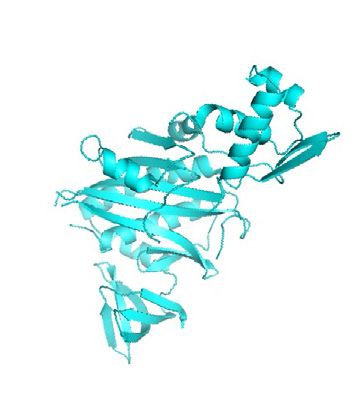
Spatial proteomics and transcriptomics Zhou and Zou 31
Figure 1
(a)
Affinity Mass spec
intensity
purification analysis
m/z
High
Proximity labeling throughput ATCACAGTGGGACTCCT
sequencing CGAAGGACCAGCAGAAA
GGACACCCAGCGGGCTG
(b) HO ATGAAACATCAAACAAT
O
O H HN GAAGAGCAATCAGTCAG
N NH ……
H
S H
H2O2 HO O
O H HN
O O OH
O H HN N NH
H
N NH S H
H OH
S H
Phenoxyl radical Chemical Photo- Chromatin Direct RNA
crosslinking crosslinking labeling with labeling
Evolved faster DTB- dCas9-APEX2
enzyme phenol APEX- Proximity APEX-seq
A134PAPEX
K14D/E112K/W41FAPX probe RIP -CLIP GLoPro
Enhanced & Split
ascorbate APEX APEX2 Spot-ID C-BERST APEX2
peroxidase
2012 2013 2014 2015 2016 2017 2018 2019 2020
A smaller biotin ligase
from A. aeolicus Contact-ID
BioID
&
BirA* = R118GBirA BioID2 Split- TurboID & Split-TurboID
BioID miniTurbo RaPID
O
O H HN Evolved faster BASU from BASU fused ChromID
HO NH
and smaller BirA* B. subtilis with chromatin
S H
readers
ATP O O H HN
O
O
N O
O P NH2 H HN
H 2N N O O NH
OH N NH
S H H
N N S H
HO OH
Bio-AMP
Current Opinion in Chemical Biology
The mechanism and evolution of enzyme-mediated proximity labeling. (a) Schematics of proximity labeling (PL) workflow. An enzyme (a biotin ligase
or a peroxidase) is targeted to a specific subcellular location (e.g. mitochondrial matrix) via fusion with protein markers or signal peptides. Proximity is
achieved through the in situ enzymatic synthesis of biotin-conjugated reactive intermediates, which subsequently diffuse away and react with nearby
proteins/RNAs. The nanometer-scale action radius of the intermediates (shown as a red contour map) covers both proteins/RNAs that tightly associate
with the bait and those that loosely interact in the same compartment, enabling PL to reach over multiple layers of protein–protein/RNA interactions. After
cell lysis, biotinylated proteins are collected by affinity purification and characterized by mass spectrometry. Biotinylated RNAs are analyzed by high-
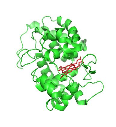
throughput sequencing. (b) The mechanism and technology development timeline of PL. In the presence of H2O2, APEX (green, PDB 1V0H) converts
biotin phenol to phenoxyl free radical, which reacts with the adjacent tyrosine residues. In the presence of ATP, BioID (cyan, PDB 2EWN) activates biotin
into bio-AMP, which reacts with lysine residues of neighboring proteins. The timeline describes a brief history of major APEX- and BirA-mediated PL
techniques. Methods highlighted in green, blue, and pink refer to protein-centered, RNA-centered, and DNA-centered profiling, respectively.
comprehensive chronology of all PL applications, inter- action radius of the reactive intermediate. Depending
ested readers may find further information in several on the nature of enzymes and chemical reactions, PL is
excellent reviews [12e15]. broadly categorized as biotin ligase mediated or peroxi-
dase mediated.
The evolution of promiscuous enzymatic
labeling BioID (also known as BirA* [16]) is a 35 kDa Escherichia
The high spatial resolution of PL technique is achieved coli biotin ligase mutant R118GBirA. In the presence of
via both genetic targeting of the enzyme and the small ATP, BioID converts biotin into biotinyl-50 -AMP (bio-
AMP), which is subsequently released into the cellular
www.sciencedirect.com Current Opinion in Chemical Biology 2021, 60:30–3832 Omics
environment and reacts with the lysine side chain of fold higher activity and reduces the labeling time
nearby proteins [17](Figure 1b). Initially developed in window down to 10 min. Meanwhile, a truncation
2012, BioID has been broadly applied to cultured variant with reduced protein size and comparable la-
mammalian cells [13], plant cells [18], mouse [19], beling efficiency was introduced as miniTurbo [24]. In
yeast [20], etc., to profile proteomes in numerous sub- the second strategy, by analyzing the sequence and
cellular structures, including the nuclear lamina [17], structural alignment of biotin ligases from different
the nuclear pore complex [21], and centriolar satellites species, Khavari and coworkers introduced mutations to
[22](Figure 2). In 2016, an improved smaller biotin a B. subtilis biotin ligase to obtain a rationally designed
ligase (BioID2) was developed from an Aquifex aeolicus PL enzyme, BASU [25]. When used to investigate the
enzyme to enable more-selective targeting of fusion RNA-binding proteome (RaPID), BASU exhibited ki-
proteins, leading to a better coverage of the nuclear pore netics more than three orders of magnitude faster than
complex components [23]. BioID [25]. However, in another study, the activity of
BASU was shown to be comparable with BioID/BioID2
BioID/BioID2 requires several-hour labeling due to slow in the cytoplasm [24].
enzymatic kinetics [24]. Two strategies have been used
to address this limitation. In the first one, Ting and Peroxidase-mediated PL offers even faster reaction ki-
coworkers applied yeast display-based directed evolu- netics by using free radical chemistry, as exemplified by
tion to enhance the catalytic efficiency of BioID. The horseradish peroxidase (HRP). Peroxidases could act on
resulting ligase mutant, termed TurboID, has up to 23- a wide variety of aromatic substrates, including luminol
Figure 2
(a) (b) (c)
Primary cilia Plasma membrane Mitochondria
APEX
BioID
Plasma
membrane
Endoplasmic
reticulum
Ciliary barrier
(d) (e)
GPCR
(g) Centriolar Microtubule (f)
satellites
Mother
centriole (h) Nuclear envelope
Pericentriolar
material Lamin A
Current Opinion in Chemical Biology
Representative applications of enzyme-mediated proximity labeling. (a) Primary cilia-targeted APEX2 identified several ciliary signaling molecules,
including PKA, AMPK, and LKB1. (b) STIM1-fused APEX2 mapped the proteome of endoplasmic reticulum (ER)–plasma membrane junctions and
uncovered an ER-localized transmembrane protein STIMATE. (c) Applications of APEX-mediated PL in mitochondria. (d) Interaction network and agonist
response of GPCR were tracked by space- and time-resolved APEX PL (e–f) Applications of BioID to analyze the protein–protein interaction networks of
two cytosolic membrane-less ribonucleoprotein complexes known as processing bodies (e) and stress granules (f). (g) A comprehensive proteomic
profiling of centriolar satellites by BioID-suggested satellite assembly is independent of centrosome. (h) Profiling nuclear lamina proteome with BioID.
Current Opinion in Chemical Biology 2021, 60:30–38 www.sciencedirect.comSpatial proteomics and transcriptomics Zhou and Zou 33
Figure 3
(a) (b) (c) Isoproterenol
Excitatory
synapse Organelle contact site
β2AR
Inhibitory
HRP synapse Recycle Endo-
Dimerization cytosis
Endosome
Lysosome APEX2
(d)
Alkyne Trypsin N
N
N B SA bead On-bead N
N N
phenol digestion N3 B incubation UV cleavage
Click
Desthiobiotin H HN
O SA bead On-bead protein O
phenol NH incubation digestion Elution H
HN NH
H
H
(e) (f) Chromatin reader domains
sgRNA
BASU
dCas9
APEX2 Biotin
Chromatin Epigenetically
modified chromatin
(g) (h) RNA (i)
APEX2 APEX2
BASU
λN λN
RBP 4SU
RNA
Chemical
Photo-crosslinking
crosslinking Biotin-phenol
RBP
RBP
BoxB BoxB
Current Opinion in Chemical Biology
Emerging trends of enzyme-mediated proximity labeling. (a–b) From membrane-bound organelles to open-space compartments. Peroxidase-based
PL was used to distinguish between the excitatory and inhibitory synaptic cleft proteome (a). Split enzymes were used to profile proteome at organelle
contact sites (b). (c) From static to dynamic view of local proteome. Time-resolved APEX PL reveals dynamics of GPCR interaction network. (d) From
protein-centric to peptide-centric analysis. Top: APEX PL with desthiobiotin-phenol whose reduced affinity to streptavidin (SA) facilitates recovery of
labeled peptides. Bottom: APEX PL with alkyne-phenol allows functionalization of alkynylated peptides with azide-conjugated biotin bearing a UV-
cleavable linker. Enriched peptides are released from streptavidin-coated beads via UV irradiation and identified by tandem mass spectrometry. Both
approaches improves detection of labeling sites (e–f) From protein-centered to DNA-centered proteomic profiling. Proteins associated with specific
genomic loci (e) or epigenetic marks (f) are labeled with dCas9-targeted APEX2 (e) or chromatin reader-fused BASU (f), respectively. (g) From protein-
centered to RNA-centered proteomic profiling. The RNA-binding proteome is captured by BASU targeted via lN peptide/BoxB interaction (h–i) From
proteomic to transcriptomic analysis. APEX PL is combined with protein-RNA crosslinking, via either formaldehyde or UV irradiation. Following purification
of biotinylated protein–RNA complex, RNA could be identified by high-throughput sequencing (h). RNA could also be directly labeled by APEX PL (i).
www.sciencedirect.com Current Opinion in Chemical Biology 2021, 60:30–3834 Omics
for chemiluminescence [26], 3,30 -diaminobenzidine for ones. This example demonstrates that peroxidase-
electron microscopy contrast [27], and tyramine for mediated PL is both applicable to primary culture and
protein labeling and signal amplification [28]. Because capable of dealing with small amount of material [11].
HRP is inactive when expressed in the cytosolic com-
partments [27], its applications have been restricted to The high spatial resolution of PL has enabled the
the cell surface [11,29]. APEX is a 28 kDa engineered investigation of proteineprotein interaction networks in
peroxidase that generates phenoxyl free radicals in living LLPS-driven membrane-less organelles, such as SGs
cells to label proximal proteins (Figure 1b). The first and processing bodies. In 2018, through fusing APEX2
application of APEX in the mitochondrial matrix to an SG marker G3BP1, Yeo and coworkers discovered a
required only 1-min labeling and captured 495 proteins pre-existing SG protein interaction network in un-
with >95% specificity, which helped reassign the proper stressed cells and further identified stress-specific and
localization of heme metabolic enzymes, PPOX and cell typeespecific SG subproteomes [36]. Coincidently,
CPOX [30]. Gingras and coworkers applied BioID to uncover 7424
unique proximity interactions and 144 protein compo-
In 2015, through yeast display-based directed evolution, nents of cytosolic RNA granules [37].
Ting and coworkers identified a point mutation of APEX
that substantially improved its catalytic efficiency To further improve the spatial specificity at organelle
(APEX2), likely by reducing its susceptibility to H2O2- interface, a split enzyme strategy has been developed
induced inhibition [31]. Interestingly, this mutation is (Figure 3b). Enzymes such as BioID [38,39], TurboID
conserved in secreted peroxidases including HRP, thus [40], and APEX2 [41] are split into two catalytically
effectively turning APEX2 more HRP-like [31]. Over inactive fragments which only recover biotinylation ca-
the past 8 years, APEX/APEX2 has been applied to a pabilities when they physically interact and reconstitute
wide variety of subcellular compartments [15], at membraneemembrane contact sites. These split
including the mitochondrial intermembrane space versions of enzymes have enabled the specific profiling
(IMS) and outer membrane [32,33], the EReplasma of proteomes at the ER-mitochondrial contact (split-
membrane contact [34], and primary cilia [35] TurboID) [40] and mitochondria-associated membrane
(Figure 2). (Contact-ID) [38].
Toward higher spatial and temporal The fast reaction kinetics of APEX has enabled mapping
resolution dynamic proteomic changes with high temporal resolu-
Recently, the applications of PL has been extended from tion (Figure 3c). In 2017, APEX2 was fused to G-protein
membrane-bound organelles to open subcellular space coupled receptors (GPCRs) to investigate signaling
(Figure 3a). Initial efforts of PL in the IMS revealed complex assembly upon ligand binding at minute-level
high cytosolic background, particularly those proteins temporal resolution. While Kruse and coworkers
located near the mitochondrial outer membrane. To revealed the spatial and temporal clues to GPCR
improve the spatial specificity of PL, a ratiometric signaling and internalization kinetics stimulated by
quantitative proteomic workflow has been designed, different ligands [42], Lobingier et al. captured the
where the enzyme is targeted to two neighboring sub- location and function of previously unknown GPCR
cellular compartments (e.g. IMS and cytosol) in two network [43].
parallel PL experiments, respectively. The ratio of bio-
tinylation extent (e.g. IMS/cytosol) is then used as a Beyond protein-centered analysis in cell
“cytological ruler” to measure the relative proximity of lines
labeled proteins to either subcellular locations. Because Target identification in PL traditionally focuses on
the effects of protein abundance, local chemical envi- protein-level quantitation (protein ID), where bio-
ronment, steric accessibility, etc., are cancelled out tinylated proteins are first enriched and then proteo-
during the ratio calculation, this ratiometric PL lytically digested into peptides for tandem mass
approach has eliminated bias toward these factors, thus spectrometry (MS/MS) analysis [30]. As the majority of
effectively creating a proximity contour map. analyzed peptides are not tagged with biotin, this
workflow is inefficient at characterizing the sites of
A prominent example of PL in open subcellular space is biotin conjugation (site ID). Unlike protein ID, which is
the study of synapse-specific proteome in cultured prone to false positives arising from nonspecific protein
neurons, where biochemical fractionation is incapable of binding to affinity purification beads, site ID could
distinguishing excitatory synapses from inhibitory ones. unambiguously assign protein targets because the
By targeting HRP to the plasma membrane (not specific labeled peptides are directly detected by MS/MS. An
to synapses), excitatory and inhibitory synapses, additional benefit of site ID is to provide information of
respectively, ratiometric PL successfully identified 199 protein structural accessibility to enzymatic labeling,
proteins at excitatory synaptic clefts and 42 at inhibitory information that can be used to derive membrane
Current Opinion in Chemical Biology 2021, 60:30–38 www.sciencedirect.comSpatial proteomics and transcriptomics Zhou and Zou 35
protein topology or protein complex conformation [44]. landmarks and correlate them to genome architecture
However, the tight interaction between biotinylated and protein localization [56]. Ingolia and coworkers
peptides and streptavidin beads has hindered site ID applied APEX-seq and APEX-mediated protein labeling
[45]. to comprehensively understand the organization of
translation-initiation complexes and SGs [57]. Through
One solution is to weaken the interaction. Rhee and screening a panel of aromatic APEX substrates, the ef-
coworkers replaced the biotin moiety in the APEX ficiency of APEX-seq was substantially enhanced by
substrate with desthiobiotin that has reduced binding replacing biotin-phenol with biotin-arylamine probes
affinity to streptavidin beads (Spot-ID) and applied this [58].
technique to map the topological direction of 135
mitochondrial membrane proteins [46] (Figure 3d). Finally, proximity-dependent labeling has been applied
Similarly, the replacement of streptavidin with anti- not only in the cell culture but also in animals. BioID has
biotin antibody has resulted in more than 30-fold higher been applied to discover inhibitory postsynaptic prote-
ID rate for biotinylated peptides, as demonstrated by ome in neonatal mice [19]. Tissue- and subcellular
Carr and coworkers [47]. Another solution is to substi- location-specific expression of APX in Caenorhabditis
tute the biotin moiety in the APEX substrate with a elegans has identified over 3000 proteins in the nucleus
clickable bioorthogonal functional handle, such as an and the cytoplasm from four tissues [59]. To investigate
alkyne group. Labeled proteins could be conjugated the molecular mechanism underlying brain wiring, the
with an affinity tag containing a photocleavable linker, cell surface proteome was profiled by HRP in the fly
which facilitates the identification of labeling sites by brain [60].
MS/MS [48] (Figure 3d). When analyzing site ID data
sets, one should be mindful of potential false negatives, Outlook for proximity labeling
as site ID reflects not only topology and complex Given the rapidly expanding PL toolkits, one may
geometry but also the presence and accessibility of wonder which method is better. The answer depends on
particular target side-chains (i.e. lysine for BioID and the specific experimental setup, including temporal
tyrosine for APEX). resolution, sensitivity to oxidative stress, probe delivery
issues, etc. The subminute temporal resolution of APEX
In addition to studying proteineprotein interactions, method remains unrivaled, and the requirement for
PL has been applied to map proteins associated with H2O2 delivery provides a means for temporal gating,
specific genomic loci (chromatin-binding proteomes) which is a prerequisite for studying dynamic processes
(Figure 3eef). Two methods (C-BERST [49] and such as ligand-triggered cellular signaling. However, if
GLoPro [50]) combine dCas9-based genome targeting the temporal resolution is not a major concern, TurboID
and APEX2 labeling to discover proteomes bound to would be a good choice as it avoids the harsh treatment
telomeres, centrosomes, and MYC promoters. Besides, of H2O2. The dependence of BP probe delivery also
ChromID has been introduced to obtain protein net- poses a challenge to APEX application in live tissue
works at DNA methylation and histone trimethylation [59,61]. In cases where both APEX and BioID are
residues, through fusing engineered chromatin readers suitable, it would be informative to compare results from
to BASU [51]. The enzymes could be targeted to spe- the two methods, which may complement each other as
cific RNA molecules in a similar fashion to investigate these enzymes target different amino acids. For
RNA-binding proteome (Figure 3g). Fusions with example, in the mitochondrial matrix, 220 out of 495
bacteriophage MS2 (RNA-BioID) [52], lN peptide/ proteins identified by APEX were also captured by
boxB RNA (RaPID with BioID or BASU) [25], or TurboID [24,30], while in the ER membrane, 313 pro-
CRISPR-Cas13 system [53] has allowed the identifica- teins were uncovered by both APEX (637 proteins) and
tion of novel RNA-protein interactions in b-actin mRNA TurboID (808 proteins) methods [24,33].
and Zika viral RNA.
As PL techniques continue to evolve, future applica-
More recently, two strategies have been used to extend tions would likely benefit from integrating with other
APEX2 labeling to profiling the subcellular organization chemical biology techniques. For example, the combi-
of RNA molecules (Figure 3hei). In the first strategy, nation of PL and photoactivation [62,63] could leverage
APEX2-mediated protein labeling was combined with the high temporal resolution of both methods, which
protein-RNA crosslinking methods, either with formal- would be particularly useful for studying subcellular
dehyde (APEX-RIP) [54] or via UV illumination (Prox- proteomic changes when a specific cellular signaling
imity-CLIP) [55]. While APEX-RIP performed well in transduction node is activated. We expect applications
profiling transcriptome of membrane-enclosed organ- to studying the dynamic process of signaling complex
elles, its spatial resolution is poorer in the cytoplasm. assembly in the context of unfolded protein response,
Alternatively, direct RNA labeling by APEX2 was oxidative and heat stress, etc. in the coming years.
achieved. APEX-seq developed by Ting and coworkers Another possibility is to combine PL with protein/RNA
could profile subcellular transcriptomes at nine
www.sciencedirect.com Current Opinion in Chemical Biology 2021, 60:30–3836 Omics
modification profiling. While the current analysis of PL 5. Chen LL: Linking long noncoding RNA localization and
function. Trends Biochem Sci 2016, 41:761–772.
focuses heavily on protein/RNA abundance at specific
subcellular locations, future development may include 6. Yates JR, Gilchrist A, Howell KE, Jjm Bergeron: Proteomics of
organelles and large cellular structures. Nat Rev Mol Cell Biol
quantitation of their chemical modifications and turn- 2005, 6:702–714.
over rates (e.g. protein post-translation modifications, 7. Jan CH, Williams CC, Weissman JS: Principles of ER
RNA epigenetic marks, etc.), which could further shed cotranslational translocation revealed by proximity-specific
light on the function of organelles and protein assem- ribosome profiling. Science 2014, 346:1257521.
blies in their native context. 8. Alberti S, Gladfelter A, Mittag T: Considerations and challenges
in studying liquid-liquid phase separation and biomolecular
condensates. Cell 2019, 176:419–434.
Finally, the concept of proximity labeling may inspire
9. Protter DSW, Parker R: Principles and properties of stress
the development of biocompatible photocatalysts. Like granules. Trends Cell Biol 2016, 26:668–679.
enzymatic labeling, photocatalysis is also characterized
10. Mercer TR, Neph S, Dinger ME, Crawford J, Smith MA,
by multiple substrate turn-over and signal amplification. Shearwood AMJ, Haugen E, Bracken CP, Rackham O,
Earlier this year, MacMillan and coworkers used anti- Stamatoyannopoulos JA, et al.: The human mitochondrial
transcriptome. Cell 2011, 146:645–658.
body-conjugated iridium photocatalyst to generate
carbene from a diazirine probe in the vicinity of specific 11. Loh KH, Stawski PS, Draycott AS, Udeshi ND, Lehrman EK,
Wilton DK, Svinkina T, Deerinck TJ, Ellisman MH, Stevens B,
receptor molecules. This method, called MicroMapping et al.: Proteomic analysis of unbounded cellular compart-
(mMap), was applied to map proteineprotein in- ments: synaptic clefts. Cell 2016, 166:1295–1307.
teractions on the cell surface [64]. Similarly, our group 12. Nguyen TMT, Kim J, Doan TT, Lee MW, Lee M: APEX proximity
has used genetically encoded photosensitizers and labeling as a versatile tool for biological Research.
Biochemistry 2020, 59:260–269.
singlet oxygen-mediated nucleobase oxidation to label
subcellular transcriptome in live cells with high spatial 13. Varnaite R, MacNeill SA: Meet the neighbors: mapping local
protein interactomes by proximity-dependent labeling with
resolution [65]. Because the delivery of light is readily BioID. Proteomics 2016, 16:2503–2518.
controllable and precise, both spatially and temporally, 14. Gingras AC, Abe KT, Raught B: Getting to know the neigh-
we anticipate more developments and applications of borhood: using proximity-dependent biotinylation to char-
photocatalytic reactions in the future, as an important acterize protein complexes and map organelles. Curr Opin
Chem Biol 2019, 48:44–54.
supplement to the PL toolbox.
15. Kim DI, Roux KJ: Filling the void: proximity-based labeling of
proteins in living cells. Trends Cell Biol 2016, 26:804–817.
Declaration of competing interest 16. Choi-Rhee E, Schulman H, Cronan JE: Promiscuous protein
The authors declare that they have no known competing biotinylation by Escherichia coli biotin protein ligase. Protein
Sci 2004, 13:3043–3050.
financial interests or personal relationships that could have
appeared to influence the work reported in this paper. 17. Roux KJ, Kim DI, Raida M, Burke B: A promiscuous biotin
* * ligase fusion protein identifies proximal and interacting pro-
teins in mammalian cells. J Cell Biol 2012, 196:801–810.
Acknowledgements Introduction of BioID, a PL method utilizing an E. coli biotin ligase
We apologize to colleagues whose work was not cited because of the space mutant R118GBirA (BirA*). BioID enables promiscuous covalent tagging
limitations of this review. We thank Gang Wang from our lab for helpful of biotin to nearby lysine residues. This paper described the application
discussions and acknowledge funding from the Ministry of Science and of BioID to the interactome of nuclear lamina.
Technology (2018YFA0507600, 2017YFA0503600), the National Natural 18. Lin Q, Zhou Z, Luo W, Fang M, Li M, Li H: Screening of proximal
Science Foundation of China (91753131), the Natural Science Foundation and interacting proteins in rice protoplasts by proximity-
of Beijing Municipality (5182011), the Interdisciplinary Medicine Seed dependent biotinylation. Front Plant Sci 2017, 8:749.
Fund of Peking University (BMU2017MC006), and Li Ge-Zhao Ning Life
Science Junior Research Fellowship. 19. Uezu A, Kanak DJ, Bradshaw TWA, Soderblom EJ, Catavero CM,
Burette AC, Weinberg RJ, Soderling SH: Identification of an
elaborate complex mediating postsynaptic inhibition. Science
References 2016, 353:1123–1129.
Papers of particular interest, published within the period of review,
have been highlighted as: 20. Opitz N, Schmitt K, Hofer-Pretz V, Neumann B, Krebber H,
Braus GH, Valerius O: Capturing the asc1p/receptor for acti-
* of special interest vated C Kinase 1 (RACK1) microenvironment at the head
* * of outstanding interest region of the 40S ribosome with quantitative BioID in yeast.
Mol Cell Proteomics 2017, 16:2199–2218.
1. Nusse R, Clevers H: Wnt/beta-Catenin signaling, Disease, and 21. Kim DI, Birendra KC, Zhu W, Motamedchaboki K, Doye V,
emerging therapeutic modalities. Cell 2017, 169:985–999. Roux KJ: Probing nuclear pore complex architecture with
proximity-dependent biotinylation. Proc Natl Acad Sci U S A
2. Crerar H, Scott-Solomon E, Bodkin-Clarke C, Andreassi C, 2014, 111:E2453–E2461.
Hazbon M, Logie E, Cano-Jaimez M, Gaspari M, Kuruvilla R,
Riccio A: Regulation of NGF signaling by an axonal untrans- 22. Gheiratmand L, Coyaud E, Gupta GD, Laurent EMN, Hasegan M,
lated mRNA. Neuron 2019, 102. 553-563.e558. Prosser SL, Goncalves J, Raught B, Pelletier L: Spatial and
proteomic profiling reveals centrosome-independent fea-
3. Jung HS, Yoon BC, Holt CE: Axonal mRNA localization and tures of centriolar satellites. EMBO J 2019, 38, e101109.
local protein synthesis in nervous system assembly, main-
tenance and repair. Nat Rev Neurosci 2012, 13:308–324. 23. Kim DI, Jensen SC, Noble KA, Birendra KC, Roux KH,
Motamedchaboki K, Roux KJ: An improved smaller biotin
4. Buxbaum AR, Haimovich G, Singer RH: In the right place at the ligase for BioID proximity labeling. Mol Biol Cell 2016, 27:
right time: visualizing and understanding mRNA localization. 1188–1196.
Nat Rev Mol Cell Biol 2015, 16. 513-513.
Current Opinion in Chemical Biology 2021, 60:30–38 www.sciencedirect.comSpatial proteomics and transcriptomics Zhou and Zou 37
24. Branon TC, Bosch JA, Sanchez AD, Udeshi ND, Svinkina T, This study identified pre-existing protein–protein interaction networks
* * Carr SA, Feldman JL, Perrimon N, Ting AY: Efficient proximity in this highly dynamic, membrane-less and liquid-like structure.
labeling in living cells and organisms with TurboID. Nat Bio-
technol 2018, 36:880–887. 37. Youn JY, Dunham WH, Hong SJ, Knight JDR, Bashkurov M,
Engineering of two promiscuous biotin ligases with improved kinetics, * Chen GI, Bagci H, Rathod B, MacLeod G, Eng SWM, et al.: High-
TurboID and minTurbo, via yeast display-based directed evolution. The density proximity mapping reveals the subcellular organiza-
high catalytic efficiency of TurboID reduces the labeling time window to tion of mRNA-associated granules and bodies. Mol Cell 2018,
10 min and enables biotin ligase-mediated PL in flies and worms. 69. 517-532.e511.
BioID was used to systematically characterize 119 proteins related to
25. Ramanathan M, Majzoub K, Rao DS, Neela PH, Zarnegar BJ, various aspects of mRNA life cycle and functions. The prey-centric
* Mondal S, Roth JG, Gai H, Kovalski JR, Siprashvili Z, et al.: RNA- analysis uncovered the spatial relationship of RNA regulatory com-
protein interaction detection in living cells. Nat Methods 2018, plexes and identified 144 proteins that constitute the core structure of
15:207–212. stress granules and processing bodies.
A new mutant of biotin ligase BASU was engineered from B. subtilis
with faster kinetics and higher signal-to-noise ratio over E. coli BioID. 38. Kwak C, Shin S, Park JS, Jung M, Nhung TTM, Kang MG, Lee C,
The combination of lN peptide/boxB RNA localizing system and Kwon TH, Park SK, Mun JY, et al.: Contact-ID, a tool for
BASU-mediated proximity labeling identifies proteins associated with profiling organelle contact sites, reveals regulatory proteins
specific RNA motifs, thus enabling the detection of RNA-protein of mitochondrial-associated membrane formation. Proc Natl
interactions. Acad Sci U S A 2020, 117:12109–12120.
26. Thorpe GHG, Kricka LJ: Enhanced chemiluminescent re- 39. De Munter S, Gornemann J, Derua R, Lesage B, Qian J,
actions catalyzed by horseradish-peroxidase. Methods Enzy- Heroes E, Waelkens E, Van Eynde A, Beullens M, Bollen M:
mol 1986, 133:331–353. Split-BioID: a proximity biotinylation assay for dimerization-
dependent protein interactions. FEBS Lett 2017, 591:415–424.
27. Hopkins C, Gibson A, Stinchcombe J, Futter C: Chimeric mole-
cules employing horseradish peroxidase as reporter enzyme 40. Cho KF, Branon TC, Rajeev S, Svinkina T, Udeshi ND,
for protein localization in the electron microscope. Methods Thoudam T, Kwak C, Rhee HW, Lee IK, Carr SA, et al.: Split-
Enzymol 2000, 327:35–45. TurboID enables contact-dependent proximity labeling in
cells. Proc Natl Acad Sci U S A 2020, 117:12143–12154.
28. Bobrow MN, Harris TD, Shaughnessy KJ, Litt GJ: Catalyzed reporter
deposition, a novel method of signal amplification - application 41. Han Y, Branon TC, Martell JD, Boassa D, Shechner D,
to immunoassays. J Immunol Methods 1989, 125:279–285. Ellisman MH, Ting A: Directed evolution of split APEX2
peroxidase. ACS Chem Biol 2019, 14:619–635.
29. Kotani N, Gu JG, Isaji T, Udaka K, Taniguchi N, Honke K:
Biochemical visualization of cell surface molecular clustering 42. Paek J, Kalocsay M, Staus DP, Wingler L, Pascolutti R, Paulo JA,
in living cells. Proc Natl Acad Sci U S A 2008, 105:7405–7409. * Gygi SP, Kruse AC: Multidimensional tracking of GPCR
signaling via peroxidase-catalyzed proximity labeling. Cell
30. Rhee HW, Zou P, Udeshi ND, Martell JD, Mootha VK, Carr SA, 2017, 169. 338-349 e311.
* * Ting AY: Proteomic mapping of mitochondria in living cells Time-resolved APEX-mediated PL for studying the dynamic assembly
via spatially restricted enzymatic tagging. Science 2013, 339: of G-protein-coupled receptor signaling complex at sub-minute tem-
1328–1331. poral resolution; also see Ref. [43].
Development of APEX method for PL in live cells. Application of APEX
in the human mitochondrial matrix identified 495 proteins with excep- 43. Lobingier BT, Huttenhain R, Eichel K, Miller KB, Ting AY, von
tionally high spatial specificity, leading to the reassignment of matrix * Zastrow M, Krogan NJ: An approach to spatiotemporally
localization of two metabolic enzymes in the heme biosynthesis Resolve protein interaction networks in living cells. Cell 2017,
pathway. 169. 350-360 e312.
Time-resolved APEX-mediated PL for studying the dynamic assembly
31. Lam SS, Martell JD, Kamer KJ, Deerinck TJ, Ellisman MH, of G-protein-coupled receptor signaling complex at sub-minute tem-
* * Mootha VK, Ting AY: Directed evolution of APEX2 for electron poral resolution; also see Ref. [42].
microscopy and proximity labeling. Nat Methods 2015, 12:
51–54. 44. Minde DP, Ramakrishna M, Lilley KS: Biotin proximity tagging
Introduction of APEX2, a catalytically more active peroxidase engi- favours unfolded proteins and enables the study of intrinsi-
neered by yeast display-based directed evolution. APEX2 resolved the cally disordered regions. Commun Biol 2020, 3:38.
localization of a calcium-binding protein MICU1 using electron
45. Green NM: Avidin and streptavidin. Methods Enzymol 1990,
microscopy.
184:51–67.
32. Hung V, Zou P, Rhee HW, Udeshi ND, Cracan V, Svinkina T,
* * Carr SA, Mootha VK, Ting AY: Proteomic mapping of the 46. Lee SY, Kang MG, Shin S, Kwak C, Kwon T, Seo JK, Kim JS,
* Rhee HW: Architecture mapping of the inner mitochondrial
human mitochondrial intermembrane space in live cells via
membrane proteome by chemical tools in live cells. J Am
ratiometric APEX tagging. Mol Cell 2014, 55:332–341.
Chem Soc 2017, 139:3651–3662.
Development of APEX-seq for the identification of local transcriptome via
A newly designed APEX substrate, desthiobiotin phenol, enables the
directly labeling RNA molecules with biotin phenol. Application of APEX-
identification of topology direction of 135 mitochondrial intermembrane
seq to nine subcellular compartments provides a comprehensive map of
space proteins, due to a more efficient recovery of biotinylated
the spatial organization of different RNA types and isoforms.
peptides.
33. Hung V, Lam SS, Udeshi ND, Svinkina T, Guzman G, Mootha VK,
47. Udeshi ND, Pedram K, Svinkina T, Fereshetian S, Myers SA,
Carr SA, Ting AY: Proteomic mapping of cytosol-facing outer * Aygun O, Krug K, Clauser K, Ryan D, Ast T, et al.: Antibodies to
mitochondrial and ER membranes in living human cells by
biotin enable large-scale detection of biotinylation sites on
proximity biotinylation. eLife 2017, 6, e24463.
proteins. Nat Methods 2017, 14:1167–1170.
34. Jing J, He L, Sun AM, Quintana A, Ding YH, Ma GL, Tan P, An anti-biotin antibody-based enrichment was introduced to dramati-
Liang XW, Zheng XL, Chen LY, et al.: Proteomic mapping of cally improve the detection of biotinylation sites in enzyme-mediated
ER-PM junctions identifies STIMATE as a regulator of Ca2+ PL.
influx. Nat Cell Biol 2015, 17:1339–1347.
48. Li Y, Tian C, Liu K, Zhou Y, Yang J, Zou P: A clickable APEX
35. Mick DU, Rodrigues RB, Leib RD, Adams CM, Chien AS, * probe for proximity-dependent proteomic profiling in yeast.
Gygi SP, Nachury MV: Proteomics of primary cilia by prox- Cell Chem Biol 2020, https://doi.org/10.1016/j.chem-
imity labeling. Dev Cell 2015, 35:497–512. biol.2020.05.006. in press. Available from:.
A clickable APEX substrate, alkyne-conjugated phenol, was used to
36. Markmiller S, Soltanieh S, Server KL, Mak R, Jin WH, Fang MY, profile the proteome and transcriptome of the yeast mitochondrial
* Luo EC, Krach F, Yang DJ, Sen A, et al.: Context-dependent matrix. By avoiding the harsh zymolase treatment, this probe extends
and disease-specific diversity in protein interactions within APEX-mediated PL to microorganisms.
stress granules. Cell 2018, 172. 590-604.e513.
Application of APEX-based PL to profile protein composition of stress 49. Gao XD, Tu LC, Mir A, Rodriguez T, Ding Y, Leszyk J, Dekker J,
granules under various stress conditions and in different cell types. * Shaffer SA, Zhu LJ, Wolfe SA, et al.: C-BERST: defining
www.sciencedirect.com Current Opinion in Chemical Biology 2021, 60:30–3838 Omics
subnuclear proteomic landscapes at genomic elements with Development of APEX-seq for the identification of local transcriptome
dCas9-APEX2. Nat Methods 2018, 15:433–436. via directly labeling RNA molecules with biotin phenol. Application of
C-BERST was developed based on CRISPR-dependent genome APEX-seq to nine subcellular compartments provides a comprehen-
targeting and APEX-mediated proximity labeling to enable proteomic sive map of the spatial organization of different RNA types and
mapping at telomere and centromere loci, thus enabling the detection isoforms.
of DNA-protein interaction; also see Ref. [50].
57. Padron A, Iwasaki S, Ingolia NT: Proximity RNA labeling by
50. Myers SA, Wright J, Peckner R, Kalish BT, Zhang F, Carr SA: APEX-seq reveals the organization of translation initiation
* Discovery of proteins associated with a predefined genomic complexes and repressive RNA granules. Mol Cell 2019, 75.
locus via dCas9-APEX-mediated proximity labeling. Nat 875-887.e875.
Methods 2018, 15:437–439.
GLoPro was developed based on CRISPR-dependent genome 58. Zhou Y, Wang G, Wang PC, Li ZY, Yue TQ, Wang JB, Zou P:
targeting and APEX-mediated proximity labeling to enable proteomic * Expanding APEX2 substrates for proximity-dependent label-
mapping at MYC promoter and telomere loci, thus enabling the ing of nucleic Acids and proteins in living cells. Angew Chem
detection of DNA-protein interaction; also see Ref. [49]. Int Ed 2019, 58:11763–11767.
The paper expanded the scope of APEX substrates. Seven novel ar-
51. Villasenor R, Pfaendler R, Ambrosi C, Butz S, Giuliani S, Bryan E, omatic substrates were synthesized and compared for their labeling
Sheahan TW, Gable AL, Schmolka N, Manzo M, et al.: ChromID performance toward protein, RNA, and DNA. Biotin-aniline was iden-
identifies the protein interactome at chromatin marks. Nat tified as the most efficient probe for RNA labeling and used for profiling
Biotechnol 2020, 38:728–736. the mitochondrial matrix transcriptome.
52. Mukherjee J, Hermesh O, Eliscovich C, Nalpas N, Franz- 59. Reinke AW, Mak R, Troemel ER, Bennett EJ: In vivo mapping of
Wachtel M, Macek B, Jansen RP: beta-Actin mRNA inter- tissue-and subcellular-specific proteomes in Caenorhabditis
actome mapping by proximity biotinylation. Proc Natl Acad Sci elegans. Sci Adv 2017, 3, e1602426.
U S A 2019, 116:12863–12872.
60. Li JF, Han S, Li HJ, Udeshi ND, Svinkina T, Mani DR, Xu CY,
53. Han S, Zhao BS, Myers SA, Carr SA, He C, Ting AY: RNA-pro- Guajardo R, Xie QJ, Li TC, et al.: Cell-surface proteomic
tein interaction mapping via MS2 or Cas13-based APEX profiling in the fly brain uncovers wiring regulators. Cell 2020,
targeting. BioRxiv February 28, 2020:968297, https://doi.org/ 180. 373-386.e315.
10.1101/2020.02.27.968297. Available from:.
61. Chen CL, Hu YH, Udeshi ND, Lau TY, Wirtz-Peitz F, He L,
54. Kaewsapsak P, Shechner DM, Mallard W, Rinn JL, Ting AY: Live- Ting AY, Carr SA, Perrimon N: Proteomic mapping in live
* cell mapping of organelle-associated RNAs via proximity Drosophila tissues using an engineered ascorbate peroxi-
biotinylation combined with protein-RNA crosslinking. eLife dase. Proc Natl Acad Sci U S A 2015, 112:12093–12098.
2017, 6, e29224.
Introduction of APEX-RIP, the first application of APEX-mediated PL of 62. Wang J, Liu Y, Liu YJ, Zheng SQ, Wang X, Zhao JY, Yang F,
local transcriptome. APEX2-mediated PL was combined with RNA- Zhang G, Wang C, Chen PR: Time-resolved protein activation by
protein formaldehyde crosslinking to study RNA composition in proximal decaging in living systems. Nature 2019, 569:509–513.
various subcellular structures, including the mitochondrial matrix, nu-
63. Dagliyan O, Hahne KM: Controlling protein conformation with
cleus, cytosol, and endoplasmic reticulum.
light. Curr Opin Struct Biol 2019, 57:17–22.
55. Benhalevy D, Anastasakis DG, Hafner M: Proximity-CLIP pro-
* 64. Geri JB, Oakley JV, Reyes-Robles T, Wang T, McCarver SJ,
vides a snapshot of protein-occupied RNA elements in sub-
White CH, Rodriguez-Rivera FP, Parker Jr DL, Hett EC,
cellular compartments. Nat Methods 2018, 15:1074–1082.
Fadeyi OO, et al.: Microenvironment mapping via Dexter
Proximity-CLIP combines APEX2-mediated PL with photoactivatable
energy transfer on immune cells. Science 2020, 367:
4SU-enhanced RNA-protein crosslinking to identify the local proteome
1091–1097.
and transcriptome in the nucleus, the cytoplasm, and the cell–cell
interface. 65. Wang P, Tang W, Li Z, Zou Z, Zhou Y, Li R, Xiong T, Wang J,
Zou P: Mapping spatial transcriptome with light-activated
56. Fazal FM, Han S, Parker KR, Kaewsapsak P, Xu J, Boettiger AN,
* * Chang HY, Ting AY: Atlas of subcellular RNA localization proximity-dependent RNA labeling. Nat Chem Biol 2019, 15:
1110–1119.
revealed by APEX-seq. Cell 2019, 178. 473-490.e426.
Current Opinion in Chemical Biology 2021, 60:30–38 www.sciencedirect.comYou can also read


