Mechnetor: a web server for exploring protein mechanism and the functional context of genetic variants
←
→
Page content transcription
If your browser does not render page correctly, please read the page content below
W366–W374 Nucleic Acids Research, 2021, Vol. 49, Web Server issue                                                      Published online 2 June 2021
https://doi.org/10.1093/nar/gkab399

Mechnetor: a web server for exploring protein
mechanism and the functional context of genetic
variants
                                                        1,2
Juan Carlos González-Sánchez                                , Mustafa F.R. Ibrahim1,2 , Ivo C. Leist1,2 , Kyle R. Weise1,2
and Robert B. Russell 1,2,*
1
 BioQuant, Heidelberg University, Heidelberg 69120, Germany and 2 Biochemistry Center (BZH), Heidelberg

                                                                                                                                                              Downloaded from https://academic.oup.com/nar/article/49/W1/W366/6291159 by guest on 08 September 2021
University, Heidelberg 69120, Germany

Received February 23, 2021; Revised April 06, 2021; Editorial Decision April 17, 2021; Accepted May 31, 2021

ABSTRACT                                                                        GRAPHICAL ABSTRACT
Advances in DNA sequencing and proteomics mean
that researchers must now regularly interrogate thou-
sands of positional gene/protein changes in order
to find those relevant for potential clinical applica-
tion or biological insights. The abundance of already
known information on protein interactions, mecha-
nism, and tertiary structure provides the possible
means to understand these changes rapidly, though
a careful and systematic integration of these di-
verse datasets is first needed. For this purpose,
we developed Mechnetor, a tool that allows users
to quickly explore and visualize integrated mecha-                              INTRODUCTION
nistic data for proteins or interactions of interest.                           High-throughput sequencing technologies permit the iden-
Central to the system is a careful cataloguing of                               tification of thousands of genetic variants in healthy and
diverse sources of protein interaction mechanism,                               diseased individuals (1–3). However, understanding which
and an efficient means to visualize interactions be-                            among them are responsible for a disease, and more specif-
tween relevant and/or known protein regions. The                                ically, the molecular mechanisms by which such changes
result is a finer resolution interaction network that                           elicit disease pathology, remains challenging (4–6). Most
provides more immediate clues as to points of in-                               popular methods for assessing variant impact (7,8) do not
tervention or mechanistic understanding. Users can                              fully exploit available protein mechanistic data which makes
                                                                                them of limited use, for instance, when making clinical rec-
import protein, interactions, genetic variants or post-
                                                                                ommendations (9). Fortunately, there has been extensive
translational modifications and see these data in the                           recent growth in many functionally relevant datasets, in-
best known mechanistic context. We demonstrate                                  cluding protein families (10), interactions (11,12), pathways
the tool with topical examples in human genetic dis-                            (13), structures (14) and post-translational modifications
eases and cancer genomics. The tool is freely avail-                            (15,16). There are moreover constant improvements in pro-
able at: mechnetor.russelllab.org.                                              tein functional annotations and information about previ-
                                                                                ously studied variants (17). There is thus great potential
                                                                                to perform systematic mechanistic analyses of new genetic
                                                                                variants.
                                                                                   Because this wealth of information is scattered across nu-
                                                                                merous databases and the literature, gathering and integrat-
                                                                                ing the data can be difficult and time-consuming. Effec-
                                                                                tive data visualization is equally important as it allows a
                                                                                more rapid synthesis of diverse information into a coher-

* To   whom correspondence should be addressed. Tel: +49 6221 54 51362; Email: robert.russell@bioquant.uni-heidelberg.de
Present address: Ivo C. Leist, CNAG-CRG, Centre for Genomic Regulation, Barcelona Institute of Science and Technology, Barcelona 08028, Spain.


C The Author(s) 2021. Published by Oxford University Press on behalf of Nucleic Acids Research.
This is an Open Access article distributed under the terms of the Creative Commons Attribution License (http://creativecommons.org/licenses/by/4.0/), which
permits unrestricted reuse, distribution, and reproduction in any medium, provided the original work is properly cited.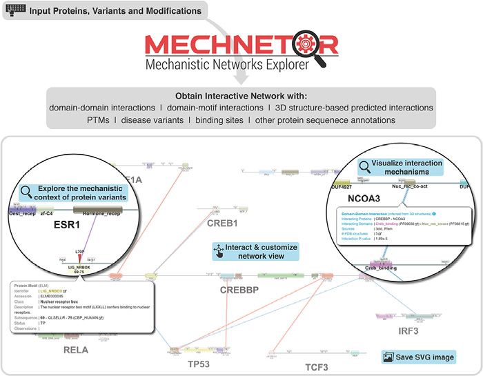
Nucleic Acids Research, 2021, Vol. 49, Web Server issue W367

ent mechanism (18,19). Resources like Pfam (10), SMART               add a more complete set of human viral proteins in the fu-
(20) and InterPro (21) provide crucial details about pro-            ture.
tein functional modules and visual representations of pro-              For each query protein, Mechnetor systematically gath-
tein modular architecture. However, these lack details re-           ers domains, linear motifs, post-translational modifications
garding interactions, and one cannot readily visualize multi-        (from various data sources; see Materials and Methods) and
ple proteins. On the other hand, resources for interrogating         other relevant sequence features (from UniProt), as well as
protein-protein interactions (PPIs), such as BioGRID (12),           interactions between those proteins (PPIs) and their ele-
STRING (22) or GeneMANIA (23), do not specify protein                ments (DDIs and DMIs), interactions predicted from 3D
segments involved in specific interactions. This information         structures, and interactions or associations extracted from
is stored in resources like 3did (24), a database of domain–         annotations. This process relies on an underlying database
domain (DDIs) and domain–motif interactions (DMIs) de-               where data from diverse sources are carefully integrated, by
rived from 3D structures; or ELM (25), a database of linear          matching different formats and descriptors, ensuring that

                                                                                                                                        Downloaded from https://academic.oup.com/nar/article/49/W1/W366/6291159 by guest on 08 September 2021
motifs and their interaction domains with a related tool,            information can be retrieved efficiently (Figure 1B).
iELM (26), for viewing them inside PPI networks.                        The collected data are then used to create an interac-
   There are many methods to assess the impact of variants           tive mechanistic network that can include variants/PTMs
on protein function, for example, based on sequence, phylo-          mapped into their corresponding protein positions, and is
genetic and/or structural information (Polyphen-2 (8), Mu-           presented to the user together with extensive interactivity
tationAssessor (27)); or using 3D structures to evaluate pro-        options to facilitate an intuitive exploration (Figure 1C).
tein stability, folding or dynamics (FoldX (28), Rossetta            Additionally, a searchable table lists in detail all interaction
(29)). These tools are useful for estimating the impact of col-      evidence contained in the network, and can be also down-
lections of mutations generally on proteins, but do not nor-         loaded in text format for local analysis (Figure 1D). The
mally consider the wider, protein-network context. Other             results page can be bookmarked for later access–results will
tools, such as Mechismo (30) or dSysMap (31), help un-               be kept for a period of no less than a month.
derstand mechanism of action by combining structure with
interaction data to predict and visualize the effect on inter-
action interfaces of known structure, though are limited in
                                                                     The mechanistic network
their reporting of mechanistic details lacking coordinate in-
formation.                                                           The Mechnetor network view shows detailed mechanistic
   Here, we present Mechnetor (mechnetor.russelllab.org),            information for every submitted protein. Proteins are rep-
a new web tool that sits between the above resources.                resented as linear diagrams (length proportional) of func-
For sets of proteins, interactions and/or variants, Mech-            tional elements (Table 1; Figure 2A). Edges in the net-
netor quickly integrates diverse mechanistic data sources            work specifically link entire proteins or the functional el-
(PPIs, DDIs, DMIs, 3D structure, post-translational mod-             ements involved in different types of interactions, which are
ifications, and numerous functional annotations) and con-            coloured accordingly (Table 2; Figure 2B). Some of them
structs an interactive network for intuitive visualization of        are weighted according to particular parameters (e.g. num-
protein mechanisms. Proteins are represented as linear ar-           ber of experimental studies, number of 3D structures, etc) to
rangements of domains, motifs and other functional ele-              indicate the extent of interaction evidence, which is reflected
ments, which permits the display of interactions between             in edge thickness. Interactions involving domains and mo-
the relevant/known protein regions. The result is a finer            tifs are given a P-value that indicates the probability of ran-
resolution interaction network that enhances mechanis-               domly observing the particular pair in the interactome of
tic interpretations of biological processes and variants of          the organism considered.
interest.                                                               Users can click on any protein element or interaction to
                                                                     display a box with more information and links to origi-
RESULTS AND DISCUSSION                                               nal data sources (Figure 2). The control panel allows for
                                                                     all nodes and edges to be toggled on or off individually or
Mechnetor: mechanistic networks explorer
                                                                     by setting thresholds (for interactions). Initial protein po-
Mechnetor is a web tool that allows for a quick and user-            sitions in the network are completely arbitrary, and they
friendly exploration of proteins and variants of interest            can be moved freely by simply dragging them. The graph
within a detailed mechanistic context. The general work-             viewer also allows to zoom in or out without any loss of im-
flow is represented in Figure 1. Users can directly input            age quality. All these interactivity options permit users to
proteins or protein pairs in the form of UniProtKB acces-            explore the data in the network, but also to customize the
sions and identifiers, or gene symbols (with the option of           view which can be exported at any time as a snapshot image
automatically adding any number of known interactors for             (PNG or JPEG), or vector graphics (SVG) containing the
those proteins); as well as their own sets of genetic vari-          full network, suitable for editing and preparing publication-
ants and/or post-translational modifications (Figure 1A).            quality figures.
Currently, Mechnetor supports eight of the most com-                    The interactive network is especially designed to investi-
mon model organisms: Homo sapiens, Mus musculus, Danio               gate mechanistic details for only a handful of proteins at the
rerio, Xenopus tropicalis, Arabidopsis thaliana, Drosophila          time. The ‘hairball’ effect is an intrinsic problem of network
melanogaster, Caenorhabditis elegans, and Saccharomyces              visualization and we do not recommend (or indeed allow)
cerevisiae. In addition, we included SARS-CoV-2 proteins,            networks involving >20 proteins. The user will be warned
which can be queried together with human proteins. We will           if the input is too big, and the network will contain only a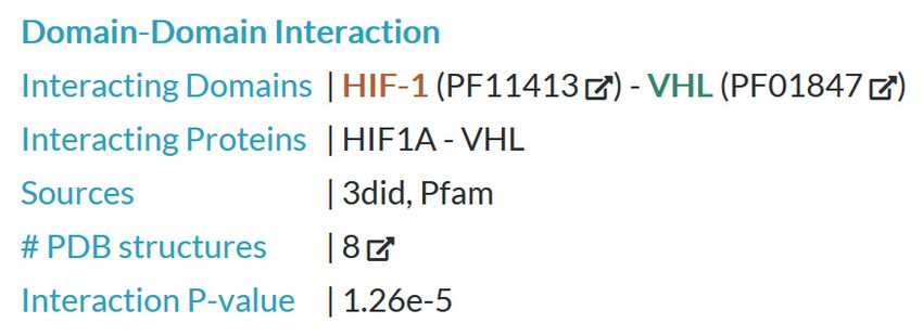
W368 Nucleic Acids Research, 2021, Vol. 49, Web Server issue
Downloaded from https://academic.oup.com/nar/article/49/W1/W366/6291159 by guest on 08 September 2021
Figure 1. Overview of the Mechnetor web server. (A) Initial query submission page, where users can input proteins, protein pairs and/or protein variants
for eight model organisms, plus SARS-CoV-2. (B) Summary of the different data sources present in Mechnetor’s database, which include protein sequence
features (domains, linear motifs, PTMs, and other functional regions) and interaction/associations between proteins (protein-protein) and their features
(domain-domain, domain-motif, etc). (C) First component of the results page: the mechanistic network, containing query proteins and variants together
with all gathered mechanistic information. Positional protein features are displayed along their sequences, while edges represent different types of interac-
tions between them. Users can manually explore the network making use of many interactivity options. (D) The second component of the results page is
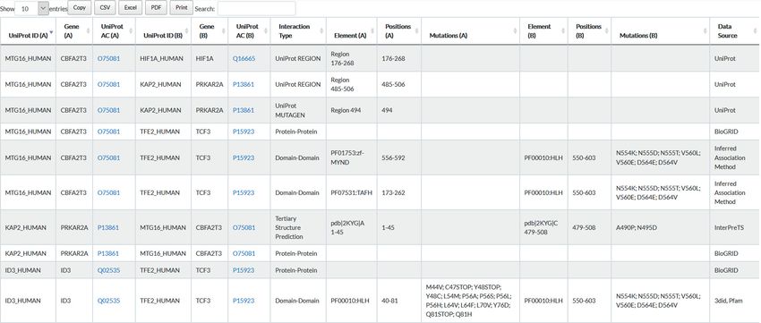
a table containing all interactions between any two proteins comprised in the network.
subset of proteins. However, the table will still contain all homeostasis and regulating blood pressure (32). Variants in
relevant data gathered for the complete input protein set. this protein are related to two genetic diseases: Bronchiec-
tasis with or without elevated sweat chloride 1 (BESC1)
and Liddle syndrome 1 (LIDLS1). A quick glance at the
Case studies network display reveals that variants for these two dis-
We interrogated a dataset of Mendelian disease variants eases are located in different regions of the protein (Figure
(17) and found instances where mechanistic differences 3A). BESC1 variants are more widespread but all within
highlighted by Mechnetor correspond to different patholo- the most conserved part of the protein which comprises
gies. For instance, protein SCNN1B (UniprotKB: P51168) the sodium channel (ASC family domain [Pfam accession:
constitutes the  subunit of the heterotrimeric epithelial PF00858]). Thus, they are more likely deleterious and re-
sodium channel ENaC, located mostly in high resistance sult in decreased channel activity (33). In contrast, LIDLS1
epithelia cells in vertebrates, and involved in maintaining variants are clustered in a small region towards the C-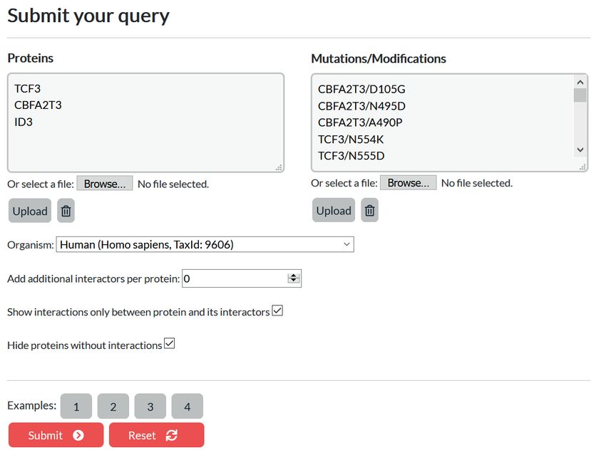
Nucleic Acids Research, 2021, Vol. 49, Web Server issue W369
Table 1. Mechnetor network protein components (nodes)
Protein component Description Source
Domains Domain architecture Pfam
Motifs Linear motifs involved in potential interactions with domains present ELM, 3did
in the network. Toggles allow to only show confirmed motif instances
and exclude motifs inside protein domains
UniProt sequence features These include diverse annotations, such as regions of interest, binding UniProt
sites for chemical groups, metals and DNA, transmembrane regions,
disulphide bonds, and sites altered by mutagenesis experiments
Post-translational modifications Phosphorylation, acetylation and glycosylation sites UniProt,
PhosphositePlus
Variants/Modifications Variants and modifications input by the user User input
Disease variants Variants involved in human genetic diseases UniProt
Cancer variants Cancer missense variants in human COSMIC
Downloaded from https://academic.oup.com/nar/article/49/W1/W366/6291159 by guest on 08 September 2021
terminus that overlaps with a WW domain binding motif Data sources and processing
(LIG WW 1 or ELM accession: ELME000003). The na-
All data required by Mechnetor were obtained from pub-
ture of these amino acid changes suggest that LIDLS1 vari-
licly available data sources and stored in our PostgreSQL
ants disrupt the motif pattern, thus affecting recognition of
database after some pre-processing and integration, that en-
SCNN1B by E3 ubiquitin ligases, like WWP2 or NEDD4.
sures information can be quickly retrieved and displayed by
This would result in a decrease of ubiquitination, which in
the tool.
turn would impair degradation of the EnAC, that would re-
Protein names, identifiers, gene, sequences, and mul-
main constitutively active, resulting in an increase of blood
tiple other annotations (post-translational modifications,
volume and pressure, and this is in fact the known molecu-
variants, mutagenesis experiments, functional and interact-
lar mechanism causing LIDLS1 (34,35).
ing regions, transmembrane regions, disulphide bonds and
To demonstrate how the tool can study somatic cancer
binding sites) are obtained from UniProt (17). All other
variants, we considered the oncogene CTNNB1 (UniPro-
data are always referenced to UniProt proteins. Protein
tKB: P35222). Using the preloaded cancer missense variant
domains are gathered from Pfam (10), or identified with
dataset from COSMIC (3), and requiring ≥5 samples
the PfamScan tool (38) against the Pfam-A database with
for a variant to be reported, a clear hotspot of highly
a 0.001 expectation value cut-off. Short linear motifs in-
recurrent variants can be located at the N terminus of
stances are obtained from ELM (25) and 3did (24), com-
the protein, in a region that also contains several GSK3B
prising 291 and 812 motif classes respectively, and their
(UniProtKB: P49841) phosphorylation sites (recognised
sequence patterns are used to identify potential new in-
by MOD GSK3 1 motifs [ELM accession: ELME000053])
stances by regex matching. Additional PTMs are extracted
targeted by these variants (S33, S37, T41 and S45; Figure
from PhosphositePlus (15). Human cancer protein missense
3B). There are other MOD GSK3 motifs that could be false
variants are obtained from COSMIC genome-wide screens
positives owing to the simplicity of its pattern, which is es-
only (3). Protein–protein interactions are gathered from Bi-
sentially just a pair of Serine/Threonine residues separated
oGRID (12). 3did (24) is used as source of domains inter-
by three amino acids (. . . [ST]. . . [ST]). Only a few, including
actions. 3did systematically charts atomic contacts between
the two within the cancer hotspot, actually correspond with
Pfam domains within 3D structures. In addition, we predict
known phosphosites supported with experimental evidence
domain-domain associations from interaction data (see be-
(obtained from UniProt and PhosphositePlus). Moreover,
low). Domain-linear motif interactions are obtained from
these phosphorylations are required for the recognition
3did, and also derived from a modified dataset from ELM
of CTNNB1 by BTRC (UniProtKB: Q9Y297), a com-
(see below). To predict 3D structure-based interactions and
ponent of E3 ubiquitin-protein ligase complex, through
interfaces, Mechnetor runs an internal version of Inter-
a diphospho-dependent degron DEG SCF TRCP1 1
PreTS (39,40), which itself uses the RCSB PDB database
(ELM accession: ELME000269) that interacts with its
(14). InterPReTs compares sequence pairs to proteins in-
WD40 -propeller. Therefore, these variants ultimately
teractions of known structure and scores (Z score, P-value)
prevent ubiquitination of CTNNB1 and its subsequent
how well the sequences fit on any identified interface.
degradation, which can then translocate to the nucleus
Data will be periodically updated. Current data versions
and continuously promote transcription of its target
can always be consulted at: mechnetor.russelllab.org/help.
genes (36).
Reviewing domain–motif interactions
MATERIALS AND METHODS The source file of ELM interaction domains data (elm.eu.
org/interactiondomains) only lists motif classes and their
Web server implementation
interaction domains but, actually, not all motifs that in-
The Mechnetor web server is built with Python3 under teract with the same domain type can interact with the
the Flask micro-web framework, and uses a PostgreSQL same proteins. For example, ELM has >30 motif classes
database. Data visualization makes extensive use of the Cy- that interact with the ‘protein kinase domain’ (Pfam iden-
toscape JavaScript library (cytoscape.js) (37). tifier: PF00069), which in the human proteome is present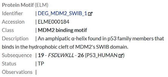
W370 Nucleic Acids Research, 2021, Vol. 49, Web Server issue

 A

 B

                                                                                                                                                              Downloaded from https://academic.oup.com/nar/article/49/W1/W366/6291159 by guest on 08 September 2021
Figure 2. Illustrative examples of network components. (A) Examples of functional elements extracted from different proteins for illustration. For some,
a popup box with additional information (that can be toggled on when clicking on the node) is displayed. From left to right: protein domain (from
Pfam); linear motif (from ELM); DNA binding site (from UniProt); phosphorylation and acetylation sites (from UniProt and PhosphositePlus); cancer
missense variants (from COSMIC); genetic disease variant (from UniProt); variants input by the user. (B) Selected pairs of human proteins to illustrate the
different types of interactions (with popup boxes). From top to bottom, and left to right: protein–protein interaction (green) between MDM2 (UniProtKB:
Q00987) and CHEK2 (UniProtKB: O96017) linking the entire proteins, supported by seven low-throughput experiments (according to BioGRID); domain-
domain interaction inferred by domain co-occurrence (yellow) between the Pas 3 domain of HIF1A (UniProtKB: Q16665) and the HLH domain of ID3
(UniProtKB: Q02535), with an association score of 4.74; domain-domain interaction (cyan) between the HIF-1 domain of HIF1A and the VHL domain of
VHL (UniProtKB: P40337) supported by eight PDB structures (according to 3did and also Pfam); domain-motif interaction (purple) between the RB B
domain of RB1 (UniProtKB: P06400) and a LIG Rb LxCxE 1 (ELM accession: ELME000007) in HDAC2 (UniProtKB: Q92769) (from ELM); interaction
predicted through tertiary structure (red) between KPNA4 (UniProtKB: O00629) and NUP50 (UniProtKB: Q9UKX7) using InterPreTS and PDB ID:
2C1M as template; interaction (black and orange) between a region of SORT1 (UniProtKB: Q99523) and GGA1 (UniProtKB: Q9UJY5) extracted from
UniProt annotations.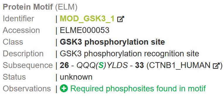
Nucleic Acids Research, 2021, Vol. 49, Web Server issue W371

Table 2. Mechnetor network interactions types (edges)
Interactions                       Description                                                                        Source
Protein-protein                    Represents current experimental evidence for the interaction between two           BioGRID
                                   proteins
Domain-domain (i)                  Domain interactions inferred from 3D structures directly or through                3did
                                   homology
Domain-domain (ii)                 Domain interactions inferred by significant co-occurrence of domain pairs in       Predicted (see Methods)
                                   known interacting proteins. A log-odds indicates the strength of the domain
                                   association.
Domain-motif (i)                   Interactions between linear motifs and their binding domains, obtained from        ELM
                                   annotated motif classes. Certain restrictions are applied based on annotation
                                   to ensure these interactions are biologically significant (see Methods)
Domain-motif (ii)                  Interactions between linear motifs and their binding domains, inferred from        3did
                                   3D structure

                                                                                                                                                Downloaded from https://academic.oup.com/nar/article/49/W1/W366/6291159 by guest on 08 September 2021
3D-based                           Links potential interfaces predicted through tertiary structure. Uses own          InterPreTS
                                   scoring system
Other associations                 Associations between certain UniProt features (regions, binding sites,             UniProt
                                   mutagenesis) and other proteins in the network

in hundreds of proteins. However, most of these mo-                          the domains, and assess the significance returning a log-
tif classes are only recognized by the kinase domains                        odds or association score. A high log-odds value indicates
of very particular and different protein kinases, e.g. the                   a strong correlation between the corresponding domains in
ELM motif MOD NEK2 1 is the specific phosphoryla-                            interacting proteins. We define a domain pair as enriched
tion site of the Serine/Threonine-protein kinase NEK2,                       if its association score is greater than or equal to 2, but
while DOC MAPK gen 1 is the docking motif of mem-                            to avoid not significant associations, only if its observed
bers of the MAP kinase family (MAPKs). Furthermore,                          count is also greater than or equal to 4, and the individ-
some motifs are exclusively located in certain proteins,                     ual counts of proteins containing each of the signatures
such as LIG PEX14 1 which mediates the interaction be-                       are greater than or equal to 4. We assigned an associa-
tween PEX5 and PEX14; or are exclusive to certain taxa,                      tion score of –5 (which is smaller than the minimum log-
like LIG PAM2 2, which is a variant of the PABP (Poly-                       odds value calculated) to those pairs where the observed
adenylate binding protein)-interacting motif specific for an-                frequency was zero. In addition, the observed frequency of
imals.                                                                       every domain–domain, but also domain-motif pair, is also
   All this information and more can be found in the cu-                     reported as a P-value for every DDI and DMI. It repre-
rated entries of motif classes at the ELM website. Based                     sents the probability of finding the particular pair in the
on this, we manually annotated each motif-domain inter-                      interaction dataset and thus can be used to estimate their
action with additional restrictions and requirements for the                 significance.
interaction to take place. These include: restriction to cer-
tain taxa, restriction of interaction domain and/or motif
to only certain genes, require the presence of other lin-
ear motifs in the same protein, and require the presence                     CONCLUSION
of phosphosites within the motif. An updated version of                      Despite the immense volume of data generated by sequenc-
these ELM interaction domains/proteins with our addi-                        ing efforts, its impact on the advancement of medical knowl-
tional annotations can be found in Supplementary File S1.                    edge and the development of patient-tailored treatments
In order to present only the most biologically relevant infor-               has been limited by our still narrow ability to interpret the
mation, Mechnetor only shows domain-motif interactions                       molecular consequences of coding variants. This task neces-
that match to protein/domain motif pairs in this revised                     sarily requires the simultaneous analysis of diverse protein
table.                                                                       data, which often implies consulting several data sources
                                                                             and applying computational approaches to take further ad-
                                                                             vantage of them.
Scoring and inferring DDI interactions
                                                                                Mechnetor facilitates this by performing a systematic and
To infer interactions between protein domains, we use the                    fast integration of diverse protein data and presenting it to
method first described by Sprinzak and Margalit in 2001                      the user in an interactive and intuitive way. One of our prior-
(41) for the identification of over-represented sequence-                    ities was to make this tool very user-friendly so, in essence,
signatures pairs in interacting proteins by comparing their                  Mechnetor can be used by simply entering a pair of pro-
expected and observed frequencies. This is done for each                     teins and/or protein variants, clicking the submit button,
organism independently, using a subset of non-redundant                      and getting mechanistic ideas in a few seconds. Further pos-
PPI reported by at least two experimental sources. For every                 sibilities include studying larger datasets of interacting pro-
possible domain-domain combination, the method com-                          tein pairs by directly downloading the integrated data for
pares the observed number of interacting proteins contain-                   local analysis. We will update Mechnetor data regularly, and
ing the pair of domains (one or more times) against the ex-                  we plan on supporting more organisms, as well as extending
pected number according to the individual frequencies of                     some of its functionalities.
W372 Nucleic Acids Research, 2021, Vol. 49, Web Server issue
A
Downloaded from https://academic.oup.com/nar/article/49/W1/W366/6291159 by guest on 08 September 2021
B
Figure 3. Case studies illustrating Mechnetor’s functionalities. (A) Network view of SCNN1B (UniprotKB: P51168), WWP2 (UniProtKB: O00308) and
NEDD4 (UniProKB: P46934), showing domain architecture, domain-motif interactions from ELM (purple lines) and UniProt’s genetic disease variants
(bronchiectasis variants in orange and Liddle syndrome variants in green). SCNN1B C-terminal region has been zoomed to enhance visualization of the
overlap of Liddle syndrome variants and the LIG WW 1 motif. Popup boxes show annotation for the ASC domain (left), and the LIG WW 1 motif in
SCNN1B (right) which contains its sequence on the protein, supporting the observation that Liddle syndrome variants (amino acid substitutions in Pro-
617, Pro-618 and Tyr-620) alter the motif. (B) Network view of CTNNB1 (UniProtKB: P35222), GSK3B (UniProtKB: P49841) and BTRC (UniProtKB:
Q9Y297), displaying protein domains, domain-motif interactions from ELM (purple lines), phosphorylation sites (small yellow flags), and cancer missense
variants from COSMIC (blue t-shaped lines, heights are proportional to number of samples). We set a minimum number of 5 samples for cancer variants
to be displayed. The N-terminal region of CTNNB1 (zoomed) shows the overlap between a cluster of cancer variants, a number of phosphosites and
three motifs: two GSK3B recognition sites (MOD GSK3) and the diphospho-degron (DEG SCF TRCP 1) recognized by BTRC. Popup boxes show more
detailed annotations and let us know that the required phosphosites are found within these motifs. This support the validity of these motifs and suggests
that cancer variants in this positions might result in the disruption of both recognition mechanisms.Nucleic Acids Research, 2021, Vol. 49, Web Server issue W373
DATA AVAILABILITY 12. Oughtred,R., Rust,J., Chang,C., Breitkreutz,B.J., Stark,C.,
Willems,A., Boucher,L., Leung,G., Kolas,N., Zhang,F. et al. (2021)
Mechnetor is a web server freely accessible without login The BioGRID database: a comprehensive biomedical resource of
requirement at mechnetor.russelllab.org. The source code is curated protein, genetic, and chemical interactions. Protein Sci., 30,
available at https://github.com/JCGonzS/mechnetor. 187–200.
13. Jassal,B., Matthews,L., Viteri,G., Gong,C., Lorente,P., Fabregat,A.,
Sidiropoulos,K., Cook,J., Gillespie,M., Haw,R. et al. (2020) The
SUPPLEMENTARY DATA reactome pathway knowledgebase. Nucleic Acids Res., 48,
D498–D503.
Supplementary Data are available at NAR Online. 14. Berman,H.M., Westbrook,J., Feng,Z., Gilliland,G., Bhat,T.N.,
Weissig,H. and Shindyalov,I.N. (2000) The Protein Data Bank
(www.rcsb.org). Nucleic Acids Res., 28, 235–242.
ACKNOWLEDGEMENTS 15. Hornbeck,P. V., Zhang,B., Murray,B., Kornhauser,J.M., Latham,V.
and Skrzypek,E. (2015) PhosphoSitePlus, 2014: mutations, PTMs
We thank Gurdeep Singh, Torsten Schmenger and and recalibrations. Nucleic Acids Res., 43, D512–D520.
Downloaded from https://academic.oup.com/nar/article/49/W1/W366/6291159 by guest on 08 September 2021
Francesco Raimondi for helpful discussions. 16. Huang,K.Y., Lee,T.Y., Kao,H.J., Ma,C.T., Lee,C.C., Lin,T.H.,
Chang,W.C. and Huang,H. Da (2019) DbPTM in 2019: exploring
disease association and cross-talk of post-translational modifications.
FUNDING Nucleic Acids Res., 47, D298–D308.
17. The UniProt Consortium (2019) UniProt: a worldwide hub of protein
German Network for Bioinformatics Infrastructure knowledge. Nucleic Acids Res., 47, D506–D515.
(de.NBI) funded by the German Federal Ministry of 18. O’Donoghue,S.I., Gavin,A.C., Gehlenborg,N., Goodsell,D.S.,
Hériché,J.K., Nielsen,C.B., North,C., Olson,A.J., Procter,J.B.,
Education and Research (BMBF). Funding for open access Shattuck,D.W. et al. (2010) Visualizing biological data––now and in
charge: German Network for Bioinformatics Infrastruc- the future. Nat. Methods, 7, S2–S4.
ture (de.NBI), funded by the German Federal Ministry of 19. O’Donoghue,S.I., Baldi,B.F., Clark,S.J., Darling,A.E., Hogan,J.M.,
Education and Research (BMBF). Kaur,S., Maier-Hein,L., McCarthy,D.J., Moore,W.J., Stenau,E. et al.
(2018) Visualization of biomedical data. Annu. Rev. Biomed. Data
Conflict of interest statement. None declared. Sci., 1, 275–304.
20. Letunic,I., Khedkar,S. and Bork,P. (2021) SMART: recent updates,
new developments and status in 2020. Nucleic Acids Res., 43,
REFERENCES D257–D260.
1. Karczewski,K.J., Francioli,L.C., Tiao,G., Cummings,B.B., Alföldi,J., 21. Blum,M., Chang,H.Y., Chuguransky,S., Grego,T., Kandasaamy,S.,
Wang,Q., Collins,R.L., Laricchia,K.M., Ganna,A., Birnbaum,D.P. Mitchell,A., Nuka,G., Paysan-Lafosse,T., Qureshi,M., Raj,S. et al.
et al. (2020) The mutational constraint spectrum quantified from (2021) The InterPro protein families and domains database: 20 years
variation in 141,456 humans. Nature, 581, 434–443. on. Nucleic Acids Res., 49, D344–D354.
2. Amberger,J.S.S., Bocchini,C.A.A., Schiettecatte,F., Scott,A.F.F. and 22. Szklarczyk,D., Gable,A.L., Lyon,D., Junge,A., Wyder,S.,
Hamosh,A. (2015) OMIM.org: Online Mendelian Inheritance in Huerta-Cepas,J., Simonovic,M., Doncheva,N.T., Morris,J.H., Bork,P.
Man (OMIM® ), an Online catalog of human genes and genetic et al. (2019) STRING v11: protein-protein association networks with
disorders. Nucleic Acids Res., 43, D789–D798. increased coverage, supporting functional discovery in genome-wide
3. Tate,J.G., Bamford,S., Jubb,H.C., Sondka,Z., Beare,D.M., Bindal,N., experimental datasets. Nucleic Acids Res., 47, D607–D613.
Boutselakis,H., Cole,C.G., Creatore,C., Dawson,E. et al. (2019) 23. Franz,M., Rodriguez,H., Lopes,C., Zuberi,K., Montojo,J.,
COSMIC: the catalogue of somatic mutations in cancer. Nucleic Bader,G.D. and Morris,Q. (2018) GeneMANIA update 2018. Nucleic
Acids Res., 47, D941–D947. Acids Res., 46, W60–W64.
4. Lyon,G.J. and Wang,K. (2012) Identifying disease mutations in 24. Mosca,R., Céol,A., Stein,A., Olivella,R. and Aloy,P. (2014) 3did: a
genomic medicine settings: Current challenges and how to accelerate catalog of domain-based interactions of known three-dimensional
progress. Genome Med., 4, 58. structure. Nucleic Acids Res., 42, D374–D379.
5. Quintáns,B., Ordóñez-Ugalde,A., Cacheiro,P., Carracedo,A. and 25. Kumar,M., Gouw,M., Michael,S., Sámano-Sánchez,H., Pancsa,R.,
Sobrido,M.J. (2014) Medical genomics: the intricate path from Glavina,J., Diakogianni,A., Valverde,J.A., Bukirova,D., Čalyševa,J.
genetic variant identification to clinical interpretation. Appl. Transl. et al. (2020) ELM––the eukaryotic linear motif resource in 2020.
Genomics, 3, 60–67. Nucleic Acids Res., 48, D296–D306.
6. Eilbeck,K., Quinlan,A. and Yandell,M. (2017) Settling the score: 26. Weatheritt,R.J., Jehl,P., Dinkel,H. and Gibson,T.J. (2012) iELM - a
variant prioritization and Mendelian disease. Nat. Publ. Gr., 18, web server to explore short linear motif-mediated interactions.
599–612. Nucleic Acids Res., 40, W364–W369.
7. Sim,N.L., Kumar,P., Hu,J., Henikoff,S., Schneider,G. and Ng,P.C. 27. Reva,B., Antipin,Y. and Sander,C. (2011) Predicting the functional
(2012) SIFT web server: predicting effects of amino acid substitutions impact of protein mutations: application to cancer genomics. Nucleic
on proteins. Nucleic Acids Res., 40, W452–W457. Acids Res., 39, e118.
8. Adzhubei,I.A., Schmidt,S., Peshkin,L., Ramensky,V.E., 28. Delgado,J., Radusky,L.G., Cianferoni,D. and Serrano,L. (2019)
Gerasimova,A., Bork,P., Kondrashov,A.S. and Sunyaev,S.R. (2010) FoldX 5.0: working with RNA, small molecules and a new graphical
A method and server for predicting damaging missense mutations. interface. Bioinformatics, 35, 4168–4169.
Nat. Methods, 7, 248–249. 29. Leaver-Fay,A., Tyka,M., Lewis,S.M., Lange,O.F., Thompson,J.,
9. Nussinov,R., Jang,H., Tsai,C.J. and Cheng,F. (2019) Precision Jacak,R., Kaufman,K., Renfrew,P.D., Smith,C.A., Sheffler,W. et al.
medicine and driver mutations: computational methods, functional (2011) Rosetta3: an object-oriented software suite for the simulation
assays and conformational principles for interpreting cancer drivers. and design of macromolecules. In Methods Enzymol., 487, 545–574.
PLoS Comput. Biol., 15, e1006658. 30. Betts,M.J., Lu,Q., Jiang,Y., Drusko,A., Wichmann,O., Utz,M.,
10. Mistry,J., Chuguransky,S., Williams,L., Qureshi,M., Salazar,G.A., Valtierra-Gutierrez,I.A., Schlesner,M., Jaeger,N., Jones,D.T. et al.
Sonnhammer,E.L.L., Tosatto,S.C.E., Paladin,L., Raj,S., (2015) Mechismo: predicting the mechanistic impact of mutations
Richardson,L.J. et al. (2021) Pfam: the protein families database in and modifications on molecular interactions. Nucleic Acids Res., 43,
2021. Nucleic Acids Res., 49, D412–D419. e10.
11. Orchard,S., Ammari,M., Aranda,B., Breuza,L., Briganti,L., 31. Mosca,R., Tenorio-Laranga,J., Olivella,R., Alcalde,V., Céol,A.,
Broackes-Carter,F., Campbell,N.H., Chavali,G., Chen,C., Soler-López,M. and Aloy,P. (2015) dSysMap: exploring the edgetic
del-Toro,N. et al. (2014) The MIntAct project–IntAct as a common role of disease mutations. Nat. Methods, 12, 167–168.
curation platform for 11 molecular interaction databases. Nucleic
Acids Res., 42, D358–D363.W374 Nucleic Acids Research, 2021, Vol. 49, Web Server issue
32. Hanukoglu,I. and Hanukoglu,A. (2016) Epithelial sodium channel 37. Franz,M., Lopes,C.T., Huck,G., Dong,Y., Sumer,O. and Bader,G.D.
(ENaC) family: phylogeny, structure-function, tissue distribution, (2015) Cytoscape.js: a graph theory library for visualisation and
and associated inherited diseases. Gene, 579, 95–132. analysis. Bioinformatics, 32, 309–311.
33. Fajac,I., Viel,M., Sublemontier,S., Hubert,D. and Bienvenu,T. (2008) 38. Madeira,F., Park,Y.M., Lee,J., Buso,N., Gur,T., Madhusoodanan,N.,
Could a defective epithelial sodium channel lead to bronchiectasis. Basutkar,P., Tivey,A.R.N., Potter,S.C., Finn,R.D. et al. (2019) The
Respir. Res., 9, 46. EMBL-EBI search and sequence analysis tools APIs in 2019. Nucleic
34. Abriel,H., Loffing,J., Rebhun,J.F., Pratt,J.H., Schild,L., Acids Res., 47, W636–W641.
Horisberger,J.D., Rotin,D. and Staub,O. (1999) Defective regulation 39. Aloy,P. and Russell,R.B. (2002) Interrogating protein interaction
of the epithelial Na+ channel by Nedd4 in Liddle’s syndrome. J. Clin. networks through structural biology. Proc. Natl. Acad. Sci. U.S.A.,
Invest., 103, 667–673. 99, 5896–5901.
35. Furuhashi,M., Kitamura,K., Adachi,M., Miyoshi,T., Wakida,N., 40. Aloy,P. and Russell,R.B. (2003) InterPreTS: protein interaction
Ura,N., Shikano,Y., Shinshi,Y., Sakamoto,K.I., Hayashi,M. et al. prediction through tertiary structure. Bioinformatics, 19, 161–162.
(2005) Liddle’s syndrome caused by a novel mutation in the 41. Sprinzak,E. and Margalit,H. (2001) Correlated sequence-signatures
proline-rich PY motif of the epithelial sodium channel -subunit. J. as markers of protein-protein interaction. J. Mol. Biol., 311, 681–692.
Clin. Endocrinol. Metab., 90, 340–344.
Downloaded from https://academic.oup.com/nar/article/49/W1/W366/6291159 by guest on 08 September 2021
36. Shang,S., Hua,F. and Hu,Z.W. (2017) The regulation of -catenin
activity and function in cancer: Therapeutic opportunities.
Oncotarget, 8, 33972–33989.You can also read