Mouse Ataxin-3 Functional Knock-Out Model
←
→
Page content transcription
If your browser does not render page correctly, please read the page content below
Neuromol Med (2011) 13:54–65
DOI 10.1007/s12017-010-8137-3

 ORIGINAL PAPER

Mouse Ataxin-3 Functional Knock-Out Model
Pawel M. Switonski • Agnieszka Fiszer •
Katarzyna Kazmierska • Maciej Kurpisz •
Wlodzimierz J. Krzyzosiak • Maciej Figiel

Received: 14 January 2010 / Accepted: 9 September 2010 / Published online: 14 October 2010
Ó The Author(s) 2010. This article is published with open access at Springerlink.com

Abstract Spinocerebellar ataxia 3 (SCA3) is a genetic                 human and mouse exons was expressed and alternatively
disorder resulting from the expansion of the CAG repeats              spliced. We detected mRNA isoforms composed of exon 1
in the ATXN3 gene. The pathogenesis of SCA3 is based on               spliced with mouse exon 4 or with human exon 7. After
the toxic function of the mutant ataxin-3 protein, but the            applying 37 PCR cycles, we also detected a very low level
exact mechanism of the disease remains elusive. Various               of the correct exon 1/exon 2 isoform. Additionally, we
types of transgenic mouse models explore different aspects            confirmed by bioinformatic analysis that the structure and
of SCA3 pathogenesis, but a knock-in humanized mouse                  power of the splicing site between mouse intron 1 and
has not yet been created. The initial aim of this study was           human exon 2 (the targeted locus) was not changed com-
to generate an ataxin-3 humanized mouse model using a                 pared with the native mouse locus. We hypothesized that
knock-in strategy. The human cDNA for ataxin-3 con-                   these splicing aberrations result from the deletion of further
taining 69 CAG repeats was cloned from SCA3 patient and               splicing sites and the presence of a strong splicing site in
introduced into the mouse ataxin-3 locus at exon 2, delet-            exon 4, which was confirmed by bioinformatic analysis.
ing it along with exon 3 and intron 2. Although the human             In summary, we created a functional ataxin-3 knock-out
transgene was inserted correctly, the resulting mice                  mouse model that is viable and fertile and does not present
acquired the knock-out properties and did not express                 a reduced life span. Our work provides new insights into
ataxin-3 protein in any analyzed tissues, as confirmed by             the splicing characteristics of the Atxn3 gene and provides
western blot and immunohistochemistry. Analyses of RNA                useful information for future attempts to create knock-in
expression revealed that the entire locus consisting of               SCA3 models.

                                                                      Keywords Ataxin-3  Mouse model  Knock-in  Knock-
Electronic supplementary material The online version of this          out  CAG repeats  Splicing
article (doi:10.1007/s12017-010-8137-3) contains supplementary
material, which is available to authorized users.

P. M. Switonski  A. Fiszer  K. Kazmierska                          Background
W. J. Krzyzosiak  M. Figiel (&)
Laboratory of Cancer Genetics, Institute of Bioorganic
Chemistry, Polish Academy of Sciences, Noskowskiego 12/14,            Spinocerebellar ataxia 3 (SCA3), which is also called
61-704 Poznan, Poland                                                 Machado–Joseph disease (MJD), is a dominant genetic
e-mail: mfigiel@ibch.poznan.pl                                        disorder that results from the expansion of the CAG repeats
                                                                      in the coding sequence of the ATXN3 gene (Coutinho and
M. Kurpisz
Institute of Human Genetics, Polish Academy of Sciences,              Andrade 1978; Kawaguchi et al. 1994) (MJD and ATXN3:
Strzeszyńska 32, 60-479 Poznan, Poland                               OMIM 109150 and 607047). The majority of patients suf-
                                                                      fering from SCA3 carry one allele of ATXN3 with 60–82
M. Figiel
                                                                      CAG repeats and a second allele containing the normal
Department of Gene Expression, Institute of Molecular Biology
and Biotechnology, Adam Mickiewicz University,                        number of repeats, which is usually between 13 and 41
Umultowska 89, Poznan, Poland                                         (Kawaguchi et al. 1994; Giunti et al. 1995). Expression of

123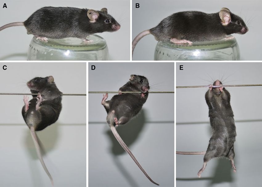
Neuromol Med (2011) 13:54–65                                                                                                 55

the mutant allele leads to the onset of neurological symp-        (NLS) or a nuclear export signal (NES) (Bichelmeier et al.
toms, typically in the third or fourth decade of life. SCA3       2007). The resulting animals containing the NLS transgene
patients commonly suffer from motor and mental abnor-             showed markedly enhanced SCA3 symptoms relative to
malities, such as gait ataxia, ocular symptoms, and demen-        animals containing the NES transgene. The role of
tia (Coutinho et al. 1982; Ishikawa et al. 2002; Kawai et al.     co-chaperones and the ubiquitin ligase C-terminus of the
2004; França et al. 2007, 2008; Riess et al. 2008).              Hsp70-interacting protein in enhancing disease progression
    Although the relevant pathogenesis of SCA3 is based on        and increasing aggregate formation was explored in the
the toxic function of the mutant ataxin-3 protein, the exact      SCA3 mouse model (Williams et al. 2009) created by Goti
mechanism of disease remains elusive (Paulson 2007; Riess         et al. (2004). Recently, Boy et al. (2009) provided evidence
et al. 2008). Expanded CAG repeats in the ataxin-3 mRNA           that the behavioral SCA3 phenotype could be reversible by
result in long polyglutamine (polyQ) tracts within the pro-       creating an inducible model of SCA3 using the Tet-off
tein, resulting in protein misfolding. These changes may          system. The same group demonstrated the late-onset
affect features, such as protein binding, subcellular locali-     transgenic model with ataxin-3 driven by huntingtin pro-
zation of the protein, and its degradation and elimination        moter (Boy et al. 2010). The mouse model where ataxin-3
from the cell (Chow et al. 2004; Lessing and Bonini 2008). In     is driven by the CMV promoter does not show intranuclear
the misfolded protein, the polyglutamine chains are exposed       inclusions (Silva-Fernandes et al. 2010). In addition, one
and can scaffold normal and mutant ataxin-3 as well as            knock-out model of mouse ataxin-3 has been created.
important proteins, such as histone acetylases, deacetylases,     These animals exhibit increased levels of total protein
transcription factors, and the proteasome complex (Evert          ubiquitination and minor behavioral changes, but they are
et al. 2006; Bilen and Bonini 2007; Ferro et al. 2007; Lessing    viable and fertile (Schmitt et al. 2007).
and Bonini 2008; Jia et al. 2008). In turn, the scaffolding and      We have created a K300 transgenic mouse model that
subsequent events can affect gene expression, change the          lacks functional ataxin-3 protein expression confirmed by
cellular localization of different proteins, and lead to the      western blotting and immunohistochemistry. The mouse
dysfunction of the protein elimination pathway in cells.          model was generated as a result of a knock-in strategy
    Knock-out, transgenic, and YAC transgenic mouse               wherein human ataxin-3 cDNA was inserted into the mouse
models have been created to explore various aspects of            ataxin-3 locus. The final mouse model exhibited the pres-
SCA3 pathogenesis. The transgenic animals in these stud-          ence of several splice forms of ataxin-3 mRNA and the
ies were generated by the random integration of a transgene       presence of deleted non-functional ataxin-3 proteins that
in the form of an artificial construct that contained either      were detected at very low levels only when using a massive
full-length human ataxin-3 cDNA or some fragment of the           excess of protein extract. The correctly spliced mRNA
gene. The transgene was usually driven by a different             isoform containing mouse exon 1/human exons was
promoter, such as a Purkinje-specific L7 promoter (Ikeda          detected at very low levels using PCR with increased
et al. 1996). Model animals containing the polyQ fragment         sensitivity, but the protein of interest was not detected even
of ataxin-3 develop the disease phenotype. However, ani-          when using a massive excess of protein extract for
mals with full-length mutant ataxin-3 protein are viable and      immunoblotting. The most prominent mRNA isoforms
healthy. To explain this surprising result, the authors           detected in mut/mut animals were mouse ataxin-3 missing
hypothesized that the lack of proteolytic cleavage of the         exons 2 and 3 and the mRNA comprising mouse exon 1
mutant transgenic protein is necessary to produce the dis-        spliced with human exon 7. We conclude that the reason
ease phenotype. Consistent with this hypothesis, transgenic       for the lack of ataxin-3 protein expression was the occur-
mice expressing higher levels of ataxin-3 with expanded           rence of alternative splicing events at the Atxn3 locus after
repeats developed a phenotype reminiscent of SCA3 and             targeting. In summary, these transgenic mice are functional
had a cleavage fragment in brain (Goti et al. 2004). The          knock-out of ataxin-3, are viable and fertile, and do not
YAC transgenic animals created by Cemal et al. (2002)             have a reduced life span.
expressed human ataxin-3 driven by its native gene pro-
moter. This strategy resulted in a close reproduction of
human SCA3 pathology in the mouse model, a result that is         Methods
probably due to the similar expression pattern of the mutant
ataxin-3 protein. This result reveals the importance of non-      Animals and Cell Line
coding sequences and native protein processing in the onset
and progression of SCA3. The impact of the cellular               The animals were kept under standard conditions with a
localization of ataxin-3 on the pathology of SCA3 has been        12-h light/dark cycle and water and food ad libitum. The
investigated using mouse models generated by introducing          animals were marked via a toe-clipping method, and the
transgenes coupled to either a nuclear localization signal        tissue from this procedure was used for genotyping

                                                                                                                     123
56 Neuromol Med (2011) 13:54–65
analyses. The animals were sacrificed by placing them in a and intron 1 in the native gene and introduce human exons
70% CO2 atmosphere. All procedures and animal handling 2–11 in place of mouse exons 2 and 3 (Fig. 1). In the final
were carried out to minimize animal stress and were construct used for homologous recombination, an SV40
approved and monitored by the Local Ethical Commission polyadenylation signal and a loxP-NEOR-loxP cassette
for Animal Experiments in Poznan. The N171-82Q were introduced downstream of the human ataxin-3 cDNA.
(Schilling et al. 1999) and B6.129P2-Htttm2Detl/J (Lin The homologous arms present in the construct were 4.5 and
et al. 2001) mouse models were obtained from The Jackson 3.5 kb for the 50 and 30 arms, respectively. The construct
Laboratory (Bar Harbor, Maine, USA). was subsequently introduced into Sv129/Pas embryonic
Primary human fibroblasts (cell line number: GM06153) stem cells, and clones were selected based on neomycin
were obtained from the Coriell Institute for Medical resistance and tested for proper recombination by PCR and
Research (Camden, NJ, USA). The cells were propagated in sequencing. The clones were then injected into blastocysts
DMEM supplemented with 10% fetal bovine serum (Invit- of C57BL/6 mice to obtain chimeras, which were then bred
rogen; Carlsbad, USA) at 37°C in a 5% CO2 atmosphere. to obtain transgenic animals. The loxP-NEOR-loxP cassette
was excised by breeding the transgenic animals with
Strategy and Generation of the K300 Mouse Model animals expressing Cre recombinase.
The K300 (ICS internal project number) SCA3 transgenic Genotyping Strategies
mouse model was generated at the Institut Clinique de la
Souris (ICS) in Strasbourg, France, as part of the ‘‘RNA All animals were genotyped using a protocol based on the
Interference Technology as Human Therapeutic Tool’’ facts that the mutant allele does not contain an intron
(RIGHT) project. The human mutant ataxin-3 cDNA between exons 2 and 3 and that the sequences of both
sequence containing 69 CAG repeats was cloned from total exons 2 and 3 are identical between humans and mice.
mRNA isolated from human fibroblasts (GM06153) that Therefore, primers binding to both human and mouse
were derived from a patient suffering from SCA3. The exons 2 and 3 would generate PCR products that differed
mouse model was generated using a knock-in strategy that only by the length of the intron. The PCR product lengths
introduced a human cDNA sequence that spanned exons 2 resulting from this approach would be 376 bp in wild-type
through 11 of the human mutant ataxin-3 mRNA into the (wt/wt) animals, 150 bp in mutant (mut/mut) animals, and
mouse ataxin-3 gene. To inactivate the mouse ataxin-3 both products in wt/mut animals. The sequences of primers
gene, the strategy was designed to preserve mouse exon 1 for genotyping are as follows: forward exon 2 CAAGG
Fig. 1 The structure of the mouse ataxin-3 gene and the targeting sequencing. The sequencing analysis of PCR products from the 50 and
strategy. The targeting vector containing human exons 2–11 (gray) 30 ends revealed correct insertion of the transgene (b). The targeted
terminated by the necessary pA signals (black) was inserted into the clones demonstrated the expression of the normal mouse ataxin-3 and
mouse Atxn3 locus by replacing and deleting mouse exon 2, intron 2, human mutant ataxin-3 protein detected by immunoblotting (c). The
and exon 3 (a). The targeted mouse Atxn3 locus contains mouse exon mouse exons are numbered with italic and human exons with regular
1, intron 1, human exons, and mouse exons 4–11. The arrows indicate font type
primers used for the amplification and verification of 50 and 30 ends by
123Neuromol Med (2011) 13:54–65 57
AGAATATTTTAGCCCTGT, reverse exon 3 GCCGCT and 100 ng of random hexamer primers (Promega) in a
GTCATCCATATTTCCA. final volume of 20 ll. PCR was performed according to the
conditions described above. Prior to each primary tran-
DNA Isolation and PCR script PCR experiment, the quality of DNase digestion was
assessed by PCR using the following templates: 1 ll of
Total DNA was isolated from mouse toes using an Invisorb digested RNA, 1 ll of undigested RNA, and 100 ng of
tissue mini kit (Invitek; Berlin, Germany) according to the digested DNA. The quality of the reverse transcription
manufacturer’s instructions. DNA concentration was esti- (RT) reaction was assessed by an amplification of the
mated using a NanoDrop (Thermo Scientific; Wilmington, Gapdh gene.
USA) spectrophotometer. PCR was performed with 1 ll of
template DNA in a total volume of 10 ll that included Immunoblotting Analyses
6 pmol of the appropriate primers and 0.25 U of recom-
binant GoTaq polymerase (Promega; Madison, USA). PCR The tissue samples and primary human fibroblasts
amplification included 28 cycles of denaturation at 94°C (GM06153) were lysed in a buffer containing 60 mM
for 30 s, annealing at 58°C for 30 s, and elongation at 72°C TRIS-base, 2% SDS, 10% sucrose, and 2 mM PMSF.
for 30 s. Primer sequences and their respective PCR Large tissue pieces were chopped and disrupted by soni-
products are listed in Table 1. The reaction products were cation. Protein concentration was estimated using a
separated on 1.5% agarose gels in TBE buffer and stained NanoDrop spectrophotometer, and 50 and 200 lg of total
with ethidium bromide. protein was diluted in a sample buffer containing
2-mercaptoethanol and boiled for 5 min. The proteins were
RNA Isolation and RT–PCR separated by SDS–polyacrylamide gel electrophoresis (5%
stacking/12% resolving gel), transferred to nitrocellulose
Total RNA was isolated from whole brain tissue samples and stained with Ponceau S solution. The blots were
using TRI REAGENT (MRC; Cincinnati, USA) according blocked with 5% nonfat milk in TBS/0.05% Tween 20 and
to the manufacturer’s instructions. RNA isolation typically subsequently incubated for 24 h at 4°C with anti-ataxin-3
yielded 20 lg of total RNA. The RNA concentration was clone 1H9 (1:3,000; wt epitope E214-L233) primary anti-
measured by a NanoDrop spectrophotometer. One micro- body (Millipore; Billerica, USA) and with anti-ubiquitin
gram of total RNA was digested by DNase I in a volume of (1:300) (Dako; Glostrup, Denmark). The blots were probed
10 ll at room temperature. After 45 min, the digestion either with combination of biotinylated antibody and
reaction was stopped by adding 1 ll of 50 mM EDTA. A AP-conjugated streptavidin or with the HRP-conjugated
total of 10 ll of digested RNA were reverse-transcribed at secondary antibody (1:3,000) (Jackson Immuno Research,
42°C for 50 min using 200 U of Superscript II (Invitrogen) Suffolk, UK). The immunoreaction was detected using the
Table 1 Primers
Forward (50 -30 ) Reverse (50 -30 ) Localization of Product (bp)
the PCR product*
CATGGGGCTTACAAAGTTTGTTTTGACTGC GTGCGATAATCTTCACTAGTAACTCCTCC 50 End of the 278
transgene (DNA)
CCCTTCAACTGTATACCGTGTGCTC CCCCACTCTAAACTGACTGTGGTAG 30 End of the 476
transgene (DNA)
CCAGACAAATAAACATGGAG TGTGGACTATTTTCACACATAC mE1/mE9 823 (wt)
613 (E1/mE4)
CCAGACAAATAAACATGGAG CCCTCTGCAAATCCTCCTCATCT mE1/hE8,mE8 498 (E1/mE4)
234 (E1/hE7)
CCAGACAAATAAACATGGAG CATTCCTGAGCCATCATT mE1/hE8 677 (E1/hE2)
205 (E1/hE7)
GCATCGACCAAAACTTAT TTTCTAAAGACATGGTCACA CAG repeats mE6, 674 (human)
hE6/mE11,hE11 473 (mouse)
The last column shows lengths of the PCR products corresponding to the particular splicing isoform
* m mouse; h human; E# exon number
12358 Neuromol Med (2011) 13:54–65
NBT-BCIP substrate (Sigma–Aldrich, St. Louis, USA) or generated from the 50 and 30 ends of the construct were
with the ECL substrate (GE Healthcare; Uppsala, Sweden). sequenced and revealed the correct insertion of the trans-
gene (Fig. 1b). Mouse exons and introns beyond exon 3
Immunohistochemistry were unchanged.
Additionally, to investigate the ataxin-3 protein
The 10-lm coronal mouse brain sections or sections from expression in targeted ES cells, the total protein extract was
testis were cut using a cryostat at -13°C and collected on analyzed by western blotting, and the expression of both
silanized glass slides. The sections were fixed in cold 4°C the mouse ataxin-3 protein (41 kDa) and human mutant
acetone for 20 min and stored at -80°C or processed ataxin-3 (57 kDa) was detected (Fig. 1c). For a positive
immediately. The sections were permeabilized by incu- control, the blots of the total protein extracted from
bating in 0.5% Tween 20 in PBS for 10 min in RT and GM06153 SCA3 human fibroblasts revealed a mutant
subsequently blocked by incubating in 2% normal goat ataxin-3 band with 69 CAG repeats that had an estimated
serum for 30 min in PBS. For immunofluorescent staining molecular weight of 57 kDa.
of intranuclear inclusions, the K300 and N171/82Q brain
sections were incubated overnight at 4°C with primary Animals
rabbit anti-ubiquitin (1:300) (Dako). The Cy-3 anti-rabbit
fluorescent secondary antibodies (Jackson ImmunoRe- The 6 mut/wt animals were generated at ICS as a result of
search) were incubated at 1:500 for 2 h at RT in PBS- backcrossing chimeras two times onto the pure C57BL/6J
Tween 20. The cryosections from testis were incubated background resulting in mixed background of 75% C57BL/
with mouse anti-ataxin-3 antibody (1:500) (Millipore) 25% Sv129/Pas. These animals were exported to Poznan
overnight at 4°C and subsequently detected by 2-h incu- and used to produce 3 generations of offspring and were
bation with the appropriate biotinylated secondary Ab and withdrawn from breeding. Next, we have generated
stained using Vectastain Elite ABC (Vector laboratory, homozygous mut/mut animals that exhibited normal body
Burlingame, CA, USA) and the DAB chromogen. The posture (Fig. 2a, b) and normal muscle strength assessed
fluorescent confocal images were acquired using the LSM by string test (Fig. 2c–e). To assess life span, 5 mut/wt
510 META system (Zeiss; Poznan, Poland). animals initially received from ICS were kept, until they
reached 2.5 years of age and then sacrificed.
Results Analyses of the Ataxin-3 Protein in K300 Mouse
Tissues
Structure of the Targeted Atxn3 Gene
and the Expression of Ataxin-3 in Targeted ES Cells To investigate the expression of the ataxin-3 protein in the
transgenic mice, total protein was extracted from the
Prior to the electroporation of ES cells, the entire cassette cerebral cortex, skeletal muscles (quadriceps), heart, lungs,
used for homologous recombination was sequenced (ICS; liver, spleen, testis, and GM06153 human fibroblasts as a
Strasburg). This verified the correct structure and sequence positive control and resolved by 12% SDS–PAGE. In wt/wt
(for a description of the cassette, see the Materials and animals, the immunoblot analyses revealed a specific band
Methods). Stable ES cell clones were selected, and PCR representing mouse ataxin-3 with an estimated molecular
and sequencing were performed to detect proper transgene weight of 41 kDa that was present at relatively high con-
integration into the Atxn3 gene locus (ICS; Strasburg). centrations in testis, cerebral cortex, lungs, liver, and
These analyses confirmed that the human cDNA was spleen and was hardly detectable in skeletal muscles and
inserted at mouse exon 2 and that the mouse genomic the heart (Fig. 3a). The band detected in wt/wt mice had a
sequence containing exon 2, intron 2, and exon 3 was lower molecular weight than the positive control band of
deleted. Mouse exon 1 and intron 1 were preserved in the wt human ataxin-3 (48 kDa). The similar mouse ataxin-3
gene structure and were followed by human exons 2–11 41-kDa band and the expected human mutant 57-kDa band
and an SV40 polyadenylation signal. The knock-in tar- were not detected in mut/mut animals in any of the tissues
geting strategy of the Atxn3 gene is depicted in Fig. 1a. To analyzed (Fig. 3a). Moreover, none of these immunoreac-
demonstrate the correct positioning of the transgene in the tive bands was observed upon increasing the protein load to
genome, two distinct pairs of primers were designed to 200 lg of mut/mut protein extract from cerebral cortex and
bind mouse intron 1/human exon 2 (50 -flank) and human testis per lane (Fig. 3b). Additionally, analysis of the
exon 10/mouse intron 3 (30 -flank). The PCR amplification ataxin-3 protein in testis from wt/wt animals revealed an
generated 2 PCR products of 278 bp and 476 bp in length, additional strongly immunoreactive band of 34 kDa that
corresponding to the expected bp count. The PCR products we describe as ataxin-3 isoform, since it was not present in
123Neuromol Med (2011) 13:54–65 59
Fig. 2 Body posture and string test of K300 mice. The 6-month-old (c) and wt/wt animal (d) were able to lift and grab the string with hind
K300 mut/mut exhibited normal body posture with normal pelvis limbs. As positive control we used the Huntington’s disease knock-in
elevation and lack of hunchback (a) like the 6-months-old non- mouse model (mut/wt; 150CAG) generated by Lin et al. (2001) and
transgenic wt/wt animal (b). The muscle strength was assessed by supplied by Jackson Laboratory (B6.129P2-Htttm2Detl/J). The HD
placing the mouse from above with only front paws on the knock-in mouse at similar age was hardly able to touch the string with
horizontally stretched string for 2 min. The K300 mut/mut animal hind limbs and often fell down (e)
testis of mut/mut animals (Fig. 3a, b). Upon increasing the inclusions positive for ubiquitin (Fig. 4a, c). Similar
total protein extract content to the large amount of 200 lg structures were not detected in cells from cortex or cere-
per lane, we were able to observe two faint immunoreac- bellum of K300 mut/mut mice stained with anti-ubiquitin
tive bands of 33 and 26 kDa, respectively, that only antibodies (Fig. 4b, d). To demonstrate the loss of ataxin-3
appeared in mut/mut animals, but exclusively in testis and expression, we cryosectioned the testis from K300 mut/
not in cerebral cortex (Fig. 3b). Note that the apparent mut animals and wt/wt animals and stained the tissue using
7-kDa molecular weight difference was also present anti-ataxin-3 antibodies and the DAB chromogen. The cells
between mouse ataxin-3 (41 kDa) and the 34-kDa ataxin-3 in testis of wt/wt animals were positively stained for
isoform detected in wt/wt testis. ataxin-3 (Fig. 4e), and no staining was detected in mut/mut
animals (Fig. 4f).
The Cellular Phenotype of the K300 Mouse The knock-out model of mouse ataxin-3 previously
created by Schmitt et al. (2007) exhibited increased levels
Nuclear inclusions are the hallmark of polyglutamine dis- of total protein ubiquitination, probably due to the absence
orders and are present in both patients and mouse models. of ataxin-3 leading to a partial loss of bulk cellular ubiq-
Therefore, we examined the brains of K300 animals for the uitin-specific protease activity. To test whether a similar
presence of intranuclear inclusions by immunofluoro- phenomenon could be observed in K300 animals, we
chemistry using anti-ubiquitin antibodies in cerebral cortex studied the level of ubiquitinated species in brain tissue
and cerebellum of the K300 mut/mut and using N171/82Q using western blotting. The immunodetection with anti-
animals as positive controls (Huntington’s disease mouse ubiquitin antibodies revealed no differences in the total
model; Schilling et al. 1999). As expected, the cortex level of ubiquitin in mut/mut compared with wt/wt animals
and cerebellum of N171/82Q mice revealed intranuclear (Fig. 4g).
12360 Neuromol Med (2011) 13:54–65
Fig. 3 Immunoblot analysis of ataxin-3 expression in tissues of K300 were not detected in any tissues from mut/mut animals. Additionally,
mice. Total protein was extracted from brain, lung, heart, spleen, we detected a 34-kDa band of the ataxin-3 isoform in testis from
testis, skeletal muscle, and liver samples collected from transgenic wt/wt animals. This isoform was not present in the testis of mut/mut
(mut/mut) and wild-type (wt/wt) animals as well as from a primary animals (a). Upon increasing the total protein load to 200 lg per lane,
culture of human SCA3 fibroblasts (GM06153), which was used as a we were still unable to detect a human mutant ataxin-3 57-kDa band,
positive control. The extracts were resolved by 12% SDS/PAGE, and but we did detect two faint immunoreactive bands of 33 and 26 kDa,
the blot was stained with a polyclonal antibody for ataxin-3. In wt/wt only in testis of mut/mut animals. The apparent 7-kDa molecular
animals, the analysis revealed a mouse ataxin-3 41-kDa band present weight difference between the 33- and 26-kDa bands is the same as
at relatively high concentrations in testis, cerebral cortex, lungs, liver, that between mouse ataxin-3 (41 kDa) and the 34-kDa ataxin-3
and spleen that was hardly detectable in skeletal muscles and heart. A isoform detected in wt/wt testis (b)
similar 41-kDa band and the human mutant ataxin-3 57-kDa band
Expression of Ataxin-3 mRNA in the Brain specific reverse primer was designed to bind exon 8 and
of K300 Animals generate a mut-specific PCR product of 677 bp. The mouse-
specific reverse primer was designed to bind exon 9 and
The examination of protein expression and the lack of generate a wt-specific product of 823 bp. Interestingly, the
ataxin-3 staining in tissue provided strong indications that standard 28 PCR cycles did not allow us to detect the mut-
the expression from the targeted Atxn3 allele was defective, specific product (677 bp) using the RT template from mut/
leading us to hypothesize that the allele had acquired mut or wt/mut animals. A detailed description of the detec-
knock-out properties. To further investigate this issue and tion of the mut-specific product and other splice forms is
the possible reasons for the lack of ataxin-3 protein in provided below. Unexpectedly, the wt-specific product was
mice, we tested Atxn3 mRNA expression. Since the present in mut/mut animals, but was shorter than the wt/wt—
expression of ataxin-3 and the pathology of SCA3 are both specific product (613 bp). Wt-specific RT–PCR of wt/mut
located in the brain, we initially examined the expression animals revealed two products, the 613-bp product described
of ataxin-3 mRNA in brain samples from transgenic ani- above and the expected 823-bp wt-specific product. As
mals, including samples of cortex, cerebellum, pons, and expected, the wt-specific product was present in wt/wt ani-
basal ganglia. A representative set of RT–PCR results mals (Fig. 5). The presence of the short 613-bp wt product in
generated using tissue samples from the cerebral cortex the mut/mut animals was even more puzzling, as this product
was selected for this publication and described below. appeared to be shortened by the combined length of mouse
RT–PCR results from other brain regions yielded similar exons 2 and 3, which had been deleted. Sequencing analysis
results (data not shown). demonstrated that the sequence of mouse exon 1 was directly
Specific primers were used to selectively detect either linked with mouse exon 4 and subsequent exons (Fig. 6).
human (mut allele) or mouse ataxin-3 mRNA (wt allele) These results indicate that the aberrant splicing of the mRNA
(Fig. 5). A single forward primer was designed for exon 1 to expressed from the targeted allele may be responsible for the
bind mRNA from both wt and mut alleles. The human- lack of ataxin-3 protein expression.
123Neuromol Med (2011) 13:54–65 61
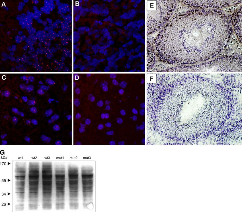
Fig. 4 The cellular phenotype of the K300 mouse model. The and wt/wt animals were stained using anti-ataxin-3 antibodies and the
cryosections prepared from cerebellum and cerebral cortex were DAB chromogen, and the nuclei were counterstained with hematox-
stained using anti-ubiquitin Ab, and confocal images were acquired. ylin dye. Testis from K300 mut/mut animals did not stain positively
The intranuclear inclusions were not detected in cryosections from for ataxin-3, and testis of wt/wt animals exhibited specific, positive
K300 mut/mut mouse brain (b, d). The positive control brains from brown staining for ataxin-3 in a subset of cells in testis (e, f). The
N171/82Q HD animals revealed a large number of intranuclear immunoblotting of total proteins with anti-ubiquitin antibodies
inclusions positive for ubiquitin in both cerebellum and cerebral cortex revealed no difference in the levels of total ubiquitin in mut/mut
(a, c). Additionally, the cryosections from testis of K300 mut/mut compared with wt/wt animals (g)
Alternative Splicing Events Present in the K300 exon 1/exon 4 splice variant that was strongly expressed in
Ataxin-3 Knock-Out Mouse testis and lungs and was less pronounced in liver, skeletal
muscles, heart, and spleen (Fig. 6b; data shown for testis
We investigated whether the mouse exon 1/exon 4 splicing and cortex). Moreover, we detected a PCR product repre-
form was the only splicing event that hindered the splicing senting a splice variant containing mouse exon 1 and part
of mouse exon 1 with human exons. We also asked whether of human exon 7 (Fig. 6c). This splice variant was spliced
a similar splicing isoform was present in the K300 model in at the splicing site that was inside human exon 7, but it was
tissues other than brain, and for this purpose, we analyzed in frame, so that the putative protein would be the exon 2–6
the total mRNA from skeletal muscles, heart, lungs, liver, deleted form of ataxin-3. A particularly pronounced
spleen, and testis of mut/mut animals. We detected two expression of the mouse exon 1/human exon 7 isoform was
major splice forms in all analyzed tissues of K300 animals detected in testis, and this isoform was detected in other
except liver where the exon 1/human exon 7 isoform was tissues at lower levels. Because some splicing events and
not present (Fig. 6). As previously demonstrated for the the resulting splice variants may be rare, we decided to
cerebral cortex, we detected the presence of the mouse increase the sensitivity of our detection by increasing the
12362 Neuromol Med (2011) 13:54–65
Fig. 5 Expression of ataxin-3 mRNA in brain samples from trans- for mouse mRNA containing exon 1 was detected in transgenic
genic animals. The 677-bp RT–PCR product, which represents animals (mut/mut and wt/mut). The product was shorter by the length
mRNA containing mouse exon 1 and human exons of ataxin-3, was of exons 2 and 3 (lines) and was 613 bp rather than the predicted
initially not detected in the mut/mut and wt/mut animals using the 823-bp wt product, suggesting the alternative splicing event. H human
standard 28 PCR cycles (H). The RT–PCR product that was specific ataxin-3 specific PCR; M mouse ataxin-3 specific PCR
number of PCR cycles. Indeed, in cortex and testis using 37
PCR cycles, we were able to detect a very faint signal of
the 677 bp band representing the mut/mut—specific splice
variant containing mouse exon 1 and the human exons. The
variety of reverse primers binding to all exons of the
ataxin-3 gene (2–11), and the forward primer for mouse
exon 1, were used to detect splicing events throughout the
primary transcript of the targeted allele. Therefore, Fig. 6
summarizes the experiments detecting splice variants with
three primers binding to exon 1, mouse exon 8, and human
exon 8 that best reflect the presence of all splice variants in
K300 tissues. Based on our experiments, we estimate that
the cellular levels of splice variants had the following
relationship: the 1/4 splice variant was the most prominent,
the level of the 1/7 variant was 10% that of the 1/4 variant,
and the 1/2 variant was hardly detectable.
Discussion
The initial goal of this project was to create a mouse model
of SCA3 using a knock-in strategy that would introduce
human ataxin-3 cDNA into the mouse ataxin-3 gene. In this
approach, intron 1, exon 1, the promoter, and other 50
regulatory sequences were left unmodified, and the human
ATXN3 cDNA was inserted into mouse exon 2. This allows
the human sequence containing 69 CAG repeats to be
expressed from the native mouse ataxin-3 promoter toge-
Fig. 6 Splicing events in mut-mut K300 animals. The diagram
ther with other putative natural regulatory sequences, such
graphically depicts the splicing events, and the primers called F, B,
A1, and A2 used to detect the various mRNA isoforms (a). The mouse as CpG methylation islands located in intron 1. After the
exon 1/exon 4 and the mouse exon 1/human exon 7 isoforms were homologous recombination cassette was checked thor-
detected in cortex, testis, and other tissues (data not shown) and were oughly by sequencing, it was electroporated into ES cells.
confirmed by sequencing (b). The exon 1/exon 2 splice variant
The selected clones were screened for proper homologous
(677 bp) was detected in the brain and testis at very low levels using
primers F/B and using 37 cycles to increase PCR sensitivity. The cassette recombination, and the clones revealed an exact
205-bp product represents exon 1/human exon 7 isoform (c) insertion of human cDNA at exon 2. Moreover, the
123Neuromol Med (2011) 13:54–65 63
targeted ES cells revealed the expression of mutant trans- et al. 2002; Bichelmeier et al. 2007; Alves et al. 2008).
gene protein, but the mutant protein was expressed at a Intranuclear inclusions positive for ubiquitin were not
lower level than the native mutant protein in human detected in cerebellum or cerebral cortex of K300 animals,
fibroblasts and also at a lower level than wt mouse ataxin- indicating the lack of mutant ataxin-3 protein accumula-
3. The mice were generated, and the transgene was vali- tion. Moreover, the direct staining of the testis, the most
dated in animals by PCR amplification and sequencing of prominent source of ataxin-3 expression, revealed a com-
50 and 30 insert flanks as well as by PCR amplification of plete loss of this protein in mut/mut K300 animals. This
the internal transgress region composed of exons 2–9, strongly indicates that the targeted allele acquired knock-
confirming correct placement in the genome in vivo. out properties and that the mut/mut K300 mice are knock-
Analyzing brain, lungs, heart, spleen, testis, muscles, and out animals. The knock-out of ataxin-3 created by Schmitt
liver from the generated animals, we did not detect the et al. (2007) exhibited enhanced total protein ubiquitina-
mutant human ataxin-3 protein that was previously detec- tion, but we did not detect this effect in K300 animals.
ted in targeted ES cells. Therefore, we increased the sen- Since the genome encodes nearly 100 deubiquitinating
sitivity of our western blotting protocol by increasing the enzymes, the changes produced by one of them may be
protein load up to 200 lg, but we were still unable to detect compensated for by other DUBs.
mutant protein expression. Instead, in testis of mut/mut To determine the reason for the lack of correct protein
mice, we detected very low levels of two proteins that were expression, we investigated the transcripts of the targeted
much shorter (33 and 26 kDa) than the wt mouse ataxin-3 Atxn3 locus and the level of ataxin-3 mRNA expression.
protein. The apparent molecular weight difference between Initially, we examined the brain and were not able to detect
the two molecules was 7 kDa, similar to the difference a common PCR product representing mRNA that contained
between wt mouse ataxin-3 protein and the second ataxin-3 mouse exon 1 and the human exons of ataxin-3. Instead, we
protein isoform, with the most pronounced expression in detected human RNA using PCR for expanded repeats
testis (41 and 34 kDa, respectively). The two isoforms (33 (Suppl. Fig. 1). This indicated that the human transgene
and 26 kDa) were probably proteins with deleted protein was transcribed. Surprisingly, in homozygous transgenic
fragments equivalent to the sequences of exons 2 and 3. animals, but not in wt animals, we detected a PCR product
The presence of splice variant (exon 1/exon 4) equivalent containing exon 1 that was specific for mouse mRNA.
to 33-kDa protein was detected in all tissues. The 33-kDa However, this PCR product was shorter than the wt product
protein must lack the segment of the catalytic Josephine by the combined length of mouse exons 2 and 3.
domain that spans from amino acids Q9–Q78 and includes Sequencing revealed that mouse exon 1 and the remaining
the C14 residue. The substitution of C14 to alanine (C14A) mouse exons 4–11 were spliced. The presence of a similar
in wild-type ataxin-3 was previously shown to be sufficient splice variant was recently detected in the blood of healthy
to render ataxin-3 nonfunctional by abolishing ubiquitin- human individuals (Bettencourt et al. 2010). Additionally,
specific protease activity (Burnett et al. 2003). we characterized the primary and mature transcripts and
The presence of the second ataxin-3 protein isoform revealed that the entire locus was expressed, and the cel-
(wt: 34 kDa; mut: 26 kDa) detected in testis was not lular levels of both the mouse and the human portions of
confirmed by the detection of an equivalent transcript. the transcript in the mut/mut mouse were relatively high.
Because we screened and designed primers in all exons, the The cellular level of human transcript was lower than the
equivalent transcript would have to appear repeatedly as a level of mouse transcript, but it was almost twice as high as
PCR product resulting from many different reverse primers the level of primary transcript (Suppl. Fig. 2).
and the forward primer for exon 1. Therefore, we conclude The lack of mutant protein expression may result from
that this protein is the ataxin-3 isoform (because it is not aberrant mRNA splicing in targeted allele where mouse
present in mut/mut mice), that it is a proteolytic fragment, exons 4–11 are still expressed and induce preferences in
and, being found at relatively high levels, that it is quite splicing that led to the exclusion of the exon 1/human exon
stable and accumulates in testis. Pronounced expression of 2 isoform. We investigated the splicing aberrations in the
a similar peptide was previously reported in C57BL mouse targeted allele in more detail and found the presence of
testis, skeletal muscles, and heart (do Carmo Costa et al. another splicing form, the splicing of exon 1 and part of
2004). exon 7. Moreover, both exon 1/exon 4 and exon 1/exon 7
Because we did not detect the mutant ataxin-3 protein splicing forms were also present in other tissues of the
using western blotting of the mut/mut protein extract, we K300 model. Additionally, we tried to increase the sensi-
investigated the expression of this protein using immuno- tivity of our PCR detection and were able to detect the
chemistry. The intranuclear inclusions often appear in the expected exon 1/exon 2 splice form at a very low level
brains of patients and in mouse models of polyglutamine after 37 PCR cycles. However, we cannot exclude the
diseases and are usually strongly ubiquitinated (Cemal possibility that other splice variants were produced from
12364 Neuromol Med (2011) 13:54–65
this targeted allele. In summary, we detected the presence and human exons with CAG repeats. The animals do not
of three splice variants and observed that the splicing exhibit changes in fertility or life span relative to their non-
favored distant splicing acceptor sites, such as mouse transgenic counterparts.
human exon 7 and exon 4, and virtually eliminated splicing
to human exon 2. This situation is surprising since the Acknowledgments This work was supported by the EU FP6 project
‘‘RNA Interference Technology as Human Therapeutic Tool
splicing acceptor site in exon 2 remained identical to the (RIGHT)’’, European Regional Development Fund within Innovative
mouse sequence up to the 24th nucleotide and contained Economy Programme (POIG.01.03.01-30-049/09), and Ministry of
only 3 single-nucleotide changes in the first 70 nucleotides Science and Higher Education (N N302 299536). We thank Krzysztof
of exon 2. We used the SROOGLE webserver, which Sobczak for useful suggestions and comments on the manuscript.
scores splice sites according to 9 different algorithms Open Access This article is distributed under the terms of the
(Schwartz et al. 2009). This analysis revealed that com- Creative Commons Attribution Noncommercial License which per-
bining mouse intron 1 and human exon 2 (like the trans- mits any noncommercial use, distribution, and reproduction in any
gene structure) returned exactly the same splice site medium, provided the original author(s) and source are credited.
characteristics and the same score as the combination of
mouse intron 1 and mouse exon 2. Further bioinformatic
characterization revealed that the mouse or human intron
1/exon 2 splice site was very weak, with a score of 0.78 References
(human) and 0.52 (mouse), according to the maximum
Alves, S., Régulier, E., Nascimento-Ferreira, I., Hassig, R., Dufour,
entropy-based scoring method (Yeo and Burge 2004). N., Koeppen, A., et al. (2008). Striatal and nigral pathology in a
Additionally, the algorithms failed to detect the branching lentiviral rat model of Machado-Joseph disease. Human Molec-
site in intron 1. Interestingly, the score for the intron 3/exon ular Genetics, 17, 2071–2083.
4 splice site was 10.55 (mouse; max. entropy). Therefore, Bettencourt, C., Santos, C., Montiel, R., Costa, M. D. C., Cruz-
Morales, P., Santos, L. R., et al. (2010). Increased transcript
the splicing aberrations present in the targeted allele most diversity: novel splicing variants of Machado-Joseph disease
likely did not result from changing the properties of the gene (ATXN3). Neurogenetics, 11, 193–202.
natural intron 1/exon 2 splice site. These aberrations may Bichelmeier, U., Schmidt, T., Hübener, J., Boy, J., Rüttiger, L.,
be attributed to the deletion of splicing sites present in Häbig, K., et al. (2007). Nuclear localization of ataxin-3 is
required for the manifestation of symptoms in SCA3: In vivo
intron 2, exon 3, and intron 3. The lack of these structures evidence. Journal of Neuroscience, 27, 7418–7428.
and therefore the lack of splicing probably exposed the Bilen, J., & Bonini, N. M. (2007). Genome-wide screen for modifiers
strong acceptor site in exon 4 to the donor site in exon 1. of ataxin-3 neurodegeneration in Drosophila. PLoS Genetics, 3,
Moreover, it is possible that the natural low-efficiency 1950–1964.
Boy, J., Schmidt, T., Schumann, U., Grasshoff, U., Unser, S.,
splicing between exons 1 and 2 of the ataxin-3 gene may be Holzmann, C., et al. (2010). A transgenic mouse model of
the limiting step that, at least to some extent, regulates the spinocerebellar ataxia type 3 resembling late disease onset and
expression of the protein. These facts provide important gender-specific instability of CAG repeats. Neurobiology of
lessons for future attempts to create a knock-in mouse Diseases, 37, 284–293.
Boy, J., Schmidt, T., Wolburg, H., Mack, A., Nuber, S., Böttcher, M.,
model of SCA3. The most important conclusion is to avoid et al. (2009). Reversibility of symptoms in a conditional mouse
the 50 region of the Atxn3 gene in future knock-in strategies model of spinocerebellar ataxia type 3. Human Molecular
due to its splicing characteristics, such as week exon Genetics, 18, 4282–4295.
1/exon 2 splicing efficiency. Burnett, B., Li, F., & Pittman, R. N. (2003). The polyglutamine
neurodegenerative protein ataxin-3 binds polyubiquitylated pro-
teins and has ubiquitin protease activity. Human Molecular
Genetics, 12, 3195–3205.
Conclusions Cemal, C. K., Carroll, C. J., Lawrence, L., Lowrie, M. B., Ruddle, P.,
Al-Mahdawi, S., et al. (2002). YAC transgenic mice carrying
pathological alleles of the MJD1 locus exhibit a mild and slowly
We have attempted to create a K300 SCA3 mice using the progressive cerebellar deficit. Human Molecular Genetics, 11,
knock-in strategy. The mouse model turned out to be 1075–1094.
nonfunctional and lacked ataxin-3 protein expression and Chow, M. K. M., Ellisdon, A. M., Cabrita, L. D., & Bottomley, S. P.
expressed very low levels of nonfunctional proteins. We (2004). Polyglutamine expansion in ataxin-3 does not affect
protein stability: implications for misfolding and disease.
have systemically demonstrated the loss of protein Journal of Biological Chemistry, 279, 47643–47651.
expression in all analyzed tissues and concluded that we Coutinho, P., & Andrade, C. (1978). Autosomal dominant system
have produced a knock-out of ataxin-3. We have provided degeneration in Portuguese families of the Azores Islands. A new
evidence that the reason for the lack of expected protein genetic disorder involving cerebellar, pyramidal, extrapyramidal
and spinal cord motor functions. Neurology, 28, 703–709.
expression was the occurrence of intensive splicing events Coutinho, P., Guimarães, A., & Scaravilli, F. (1982). The pathology
present in the targeted locus, since the model (mut/mut) of Machado-Joseph disease. Report of a possible homozygous
expresses a long primary transcript containing mouse exons case. Acta Neuropathologica, 58, 48–54.
123Neuromol Med (2011) 13:54–65 65
do Carmo Costa, M., Gomes-da-Silva, J., Miranda, C. J., Sequeiros, Machado-Joseph disease at chromosome 14q32.1. Nature Genet-
J., Santos, M. M., & Maciel, P. (2004). Genomic structure, ics, 8, 221–228.
promoter activity, and developmental expression of the mouse Kawai, Y., Takeda, A., Abe, Y., Washimi, Y., Tanaka, F., & Sobue,
homologue of the Machado-Joseph disease (MJD) gene. Genom- G. (2004). Cognitive impairments in Machado-Joseph disease.
ics, 84, 361–373. Archives of Neurology, 61, 1757–1760.
Evert, B. O., Araujo, J., Vieira-Saecker, A. M., de Vos, R. A. I., Lessing, D., & Bonini, N. M. (2008). Polyglutamine genes interact to
Harendza, S., Klockgether, T., et al. (2006). Ataxin-3 represses modulate the severity and progression of neurodegeneration in
transcription via chromatin binding, interaction with histone Drosophila. PLoS Biology, 6, e29.
deacetylase 3, and histone deacetylation. Journal of Neurosci- Lin, C., Tallaksen-Greene, S., Chien, W., Cearley, J. A., Jackson, W.
ence, 26, 11474–11486. S., Crouse, A. B., et al. (2001). Neurological abnormalities in a
Ferro, A., Carvalho, A. L., Teixeira-Castro, A., Almeida, C., Tomé, knock-in mouse model of Huntington’s disease. Human Molec-
R. J., Cortes, L., et al. (2007). NEDD8: a new ataxin-3 interactor. ular Genetics, 10, 137–144.
Biochimica et Biophysica Acta, 1773, 1619–1627. Paulson, H. L. (2007). Dominantly inherited ataxias: lessons learned
França, M. C., D’Abreu, A., Friedman, J. H., Nucci, A., & Lopes- from Machado-Joseph disease/spinocerebellar ataxia type 3.
Cendes, I. (2007). Chronic pain in Machado-Joseph disease: a Seminars in Neurology, 27, 133–142.
frequent and disabling symptom. Archives of Neurology, 64, Riess, O., Rüb, U., Pastore, A., Bauer, P., & Schöls, L. (2008). SCA3:
1767–1770. Neurological features, pathogenesis and animal models. Cere-
França, M. C., D’Abreu, A., Nucci, A., & Lopes-Cendes, I. (2008). bellum, 7, 125–137.
Muscle excitability abnormalities in Machado-Joseph disease. Schilling, G., Becher, M. W., Sharp, A. H., Jinnah, H. A., Duan, K.,
Archives of Neurology, 65, 525–529. Kotzuk, J. A., et al. (1999). Intranuclear inclusions and neuritic
Giunti, P., Sweeney, M. G., & Harding, A. E. (1995). Detection of the aggregates in transgenic mice expressing a mutant N-terminal
Machado-Joseph disease/spinocerebellar ataxia three trinucleo- fragment of huntingtin. Human Molecular Genetics, 8, 397–407.
tide repeat expansion in families with autosomal dominant motor Schmitt, I., Linden, M., Khazneh, H., Evert, B. O., Breuer, P.,
disorders, including the Drew family of Walworth. Brain, 118(Pt Klockgether, T., et al. (2007). Inactivation of the mouse Atxn3
5), 1077–1085. (ataxin-3) gene increases protein ubiquitination. Biochemical
Goti, D., Katzen, S. M., Mez, J., Kurtis, N., Kiluk, J., Ben-Haiem, L., and Biophysical Research Communications, 362, 734–739.
et al. (2004). A mutant Ataxin-3 Putative-Cleavage fragment in Schwartz, S., Hall, E., & Ast, G. (2009). SROOGLE: Webserver for
brains of Machado-Joseph disease patients and transgenic mice integrative, user-friendly visualization of splicing signals.
is cytotoxic above a critical concentration. Journal of Neurosci- Nucleic Acids Research, 37, W189–W192.
ence, 24, 10266–10279. Silva-Fernandes, A., Costa, M. D. C., Duarte-Silva, S., Oliveira, P.,
Ikeda, H., Yamaguchi, M., Sugai, S., Aze, Y., Narumiya, S., & Botelho, C. M., Martins, L., et al. (2010). Motor uncoordination
Kakizuka, A. (1996). Expanded polyglutamine in the Machado- and neuropathology in a transgenic mouse model of Machado-
Joseph disease protein induces cell death in vitro and in vivo. Joseph disease lacking intranuclear inclusions and ataxin-3
Nature Genetics, 13, 196–202. cleavage products. Neurobiology of Diseases, 40, 163–176.
Ishikawa, A., Yamada, M., Makino, K., Aida, I., Idezuka, J., Ikeuchi, Williams, A. J., Knutson, T. M., Colomer Gould, V. F., & Paulson, H.
T., et al. (2002). Dementia and delirium in 4 patients with L. (2009). In vivo suppression of polyglutamine neurotoxicity by
Machado-Joseph disease. Archives of Neurology, 59, 1804–1808. C-terminus of Hsp70-interacting protein (CHIP) supports an
Jia, N., Fei, E., Ying, Z., Wang, H., & Wang, G. (2008). PolyQ- aggregation model of pathogenesis. Neurobiology of Diseases,
expanded ataxin-3 interacts with full-length ataxin-3 in a polyQ 33, 342–353.
length-dependent manner. Neuroscience Bulletin, 24, 201–208. Yeo, G., & Burge, C. B. (2004). Maximum entropy modeling of short
Kawaguchi, Y., Okamoto, T., Taniwaki, M., Aizawa, M., Inoue, M., sequence motifs with applications to RNA splicing signals.
Katayama, S., et al. (1994). CAG expansions in a novel gene for Journal of Computational Biology, 11, 377–394.
123You can also read