Analysis of the Role of Bradysia impatiens (Diptera: Sciaridae) as a Vector Transmitting Peanut Stunt Virus on the Model Plant Nicotiana ...
←
→
Page content transcription
If your browser does not render page correctly, please read the page content below
cells
Article
Analysis of the Role of Bradysia impatiens (Diptera: Sciaridae)
as a Vector Transmitting Peanut Stunt Virus on the Model Plant
Nicotiana benthamiana
Marta Budziszewska, Patryk Frackowiak
                              ˛                              and Aleksandra Obr˛epalska-St˛eplowska *

                                          Department of Molecular Biology and Biotechnology, Institute of Plant Protection—National Research Institute,
                                          Władysława W˛egorka 20, 60-318 Poznań, Poland; m.budziszewska@iorpib.poznan.pl (M.B.);
                                          p.frackowiak@iorpib.poznan.pl (P.F.)
                                          * Correspondence: ao.steplowska@iorpib.poznan.pl or olaob@o2.pl

                                          Abstract: Bradysia species, commonly known as fungus gnats, are ubiquitous in greenhouses, nurs-
                                          eries of horticultural plants, and commercial mushroom houses, causing significant economic losses.
                                          Moreover, the insects from the Bradysia genus have a well-documented role in plant pathogenic
                                          fungi transmission. Here, a study on the potential of Bradysia impatiens to acquire and transmit the
                                          peanut stunt virus (PSV) from plant to plant was undertaken. Four-day-old larvae of B. impatiens
                                          were exposed to PSV-P strain by feeding on virus-infected leaves of Nicotiana benthamiana and then
                                          transferred to healthy plants in laboratory conditions. Using the reverse transcription-polymerase
                                chain reaction (RT-PCR), real-time PCR (RT-qPCR), and digital droplet PCR (RT-ddPCR), the PSV

                                          RNAs in the larva, pupa, and imago of B. impatiens were detected and quantified. The presence of PSV
Citation: Budziszewska, M.;               genomic RNA strands as well as viral coat protein in N. benthamiana, on which the viruliferous larvae
Frackowiak,
   ˛        P.;
                                          were feeding, was also confirmed at the molecular level, even though the characteristic symptoms of
Obr˛epalska-St˛eplowska, A. Analysis
                                          PSV infection were not observed. The results have shown that larvae of B. impatiens could acquire the
of the Role of Bradysia impatiens
                                          virus and transmit it to healthy plants. Moreover, it has been proven that PSV might persist in the
(Diptera: Sciaridae) as a Vector
                                          insect body transstadially. Although the molecular mechanisms of virion acquisition and retention
Transmitting Peanut Stunt Virus on
the Model Plant Nicotiana benthamiana.
                                          during insect development need further studies, this is the first report on B. impatiens playing a
Cells 2021, 10, 1546. https://doi.org/    potential role in plant virus transmission.
10.3390/cells10061546
                                          Keywords: virus transmission; cucumovirus; fungus gnat; plant-insect-virus interactions; virus
Academic Editors: Henryk                  detection; virus vector
Hanokh Czosnek and Ahmed Hadidi

Received: 20 March 2021
Accepted: 17 June 2021                    1. Introduction
Published: 18 June 2021
                                               Bradysia species, commonly referred to as mushrooms fly, nuisance fly, black fungus
                                          gnat, or dark-winged fungus gnat, belong to the Sciaridae family and are considered to
Publisher’s Note: MDPI stays neutral
                                          be harmful in greenhouses [1]. These insects are especially ubiquitous in nurseries of
with regard to jurisdictional claims in
                                          horticultural plants, such as cucumber, tomatoes, ornamental plants productions, and
published maps and institutional affil-
                                          edible mushrooms, causing significant damages and thus economic loss. Sciarid larvae are
iations.
                                          known to be polyphagous. Primarily, they feed on algae, fungi, decomposed plant debris,
                                          but they have also been reported to attack healthy plants (i.e., seedlings of carnations,
                                          cucumber, as well field plants like soybean, wheat, corn, pea) by feeding on roots and
                                          stems of young seedlings [2–5], or occasionally on leaf tissues and cuttings [5,6].
Copyright: © 2021 by the authors.              The life cycle of Bradysia spp. lasts 15–49 days, depending on the environmental
Licensee MDPI, Basel, Switzerland.
                                          conditions, and includes eggs, four larval instars, the pupal stage, and imagoes [7]. Adult
This article is an open access article
                                          females deposit eggs usually very close to the stem of young plants [3,8], which may be
distributed under the terms and
                                          of importance for possible pathogen transmission. Herbivorous larvae cause physical
conditions of the Creative Commons
                                          damages to the stem and root tissues, disturbing water and nutrients uptake, which, in
Attribution (CC BY) license (https://
                                          consequence, leads to wilting, stunting [3], chlorosis, premature foliage loss, or eventually,
creativecommons.org/licenses/by/
                                          death [9,10]. Larvae may also affect plants indirectly, by allowing the entry of soil-borne
4.0/).

Cells 2021, 10, 1546. https://doi.org/10.3390/cells10061546                                                      https://www.mdpi.com/journal/cells
Cells 2021, 10, 1546                                                                                             2 of 15

                       pathogens. Notably, fungus gnats are known to be vectors of fungal pathogens. The
                       role of Bradysia genus representatives in the transmission of oomycete and fungi, such as
                       Pythium spp., Fusarium spp., and Verticillium spp., is well documented [11–15]. In 2017,
                       Park et al. characterized the microbes of B. agrestis, indicating that this insect was associated
                       with fungal and bacterial species. The dominant genera were Bacillus, Rhodococcus, and
                       Pseudomonas, but only the latter might be dangerous for the agricultural environments in
                       favourable conditions [16].
                              The viral gene expression data, obtained in our previous experiments on N. benthami-
                       ana-virus interactions, indicated the PSV genes were detectable in some healthy plants
                       (non-infected), suggesting accidental transmission of the virus from infected plants to
                       remote healthy plants growing inside the same cabin. The only factor that might have
                       contributed to these infections were insects of B. impatiens, incidentally brought to strictly
                       controlled glasshouse cabins, most probably with the soil. In the absence of other potential
                       vectors, we hypothesized that these insects might be the agents transmitting the viral
                       pathogen to healthy plants.
                              Peanut stunt virus (PSV) belongs to the Cucumovirus genus in the family Bromoviridae.
                       It is a severe pathogen distributed worldwide with a broad host range, mainly of legumes
                       such as pea (Pisum sativum L.), bean (Phaseolus vulgaris L.), and yellow lupine (Lupinus
                       luteus L.) [17]. Its genome (~8.3 kb in size) consists of three genomic positive-sense single-
                       stranded RNAs and two subgenomic RNAs. RNA1 and 2 encode proteins 1a and 2a,
                       respectively, which are components of the viral replication complex. RNA 3 encodes protein
                       3a, which is involved in the virus movement. Subgenomic RNA4 acts as a messenger
                       RNA for the viral coat protein (CP), whereas RNA4A encodes protein 2b, known as
                       the post-transcriptional gene silencing (PTGS) suppressor [18]. Some of the PSV strains,
                       including the one used in our study, PSV-P, contain satellite RNA [19]. In the experimental
                       conditions, PSV is mechanically transmissible via the plant sap. In contrast, in the field,
                       it is known to be transmitted in a non-persistent manner by several aphid species and
                       mechanically [14,17,20].
                              This study is aimed at verification of the hypothesis of the potential involvement of B.
                       impatiens in the transmission of PSV. Using the two highly sensitive molecular biology tools,
                       RT-qPCR, and RT-ddPCR, and a biological test based on the plant inoculation method, we
                       have undertaken a study on the possibility of the virus acquisition by B. impatiens. Then,
                       we explored PSV retention in the body of particular insect’s developmental stages and its
                       further transmission to healthy plants.
                              The results have shown that B. impatiens might be considered as a vector of not only
                       fungal phytopathogens, but also plant RNA viruses.

                       2. Materials and Methods
                       2.1. Virus-Free Insect Cultivation
                            For experimental purposes, the colony of B. impatiens originating from the slugs
                       breeding was kindly provided by the Department of Entomology and Agricultural Pests,
                       Institute of Plant Protection—NRI (Poznań, Poland). The larvae, pupae, and adults were
                       cultivated in moist soil, in a 1-L plastic box with a perforated lid for ventilation. Larvae
                       were fed on Chinese cabbage leaves or carrot roots. In the next step, 20 adult fungus
                       gnats were collected by exhauster aspiration and placed on a Petri dish with 2% (w/v) agar
                       medium, at room temperature, in the laboratory conditions. The females laid eggs, of which
                       100–150 were taken and applied directly to a fresh 2% (w/v) agar plate with a small brush.
                       To confirm that insect material is PSV-free, the 3-day-old eggs (10 eggs/sample) were taken
                       with a brush and placed in an Eppendorf tube, washed with sterile water, and stored in
                       70% ethanol at −80 ◦ C for further RT-PCR detection with virus-specific primers (Table 1).
                       Once the remaining eggs had hatched, the 2–3 second-instar larvae (about 4-day-old), and
                       subsequently 2–3 3-day-old pupae and 2–3 adult insects were also collected with small
                       brush and exhauster (for adults) and placed into Eppendorf tubes, as mentioned above, for
                       RT-PCR tests.
Cells 2021, 10, 1546                                                                                             3 of 15

                       2.2. The Insect Species Identification Using PCR
                            The taxonomic identification of cultivated fungus gnats belonging to the Bradysia
                       genus was based on the sequence analysis of the I subunit of mitochondrial cytochrome
                       c oxidase gene (mtCOI). For this purpose, the DNA was isolated from six samples, each
                       containing a single adult insect, using a NucleoSpin Tissue Kit (Macharey-Nagel, Dueren
                       Germany) followed by PCR reaction with primers, designed by Folmer et al. [20], and
                       hybridizing to the fragment of mtCOI gene. PCR profiles were as follows: denaturation
                       at 95 ◦ C for 3 min, 35 cycles: 95 ◦ C for 30 s, 45 ◦ C for 30 s, 72 ◦ C for 40 s, and final
                       elongation at 72 ◦ C for 5 min. Products of amplification were sequenced and submitted
                       to the GenBank database with the following accession numbers MW798234, MW798235,
                       MW798236, MW798237, MW798238, MW798239. The obtained results were analysed using
                       the Nucleotide BLAST tool (https://blast.ncbi.nlm.nih.gov/Blast.cgi. The National Center
                       for Biotechnology Information, NCBI, Bethesda, MD, USA).

                       2.3. Viral Strain and Plant Material
                            As a viral source, the peanut stunt virus strain P (PSV-P, from Lupinus luteus L.) from
                       our laboratory collection was used [19]. PSV was maintained and propagated in Pisum
                       sativum. To prepare inoculum for mechanical transmission, the infected pea plants, showing
                       systemic symptoms, were homogenized in 0.05 M phosphate buffer, pH 7.5, and the sap
                       was rubbed in the previously carborundum-dusted leaves of 3–4-week-old N. benthamiana
                       seedlings [17]. The plants were grown in the greenhouse with a 16 h light/8 h dark cycle
                       at 21/18 ◦ C day/night temperature. Plants were observed for symptom development
                       for 2–4 weeks after mechanical inoculation. After 10–14 days, the presence of PSV RNAs
                       was verified in the systemic leaves by RT-PCR with specific primers (Table 1). Infected
                       plants were used as a virus source for the transmission studies by fungus gnats. All
                       biological experiments were conducted in the glasshouse of the Department of Molecular
                       Biology and Biotechnology of the Institute of Plant Protection—National Research Institute
                       (Poznań, Poland).

                       2.4. Studies on the Fungus Gnats Ability to Acquire a Virus
                             To analyse the potential of the fungus gnat to acquire the virus, we collected 30–40 larvae
                       from a virus-free colony of B. impatiens (4-day-old) and placed them for 24 h, on a sterile
                       Petri dish with 2% (w/v) agar medium and leaf disks of PSV-infected N. benthamiana plants,
                       as a food source. In negative controls, 30–40 specimens of virus-free larvae were placed for
                       24 h on a separate Petri dish, with agar and disks of a healthy N. benthamiana (Figure 1).
                       After this time, virus-exposed and non-exposed larvae were transferred separately to
                       sterile plates with 2% (w/v) agar, according to Gardiner et al. [21], for the next 24 h. Then,
                       2–3 larvae of B. impatiens, PSV-exposed and non-exposed, were caught with a brush and put
                       respectively in Eppendorf tubes with 70% ethanol and frozen at −80 ◦ C, for further RT-PCR
                       reactions with PSV-specific primers (Table 1). To verify the possibility of virus persistence
                       at the insect’s developmental stages, 3-day-old pupae (2–3 specimens), and emerging
                       adults (2–3 individuals), developed from virus-exposed and respectively virus-free larvae
                       were collected. RT-PCR detection of PSV was subsequently performed. Additionally, the
                       PSV detection and quantification analyses of viral RNAs in the single larva, pupa, and
                       imago (from PSV-exposed and non-exposed specimens) were also conducted. For this
                       purpose, 4–5 specimens of 4-day-old larvae, 3-day-old pupae, and newly emerged imago
                       were caught and put individually in Eppendorf tubes for quantitative analyses using the
                       digital droplet PCR (ddPCR) approach preceded by RT-qPCR-based virus detection. A
                       virus acquisition study was performed in triplicate.

                       2.5. Virus Transmission Assay
                            After the acquisition access period (AAP), the virus-exposed larvae were transferred
                       to a new, sterile Petri dish for 24 h [21], as was described above. Then, 5–7 randomly
                       collected larvae were placed in a soil pot (a soil used in all biological tests was previously
Cells 2021, 10, 1546                                                                                                          4 of 15

                                  heat sterilised at 100–120 ◦ C) near to the root of two-leaf stage N. benthamiana healthy
                                  seedlings, previously molecularly tested to exclude PSV infection. As negative controls, we
                                  used larvae feeding on healthy leaf disks and proceeded as described above. The analysis
                                  included five biological replicates.

                         Table 1. The sequences of diagnostic primers hybridizing to peanut stunt virus RNAs.

                                                                                                                Amplicon Length,
   Genomic                                                                                                        Annealing
                        Primer                       Sequence 50 > 30                                            Temperature
    Strand
                                                                                                                     Ta ◦ C
                         P1a                CACAAATCCGGCTGAGAAATG                                                   426 bp,
                                                                                            this study
                         P1b                 CAAGATACCAGCGTAGATCAC                                                   57.3
     RNA1
                         q1F                 CTTCTGCCCTCGTTGATAAAG                                                   131bp,
                                                                                               [22]
                         q1R                CATACCGATTTCGAATCACTTC                                                     57
                                      TGAGAATTCAAAAAAAAAACAATGTCGAG
                        v2b1
                                                TGTCGAGCAG                                  this study              320 bp,
                                                                                                                      59
                        v2b2          CGCCTGCAGTTATCAGGAATAACTACCCTC
     RNA2
                        q2aF                  CTTCTAGGTATCCCCGTAAG                                                  130 bp,
                                                                                               [22]
                        q2aR                 CAAGCACATTGATACCCTATC                                                    56
                       P3MPCP1              GAGGTATGGTTATCTTGGACATC                                                 958 bp,
                                                                                               [19]
                       P3MPCP2             GAAGTTGAACACAGGAAACCTTC                                                    60
     RNA3
                        qCP F                ACACATACACTTCGTTGGATG                                                  130 bp,
                                                                                               [22]
                        qCP R                CCTCWTCTTCGGAAATTCAG                                                     56

                                       The plants were kept in insect-rearing cages (BugDorm, Taichung, Taiwan) under
                                  controlled conditions, as was mentioned above. Three weeks post-inoculation, two leaf
                                  disks were collected from all tested plants, and the total RNA was isolated. Afterward, the
                                  virus RT-PCR detection followed by real-time PCR and ddPCR were performed (Figure 1).

                                  2.6. Total RNA Isolation
                                       Total RNA was isolated from virus-exposed or non-exposed insect samples and small
                                  two leaf disks (about 25 mg) of all tested N. benthamiana plants. Plant material, as well as
                                  whole insect body samples, were homogenized with a small plastic pestle. RNA isolation
                                  was carried out using a Tri-Reagent solution (Ambion, Life Technologies, Naugatuck, CT,
                                  USA), followed by 2-propanol precipitation. Total RNA from insect body and plant material
                                  was dissolved, respectively, in 10 µL and 20 µL of RNase-free water. The RNA quantity
                                  was assessed with spectrophotometer NanoDrop ND-1000 (Thermo Scientific, Lenexa, KS,
                                  USA) and analysed under non-denaturing conditions by electrophoresis. Aliquots were
                                  stored at −80 ◦ C until use.

                                  2.7. RT-PCR
                                        For the reverse transcription step, 0.5 µg of total RNA from individual insect, 1 µg
                                  of total RNA from remaining insect samples, and 1 µg from plant tissue were combined
                                  with 200 ng of random hexamer primers and incubated at 65 ◦ C for 5 min. Afterward, the
                                  mixture was cooled on ice and 4 µL of 5X Reaction Buffer, 2 µL of 10 mM dNTPs, 20 U/µL
                                  RiboLock RNase Inhibitor and 200 U/µL of RevertAid M-MuLV Reverse Transcriptase
                                  (Thermo Scientific, Lenexa, KS, USA) were added. The reaction was carried out in a
                                  total volume of 20 µL. The thermal profile was as follows: 10 min at 25 ◦ C, 60 min at
                                  42 ◦ C, termination at 70 ◦ C for 5 min. In the case of insects, the synthesized cDNAs were
                                  additionally amplified using PCR with mtCOI specific primers using above mentioned
                                  thermal conditions. The obtained cDNA served also as a template for PCR reactions with
Cells 2021, 10, 1546                                                                                                                        5 of 15

                                    primers amplifying three PSV genes: 1a, 2b as well the fragment spanning the movement
                                    and coat protein (MPCP) (Table 1). PCR was performed in 15 µL of the reaction mixture that
                                    contained 1X DreamTaq Master mix (ThermoScientific, Lenexa, KS, USA), 0.5 µM primers,
                                    1 µL of cDNA template (reverse transcribed on the total RNA isolated from 2–3 individuals)
                                    and sterile water to the final volume. Thermal profiles consisted of initial denaturation
                                    at 95 ◦ C for 3 min followed by 40 cycles of denaturation at 95 ◦ C for 30 s, annealing step
                                    at a temperature appropriate for used primer pairs (Table 1) for 30 s, elongation at 72 ◦ C
                                    for 40 s, and final extension at 72 ◦ C for 5 min. As positive controls, we used 1 µL of
                                    cDNA synthesized from 1 µg of total RNA isolated from the virus-infected N. benthamiana
                                    plant. The RT-PCR was electrophoretically analysed, and products of randomly chosen
                                    plant and insect samples were extracted from the gel using The Wizard® SV Gel and PCR
    Cells 2021, 10, 1546            Clean-Up System (Promega, Madison, WI, USA). RT-PCR products were Sanger sequenced 4 of 16
                                    (Genomed S.A., Warsaw, Poland) and then analysed in the Nucleotide BLAST tool.

          Figure
      Figure      1. The
              1. The     scheme
                      scheme   of of experimentalsetup
                                  experimental    setupdesigned
                                                          designed to
                                                                    to analyse
                                                                       analyse the
                                                                                theB.
                                                                                    B.impatiens
                                                                                       impatienslarvae’s ability
                                                                                                  larvae’s       to to
                                                                                                           ability  acquire a virus,
                                                                                                                       acquire       virus
                                                                                                                               a virus, virus
          transstadial persistence,  and the potential to spread the virus to healthy plants.
      transstadial persistence, and the potential to spread the virus to healthy plants.

                                      2.5. Virus Transmission Assay
                                           After the acquisition access period (AAP), the virus-exposed larvae were transferred
                                      to a new, sterile Petri dish for 24 h [21], as was described above. Then, 5–7 randomly col-
                                      lected larvae were placed in a soil pot (a soil used in all biological tests was previously
                                      heat sterilised at 100–120 °C) near to the root of two-leaf stage N. benthamiana healthy
Cells 2021, 10, 1546                                                                                              6 of 15

                       2.8. Virus Detection in Insect Individuals Using Real-Time PCR
                           The reaction was carried out in the 10 µL of reaction mixture containing 1X iTaq
                       Universal SYBR Green Supermix (Bio-Rad, Hercules, CA, USA), 0.3 µM each reverse and
                       forward primers q1F/q1R, q2aF/q2aR, qCPF/qCPR complementary, respectively, to the
                       PSV 1a, 2a and CP gene (Table 1), 1 µL of cDNA template (transcribed on total RNA isolated
                       from tested individual larva, pupa, imago, healthy and PSV-infected N. benthamiana),
                       and the sterile water. The amplification was performed in a LightCycler 96 real-time
                       thermocycler (Roche Applied Science, Mannheim, Germany) under the following cycling
                       conditions: 95 ◦ C for 3 min, followed by 40 cycles of 95 ◦ C for 15 s, 55 ◦ C for 30 s (Table 1)
                       and 72 ◦ C for 30 s. All samples were analysed in three technical replicates.

                       2.9. Quantitative Analysis of Viral RNAs Using Digital Droplet PCR (RT-ddPCR)
                             The digital droplet PCR (ddPCR) technique has provided an alternative method for
                       absolute quantitation of nucleic acids based on fractionating each sample into water-oil
                       emulsion before the PCR reaction that occurs within each droplet. The copy number of
                       viral targets can be assessed by direct counting PCR-positive droplets among all reactions
                       according to Poisson’s distribution. The reactions were performed using BioRad QX200
                       System (Bio-Rad, Hercules, CA, USA). The reaction mixture contained 1X QX200 ddPCR
                       EvaGreen Supermix (Bio-Rad, Hercules, CA, USA), 0.25 µM of PSV specific primer mix
                       (q1F/q1R, q2aF/q2aR) (Table 1), 1 µL of previously synthesized cDNA, and sterile water
                       to a final volume of 20 µL. The cDNA of positive control was diluted 1x 10⁻⁴ –10−6 (PSV-
                       inoculated N. benthamiana). Vortexed samples were loaded into DG8TM Cartridge for QX200
                       Droplet Generation (Bio-Rad) sample wells, and 70 µL of QX200 Droplet Generation Oil for
                       Eva Green (Bio-Rad) was added into oil wells. The reaction mixture was then emulsified
                       with Droplet Generation Oil for EvaGreen (Bio-Rad, Hercules CA, USA). Afterward, the
                       prepared emulsions were carefully transferred into a 96-well plate, heat-sealed using a
                       PX1™PCR Plate Sealer (Bio-Rad, Hercules, CA, USA), and proceed to thermal cycling
                       using C1000 Thermal Cycler (Bio-Rad). The thermal conditions were as follows: an initial
                       denaturation cycle of 10 min at 95 ◦ C, followed by 40 cycles of denaturation for 30 s at 94 ◦ C,
                       annealing for 60 s at 57 ◦ C (ramping rate set to 2 ◦ C/s), and a final incubation for 10 min at
                       98 ◦ C, ending at 4 ◦ C. After amplification, the 96-well plate was placed into the Droplet
                       Reader (Bio-Rad, Hercules, CA, USA). The reader measured the intensity of green fluoresce
                       signals of each droplet and proceeded with the QuantaSoft™ analysis software version
                       1.7 (Bio-Rad). Positive droplets with higher fluorescent signals and negative droplets with
                       lower fluorescent signals were divided by applying a fluorescence amplitude threshold.
                       The absolute concentration of each sample was automatically reported by the ddPCR
                       software by calculating the ratio of the positive droplets over the total droplets combined
                       with Poisson distribution https://www.bio-rad.com/webroot/web/pdf/lsr/literature/
                       Bulletin_6407.pdf. Thus, the final concentration of templates was equal to the results, as
                       calculated by the software.

                       2.10. Western Blot Assay
                            Immunodetection of PSV coat protein in N. benthamiana plants was performed using
                       Western blot analysis after the plant exposure to the virus-infected larvae of B. impatiens.
                       Seven days after the contact with viruliferous larvae, one disc of the systemic leaf from each
                       plant was taken. As a negative control, we used plant material from the healthy, untreated
                       N. benthamiana. As a positive control, we used a leaf disc from N. benthamiana mechanically
                       inoculated with PSV, as well as the two samples of purified particles of PSV-P from our
                       collection. The tested plant samples were homogenized in 100 µL 2X Laemmli sample
                       buffer. Two microliters of virus preparation were mixed with 8 µL of sterile water and 10 µL
                       of 2X sample buffer. Then, all samples were denatured at 99 ◦ C for 10 min, centrifuged at
                       16,500× g for 10 min to remove plant debris. The PSV CP protein was detected by Western
                       blotting as follows: 15 µL of the protein lysates were separated in 12% sodium dodecyl
                       sulphate-polyacrylamide gel electrophoresis (SDS–PAGE), followed by protein transfer
Cells 2021, 10, 1546 7 of 15
onto a PVDF membrane. The filter was blocked for 1 h at room temperature with 5%
non-fat milk in Tris-buffered saline with 0.1% Tween (TBS-T), followed by incubation (1 h
at room temperature) with antiserum against native PSV virion preparations (produced
in rabbit) (kindly provided by Prof. H. Pospieszny from The Department of Virology
and Bacteriology of IPP-NRI, Poznań, Poland) at a dilution of 1:1000 in blocking buffer.
The membrane was washed three times with TBS-T, followed by incubation (1 h at room
temperature) with a secondary antibody conjugated with phosphate alkaline (goat anti-
rabbit IgG, Sigma-Aldrich, Darmstadt, Germany) at a dilution of 1:25,000. Afterward,
the membrane was washed three times with TBS-T and the reaction was developed with
1 mL of Western Blue® Stabilized Substrate for Alkaline Phosphatase (Promega, Madison,
WI, USA).
3. Results
3.1. Insect Taxonomic Classification
The species of a cultivated population of fungus gnats were analysed based on the
DNA sequence of the mtCOI gene. The amplified and subsequently sequenced 681 bp
fragments of the mtCOI gene of the fungus gnats were compared with Bradysia sequences
already deposited in the GenBank database and showed 99–100% of nucleotide sequence
identity to Bradysia impatiens species, also known as B. difformis or B. paupera [23].
3.2. PSV Acquisition by B. impatiens Larvae
The exposure of B. impatiens larvae to PSV-infected plant tissue, followed by the RT-
PCR test, confirmed the presence of PSV in fungus gnat’s body. Therefore, the possibility of
PSV persistence in the subsequent insect developmental stages was also molecularly tested.
The specific RT-PCR amplification products for viral genes 1a, 2b, and MPCP segment of
respective size 426, 320, 958 bp were obtained in PSV-exposed larvae, and no products
were observed in larvae not-exposed to the virus (Figure 2). The analysis of the virus
persistence in the following developmental stages showed that both pupae and imago
samples of B. impatiens maintained PSV as well, while in insects without previous contact
with the virus (negative controls), the viral RNA was undetectable (Figure 2). Additionally,
using the RT-PCR, we detected the specific products of the 1a PSV gene in the total RNA
isolated from the second generation (F2) of eggs of B. impatiens (Figure S1). However,
because there was no repeatability, this data remained inconclusive. Chosen amplicons
of 1a, 2b, and MPCP region obtained for virus-exposed insects were Sanger sequenced.
The nucleotide sequence analyses using BLAST revealed their high nucleotide sequence
identity in the range of 98.5–99.2%, 99.66–100%, and 99.2–100%, to corresponding PSV-P
RNAs, previously deposited in the GenBank (NCBI, Bethesda, MD, USA) under accession
numbers EU570236 (RNA1), EU570237 (RNA2), and EU570238 (RNA3), respectively.
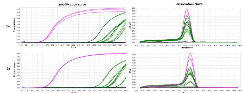
3.3. Analysis of Virus RNA Accumulation in Individuals of Fungus Gnats at Various
Developmental Stages
The virus was detected in two successive developmental stages of B. impatiens obtained
from larvae previously exposed to PSV-infected plants. No amplification products were
obtained for the control B. impatiens specimens fed on healthy plants. The melting peaks
of the amplified fragment of the PSV 1a gene in the PSV-infected N. benthamiana (positive
control) coincided with the dissociation curves of analysed insect samples exposed to
PSV (Figure 3). The obtained Ct values for pupae reached the highest values between
24 and 27, in the case of larva from 27 to 28, whereas the Ct for imago individuals were
registered between 30–33 (Figure 3). Quantitative analyses based on ddPCR indicated that
the highest copy number of viral gene per 1 µL of the reaction mixture was detected for
pupae samples, a bit lower in the case of larvae, whereas in imago specimens, the level of
virus RNA accumulation was the lowest in comparison to the remaining developmental
stages (Figure 4).Cells 2021, 10, 1546 8 of 15
Cells 2021, 10, 1546 8 of 16
FigureFigure 2. RT-PCR
2. RT-PCR detection
detection of peanut
of peanut stuntstunt
virusvirus
RNAs RNAs in developmental
in developmental stages
stages of Bradysia
of Bradysia impatiens.
impatiens. The RT-PCR
The RT-PCR products
products amplified with three PSV-specific primer pairs hybridizing to 1a, 2b, and fragment spanning 3a gene (movement
amplified with three PSV-specific primer pairs hybridizing to 1a, 2b, and fragment spanning 3a gene (movement protein,
protein, MP) and coat protein (CP); total RNA isolated from larvae, pupae, and adults (2–3 specimens) of B. impatiens
MP) and coat protein
population (CP);intotal
cultivated RNA isolated
laboratory from
conditions larvae,
on Petri pupae,
dishes wereand adults
used (2–3 specimens)
as a template. M—marker of B. impatiens
Gene population
Ruler 100 bp
cultivated
(Thermoin laboratory conditions
Scientific, Lenexa, on Petri
KS, USA); dishes
NTC—no were used
template as a template. M—marker Gene Ruler 100 bp (Thermo
control.
Cells 2021, 10, 1546 9 of 16
Scientific, Lenexa, KS, USA); NTC—no template control.
3.3. Analysis of Virus RNA Accumulation in Individuals of Fungus Gnats at Various
Developmental Stages
The virus was detected in two successive developmental stages of B. impatiens
obtained from larvae previously exposed to PSV-infected plants. No amplification
products were obtained for the control B. impatiens specimens fed on healthy plants. The
melting peaks of the amplified fragment of the PSV 1a gene in the PSV-infected N.
benthamiana (positive control) coincided with the dissociation curves of analysed insect
samples exposed to PSV (Figure 3). The obtained Ct values for pupae reached the highest
values between 24 and 27, in the case of larva from 27 to 28, whereas the Ct for imago
individuals were registered between 30–33 (Figure 3). Quantitative analyses based on
ddPCR indicated that the highest copy number of viral gene per 1 µ L of the reaction
mixture was detected for pupae samples, a bit lower in the case of larvae, whereas in
imago specimens, the level of virus RNA accumulation was the lowest in comparison to
the remaining developmental stages (Figure 4).
Figure 3. Results
Figure of PSV
3. Results detection
of PSV byby
detection RT-qPCR
RT-qPCRininsingle
single specimens oflarva,
specimens of larva,pupa,
pupa, and
and imago
imago B. impatiens
ofimpatiens
of B. after after the virus
the virus
acquisition by larvae feeding on PSV-infected N. benthamiana plants. Panel (a) The amplification plots of PSV 1a gene
acquisition by larvae feeding on PSV-infected N. benthamiana plants. Panel (a) The amplification plots of PSV 1a gene detected
detected in a single larva—red line, pupa—blue line, imago—green line; positive control (PSV-infected N. benthamiana)—
pink line (upper). Below, melting (dissociation) curves for amplicons of the PSV ORF 1a (replicase gene). For each devel-
opmental stage of fungus gnat, four single specimens were analysed, in three replicates. No amplification plots were ob-
tained in the case of negative controls, including healthy larva, pupa, and imago, which were not exposed to PSV as well
in NTC—no template control—grey line. Panel (b) Table with Ct values obtained by using in RT-qPCR analysis for PSV
1a gene for viruliferous larvae, pupae, and imago (four samples). Provided data concern the PSV-exposed larvae, pupae,
and imago, which developed from PSV-exposed larvae, in three replicates. The Ct value for the positive control, the PSV-Cells 2021, 10, 1546 9 of 15
in a single larva—red line, pupa—blue line, imago—green line; positive control (PSV-infected N. benthamiana)—pink line
(upper). Below, melting (dissociation) curves for amplicons of the PSV ORF 1a (replicase gene). For each developmental
stage of fungus gnat, four single specimens were analysed, in three replicates. No amplification plots were obtained in the
case of negative controls, including healthy larva, pupa, and imago, which were not exposed to PSV as well in NTC—no
template control—grey line. Panel (b) Table with Ct values obtained by using in RT-qPCR analysis for PSV 1a gene for
Cells viruliferous
2021, 10, 1546 larvae, pupae, and imago (four samples). Provided data concern the PSV-exposed larvae, pupae, and imago, 10 of 16
which developed from PSV-exposed larvae, in three replicates. The Ct value for the positive control, the PSV-infected N.
benthamiana (K + PSV), is also included.
Figure4.4.Detection
Figure Detection of
of PSV 1a gene in the
the subsequent
subsequentdevelopmental stagesofofB.B.impatiens
developmentalstages impatiensusing
usingEvaGreen
EvaGreendigital
digitaldroplet
droplet
PCR approach. (Upper):
PCR approach. (Upper): 1-D plot diagram
diagram presents differences in amplitudes of fluorescence signals between positiveand
presents differences in amplitudes of fluorescence signals between positive and
negative droplets. Blue dots: positive droplets
negative droplets. Blue dots: positive droplets with
with amplification,
amplification, grey
greydots:
dots:negative
negativedroplets
dropletswith
withno
noamplification.
amplification.Pink
Pink
line:threshold
line: threshold separating
separating population
population of
of negative
negative from
from positive
positive droplets.
droplets. (Below):
(Below):the thediagram
diagramshowing
showingthe
theconcentration
concentration
of viral copies per 1 μL of the reaction mixture. L—larva, P—pupa, IM—adult stage, E—egg from a virus-free B. impatiens
of viral copies per 1 µL of the reaction mixture. L—larva, P—pupa, IM—adult stage, E—egg from a virus-free B. impatiens
colony, (−)—negative control samples: PSV-non-exposed, NTC—no template control, K(+) —positive control, N. bentham-
colony, (−)—negative
iana plants control
infected with PSVsamples:
inoculum.PSV-non-exposed, NTC—no template control, K(+) —positive control, N. benthamiana
plants infected with PSV inoculum.
3.4. Bradysia impatiens Ability to Vector PSV to Healthy Plants
To verify the virus vectoring ability of B. impatiens, the larvae which fed on PSV-in-
oculated leaves and parallel on the virus-free plant were put into soil pots with young N.
benthamiana. It is worth adding that, before the transmission assay, all plant seedlings were
molecularly tested to exclude PSV infection.
The plant’s visual inspection, three weeks after the inoculation by PSV-infected B.
impatiens larvae, did not reveal any characteristic disease symptoms like chlorosis, mosa-Cells 2021, 10, 1546 10 of 15
3.4. Bradysia impatiens Ability to Vector PSV to Healthy Plants
To verify the virus vectoring ability of B. impatiens, the larvae which fed on PSV-
inoculated leaves and parallel on the virus-free plant were put into soil pots with young N.
benthamiana. It is worth adding that, before the transmission assay, all plant seedlings were
molecularly tested to exclude PSV infection.
Cells 2021, 10, 1546 11 of 16
The plant’s visual inspection, three weeks after the inoculation by PSV-infected B.
impatiens larvae, did not reveal any characteristic disease symptoms like chlorosis, mosaics,
stunting, or leave malformations. Thus, they were tested for the presence of viral RNAs
RNAs using molecular
using molecular tools.detection
tools. PSV PSV detection was performed
was performed utilizing
utilizing RT-qPCR.
RT-qPCR. The anal-
The analyses were
yses were based on the amplification of three viral targets 1a, 2a, and CP genes.
based on the amplification of three viral targets 1a, 2a, and CP genes. The melting peaks The melt- of
ing
the peaks of the
amplified amplified
fragments forfragments for PSV-infected
PSV-infected N. benthamiana N.(positive
benthamiana (positive
control) control)
coincided with
coincided with the
the dissociation dissociation
curves obtainedcurves obtained
for tested for tested plants,
N. benthamiana N. benthamiana
previouslyplants, previ-
treated with
ously treated with
PSV-exposed PSV-exposed
B. impatiens larvae.B.No
impatiens larvae. No
amplification amplification
plots were observed plots
forwere observed
healthy control
for healthy
plants control
(Figure 5). plants (Figure 5).
Figure 5.
Figure 5. The
The RT-qPCR
RT-qPCR detection
detectionof
ofPSV
PSVRNAs
RNAsininN.
N.benthamiana
benthamianaplants
plantstreated
treatedwith
withPSV-exposed
PSV-exposed larvae
larvae ofof
B. B. impatiens.
impatiens. The
The diagrams
diagrams present
present the amplification
the amplification (left)
(left) andand dissociation
dissociation curves
curves (right)
(right) for for amplification
amplification products
products of 1a,
of 1a, 2a,2a,
andand
CPCPPSV
PSV genes, detected in N. benthamiana plants; pink line—PSV-inoculated N. benthamiana (positive control); green lines—
genes, detected in N. benthamiana plants; pink line—PSV-inoculated N. benthamiana (positive control); green lines—tested
tested N. benthamiana plants treated with PSV-exposed larvae; black line—negative controls, healthy plant as well as
N. benthamiana plants treated with PSV-exposed larvae; black line—negative controls, healthy plant as well as NTC—no
NTC—no template control.
template control.
The
Theabsolute
absolutequantification,
quantification,basedbasedon on1a1aand
and2a2aviral
viraltargets,
targets,was
wasperformed
performed using
using a a
ddPCR
ddPCRreaction.
reaction.EvaGreen
EvaGreenddPCR ddPCR results showed
results showed that thethe
that accumulation
accumulationof the
of 1a
thegene is
1a gene
higher thanthan
is higher the 2a
thegene. The EvaGreen
2a gene. The EvaGreenddPCRddPCRshowedshowed
positivepositive
dropletsdroplets
for tested plants
for tested
detecting 20–554 copies of 1a gene in the 20 μL reaction mixture that
plants detecting 20–554 copies of 1a gene in the 20 µL reaction mixture that correspondscorresponds to 16– to
443 copies
16–443 per per
copies 1 mg plant
1 mg tissue.
plant TheThe
tissue. absolute quantity
absolute quantityof 2aof gene waswas
2a gene a bit lower,
a bit lower,indi-
indi-
cating
cating20–190
20–190copies
copiesinin20
20μL µLofofPCR
PCRvolume,
volume,which
whichcorresponds
correspondstoto16–152
16–152copies/1
copies/1 mg mg
plant
planttissue
tissueininthree
threeout
outofoffive
fivetested
testedplants,
plants,and
andininthe
thetwotworemaining
remainingplants
plants (Nb1
(Nb1 andand
Nb5),
Nb5),we weindicated
indicatedonlyonly~3–5
~3–5copies/1
copies/1 μLµLof of
reaction, thethe
reaction, equivalent
equivalentof 2–4 copies
of 2–4 per per
copies 1
mg
1 mg of of
plant tissue
plant (Figure
tissue (Figure6). 6).Cells 2021, 10, 1546 11 of 15
Cells 2021, 10, 1546 12 of 16
Figure6.6.The
Figure TheddPCR
ddPCRdetection
detectionofofviral
viral 1a
1a and 2a genes
genes in N.benthamiana
inN. benthamianaplants
plantsafter
afterinoculation
inoculation with
with PSV-exposed
PSV-exposed larvae.
larvae.
(Upper)
(Upper) 1-Dplot
1-D plotdiagrams
diagramspresent
presentdifferences
differencesininamplitudes
amplitudesofoffluorescence
fluorescencesignals
signalsbetween
betweenpositive
positiveand
andnegative
negativedroplets
drop-
forlets
1afor 1a (left)
(left) and 2a and 2a (right).
(right). The black
The black droplets
droplets represent
represent negative
negative droplets
droplets withwith no templates,
no templates, andand blue
blue represent
represent pos-
positive
itive droplets containing amplified templates; pink line: threshold separating negative from positive droplets. Nb1–Nb5
droplets containing amplified templates; pink line: threshold separating negative from positive droplets. Nb1–Nb5 samples
samples of tested plants inoculated with the viruliferous insect; H1–H4 healthy plants of N. benthamiana; K(+) positive
of tested plants inoculated with the viruliferous insect; H1–H4 healthy plants of N. benthamiana; K(+) positive controls
controls cDNA from PSV infected plants (10—fold diluted, 1 × 10⁻⁴, 10−5, 10−6); NTC—no template control. (Below) The
cDNA from PSV infected plants (10—fold diluted, 1 × 10 −4 , 10−5 , 10−6 ); NTC—no template control. (Below) The EvaGreen
EvaGreen ddPCR absolute quantification of copies number of PSV 1a and 2a genes per 1 μL of ddPCR reaction mixture,
ddPCR
in 20 μLabsolute quantification
of the reaction mixtureofand
copies number of of
the equivalent PSV 1a number
copy and 2a genes per
of viral 1 µL and
RNA1 of ddPCR
RNA2 reaction
per 1 mgmixture, in 20 µL of
of plant tissue.
the reaction mixture and the equivalent of copy number of viral RNA1 and RNA2 per 1 mg of plant tissue.
3.5. Detection of PSV by Western Blotting
3.5. Detection
To finallyofconfirm
PSV by that
Western
PSVBlotting
was transmitted by viruliferous larvae of B. impatiens, the
PSVTo finallyantisera
specific confirmwas
thatused
PSV for
wasimmunodetection.
transmitted by viruliferous larvae
Western blot on of B. impatiens,
crude the
plants ex-
PSV specific
tracts from N.antisera was used
benthamiana forinoculated
plants immunodetection. Western blot
with PSV-infected larvaeon confirmed
crude plantstheextracts
pres-
ence N.
from benthamiana
of PSV plantsWe
coat protein. inoculated
detected with
the CPPSV-infected
(ca. 24 kDa)larvae
and itsconfirmed
dimers in the presence
plants inocu-of
PSV
latedcoat
withprotein. We detected
B. impatiens the CPlarvae,
PSV-exposed (ca. 24 the
kDa) andsignal
same its dimers in plants in
was detected inoculated with
the positive
B. impatiens
controls: N. PSV-exposed larvae,
benthamiana plants the same signal
mechanically was detected
inoculated in and
with PSV, the positive virionsN.
controls:
in purified
benthamiana
preparations. plants mechanically
No signal inoculated
was detected with PSV,
for healthy, and inN.purified
untreated virions
benthamiana preparations.
plants (Figure
7). signal was detected for healthy, untreated N. benthamiana plants (Figure 7).
NoCells 2021, 10, 1546 12 of 15
Cells 2021, 10, 1546 13 of 16
Figure
Figure 7.
7. Detection of PSV
Detection of PSV coat
coatprotein
proteinininN.
N.benthamiana
benthamianaplants
plantsinoculated
inoculated with
with virus-exposed
virus-exposed lar-
larvae
vae of B. impatiens by western blotting. K−—healthy plant, untreated N. benthamiana; samples
of B. impatiens by western blotting. K−—healthy plant, untreated N. benthamiana; samples Nb1-5: N. Nb1-
5: N. benthamiana plants inoculated with B. impatiens larvae; M—protein marker PageRuler™ Pres-
benthamiana plants inoculated with B. impatiens larvae; M—protein marker PageRuler™ Prestained
tained Protein Ladder Plus (Thermo Scientific, Lenexa, KS, USA), K+—positive control, PSV-inocu-
Protein Ladder Plus (Thermo Scientific, Lenexa, KS, USA), K+—positive control, PSV-inoculated
lated plant; purified virions: as positive controls protein extracts from native virus preparations
plant;also
were purified
used.virions:
The PSV ascoat
positive controls
protein protein
and its extracts
dimers from native
are indicated virusarrows.
by black preparations were also
used. The PSV coat protein and its dimers are indicated by black arrows.
4.
4. Discussion
Discussion
The
The primary
primary focusfocus ofof this
this study
study waswas toto determine
determine the the ability
ability of
of B.
B. impatiens
impatiens larvae,
larvae, inin
laboratory trials, to acquire and then to vector PSV. Our previous
laboratory trials, to acquire and then to vector PSV. Our previous observations, performed observations, performed
in
in the controlled experimental
the controlled experimental conditions,
conditions, indicated
indicated the the accidental
accidental contamination
contamination with with
PSV of previously healthy plants that were grown in the
PSV of previously healthy plants that were grown in the same glasshouse cabin withsame glasshouse cabin with vi-
rus-infected
virus-infected N.N.benthamiana.
benthamiana.Thus, Thus,we wehave
haveundertaken
undertakentotoanalyse
analysethe thepotential
potential involve-
involve-
ment of Bradysiaspp., which was the only factor present in greenhouse
ment of Bradysia spp., which was the only factor present in greenhouse soil that could soil that could have
contributed to the virus transmission. The RT-PCR analyses
have contributed to the virus transmission. The RT-PCR analyses carried out on RNAs carried out on RNAs isolated
from
isolatedsamples of larvae,
from samples ofpupae,
larvae, andpupae,adults
andof fungus
adults gnat, collected
of fungus from soil
gnat, collected frompots,
soilunex-
pots,
pectedly
unexpectedly confirmed the presence of PSV genomic RNAs in all insect stages. These data
confirmed the presence of PSV genomic RNAs in all insect stages. These
prompted us to explore this observation.
Indeed,
Indeed, the theanalyses
analysesperformed
performedinin this study,
this study,confirmed
confirmed ourour
preliminary
preliminary results. We
results.
confirmed
We confirmed the ability of virus
the ability acquisition
of virus by theby
acquisition larval stage of
the larval B. impatiens
stage and its and
of B. impatiens persis-
its
tence withinwithin
persistence the insect body body
the insect during its development.
during its development. The Theobservation
observation suggests thatthat
suggests the
virus persists
the virus transstadially.
persists transstadially. In In
this respect,
this respect, these
thesedata
dataareareininagreement
agreementwith with the
the results
of the experiments on fungi vectoring capacity of B. impatiens [15]. It It is
is worth
worth adding
adding that,
that,
when we started work on this issue, we also detected the presence of PSV RNA in the
second generation (F2) of B.
(F2) of B. impatiens,
impatiens,namely,
namely,ininfreshly
freshlylaidlaideggs
eggsfrom
from adult
adultfemales
females B.
of of
impatiens
B. impatiens (whose
(whose larval stadium
larval stadium of of
F1 F1was
was exposed
exposed to to
PSV)PSV)as as
well as as
well in the specimens
in the specimens of
larvae
of from
larvae thethe
from second
second generation
generation (F2)(F2)
hatched
hatched fromfromthese eggs
these (Supplementary
eggs (Supplementary Figure S1).
Figure
However, we did not obtain repeatability, which may have
S1). However, we did not obtain repeatability, which may have resulted from the low resulted from the low level
of virus
level RNARNA
of virus accumulation
accumulation or possibly fromfrom
or possibly the fact thatthat
the fact PSVPSV waswas not not
detectable in all
detectable in
laidlaid
all eggs.
eggs.That
That is iswhy
whywe wecannot
cannotconclude
concludethat thatthe
thevirus
virus persists
persists transgenerationally
transgenerationally
in B. impatiens. However, these results showed that PSV-infested larvae developed into
PSV-infested pupae and then into PSV-infested adults.Cells 2021, 10, 1546 13 of 15
in B. impatiens. However, these results showed that PSV-infested larvae developed into
PSV-infested pupae and then into PSV-infested adults.
In the light of these data, a further question that arises is about the mode of virus
transmission in the fungus gnat’s body. Thus far, cucumoviruses have been known
to be transmitted by aphids (subfamily Aphidinae) in a non-persistent manner. Over
80 species of aphids have been identified to vector CMV [24,25], whereas PSV is known
to be transmitted by Aphis craccivora, A. solanella, A. spiraecola, Myzus persicae, and Liaphis
erysimi [26,27]. Currently, many authors focus on clarifying the cucumovirus-aphid-plant
interactions [24,28–30]. Noteworthy, Kameya-Iwaki et al. have proven that PSV and CMV
might be transmitted by M. persicae in a non-persistent but also in a semi-persistent manner,
depending on the plant species used for the assay [25,31].
Viruses vectored by insects are divided into two categories: non-circulative (NC) and
circulative viruses (CVs). In the first category, the viral particles attach to the cuticle of
the insect vector without circulation in the insect body [32]. The NC viruses are divided
into subcategories, transmitted in a non-persistent and semi-persistent manner, depending
on the duration of virus retention in the insect’s body. Non-persistent virus transmission
is characterized by very short acquisition and inoculation times of seconds to minutes.
In the semi-persistent transmission, the acquisition can occur within minutes, but the
transmission efficiency increases with prolonged insect feeding. Moreover, the second
distinguishing feature is the retention period of hours to days [33]. In the second group,
CVs need to circulate in the insect tissue before transmission to healthy plants. In some
cases, these viruses can also replicate in the vector’s body [32,34]. The quantitative analyses
of viral RNAs in developmental stages of fungus gnat showed that the amount of viral
RNA1 encoding 1a ORF in the pupa stage was in a few cases higher than in the larvae
that acquired viral particles. The obtained data suggested a far-reaching hypothesis that
PSV may circulate or even replicate in fungus gnats tissues in contrast to the above-
mentioned transmission in the aphid. On the other hand, the differences in virus RNA
accumulation are an individual matter of specimens, and the larvae (from which the
analysed pupae developed) previously acquired much more virus compared to the others.
However, only further experiments will shed light on the replication of this virus in
different developmental stages of B. impatiens.
On the basis of the available literature, Bradysia species are known to be vectoring soil-
borne pathogens [21]. In 1990 Gardiner et al. showed that the larval stage of B. impatiens
might act as an important vector for Pythium spp. [21]. However, there are no literature
data on the role of Bradysia spp. in virus spreading. The transmission assay performed in
this study has shown that B. impatiens larvae exposed to PSV are able to transmit the virus
to a healthy plant. In 2010, Braun et al. have reported transstadial transmission of Pythium
and proved the lack of B. impatiens adults vectoring ability [15]. The vectoring capacity
of adult stages has been poorly studied. Nevertheless, a few reports have indicated the
role of adults of B. impatiens in pathogen transmission [12]. Kapongo and co-workers
in 2020 showed that not only larva, but also adults of B. impatiens might be a vector of
Fusarium oxysporum and Pythium aphanidermatum [35]. In this study, we have not confirmed
the ability of an adult insect to transmit the virus to healthy plants. On the basis of the
obtained data, the virus acquired in the larval stage persists transstadially, which is also in
agreement with the previous report published by Braun and co-workers [15]. Hence, the
most likely possibility is that a virus is transmitted by the larva in the second generation
(F2). As mentioned above, PSV was detected in the eggs and larvae of the F2 generation,
but only in one experiment. In general, we suppose that virus might be carried on a very
low level in F2 eggs or might be present only in some of them, or it may not always be
transferred to the next generation. Hence, this issue needs further experiments that will
include a larger number of samples, to draw reliable conclusions.
In the molecular experiments complemented with biological test on PSV-exposed
larvae and healthy plants of N. benthamiana, we have shown that B. impatiens larvae
have the vectoring capacity. However, after the inoculation access period, the level ofCells 2021, 10, 1546 14 of 15
the viral RNA accumulation in plant tissues, in almost all cases, was rather low. Visual
observation of plants inoculated by viruliferous B. impatiens revealed no characteristic
symptoms of PSV infection. The typical symptoms of PSV infection on N. benthamiana
are stunting, leaves malformation, and chlorosis. However, in some cases, the symptoms
may be attenuated, depending on various conditions, among other temperature [22,36].
Herein, the symptomless infection might result from a small number of virus particles
or low transmission efficiency caused by a small number of insect specimens used in
these experiments. However, using the Western blotting approach, we have confirmed the
presence of viral coat protein, which proves that the genomic strands of PSV, detected in
the plants exposed to viruliferous larvae of B. impatiens, are infectious.
Thus, on the basis of the above-described findings, B. impatiens role as a plant virus
vector, or a reservoir of this pathogen, should be taken into account. The obtained data help
to better understand the biology of B. impatiens and its role in plant pathogen spreading.
Supplementary Materials: The following are available online at https://www.mdpi.com/article/10
.3390/cells10061546/s1, Figure S1: RT-PCR detection of PSV 1a gene of chosen developmental stages
of Bradysia impatiens.
Author Contributions: Conceptualization, M.B., P.F., and A.O.-S.; methodology, M.B., P.F., and
A.O.-S.; formal analysis, M.B., P.F., and A.O.-S.; investigation, M.B. and P.F.; resources, A.O.-S.;
data curation, M.B. and P.F.; writing—original draft preparation, M.B. and A.O.-S.; writing—review
and editing, M.B., P.F., and A.O.-S.; supervision, A.O.-S.; project administration, A.O.-S.; funding
acquisition, A.O.-S. All authors have read and agreed to the published version of the manuscript.
Funding: This work was supported by the Polish Ministry of Science and Higher Education, statutory
activity project: BIOTECH01.
Institutional Review Board Statement: Not applicable.
Informed Consent Statement: Not applicable.
Data Availability Statement: All data are provided in the manuscript and in the GenBank database.
Acknowledgments: We wish to thank Marek Tomalak from the Department of Biological Pest
Control and Organic Agriculture, IPP-NRI (Poznań, Poland) for his valuable comments. We thank
also Irena Nowacka from the Department of Entomology and Agricultural Pests, IPP-NRI (Poznań,
Poland) for providing the population of Bradysia spp.
Conflicts of Interest: The authors declare no conflict of interest.
References
1. Katumanyane, A.; Ferreira, T.; Malan, A.P. A Review of Bradysia spp. (Diptera: Sciaridae) as Pests in Nursery and Glasshouse
Crops, with Special Reference to Biological Control Using Entomopathogenic Nematodes. Afr. Entomol. 2018, 26, 1–13. [CrossRef]
2. Menzel, F.; Smith, J.E.; Colauto, N.B. Bradysia difformis Frey and Bradysia ocellaris (Comstock): Two additional neotropical
species of black fungus gnats (Diptera: Sciaridae) of economic importance: A redescription and review. Ann. Entomol. Soc. Am.
2003, 96, 448–457. [CrossRef]
3. Rebora, M.; Salerno, G.; Piersanti, S.; Gorb, E.; Gorb, S. Entrapment of Bradysia paupera (Diptera: Sciaridae) by Phaseolus
vulgaris (Fabaceae) plant leaf. Arthropod-Plant Interact. 2020, 14, 499–509. [CrossRef]
4. Cloyd, R.A. Ecology of fungus gnats (Bradysia spp.) in greenhouse production systems associated with disease-interactions and
alternative management strategies. Insects 2015, 6, 325–332. [CrossRef] [PubMed]
5. Wilkinson, J.D.; Daugherty, D.M. The biology and immature stages of Bradysia impatiens (Diptera: Sciaridae). Ann. Entomol. Soc.
Am. 1970, 63, 656–660. [CrossRef]
6. Woelke, J.B.; Pham, K.; Humala, A.E. New species of Stenomacrus (Hymenoptera: Ichneumonidae: Orthocentrinae) reared from
Bradysia impatiens (Diptera: Sciaridae) in The Netherlands. J. Nat. Hist. 2020, 54, 1603–1616. [CrossRef]
7. Hall, R.D.; Gerhardt, R.R. Medical and Veterinary Entomology, 2nd ed.; Mullen, G.R., Durden, L.A., Eds.; Academic Press:
Cambridge, MA, USA, 2009.
8. Chabannes, M.; Hatt, G.; Thébaud, G.; Bedford, I.D.; Lamb, C. Establishment of an in vitro sciarid fly larvae assay to study plant
resistance. Ann. Appl. Biol. 2009, 155, 293–296. [CrossRef]
9. James, R.L.; Dumroese, R.K.; Wenny, D.L. Botrytis cinerea carried by adult fungus gnats (Diptera: Sciaridae) in container nurseries.
Tree Plant Notes 1995, 46, 48–53.Cells 2021, 10, 1546 15 of 15
10. Jagdale, G.B.; Casey, M.L.; Cañas, L.; Grewal, P.S. Effect of entomopathogenic nematode species, split application and potting
medium on the control of the fungus gnat, Bradysia difformis (Diptera: Sciaridae), in the greenhouse at alternating cold and
warm temperatures. Biol. Control 2007, 43, 23–30. [CrossRef]
11. Leath, K.T.; Newton, R.C. Interaction of a fungus gnat Bradysia sp (Sciaridae) with Fusaium spp on alfaalfa and red clover.
Phytopathology 1969, 59, 257.
12. Kalb, D.W. Dispersal of Verticillium albo-atrum by the Fungus Gnat (Bradysia impatiens). Plant Dis. 1986, 70, 752. [CrossRef]
13. Jarvis, W.R.; Shipp, J.L.; Gardiner, R.B. Transmission of Pythium aphanidermatum to greenhouse cucumber by the fungus gnat
Bradysia impatiens (Diptera: Sciaridae). Ann. Appl. Biol. 1993, 122, 23–29. [CrossRef]
14. Edwardson, J.R.; Christie, R.G. CRC Handbook of Viruses Infecting Legumes; CRC Press: Boca Raton, FL, USA, 1991.
15. Braun, S.E.; Castrillo, L.A.; Sanderson, J.P.; Daughtrey, M.L.; Wraight, S.P.; Braun, S.E. Transstadial Transmission of Pythium in
Bradysia impatiens and Lack of Adult Vectoring Capacity. Phytopathology 2010, 100, 1307. [CrossRef]
16. Park, J.M.; You, Y.H.; Park, J.H.; Kim, H.H.; Ghim, S.Y.; Back, C.G. Cutaneous microflora from geographically isolated groups of
bradysia agrestis, an insect vector of diverse plant pathogens. Mycobiology 2017, 45, 160–171. [CrossRef]
17. Obrepalska-Steplowska, A.; Nowaczyk, K.; Budziszewska, M.; Czerwoniec, A.; Pospieszny, H. The sequence and model structure
analysis of three Polish peanut stunt virus strains. Virus Genes 2008, 36, 221–229. [CrossRef] [PubMed]
18. Obrȩpalska-Stȩplowska, A.; Budziszewska, M.; Wieczorek, P.; Czerwoniec, A. Analysis of two strains of Peanut stunt virus:
SatRNA-associated and satRNA free. Virus Genes 2012, 44, 513–521. [CrossRef]
19. Obrepalska-Steplowska, A.; Budziszewska, M.; Pospieszny, H. Complete nucleotide sequence of a Polish strain of Peanut stunt
virus (PSV-P) that is related to but not a typical member of subgroup I. Acta Biochim. Pol. 2008, 55, 731–739. [CrossRef]
20. Fereres, A.; Raccah, B. Plant Virus Transmission by Insects. eLS 2015, 1–12. [CrossRef]
21. Gardiner, R.B.; Jarvis, W.R.; Shipp, J.L. Ingestion of Pythium spp. by larvae of the fungus gnat Bradysia impatiens (Diptera:
Sciaridae). Ann. Appl. Biol. 1990, 116, 205–212. [CrossRef]
22. Obrepalska-Steplowska, A.; Renaut, J.; Planchon, S.; Przybylska, A.; Wieczorek, P.; Barylski, J.; Palukaitis, P. Effect of temperature
on the pathogenesis, accumulation of viral and satellite RNAs and on plant proteome in peanut stunt virus and satellite
RNA-infected plants. Front. Plant Sci. 2015, 6, 903. [CrossRef]
23. Vilkamaa, P. Checklist of the family Sciaridae (Diptera) of Finland. Zookeys 2014, 441, 151–164. [CrossRef]
24. Tungadi, T.; Donnelly, R.; Qing, L.; Iqbal, J.; Murphy, A.M.; Pate, A.E.; Cunniffe, N.J.; Carr, J.P. Cucumber mosaic virus 2b proteins
inhibit virus-induced aphid resistance in tobacco. Mol. Plant Pathol. 2020, 21, 250–257. [CrossRef]
25. Kameya-Iwaki, M.; Murakami, K.; Ito, S.; Hanada, K.; Tanaka, S. Semipersistency of Myzus persicae transmission of cucu-
moviruses systemically infecting leguminous plants. J. Gen. Plant Pathol. 2000, 66, 64–67. [CrossRef]
26. Hebert, T.T. Epidemiology of the peanut stunt virus in North Carolina. Phytopathology 1967, 57, 461.
27. El Sadiq, E.O.; Ahmed, A.H. Comparative studies on aphid transmission of the Sudanese strain of peanut stunt virus. J.
Phytopathol. 1986, 115, 160–164. [CrossRef]
28. Liang, Y.; Gao, X.-W. The cuticle protein gene MPCP4 of Myzus persicae (Homoptera: Aphididae) plays a critical role in cucumber
mosaic virus acquisition. J. Econ. Entomol. 2017, 110, 848–853. [CrossRef]
29. Krenz, B.; Bronikowski, A.; Lu, X.; Ziebell, H.; Thompson, J.R.; Perry, K.L. Visual monitoring of Cucumber mosaic virus infection
in Nicotiana benthamiana following transmission by the aphid vector Myzus persicae. J. Gen. Virol. 2015, 96, 2904–2912.
[CrossRef]
30. Wamonje, F.O.; Donnelly, R.; Tungadi, T.D.; Murphy, A.M.; Pate, A.E.; Woodcock, C.; Caulfield, J.; Mutuku, J.M.; Bruce, T.J.A.;
Gilligan, C.A.; et al. Different Plant Viruses Induce Changes in Feeding Behavior of Specialist and Generalist Aphids on Common
Bean That Are Likely to Enhance Virus Transmission. Front. Plant Sci. 2020, 10, 1811. [CrossRef]
31. Kameya-Iwaki, M.; Shigeyoshi, M.; Ito, S.; Tanaka, S. Effects of susceptibility of test plants on modes of Cucumovirus transmission
by Myzus persicae. J. Gen. Plant Pathol. 2001, 67, 69–72. [CrossRef]
32. Garzo, E.; Moreno, A.; Plaza, M.; Fereres, A. Feeding Behavior and Virus-transmission Ability of Insect Vectors Exposed to
Systemic Insecticides. Plants 2020, 9, 895. [CrossRef]
33. Ng, J.C.K.; Perry, K.L. Transmission of plant viruses by aphid vectors. Mol. Plant Pathol. 2004, 5, 505–511. [CrossRef] [PubMed]
34. Yadav, D.; Rana, R. Transmission of plant virus through arthropod vector. J. Entomol. Zool. Stud. 2020, 8, 1934–1939.
35. Kapongo, J.-P.; Kevan, P.G.; Shipp, L.; Taki, H. Making a Pest Beneficial: Fungus Gnats [Bradysia impatiens (Diptera: Sciaridea)] as
Potential Vectors of Microbial Control Agents to Suppress Pathogens they Also Spread. In Entomovectoring for Precision Biocontrol
and Enhanced Pollination of Crops; Springer: Berlin/Heidelberg, Germany, 2020; pp. 239–250.
36. Obrepalska-Steplowska, A.; Wieczorek, P.; Budziszewska, M.; Jeszke, A.; Renaut, J. How can plant virus satellite RNAs alter the
effects of plant virus infection? A study of the changes in the Nicotiana benthamiana proteome after infection by Peanut stunt
virus in the presence or absence of its satellite RNA. Proteomics 2013, 13, 2162–2175. [CrossRef] [PubMed]You can also read