Comparative microRNAome analysis of the testis and ovary of the Chinese giant salamander
←
→
Page content transcription
If your browser does not render page correctly, please read the page content below
REPRODUCTION
    RESEARCH

Comparative microRNAome analysis of the testis and ovary of
the Chinese giant salamander
Rui Chen1, Jian Du1, Lin Ma1, Li-qing Wang1, Sheng-song Xie2, Chang-ming Yang3,
Xian-yong Lan1, Chuan-ying Pan1 and Wu-zi Dong1
1
 College of Animal Science and Technology, Northwest A& F University, Yangling, China, 2Key Lab of Agricultural
Animal Genetics, Breeding, and Reproduction of Ministry of Education, Huazhong Agricultural University, Wuhan,
China and 3Animal Husbandry and Veterinary Station of Chenggu County, Hanzhong, China
Correspondence should be addressed to C Pan or W Dong; Email: chuanyingpan@126.com or dongwuzi@nwsuaf.edu.cn

Abstract
MicroRNAs (miRNAs) are 18–24 nucleotides non-coding RNAs that regulate gene expression by post-transcriptional suppression of
mRNA. The Chinese giant salamander (CGS, Andrias davidianus), which is an endangered species, has become one of the important
models of animal evolution; however, no miRNA studies on this species have been conducted. In this study, two small RNA libraries
of CGS ovary and testis were constructed using deep sequencing technology. A bioinformatics pipeline was developed to distinguish
miRNA sequences from other classes of small RNAs represented in the sequencing data. We found that many miRNAs and other small
RNAs such as piRNA and tsRNA were abundant in CGS tissue. A total of 757 and 756 unique miRNAs were annotated as miRNA
candidates in the ovary and testis respectively. We identified 145 miRNAs in CGS ovary and 155 miRNAs in CGS testis that were
homologous to those in Xenopus laevis ovary and testis respectively. Forty-five miRNAs were more highly expressed in ovary than in
testis and 21 miRNAs were more highly expressed in testis than in ovary. The expression profiles of the selected miRNAs (miR-451,
miR-10c, miR-101, miR-202, miR-7a and miR-499) had their own different roles in other eight tissues and different development
stages of testis and ovary, suggesting that these miRNAs play vital regulatory roles in sexual differentiation, gametogenesis and
development in CGS. To our knowledge, this is the first study to reveal miRNA profiles that are related to male and female CGS
gonads and provide insights into sex differences in miRNA expression in CGS.
Reproduction (2017) 154 269–279

Introduction                                                       increasing prevalence of infectious diseases (Dong et al.
                                                                   2010, Du et al. 2016), the CGS population has declined
Amphibians are an important evolutionary bridge                    sharply in last half-century (Wang et al. 2004, Che et al.
between aquatic and terrestrial vertebrates (Vogel et al.          2014, Li et al. 2015a,b). At present, artificial breeding
1999). The Chinese giant salamander (CGS, Andrias                  with mixed-sex cultures is used to increase the CGS
davidianus) is the largest extant amphibian in the world           population (Fan et al. 2015), but not wholly successfully.
(Zhou et al. 2013) and belongs to the Cryptobranchidae             For example, the reproductive rate is low, and less
family, which only contains three species (Cryptobranchus          than 15% of CGSs produce offspring every year (Shi
alleganiensis in North America, Andrias japonicus in               2011). Because of the difficulty in determining CGS
Japan and Andrias davidianus in China). It is known                sex at the immature stage, mixed-sex cultures have
as a ‘living fossil’ because it has existed for more than          not satisfactorily increased the CGS population. The
350 million years (Gao & Shubin 2003). It was listed in            reproductive physiology and gonadal development of
the China Red Data Book as an endangered species in                CGSs are currently unknown. However, recent advances
1986 and has been included on the International Union              have revealed that microRNAs (miRNAs) are essential
for Conservation of Nature and Natural Resources Red               for sexual differentiation, gonadal development,
List of Threatened Species since 2004 (Hu et al. 2016).            gametogenesis        and    reproductive     performance
The phylogenetic position of the CGS makes it as an                (Grossman & Shalgi 2016, Kwekel et al. 2017).
invaluable model organism, and it has received a great                The miRNAs belong to a group of endogenous small
deal of attentions in studies on evolution, comparative            non-coding RNAs that are 18–24 nucleotides (nt) in
biology and other studies (Fan et al. 2015). However,              length (Bartel et al. 2004, Dong et al. 2014), exist in all
due to the deterioration of its habitat, over-harvesting,          known animal species and have different spatiotemporal
environmental pollution, climate change and the                    expression patterns (Li et al. 2010, Kadri et al. 2011,
© 2017 Society for Reproduction and Fertility                                                                  DOI: 10.1530/REP-17-0109
ISSN 1470–1626 (paper) 1741–7899 (online)                                                 Online version via www.reproduction-online.org
                                                                                            Downloaded from Bioscientifica.com at 09/16/2020 10:39:23PM
                                                                                                                                          via free access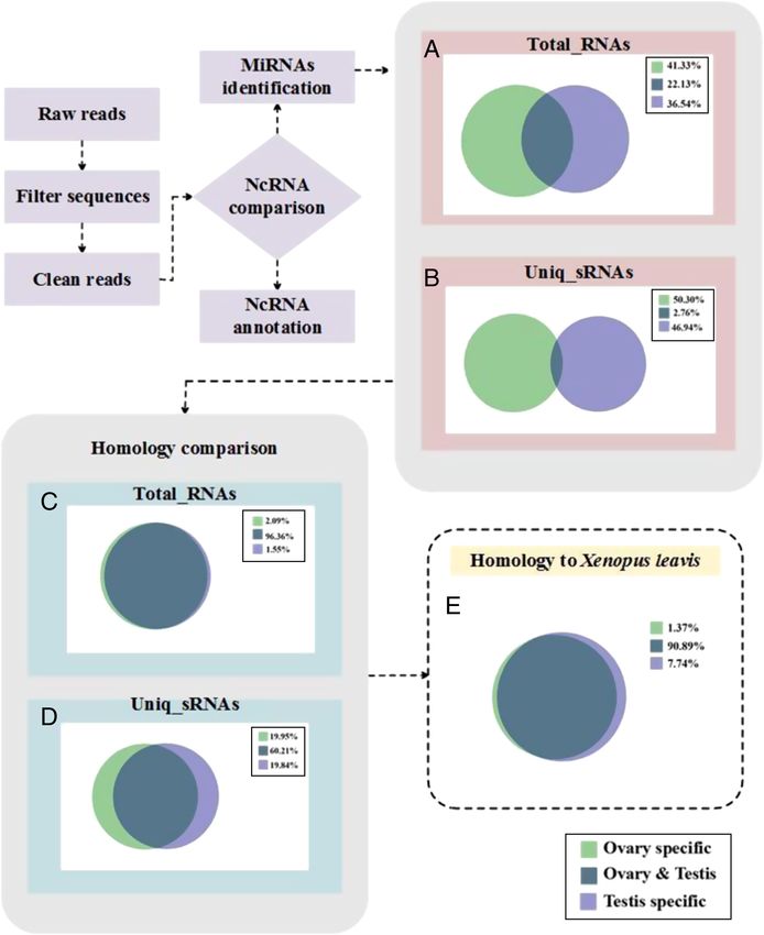
270     R Chen and others

Ji et al. 2012). In animals, almost all miRNAs regulate               For Illumina sequencing, tissue RNA from 4-year-old CGSs
their gene expression at the post-transcriptional level            was extracted, and RNA samples from three ovaries and
by binding to complementary target sites in the 3′                 three testes tissues were pooled prior to the construction of
untranslated region of mRNA (He & Hannon 2004,                     indexed libraries for Illumina sequencing (Beijing Biomarker
Leung & Sharp 2010). Over one-third of protein-coding              Technologies, China).
genes in humans are regulated by miRNAs, which                        Total RNA was extracted using the TRIzol reagent (TaKaRa)
open a new perspective on gene regulatory networks                 following the manufacturer’s protocol. The quantity and
(Kim & Nam 2006). The miRNAs are involved in a                     quality of the total RNA were determined using a NanoDrop
                                                                   ND-1000 spectrophotometer (Thermo Fisher Scientific) at
variety of biological processes, such as development
                                                                   260/280 nm (ratio >2.0), and its integrity was tested using a
(Ambros et al. 2003, Chen et al. 2004), cell proliferation
                                                                   2100 Bioanalyzer and a RNA 6000 Nano LabChip Kit (Agilent)
and death (Brennecke et al. 2003), cell differentiation,
                                                                   with an RNA integrity number greater than 8.0.
cell survival, cell-cycle control, apoptosis (Beilharz et al.
2009), immune responses (Pedersen et al. 2007, Li et al.
2008), as well as diseases (Poy et al. 2004); however, to          Histological analysis
our knowledge, there have been no reports of miRNAs                Ovary and testis tissues from 1-, 2-, 3- and 4-year-old CGSs
in the CGS.                                                        were fixed in Bouin’s buffer for 12 h, stored in 70% (v/v) ethanol
   High-throughput sequencing of RNA (RNA-Seq) is an               and embedded in paraffin. The 7 μm thick sections were
efficient way of mapping and quantifying transcriptomes            deparaffinized with xylene and rehydrated with an ethanol
and has been developed to analyze global gene expression           series from 100% (v/v) to 70% (v/v). The slides were washed
in different tissues. In this study, Illumina sequencing           for 5 min with phosphate-buffered saline (PBS) three times,
technology was used to characterize miRNA expression               followed by a hematoxylin wash for 30 s at room temperature.
profiles in the gonadal tissues of male and female CGSs.           The slides were then washed and stained with 5% acid alcohol
A miRNA database would not only significantly advance              for 30 s. Subsequently, the slides were washed for 10 min in PBS
our knowledge of the miRNA population presented in                 to change the stain from purple to blue, before being stained
CGSs but also improve our understanding of the roles               with eosin for 30 s and dehydrated with an ethanol series from
that miRNAs play in biological processes, such as                  70% (v/v) to 100% (v/v). Digital images were captured using a
sexual differentiation, reproductive performance, and              Nikon Eclipse 80i microscope camera (Nikon).
the annual cycle of gonadal development in the CGS.
Furthermore, the identified miRNAs and differentially              Small RNA sequence analysis
expressed miRNAs would be excavated for revealing
their regulatory roles in the sexual differentiation and           The original image data obtained by the Illumina sequencing
reproduction in this species.                                      analyzer were automatically transformed into raw reads
                                                                   using base calling. After removing adaptor sequences, low-
                                                                   quality reads, sequences smaller than 18 bp and reads with
Materials and methods                                              no insertion, clean reads were obtained and used for further
                                                                   analysis. The sequences were classified by comparing them
CGSs were obtained from artificial breeding farms in Chenggu       with the following non-coding RNAs that are deposited in
County, Shaanxi Province, China in November. All of the            the US National Center for Biotechnology Information (NCBI)
experimental animals were the second generation individuals        GenBank database (https://www.ncbi.nlm.nih.gov/genbank/)
that were completely permitted for use in research by the          and the Rfam database (http://rfam.xfam.org/): ribosomal RNA
Wildlife Conservation Bureau of Shaanxi Province, China. The       (rRNA), tRNA-derived small RNA (tRNA), small cytoplasmic
experimental procedures used in this study were approved by        RNA (scRNA), small nuclear RNA (snRNA) and small nucleolar
the Faculty Animal Policy and Welfare Committee of Northwest       RNA (snoRNA), using BLAST to annotate the small RNA
A&F University. The CGSs were anesthetized with 0.6 mg/L           sequences. We also compared small RNA expression levels
tricaine methane sulfonate (MS-222) before being killed by         between the ovary and testis.
severing the spinal cord with a needle. Tissues samples were
collected immediately after death.
                                                                   Identification and expression analysis of miRNAs
                                                                   Genomic and transcriptomic CGS characteristics are currently
Sample collection and RNA extraction
                                                                   unknown; therefore, all the sequencing reads were aligned
Tissues samples from the heart, liver, spleen, lung, kidney,       against miRNA sequences in miRBase (version 20.0) (http://
brain, muscle, pancreas, bladder, ovary and testis were            www.mirbase.org/) by the homology comparison method
collected at different developmental stages (1, 2, 3 and 4 years   (the default allows one or two base mismatches). The model
old) in November. Three female and three male CGSs were            amphibian Xenopus laevis, which has a close genetic
dissected to obtain tissue samples at each developmental           relationship with the CGS and its transcriptomic data being
stage. Some aliquots of tissue were fixed in Bouin’s buffer        available, was used as a reference to analyze CGS miRNA
for sectioning, and others were immediately frozen in liquid       sequences and expression profiles. Homologous miRNAs
nitrogen and stored at −80°C.                                      were identified by comparing clean tags with mature miRNAs

Reproduction (2017) 154 269–279www.reproduction-online.org

                                                                                            Downloaded from Bioscientifica.com at 09/16/2020 10:39:23PM
                                                                                                                                          via free access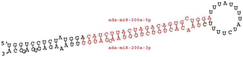
MicroRNAome of CGSs’ gonads 271
in miRBase. A differential expression analysis of miRNAs 3 years (Fig. 1A, B, C, A′, B′ and C′). At 4 years, the
between the ovary and testis was conducted using miRDeep seminiferous tubules contained various types of germ cell
2.0 (Wu et al. 2013). The significance level was set at |log2 including primary germ cells, primary spermatocytes,
(fold-change)| > 1, which ensured an accurate selection of secondary spermatocytes and sperm (Fig. 1D and D′).
differentially expressed miRNAs. All the sequence data were Only primary oocytes and primordial follicles were
submitted to the NCBI Sequence Read Archive (https://submit. observed in the ovaries of 1-, 2- and 3-year-old CGSs
ncbi.nlm.nih.gov/subs/sra/) with accession no. SRP097571. (Fig. 1E, F and G), whereas in 4-year-olds, in addition to
the above, large antral follicles were present (Fig. 1H).
Real-time quantitative validation Therefore, we chose the gonadal tissues of 4-year-old
CGSs for sequencing.
Reverse transcription PCRs (RT-PCRs) of the separate RNA
samples used for sequencing were performed. The stem-
loop RT-PCR method was developed by Chen and coworkers Overview of the sequencing data
(Chen et al. 2005) and has been used by other researchers
(Sun et al. 2014). ReverTra Ace reverse transcriptase (TaKaRa) In order to identify differentially expressed miRNAs
and miRNA-specific stem-loop RT primers were used to between the ovary and testis, two small RNA libraries
synthesize cDNA. The amplification program was as follows: of 4-year-old CGSs were constructed. The Illumina
incubation at 37°C for 15 min, 85°C for 5 s and then stored at sequencing of which provided a total dataset of
4°C. A SYBR Green Real-Time PCR Master Mix (TaKaRa) and a 31,406,812 raw reads (15,464,561 and 15,942,251
Bio-Rad CFX96 Real-Time PCR system (Bio-Rad) were used to reads from the ovary and testis libraries respectively).
conduct real-time quantitative PCRs (qPCRs) according to the After removing low-quality sequences, simple
standard protocol. Each 20 μL qPCR system contained 10 μL sequences, contaminants that were formed by adapter–
SYBR Premix Ex Taq II (Tli RNaseH Plus) (2×), 1 μL cDNA (blank adapter ligation and sequences longer than 30 nt or
control using ddH2O rather than a cDNA template), 0.8 μL shorter than 18 nt, 13,018,310 and 11,796,754 clean
forward miRNA primer and 0.8 μL reverse miRNA primer. All reads were ultimately obtained from the ovary and
the reactions were run in triplicate. The qPCR amplification testis libraries respectively (Table 2). We compared the
program was as follows: pre-denaturation at 95°C for 2 min, small RNA sequences with those in GenBank and the
followed by 35 cycles of 30 s at 95°C, 30 s at 60°C and 15 s at
Rfam database to obtain the annotation information
72°C (Chen et al. 2015). Relative quantification was calculated
of other non-coding RNAs (Table 3). We found a large
using the 2−ΔΔCT formula (Sun et al. 2014), with U6 snRNA
amount of rRNA, which is consistent with that reported
included as an internal control. The data were compared by
Student t-test, using the SPSS (version 17.0) (SPSS), and the
by previous studies (Kadri et al. 2011). This was not
results are expressed as the mean ± 1 s.d. of duplicates values. surprising, because rRNA is the most abundant small
P < 0.05 was considered statistically significant. All the primers RNA and regulates protein biosynthesis by binding
for the RT-PCRs and qPCRs are presented in Table 1. ribosomes to a variety of proteins. After removing the
rRNA and other non-coding RNA sequences, the two
small RNA libraries were analyzed to find tissue-specific
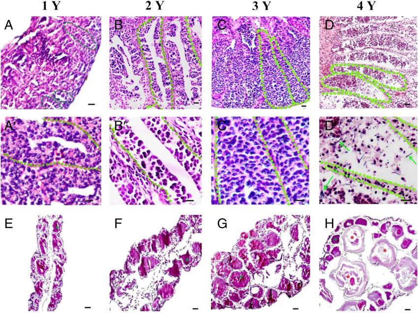
Results small RNAs. The percentages of the ovary-specific and
testis-specific sequences were 41.33% and 36.54% of
Gonadal development
the total small RNAs in the two libraries respectively
Testis and ovary tissues at different developmental stages (Fig. 2A). Ovary-specific unique sequences accounted
(1, 2, 3 and 4 years) were examined by hematoxylin and for 50.30% of all sequencing reads, and testis-specific
eosin staining (Fig. 1), which revealed that the male unique sequences accounted for 46.94% of the
CGSs had not reached sexually maturity at 1, 2 and sequencing reads (Fig. 2B).
Table 1 Stem-loop RT-PCR and qPCR primers of miRNAs of CGS.
miRNA ID RT primer (5′–3′) Forward primer (5′–3′) Reverse primer (5′–3′)
ada-miR-451 GTCGTATCCAGTGCAGGGTCCGAGGTATTCGCACTG- AACACGTGAAACCGTTACCATT CAGTGCAGGGTCCGAGGT
GATACGACACTCAG
ada-miR-10c GTCGTATCCAGTGCAGGGTCCGAGGTATTCGCACTG- ACGGAACCACCCTGTAGAATC CAGTGCAGGGTCCGAGGT
GATACGACACAAAT
ada-miR-7a GTCGTATCCAGTGCAGGGTCCGAGGTATTCGCACTG- AACAAGCAAAGTGCTGTTCGT CAGTGCAGGGTCCGAGGT
GATACGACACAACA
ada-miR-101 GTCGTATCCAGTGCAGGGTCCGAGGTATTCGCACTG- CACCGTGGTACAGTACTGTGA CAGTGCAGGGTCCGAGGT
GATACGACTCAGTT
ada-miR-499 GTCGTATCCAGTGCAGGGTCCGAGGTATTCGCACTG- ACGGAACTTAAGACTTGCAGTG CAGTGCAGGGTCCGAGGT
GATACGACTAAACA
ada-miR-202 GTCGTATCCAGTGCAGGGTCCGAGGTATTCGCACTG- TCGCGCATTCCTATGCATATAC CAGTGCAGGGTCCGAGGT
GATACGACCAAAGA
U6 TTACATTGCTATCCACAGAACGG CTATGCTGCTGCTTTTTGCTC
www.reproduction-online.org Reproduction (2017) 154 269–279
Downloaded from Bioscientifica.com at 09/16/2020 10:39:23PM
via free access272 R Chen and others
Figure 1 The histology of testis and ovary tissues from different development stages (1, 2, 3 and 4 years) of CGSs by H&E staining. A, B, C, D, A′,
B′, C′, D′ and E, F, G, H were represented testis and ovary tissues of 1, 2, 3 and 4 years old CGSs respectively. 1 Y: 1-year old; 2 Y: 2-year old;
3 Y: 3-year old; 4 Y: 4-year old. Sectors of green dots represented seminiferous tubules in A, B, C, D and A′, B′, C′, D′. Green arrows indicated
sperm in D′. Green asterisk indicated primordial follicles in E, F, G and H and red asterisk displayed large antral follicles in H. Bar = 60 μm.
The miRNAs found by homology comparisons sequences (isomiRs or SNPs) found by homology
comparisons in the ovary and testis were 18,182 and
Many miRNAs varied in sequence length and in the
16,719 respectively (Supplementary Tables 1 and
number of single-nucleotide polymorphisms (SNPs)
2, see section on supplementary data given at the
they contained, possibly due to post-transcriptional
end of this article). When all the identical sequence
RNA modifications. These miRNA variations are
reads were classified as a group, 757 unique miRNAs
referred as miRNA isoforms (isomiRs) which vary
in length and/or sequence (Morin et al. 2008,
Table 3 Distribution of the genome-mapped sequencing reads in
Neilsen et al. 2012). The total numbers of miRNA ovary and testis small RNA libraries of CGSs.
Table 2 Summary of small RNA sequencing data in ovary and testis Ovary Testis
libraries of CGSs. Type Count Percent (%) Count Percent (%)
Ovary Testis Total 13,018,310 100 11,796,754 100
Genome 2,181,677 16.77 2,420,409 20.51
Type Count Percent (%) Count Percent (%)
rRNA 1,612,098 12.38 1,560,497 13.23
Total_reads 15,464,561 15,942,251 scRNA 2 0.00 1 0.00
High_quality 15,464,561 100 15,942,251 100 snRNA 3062 0.02 12,420 0.11
N′ reads 2801 0.02 2702 0.02 snoRNA 4253 0.03 2309 0.02
Length 30 1,333,994 8.63 758,449 4.75 Repbase 12,252 0.09 18,984 0.16
Clean reads 13,018,310 84.18 11,796,754 74.00 Other 9,094,686 69.86 7,669,080 65.01
Reproduction (2017) 154 269–279www.reproduction-online.org
Downloaded from Bioscientifica.com at 09/16/2020 10:39:23PM
via free accessMicroRNAome of CGSs’ gonads 273
Figure 2 Flow chart of miRNA sequencing. A and B represented the percentage of the common and specific tags of total and unique sRNAs of
ovary and testis tissues respectively. (C) and (D) displayed the percentage of the common and specific tags of total and unique sRNAs of ovary
and testis tissues by homology comparison respectively. (E) showed the proportion of common and specific miRNAs of CGS ovary and testis
tissues when compared with Xenopus laevis.
www.reproduction-online.org Reproduction (2017) 154 269–279
Downloaded from Bioscientifica.com at 09/16/2020 10:39:23PM
via free access274 R Chen and others
Figure 4 The predicted hairpin structure of ada-miR-200a.
Differentially expressed miRNAs between the
ovary and testis
The expression profiles of each miRNAs in the ovary
and testis by using Xenopus laevis miRNA sequences
as a reference are presented in Supplementary Table 7.
A total of 120 miRNAs were differentially expressed
between the ovary and testis, 45 of which were more
highly expressed in ovary and 21 of which were more
Figure 3 The size distribution of small RNAs of CGS by homology highly expressed in the testis (Supplementary Tables 8
comparison.
and 9). For example, miR-451 and miR-10c were mainly
expressed in the ovary, whereas miR-101, miR-202,
were annotated as miRNA candidates in the ovary miR-7a and miR-499 were mainly expressed in the
library and 756 unique miRNAs were annotated as testis. This suggests that these miRNAs may affect the
miRNA candidates in the testis library (Supplementary development of gonadal tissue.
Tables 3 and 4). The ovary-specific and testis-specific
sequences accounted for 2.09% and 1.55% of the Validation of differentially expressed miRNAs
total miRNAs in the two libraries respectively (Fig. 2C).
Ovary-specific unique miRNAs accounted for 19.95% Six differentially expressed miRNAs between the ovary
of all the sequencing reads and testis-specific unique and testis libraries were selected: ada-miR-10c and
miRNAs accounted for 19.84% of all the sequencing ada-miR-451 from the ovary library and ada-miR-7a,
reads (Fig. 2D). ada-miR-499, ada-miR-101 and ada-miR-202 from the
We found that 145 (1.37%) and 155 (7.74%) testis library. The primers for these miRNAs are shown
miRNA sequences in the ovary and testis of CGSs in Table 1. The qPCR and deep sequencing results were
respectively were homologous to those in the ovary similar for the selected miRNAs (Fig. 5) and indicated
and testis respectively, of Xenopus laevis (Fig. 2E and that they coexisted in the different tissues.
Supplementary Tables 5 and 6). Size distributions of the The miRNAs expression levels at different
small RNAs (from 18 nt to 30 nt) were similar between the developmental stages (1, 2, 3 and 4 years) in the ovary
male and female libraries (Fig. 3). There were small peaks and testis are presented in Fig. 6. The relative expression
at 22 nt and 29–30 nt; however, most of the small RNAs levels of the selected miRNAs differed between the testis
were of different lengths. However, size distributions and ovary. During development, the relative expression
are only a rough and preliminary screening, and small levels of miR-451, miR-202 and miR-499 in the testes
RNAs should be mapped to the genome and known gradually decreased, whereas no specific trends were
pre-miRNAs and their secondary structures predicted observed in the ovary. The expression levels of miR-7a,
for identification and characterization. Variations in miR-101, miR-202 and miR-499 in the testis were
length are mainly caused by enzymatic modifications, significantly higher than those in the ovary, whereas the
such as RNA editing, 3′-editing or exonuclease activity miR-451 expression level was higher in the ovary, but
(Li et al. 2010). only in the 4-year-old CGSs.
Deep sequencing revealed that the individual
miRNAs exhibited heterogeneous 5′ or 3′ ends or post-
Discussion
transcriptional end additions, deletions or substitutions,
as exemplified by miR-200a-5p and miR-200a-3p, The CGS is an invaluable animal model for research in
the predicted precursors, which had a typical hairpin genetics, phylogenetics and evolution (Fan et al. 2015).
structure (Fig. 4). Their isoforms had different sequence However, environmental degradation, over-harvesting
reads, which suggests that they exhibit differential and other reasons have made the CGS rare. As a result,
expression in male and female gonads. The other it is artificially farmed in mesocosms for research
predicted miRNA hairpin structures in the ovary and and conservation. However, because of the difficulty
testis are shown in Supplementary Files 1 and 2. in determining the species’ sex at immature stage,
Reproduction (2017) 154 269–279www.reproduction-online.org
Downloaded from Bioscientifica.com at 09/16/2020 10:39:23PM
via free accessMicroRNAome of CGSs’ gonads 275
Figure 5 The expression profiles of miRNAs in different tissues of CGS were detected by qPCR. Note: the values with different letters (a, b, c, d,
e, f and g) differ significantly at P < 0.05 or P < 0.01 level.
mixed-sex cultures have not satisfactorily increased RNA modifications, such as shifts in Drosha and Dicer
the CGS population. Recent advances have revealed cleavage sites, exonuclease-mediated trimming, miRNA
that miRNAs are essential for sexual differentiation, editing or 3′-end non-templated nucleotide additions
gonadal development and reproductive performance (Morin et al. 2008, Neilsen et al. 2012). In addition to
(McEwen et al. 2016, Tyler et al. 2017); however, no the homologous miRNA, isomiR expression could also
studies have been conducted on the regulatory roles of have a post-transcriptional regulatory function in cells,
miRNAs in CGSs. Here, we present CGS ovary and testis tissues or at specific developmental stages (Fernandez-
miRNAs profiles, which will increase our understanding Valverde et al. 2010, Bizuayehu et al. 2012a,b).
of the mechanisms underlying sexual differentiation in A novel class of small RNAs (piRNAs) was found
this species and provide a preliminary theoretical basis in 2006 that differed from miRNAs in size (26–32 nt
for the further study of the CGS’s reproductive biology. long rather than 18–24 nt long), mostly in 29–30 nt
Lower values were obtained when using Xenopus (Lau et al. 2006, Rastetter et al. 2015) and could
laevis as a reference (1.37% and 7.74%) than when silence transposons and retroposons at the epigenetic
using the homology comparisons (19.95% and and post-transcriptional levels, maintain the genomic
19.84%), verifying the accuracy of the data analysis. stability and integrity of germ cells (Bao & Yan 2012)
That part of less (18.58% and 12.10%) possible and regulate cell proliferation (Klattenhoff & Theurkauf
matching other homologous species, such as frog, 2008), and meiosis, particularly during spermatogenesis
highlighting the difference between CGSs and Xenopus (Goh et al. 2015). Twenty-eight-nucleotide-long piRNAs
laevis, demonstrated the particularity of CGS. The size are highly expressed in both the ovary and testis of
distributions of the sequences included two peaks zebrafish (Houwing et al. 2007, Kamminga et al.
(22 nt and 29–30 nt), which were probably miRNAs 2010). In Xenopus laevis and Oreochromis niloticus,
and piwi-interacting RNAs (piRNAs) respectively piRNAs are abundant in both female and male gonads
(Grossman & Shalgi 2016, Marie et al. 2016). The size (Wilczynska et al. 2009, Xiao et al. 2014). The piRNAs
distribution of the miRNAs peaked at 22 nt, which is are present in the gonads of lower vertebrates and are
the typical size of Dicer-derived products (Mi et al. involved in the regulation of sexual differentiation,
2014). Meanwhile, miRNAs (isomiRs and SNPs) were gonadal development and gametogenesis; therefore,
found by homology comparisons in the ovary and testis. potential piRNAs were also investigated in the present
MiRNA variations can be caused by post-transcriptional study. BLAST software was used to compare the CGS
www.reproduction-online.org Reproduction (2017) 154 269–279
Downloaded from Bioscientifica.com at 09/16/2020 10:39:23PM
via free access276 R Chen and others
Figure 6 The expression profiles of miRNAs in CGSs different development stages of ovary and testis were detected by qPCR. 1, 2, 3 and 4 were
represented 1-, 2-, 3- and 4-year-old CGSs respectively. T and O were represented testis and ovary of CGSs respectively. Asterisk * and **
indicate significant differences between the two groups at P < 0.05 and P < 0.01 respectively. NS means not significant.
raw data with human, mouse and rat piRNA sequences gonads of the CGS and may play an important role in
(Sai et al. 2008), and sequences with a high similarity gonadal development in this species.
(≥96%) were selected for homology analysis. We found The qPCR and deep sequencing revealed similar
that 182, 114 and 113 piRNA-like sequences in the trends for all of the miRNAs selected. At 4 years, ada-
small CGS ovary library were homologous with piRNA miR-10c and ada-miR-451 were more highly expressed
sequences in the human, mouse and rat respectively in the ovary than the testis, whereas ada-miR-7a, ada-
(Supplementary Tables 10, 11 and 12). And 168, 129 miR-499, ada-miR-101 and ada-miR-202 were more
and 120 piRNA-like sequences in the small CGS testis highly expressed in the testis. MiR-202 is abundant in
library were homologous with piRNA sequences in the mouse and Xenopus gonads (Ro et al. 2007, Armisen et al.
human, mouse and rat respectively (Supplementary 2009), and in the chicken, is more highly upregulated in
Tables 13, 14 and 15). These piRNA-like sequences the testis than the ovary (Bannister et al. 2009, 2011), as is
require further investigation. the case in Atlantic halibut (Hippoglossus hippoglossus),
We observed only small peaks in size distribution of which is a teleost vertebrate ItheIt (Bizuayehu et al.
the small RNAs possibly because other types of non- 2012a,b). In cattle, miR-202 is highly expressed in
coding small RNAs of different sizes increased the basic sperm and could improve embryonic development
level of the size distribution. Recently, tRNA-derived and nuclear reprogramming (Gao & Zhang 2016). Our
small RNAs (tsRNAs) that are 14–32 nt in length have results for CGSs agree with these findings. The results
been discovered in various organisms (Haussecker et al. of these studies suggest that the gonadal expression
2010, Peng et al. 2012, Kumar et al. 2014). The tsRNAs of miR-202 is conserved among vertebrates and that
could maintain their stability by means of the nucleic miR-202 plays a crucial role in reproduction. Similar
acid sequence modification and are also sensitive to to our results, miR-451 has been reported to be highly
stress. The tsRNA and their RNA modifications could expressed in female halibut (Bizuayehu et al. 2012a,b)
contain epigenetic information. In mice, sperm tsRNAs and plays a vital role during bovine follicle development
could contribute to the intergenerational inheritance of (Sontakke et al. 2014). The miRNA-451 and its target
an acquired metabolic disorder (Chen et al. 2016). It gene Ankrd46 are vital for embryo implantation (Li et al.
would be reasonable to expect that tsRNAs exist in the 2015a,b). The differentially expressed miRNAs in CGS
Reproduction (2017) 154 269–279www.reproduction-online.org
Downloaded from Bioscientifica.com at 09/16/2020 10:39:23PM
via free accessMicroRNAome of CGSs’ gonads 277
gonads are functionally conserved in other animals and investigations into gonadal developmental regulation in
may play crucial roles in gonadal development and CGSs, and provide a theoretical basis for the artificial
reproductive physiology. These miRNAs were also found breeding of CGSs.
in eight other tissues (heart, liver, lung, kidney, brain,
muscle, pancreas and bladder), demonstrating that they
not only play a role in gonads, but also play regulatory Supplementary data
roles in other organs, which should be investigated This is linked to the online version of the paper at http://dx.doi.
further. Little information is available concerning the org/10.1530/REP-17-0109.
CGS genome; therefore, it is difficult to predict its target
genes. The role of the miRNA-451 target gene Ankrd46
in murine suggests that it may also play an important role Declaration of interest
in CGS development. Future studies should investigate
The authors declare that there is no conflict of interest that
the functions of these CGS miRNAs target genes. could be perceived as prejudicing the impartiality of the
The expression levels of miR-7a, miR-101, miR-202 research reported.
and miR-499 in the testis were significantly higher than
those in the ovary at all developmental stages, whereas
the miR-451 expression level was higher in the ovary Funding
than that in the testis, but only at 4 years. The relative
expression levels of miR-451, miR-202 and miR-499 This research did not receive any specific grant from any
funding agency in the public, commercial or not-for-profit
in the testes gradually decreased, whereas in the
sector.
ovary, there was no relationship between expression
level and developmental stage. This may have been
related to the histological analysis of the gonads at Author contribution statement
different developmental stages (Fig. 1). In CGSs, sexual
differentiation is complete at 1 year (Yang et al. 1983). Wu-zi Dong was the primary corresponding author and
Subsequently, the testis and ovary are immature until Chuan-ying Pan was the secondary corresponding author who
3 years of age. In 2- and 3-year-old CGSs, there are designed the overall studies. Wu-zi Dong and Chang-ming
many developmentally arrested oocytes in the ovary Yang collected and prepared samples. Sheng-song Xie, Lin Ma
and Wu-zi Dong performed bio-statistical analyses. Rui Chen
(Huang 2009), and few male germ cells in the testis
and Li-qing Wang performed to observe the histomorphology.
(Fig. 1). However, at 4 years of age, there are different
Rui Chen and Jian Du did the experimental verification. Rui
types of germ cells in the testis and the ovary. There were
Chen and Wu-zi Dong wrote the manuscript. Wu-zi Dong,
no significant differences between 2- and 3-year-old Chuan-ying Pan and Xian-yong Lan edited the manuscript.
CGSs in the ovary and testis expression levels of some All authors discussed the results and commented on the
miRNAs, such as miR-451 and miR-7a. This confirms that manuscript.
miRNAs play individual as well as synergistic roles in
regulating the gonadal development and gametogenesis
(Bizuayehu et al. 2012a,b, Gao & Zhang 2016). Acknowledgments
Although tissue-specific miRNAs were not found
This work was supported by the Fund of the Agriculture Sci-
in this study, the differentially expressed miRNAs
Tech Project of Shaanxi Province (No. 2014K01-20-01) and
identified could be used to distinguish male and female
the National Natural Science Foundation China (NSFC) (No.
CGSs during development, which will improve CGS
C170104-31172205). They thank Prof. Wen-xian Zeng and
artificial breeding. Xian-yong Lan at College of Animal Science and Technology,
Northwest A&F University who commented on their
manuscript.
Conclusion
Many miRNAs and other small RNAs, such as piRNAs
and tsRNA, were abundant in the ovary and testis of References
CGSs. We identified 145 miRNAs in the CGS ovary and Ambros V 2003 MicroRNA pathways in flies and worms: growth,
155 miRNAs in the CGS testis that were homologous to death, fat, stress, and timing. Cell 113 673–676. (doi:10.1016/S0092-
those in the Xenopus laevis ovary and testis respectively. 8674(03)00562-2)
Armisen J, Gilchrist MJ, Wilczynska A, Standart N & Miska EA 2009
Forty-five miRNAs were more highly expressed in the Abundant and dynamically expressed miRNAs, piRNAs, and other
ovary than that in the testis, and 21 were more highly small RNAs in the vertebrate Xenopus tropicalis. Genome Research 19
expressed in the testis. The selected miRNAs exhibited 1766–1775. (doi:10.1101/gr.093054.109)
Bannister SC, Smith CA, Roeszler KN, Doran TJ, Sinclair AH & Tizard
differential expression levels in the ovary and testis MLV 2011 Manipulation of estrogen synthesis alters mir202* expression
during different CGS developmental stages. These results in embryonic chicken gonads. Biology of Reproduction 85 22–30.
increase our knowledge of CGS miRNAs, facilitate (doi:10.1095/biolreprod.110.088476)
www.reproduction-online.org Reproduction (2017) 154 269–279
Downloaded from Bioscientifica.com at 09/16/2020 10:39:23PM
via free access278 R Chen and others
Bannister SC, Tizard MLV, Doran TJ, Sinclair AH & Smith CA 2009 Sexually Haussecker D, Huang Y, Lau A, Parameswaran P, Fire AZ & Kay MA 2010
dimorphic microRNA expression during chicken embryonic gonadal Human tRNA-derived small RNAs in the global regulation of RNA
development. Biology of Reproduction 81 165–176. (doi:10.1095/ silencing. RNA 16 673–695. (doi:10.1261/rna.2000810)
biolreprod.108.074005) He L & Hannon GJ 2004 MicroRNAs: small RNAs with a big role in gene
Bao JQ & Yan W 2012 Male germline control of transposable elements. regulation. Nature Reviews Genetics 5 522–531. (doi:10.1038/nrg1379)
Biology of Reproduction 86 162. Houwing S, Kamminga LM, Berezikov E, Cronembold D, Girard A, van
Bartel DP 2004 MicroRNAs: genomics, biogenesis, mechanism, and den Elst H, Filippov DV, Blaser H, Raz E, Moens CB et al. 2007 A role
function. Cell 116 281–297. for piwi and piRNAs in germ cell maintenance and transposon silencing
Beilharz TH, Humphreys DT, Clancy JL, Thermann R, Martin DIK, Hentze in zebrafish. Cell 129 69–82. (doi:10.1016/j.cell.2007.03.026)
MW & Preiss T 2009 MicroRNA-mediated messenger RNA deadenylation Hu QM, Xiao HB, Tian HF & Meng Y 2016 Characterization and expression
contributes to translational repression in mammalian cells. PLoS ONE 4 of cyp19a gene in the Chinese giant salamander Andrias davidianus.
e6783. (doi:10.1371/journal.pone.0006783) Comparative Biochemistry and Physiology B Biochemistry and Molecular
Bizuayehu TT, Babiak J, Norberg B, Fernandes JMO, Johansen SD & Babiak Biology 192 21–29. (doi:10.1016/j.cbpb.2015.11.005)
I 2012a Sex-biased miRNA expression in Atlantic halibut (Hippoglossus Huang XY 2009 The process of embryonic development of giant
hippoglossus) brain and gonads. Sexual Development 6 257–266. salamander. Nature and Science 6 52–53. (in Chinese)
(doi:10.1159/000341378) Ji ZB, Wang GZ, Xie ZJ, Zhang CL & Wang JM 2012 Identification and
Bizuayehu TT, Lanes CFC, Furmanek T, Karlsen BO, Fernandes JMO, characterization of microRNA in the dairy goat (Capra hircus) mammary
Johansen SD & Babiak I 2012b Differential expression patterns of gland by Solexa deep sequencing technology. Molecular Biology Reports
conserved miRNAs and isomiRs during Atlantic halibut development. 39 9361–9371. (doi:10.1007/s11033-012-1779-5)
BMC Genomics 13 11. (doi:10.1186/1471-2164-13-11) Kadri S, Hinman VF & Benos PV 2011 RNA deep sequencing reveals
Brennecke J, Hipfner DR, Stark A, Russell PB & Cohen SM 2003 Bantam differential microRNA expression during development of sea urchin and
encodes a developmentally regulated microRNA that controls cell sea star. PLoS ONE 6 e29217. (doi:10.1371/journal.pone.0029217)
proliferation and regulates the proapoptotic gene hid in Drosophila. Cell Kamminga LM, Luteijn MJ, den Broeder MJ, Redl S, Kaaij LJ, Roovers
113 25–36. (doi:10.1016/S0092-8674(03)00231-9) EF, Ladurner P, Berezikov E & Ketting RF 2010 Hen1 is required for
Che RB, Sun YN, Wang RX & Xu TJ 2014 Transcriptomic analysis of oocyte development and piRNA stability in zebrafish. EMBO Journal 29
endangered Chinese salamander: identification of immune, sex and 3688–3700. (doi:10.1038/emboj.2010.233)
reproduction-related genes and genetic markers. PLoS ONE 9 e87940. Kim VN & Nam J 2006 Genomics of microRNA. Trends in Genetics 22
(doi:10.1371/journal.pone.0087940) 165–173. (doi:10.1016/j.tig.2006.01.003)
Chen CZ, Li L, Lodish HF & Bartel DP 2004 MicroRNAs modulate Klattenhoff C & Theurkauf W 2008 Biogenesis and germline functions of
hematopoietic lineage differentiation. Science 303 83–86. (doi:10.1126/ piRNAs. Development 135 3–9. (doi:10.1242/dev.006486)
science.1091903) Kumar P, Anaya J, Mudunuri SB & Dutta A 2014 Meta-analysis of tRNA
Chen Q, Ma J, Fan YD, Meng Y, Xu J, Zhou Y, Liu WZ, Zeng XH & Zeng LB derived RNA fragments reveals that they are evolutionarily conserved
2015 Identification of type I IFN in Chinese giant salamander (Andrias and associate with AGO proteins to recognize specific RNA targets.
davidianus) and the response to an iridovirus infection. Molecular BMC Biology 12 78. (doi:10.1186/s12915-014-0078-0)
Immunology 65 350–359. (doi:10.1016/j.molimm.2015.02.015) Kwekel JC, Vijay V, Han T, Moland CL, Desai VG & Fuscoe JC 2017 Sex
Chen C, Ridzon DA, Broomer AJ, Zhou Z, Lee DH, Nguyen JT, Barbisin M, and age differences in the expression of liver microRNAs during the life
Xu NL, Mahuvakar VR, Andersen MR et al. 2005 Real-time quantification span of F344 rats. Biology of Sex Differences 8 6. (doi:10.1186/s13293-
of microRNAs by stem-loop RT-PCR. Nucleic Acids Research 33 e179. 017-0127-9)
(doi:10.1093/nar/gni178) Lau NC, Seto AG, Kim J, Kuramochi-Miyagawa S, Nakano T, Bartel DP
Chen Q, Yan MH, Cao ZH, Li X, Zhang YF, Shi JC, Feng GH, Peng HY, Zhang & Kingston RE 2006 Characterization of the piRNA complex from rat
XD, Zhang Y et al. 2016 Sperm tsRNAs contribute to intergenerational testes. Science 313 363–367. (doi:10.1126/science.1130164)
inheritance of an acquired metabolic disorder. Science 351 397–400. Leung AK & Sharp PA 2010 MicroRNA functions in stress responses.
(doi:10.1126/science.aad7977) Molecular Cell 40 205–215. (doi:10.1016/j.molcel.2010.09.027)
Dong WZ, Zhang XM, Yang CM & Zeng WX 2010 Iridovirus outbreak Li ZJ, Lu J, Sun M, Mi SL, Zhang H, Luo RT, Chen P, Wang YG, Yan M, Qian
in Chinese giant salamanders. Emerging Infectious Diseases 17 ZJ et al. 2008 Distinct microRNA expression profiles in acute myeloid
2388–2389. (doi:10.3201/eid1712.101758) leukemia with common translocations. PNAS 105 15535–15540.
Du J, Wang LQ, Wang YX, Shen JA, Pan CY, Meng Y, Yang CM, Ji H & Dong (doi:10.1073/pnas.0808266105)
WZ 2016 Autophagy and apoptosis induced by Chinese giant salamander Li MZ, Xia YL, Gu YR, Zhang K, Lang QL, Chen L, Guan JQ, Luo ZG,
(Andrias davidianus) iridovirus (CGSIV). Veterinary Microbiology 195 Chen HS, Li Y et al. 2010 MicroRNAome of porcine pre- and
87–95. (doi:10.1016/j.vetmic.2016.09.011) postnatal development. PLoS ONE 5 e11541. (doi:10.1371/journal.
Dong F, Zhang Y, Xia F, Yang Y, Xiong S, Jin L & Zhang J 2014 Genome- pone.0011541)
wide miRNA profiling of villus and decidua of recurrent spontaneous Li ZY, Jia J, Gou JH, Zhao X & Yi T 2015a MicroRNA-451 plays a role in
abortion patients. Reproduction 148 33–41. (doi:10.1530/REP-14-0095) murine embryo implantation through targeting Ankrd46, as implicated
Fan YD, Chang MX, Ma J, LaPatra SE, Hu YW, Huang LL, Nie P & Zeng by a microarray-based analysis. Fertility and Sterility 103 834–844.
LB 2015 Transcriptomic analysis of the host response to an iridovirus (doi:10.1016/j.fertnstert.2014.11.024)
infection in Chinese giant salamander, Andrias davidianus. Veterinary Li FG, Wang LX, Lan QJ, Yang H, Li Y & Liu XL 2015b RNA-seq analysis
Research 46 136. (doi:10.1186/s13567-015-0279-8) and gene discovery of Andrias davidanus using Illumina short
Fernandez-Valverde SL, Taft RJ & Mattick JS 2010 Dynamic isomiR read sequencing. PLoS ONE 10 e0123730. (doi:10.1371/journal.
regulation in Drosophila development. RNA 16 1881–1888. pone.0123730)
(doi:10.1261/rna.2379610) Marie PP, Ronsseray S & Boivin A 2016 From embryo to adult: piRNA-
Gao KQ & Shubin NH 2003 Earliest known crown-group salamanders. mediated silencing throughout germline development in Drosophila. G3
Nature 422 424–428. (doi:10.1038/nature01491) 7 505–516. (doi:10.1534/g3.116.037291)
Gao Y & Zhang Y 2016 A preliminary study of bovine sperm-borne miR- McEwen TJ, Yao Q, Yun S, Lee CY & Bennett KL 2016 Small RNA in situ
202 on early embryonic development. Thesis for Master’s Degree. hybridization in Caenorhabditis elegans, combined with RNA-seq,
Northwest A&F University. identifies germline-enriched microRNAs. Developmental Biology 418
Goh WSS, Falciatori I, Tam OH, Burgess R, Meikar O, Kotaja N, Hammell 248–257. (doi:10.1016/j.ydbio.2016.08.003)
M & Hannon GJ 2015 PiRNA-directed cleavage of meiotic transcripts Mi X, Wei ZL, Zhou ZC & Liu XL 2014 Identification and profiling of sex-
regulates spermatogenesis. Genes and Development 29 1032–1044. biased microRNAs from sea urchin Strongylocentrotus nudus gonad by
(doi:10.1101/gad.260455.115) Solexa deep sequencing. Comparative Biochemistry and Physiology D
Grossman H & Shalgi R 2016 A role of microRNAs in cell differentiation Genomics and Proteomics 10 1–8. (doi:10.1016/j.cbd.2014.01.001)
during gonad development. Results and Problems in Cell Differentiation Morin RD, O’Connor MD & Griffith M 2008 Application of massively
58 309–336. (doi:10.1007/978-3-319-31973-5_12) parallel sequencing to microRNA profiling and discovery in human
Reproduction (2017) 154 269–279www.reproduction-online.org
Downloaded from Bioscientifica.com at 09/16/2020 10:39:23PM
via free accessMicroRNAome of CGSs’ gonads 279
embryonic stem cells. Genome Research 18 610–621. (doi:10.1101/ neural stem cell fate in a sex-dependent manner. Neurotoxicology and
gr.7179508) Teratology 59 1–15. (doi:10.1016/j.ntt.2016.10.004)
Neilsen CT, Goodall GJ & Bracken CP 2012 IsomiRs-the overlooked Vogel G 1999 Frog is a prince of a new model organism. Science 285 25.
repertoire in the dynamic microRNAome. Trends in Genetics 28 (doi:10.1126/science.285.5424.25)
544–549. (doi:10.1016/j.tig.2012.07.005) Wu J, Liu Q, Wang X, Zheng J, Wang T, You M, Sheng Sun Z & Shi Q 2013
Pedersen IM, Cheng G, Wieland S, Volinia S, Croce CM, Chisari FV & mirTools 2.0 for non-coding RNA discovery, profiling, and functional
David M 2007 Interferon modulation of cellular microRNAs as an annotation based on high-throughput sequencing. RNA Biology 10
antiviral mechanism. Nature 449 919–922. (doi:10.1038/nature06205) 1087–1092. (doi:10.4161/rna.25193)
Peng HY, Shi JC, Zhang Y, Zhang H, Liao SY, Li W, Lei L, Han CS, Wilczynska A, Minshall N, Armisen J, Miska EA & Standart N 2009 Two
Ning LN, Cao YJ et al. 2012 A novel class of tRNA-derived small Piwi proteins, Xiwi and Xili, are expressed in the Xenopus female
RNAs extremely enriched in mature mouse sperm. Cell Research 22 germline. RNA 15 337–345. (doi:10.1261/rna.1422509)
1609–1612. (doi:10.1038/cr.2012.141) Wang XM, Zhang KJ, Wang ZH, Ding YZ, Wu W & Huang S 2004 The
Poy MN, Eliasson L, Krutzfeldt J, Kuwajima S, Ma X, Macdon-ald PE, decline of the Chinese giant salamander Andrias davidianus and
Pfeffer S, Tuschl T, Rajewsky N, Rorsman P et al. 2004 A pancreatic implications for its conservation. Oryx 38 197–202.
islet-specific microRNA regulates insulin secretion. Nature 432 Xiao J, Zhong H, Zhou Y, Yu F, Gao Y, Luo YJ, Tang ZY, Guo ZB, Guo EY,
226–230. (doi:10.1038/nature03076) Gan X et al. 2014 Identification and characterization of microRNAs in
Rastetter RH, Smith CA & Wilhelm D 2015 The role of non-coding RNAs in ovary and testis of Nile tilapia (Oreochromis niloticus) by using solexa
male sex determination and differentiation. Reproduction 150 R93–R107. sequencing technology. PLoS ONE 9 e86821. (doi:10.1371/journal.
Ro S, Song R, Park C, Zheng H, Sanders KM & Yan W 2007 Cloning and pone.0086821)
expression profiling of small RNAs expressed in the mouse ovary. RNA Yang AS, Bian W, Liu YQ & Liu GJ 1983 Preliminary studies on the
13 2366–2380. (doi:10.1261/rna.754207) embryonic development of giant salamander. Acta Zoologica Sinica 29
Sai LS & Agrawal S 2008 piRNABank: a web resource on classified and 42–47. (in Chinese).
clustered Piwi-interacting RNAs. Nucleic Acids Research 36 D173– Zhou ZY, Geng Y, Liu XX, Ren SY, Zhou Y, Wang KY, Huang XL, Chen DF,
D177. (doi:10.1093/nar/gkm696) Peng X & Lai WM 2013 Characterization of a ranavirus isolated from
Shi XW 2011 Preliminary study of artificial propagation technology of the Chinese giant salamander (Andrias davidianus, Blanchard, 1871) in
Andrias davidianus. Journal of Fujian Fisheries 33 48–51. (in Chinese) China. Aquaculture 384 66–73.
Sontakke SD, Mohammed BT, McNeilly AS & Donadeu FX 2014
Characterization of microRNAs differentially expressed during bovine follicle
development. Reproduction 148 271–283. (doi:10.1530/REP-14-0140)
Sun JJ, Zhang B, Lan XY, Zhang CL, Lei CZ & Chen H 2014 Comparative
transcriptome analysis reveals significant differences in MicroRNA Received 23 February 2017
expression and their target genes between adipose and muscular tissues
First decision 6 April 2017
in cattle. PLoS ONE 9 e102142. (doi:10.1371/journal.pone.0102142)
Tyler CR, Labrecque MT, Solomon ER, Guo X & Allan AM 2017 Prenatal Revised manuscript received 23 May 2017
arsenic exposure alters REST/NRSF and microRNA regulators of embryonic Accepted 19 June 2017
www.reproduction-online.org Reproduction (2017) 154 269–279
Downloaded from Bioscientifica.com at 09/16/2020 10:39:23PM
via free accessYou can also read