Case Report: Visual Rehabilitation in Hemianopia Patients. Home-Based Visual Rehabilitation in Patients With Hemianopia Consecutive to Brain Tumor ...
←
→
Page content transcription
If your browser does not render page correctly, please read the page content below
CASE REPORT
                                                                                                                                                  published: 21 July 2021
                                                                                                                                         doi: 10.3389/fneur.2021.680211

                                               Case Report: Visual Rehabilitation in
                                               Hemianopia Patients. Home-Based
                                               Visual Rehabilitation in Patients With
                                               Hemianopia Consecutive to Brain
                                               Tumor Treatment: Feasibility and
                                               Potential Effectiveness
                                               Monica Daibert-Nido 1,2 , Yulia Pyatova 1 , Kyle Cheung 2 , Camilus Nayomi 2 ,
                                               Samuel N. Markowitz 1,2 , Eric Bouffet 3 and Michael Reber 1,2,4*
                                               1
                                                 Department of Ophthalmology and Vision Sciences, University of Toronto, Toronto, ON, Canada, 2 Donald K. Johnson Eye
                                               Institute, Krembil Research Institute, University Health Network, Toronto, ON, Canada, 3 Division of Hematology/Oncology,
                                               Research Institute, The Hospital for Sick Children, Toronto, ON, Canada, 4 Laboratory Medicine and Pathobiology, Cell and
                         Edited by:            System Biology, University of Toronto, Toronto, ON, Canada
                       Jiawei Zhou,
   Wenzhou Medical University, China
                                               Background/Objectives: Visual field loss is frequent in patients with brain tumors,
                      Reviewed by:
                    Christine Rogers,          worsening their daily life and exacerbating the burden of disease, and no supportive care
University of Cape Town, South Africa          strategies exist. In this case series, we sought to characterize the feasibility and potential
                Benjamin A. Rowland,
Wake Forest University, United States
                                               effectiveness of a home-based visual rehabilitation program in hemianopia patients using
                 *Correspondence:
                                               immersive virtual-reality stimulation.
                      Michael Reber
                                               Subjects/Methods: Two patients, one with homonymous hemianopia and the other
       michael.reber@uhnresearch.ca
                                               with bitemporal hemianopia, consecutive to pediatric brain tumors, with no prior visual
                    Specialty section:         rehabilitation performed 15 min of home-based audiovisual stimulation every 2 days for 6
          This article was submitted to        weeks (case 2) and 7 weeks (case 1) between February and August 2020. Patients used a
                     Neurorehabilitation,
                a section of the journal       virtual-reality, stand-alone, and remotely controlled device loaded with a non-commercial
                 Frontiers in Neurology        audiovisual stimulation program managed in real time from the laboratory. Standard visual
           Received: 13 March 2021             outcomes assessed in usual care in visual rehabilitation were measured at the clinic.
            Accepted: 15 June 2021
                                               Following a mixed method approach in this pragmatic study of two cases, we collected
            Published: 21 July 2021
                                               quantitative and qualitative data on feasibility and potential effectiveness and compared
                              Citation:
Daibert-Nido M, Pyatova Y, Cheung K,           the results pre- and post-treatment.
  Nayomi C, Markowitz SN, Bouffet E
    and Reber M (2021) Case Report:
                                               Results: Implementation and wireless delivery of the audiovisual stimulation, remote
   Visual Rehabilitation in Hemianopia         data collection, and analysis for cases 1 and 2 who completed 19/20 and 20/20
         Patients. Home-Based Visual
                                               audiovisual stimulation sessions at home, respectively, altogether indicated feasibility.
        Rehabilitation in Patients With
    Hemianopia Consecutive to Brain            Contrast sensitivity increased in both eyes for cases 1 and 2. Visual fields, measured
      Tumor Treatment: Feasibility and         by binocular Esterman and monocular Humphrey full-field analyses, improved in case 1.
               Potential Effectiveness.
            Front. Neurol. 12:680211.
                                               A minor increase was observed in case 2. Cases 1 and 2 enhanced reading speed. Case
     doi: 10.3389/fneur.2021.680211            2 strongly improved quality of life scores.

Frontiers in Neurology | www.frontiersin.org                                         1                                             July 2021 | Volume 12 | Article 680211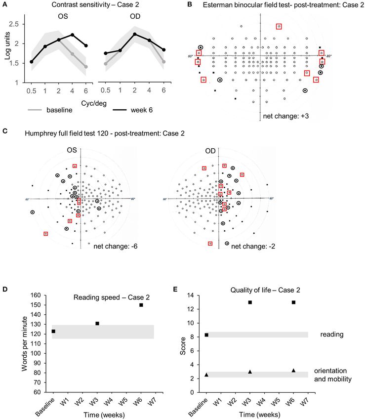
Daibert-Nido et al. Visual Rehabilitation in Hemianopia
Conclusion: This is the first report of a home-based virtual-reality visual rehabilitation
program for adult patients with hemianopia consecutive to a pediatric brain tumor.
We show the feasibility in real-world conditions and potential effectiveness of such
technology on visual perception and quality of life.
Keywords: low-vision, rehabilitation, virtual-reality, personalized medicine 2, hemianopia or hemianopsia,
hemianopia rehabilitation
INTRODUCTION eye-tracking, field of view, and even decision-making in healthy
and pathological participants (17–19). Using a mixed method
Central nervous system tumors are the second most common approach, our objective was to test the feasibility (treatment
malignancies in childhood (1). Brain tumor and its treatment can implementation and wireless delivery, remote collection and
affect the visual system at different levels, from the optic nerves analysis of data, compliance and adherence, and adverse effects)
(through compression or infiltration), to subcortical structures and potential effectiveness (visual function and functional vision)
like the superior colliculus (SC) and lateral geniculate nuclei of a home-based, IVR, audiovisual stimulation program on
(LGN) to optic tracts, optic radiations, and visual cortices (1– quality of life and visual field perception. This treatment was
4). Children with brain tumors can present visual impairments performed between March and August 2020, during the stay-
like decreased visual acuity and contrast sensitivity (CS), loss at-home order (Ontario Government—Emergency Management
of color vision, and visual field loss such as hemianopias (1–5). and Civil Protection Act, Order in Council 518/2020) due to the
These visual field defects in children affect their psycho-social COVID-19 pandemic. The audiovisual stimulation protocol was
and educational development with long-term debilitating effects. implemented remotely (telerehabilitation) in the patient’s device
Hemianopia patients present difficulties in detecting stimuli and data were collected every 2 days. The patients were able
in the defective visual field and show defective scanning and to comply and adhere to the audiovisual rehabilitation protocol
exploration (6). Moreover, they show a rotation and compression without any interruptions in the treatment.
of the auditory space leading to imprecise localization of
sound across both hemispaces (7). Patients with hemianopia
naturally develop oculomotor strategies (more saccades toward
CASE DESCRIPTION
the blind field) to compensate for visual field loss (8, 9), Case 1
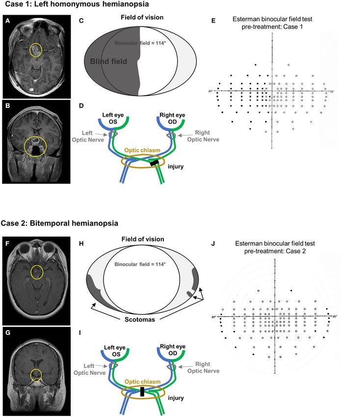
but visual rehabilitation procedure must still be developed A 29-year-old male was diagnosed with a left homonymous
to optimize/improve visual perception in the blind field and hemianopia consecutive to a low-grade glioma diagnosed at
enhance the quality of life. Here, we report, for the first time, the age of 8 (Figures 1A–E) (Table 1). Initial symptoms were
the feasibility and potential effectiveness of a visual rehabilitation related to hydrocephalus and MRI scan identified disseminated
procedure consecutive to a pediatric brain tumor in two young nodules in the brain, the cerebellum, and along the spinal
adult patients with hemianopia. We developed a home-based cord. A biopsy of a spinal nodule was performed, and findings
audiovisual stimulation protocol (currently not commercialized) were in keeping with the diagnosis of low disseminated
using immersive virtual reality (IVR) in the stand-alone and grade glioma. He was treated with chemotherapy with a
remotely controlled head-mounted display (HMD) Oculus Go. good clinical and radiological response. At the age of 16, he
The basic concept relies on the stimulation of residual vision progressively developed left homonymous hemianopia and his
and multisensory-induced visual plasticity using specific visual MRI scan showed tumor progression in the optic chiasm.
tasks, combined to spatially and temporally congruent auditory There is a heterogeneous enhancing lesion involving the
stimuli (10, 11). This approach is based on the multisensory floor of the third ventricle hypothalamus and suprasellar
integration properties of the brain for the detection and cistern (Figures 1A,B). The pituitary stalk and optic chiasm
tracking of elements in the surroundings (12, 13). Recent cannot be differentiated from the mass, which are most
evidence shows that static audiovisual stimulation reorganizes likely infiltrated (Figures 1A,B). Despite further chemotherapy
the functional connectivity in subcortical and cortical visual that prevented further tumor progression, his visual deficit
areas in hemianopia patients improving visual perception in remained unchanged, with mild impact on his daily activities
the blind hemifield (11, 14). Here, we used the 3D-multiple (Figures 1C–E).
object tracking (3D-MOT) paradigm, which closely matches
attentionally demanding real-life situations (15, 16). The setup Case 2
of the task is highly dynamic as the objects change their A 32-year-old female was diagnosed with bitemporal hemianopia
location over time, requiring a continuous deployment of visual and left exotropia consecutive to a suprasellar non-germinatous
attention and oculomotor control to avoid confusions between germ cell tumor diagnosed at the age of 13 (Figures 1F–J)
the objects (16). 3D-MOT stimulation programs displayed (Table 1). She was treated with a combination of chemotherapy
on monitors or TV screens have been shown to increase and focal radiotherapy and achieved an excellent response. The
brain capacity for complex processes such as anticipation, end of treatment MRI still showed a 1.5-cm residual mass
Frontiers in Neurology | www.frontiersin.org 2 July 2021 | Volume 12 | Article 680211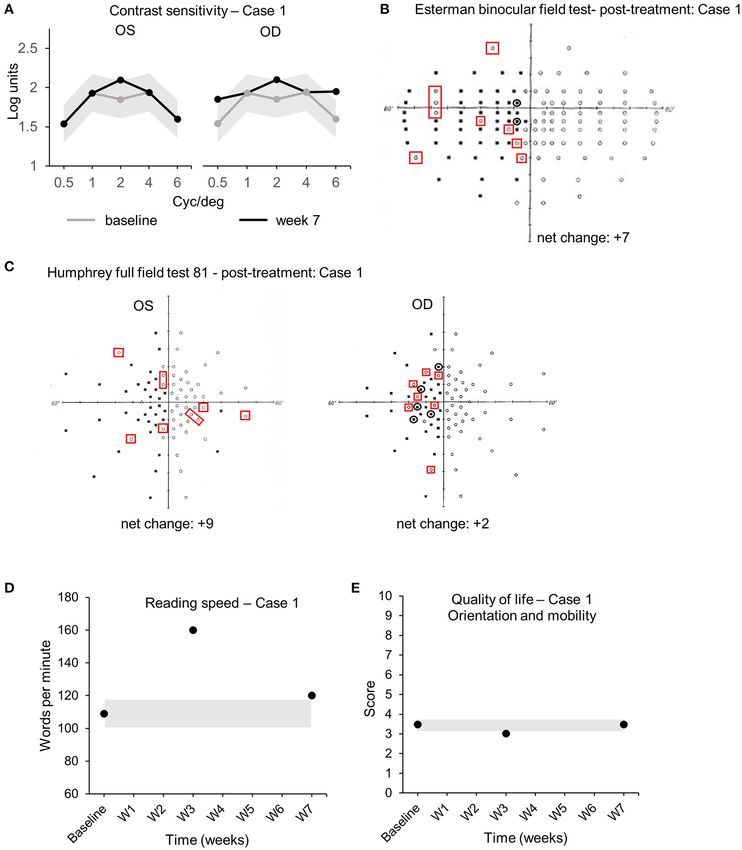
Daibert-Nido et al.                                                                                                                   Visual Rehabilitation in Hemianopia

 FIGURE 1 | Case presentation. (A–E) Case 1 MRI axial (A) and coronal (B) view (T1 + contrast). Yellow circles indicate the remaining tumor mass after chemotherapy.
 (C) Schematic of the visual field defects. (D) Diagram of the optic pathway indicating the location of the injury. (E) Esterman binocular field test at baseline
 (pre-treatment). Black dots correspond to unseen point of light. (F–J) Case 2 MRI axial (F) and coronal (G) view (T1 + contrast). Yellow circles indicate the remaining
 tumor mass after chemo- and focal radiotherapy. (H) Schematic of the visual field defects. (I) Diagram of the optic pathway indicating the location of the injury. (J)
 Esterman binocular field test at baseline (pre-treatment). Black dots correspond to unseen point of light.

in the suprasellar region, inseparable from the hypothalamus                           bitemporal hemianopia (Figures 1H–J), was detected at the
and optic chiasm, which remained unchanged over time                                   time of initial diagnosis did not improve despite the successful
(Figures 1F,G). Her visual impairment, corresponding to a                              management of her tumor.

Frontiers in Neurology | www.frontiersin.org                                       3                                              July 2021 | Volume 12 | Article 680211
Daibert-Nido et al. Visual Rehabilitation in Hemianopia
TABLE 1 | Summary of the episode of care.
Case 1
Timeline/age 8 years old (1999) 16 years old (2007) 29 years old (2020)
Episode of care Grade glioma in brain, cerebellum, and - Tumor progression in the optic chiasm - No improvement of the left hemianopia
spinal cord - Left homonymous hemianopsia worsening - 7-week home-based visual rehabilitation program
- Chemotherapy
- Diagnosis of left homonymous hemianopsia
Case 2
Timeline/age 13 years old (2001) 32 years old (2020)
Episode of care Diagnosis of suprasellar non-germinatous germ cell tumor - No improvement of bitemporal hemianopia
- Chemotherapy and focal radiotherapy - 6-week home-based visual rehabilitation program
- Diagnosis of bitemporal hemianopia and left exotropia
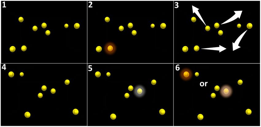
Assessments The audiovisual stimulation task corresponds to the 3D-MOT
Visual assessments investigating both visual function and paradigm composed of eight high-contrast yellow spheres on
functional vision were performed at the Ophthalmology Low a black background whose features were adapted to the visual
vision Clinic at the Toronto Western Hospital (University Health ability of low-vision patients (luminosity = 100 cd/m2 , size
Network, Toronto, Canada) following standard procedures in = 1.57◦ of visual angle). After one of the spheres was cued
low-vision rehabilitation (20–23). These assessments include (turning red for 5 s and switching back to yellow, Figure 2), the
visual acuity [best corrected visual acuity (BCVA)] measured spheres move for 20 s following random linear paths, bouncing
using the Early Treatment Diabetic Retinopathy Study (ETDRS) on one another and on the walls of a virtual 3D cube when
charts at 4 m and CS measured using the Functional Acuity collisions occur. The overall span of the movement of the
Contrast Test (FACT). Retinal sensitivity (RS) and fixation spheres covers 78◦ and 50◦ of horizontal and vertical visual
stability [FS; bivariate contour ellipse area (BCEA), 63%] were angle, respectively (Figure 2). The initial speed of the spheres
measured using the Macular Integrity Assessment (MAIA) is adjustable, from 3◦ /s to 240◦ /s. and determined at baseline.
microperimeter (CentreVue, Padova, Italy). Field of vision A spatial sound (50 Hz, 25–65 dBHL) is correlated to the
was evaluated by the monocular Humphrey full field and movement of the cued target. After 20 s, the movement stops
Esterman binocular field of vision, which were assessed using the and the patient is asked to select, using a virtual laser pointer,
Humphrey Full Field Analyzer 3 standard automated perimeter the cued sphere among the distractors (mark-all procedure)
(HFA 3, Zeiss, Heilberg, Germany). Quality of life was measured (16). If the selection is correct (i.e., corresponding to the cued
in two subdomains (orientation and mobility—reading) using target), a positive feedback sound is provided and the speed
the Veteran’s affairs low-vision visual functioning questionnaire of the spheres in the next trial is increased by 0.05log. If the
(LV-VFQ) (24). Reading speed was measured at critical print selection is incorrect (i.e., corresponding to a distractor), a
size using the Minnesota Low Vision Reading (MNREAD) test negative feedback sound is provided and the speed of the spheres
(25). Results were analyzed using the coefficient of repeatability in the next trial is decreased by 0.05log (adaptative simple up-
(COR), specific to each assessment, to compare pre- and post- down staircase) (29). After each block of 5 min, the system
treatment data (26). The COR, also referred as the smallest sends data relative to the performance of the patient to our
real difference (SRD), quantifies an absolute reliability in the secured laboratory computer through Wi-Fi. Patient consent
same unit as the assessment tool. The COR is directly related was obtained following UHN policies and according to the
to the 95% limits of agreement proposed by Bland and Altman Declaration of Helsinki.
(27). It corresponds to the value below which the absolute
differences between two measurements would lie within 95%
of probability. Therefore, measurement values strictly different Case 1
from COR indicate an effect of the treatment (26, 28). The audiovisual stimulation parameters (initial speed, trial
duration, number of targets, total number of spheres, number
of blocks, and number of sessions) were updated and uploaded
DIAGNOSTIC ASSESSMENT remotely into the patient’s device from the laboratory’s
computers every 2 days for 7 weeks. Real-time data (date/time,
Treatment average/maximum speed of the spheres, positive hits, number
The patients followed an IVR audiovisual stimulation protocol of trials/blocks/sessions performed, total time, and response
at home using the stand-alone and remotely controlled HMD time) were collected remotely from the device and indicated
Oculus Go for 7 weeks (case 1) or 6 weeks (case 2). Every that case 1 completed the audiovisual stimulation protocol at
2 days, the patient performed one session of IVR audiovisual home by performing 19 sessions of 3 blocks of 15 trials of 20 s
stimulation composed of three blocks of 15 trials of 20 s, each, every 2 days (±1 day), for a total continuous audiovisual
equivalent to 3 × 5 min of continuous audiovisual stimulation. stimulation of 4 h and 45 min. Visual assessments performed
Frontiers in Neurology | www.frontiersin.org 4 July 2021 | Volume 12 | Article 680211Daibert-Nido et al. Visual Rehabilitation in Hemianopia FIGURE 2 | Principle of audiovisual stimulation program (NeurofyResearch). Sequence of the visual task. (1) Eight yellow still spheres are present in a virtual cube. (2) One of these spheres turns red for 5 s (cued target) and returns to yellow. (3) All spheres randomly move following linear paths across the visual field encompassing the blind field and bouncing on one another and on the walls of the virtual 3D cube when collisions occurred. (4) After 30 s, the spheres stopped moving. (5) The patient had to select the cued target using a hand-guided virtual laser pointer. (6) A correct selection is considered a positive hit. at the clinic comparing baseline (pre-treatment) and week 7 Case 2 (post-treatment) demonstrated an enhanced CS in both left The audiovisual stimulation parameters (same as case 1) were (Oculus Sinister, OS) and right (Oculus Dexter, OD) eyes. CS updated and uploaded remotely into the home-based patient’s improved at 2 cyc/deg from 1.65 to 2.1 logits [COR ±0.24 device from the laboratory computers every 2 days for 6 weeks. logits (30)] in OS (Figure 3A). CS increased at 0.5, 2, and Real-time data (same as case 1) were collected remotely from 6 cyc/deg for OD from 1.54 to 1.85 logits, from 1.85 to 2.1 the device to the laboratory’s computer and indicated that case logits, and from 1.6 to 1.95 logits [COR ±0.24 logits (30)], 2 completed the audiovisual stimulation protocol at home by respectively (Figure 3A). Visual field analysis revealed that the performing 20 sessions of three blocks of 15 trials of 20 s number of points seen in the binocular Esterman field of vision each, every 2 days (±1 day), for a total continuous audiovisual test increased from 66 points seen at baseline to 69 at week stimulation of 4 h and 50 min. Visual assessments performed at 3 and 73 at week 7 with some reorganization (black circles week 3 and week 6 were compared to baseline. CS improved at indicate loss of points, red squares indicate acquired points, net 4 and 6 cyc/deg from 1.74 to 2.23 logits and from 1.41 to 1.95 change: +7 points, all in the blind left hemifield, +5.8% from logits [COR ±0.24 logits (30)], respectively, in OS (Figure 4B). baseline, Table 2, Figure 3B). Importantly, 3 adjacent points at CS improved at 0.5 and 6 cyc/deg for OD from 1.54 to 1.85 60◦ eccentricity in the left blind hemifield were detected at logits and from 1.6 to 1.95 logits [COR ±0.24 logits (30)], week 7, suggesting an improved peripheral visual perception respectively (Figure 4A). The Esterman binocular field testing in the blind field (Figure 3B red squares). Monocular visual showed a minor increase in the number of points seen between field analysis using Humphrey full-field analysis (81 points) also baseline and week 6 with some reorganization (black circles revealed a valid (loss of fixation < 20%) increase in the number indicate loss of points, red squares indicate acquired points, net of points seen in the full field with the left eye (OS) with change: +3 points in the scotomas, +2.5% from baseline, Table 3, an addition of 6 points between baseline (37/81) and week 3 Figure 4B). Monocular visual field analysis using Humphrey full- (43/81) and +3 points between week 3 and week 7 (46/81) (net field analysis (120 points) indicated a decrease in the number of change: +9 points total, +5 in the blind left hemifield, +11.1% perceived points for both OS (net change: −6 points, 4 of them in from baseline, loss of fixation 0/20–0%, Table 2, Figure 3C). the blind left hemifield, −5% from baseline, loss of fixation 2/15– There was a minor increase in the number of points seen 13.3%, Table 3, Figure 4C) and OD (net change: −2 points in the in OD with some reorganization (net change: +2 points in blind right hemifield, −1.6% from baseline, loss of fixation 2/17– the blind left hemifield, +2.5% from baseline, loss of fixation 11.7%, Table 3, Figure 4C). Such discrepancy (−6 net change 1/19–5.3%, Table 2, Figure 3C). Reading speed improved from in OS) is due to the unstable fixation of case 2 at baseline with 109 words per minute (wpm) to 160 at week 3 and to 120 >20% of fixation losses (4/17, 23%), decreasing the reliability of at week 7 [+10% between baseline and week 7, Figure 3D, these particular measures (32). Clinically meaningful increase in black dots, COR ±8.6 wpm (31), Table 2]. No variations in reading speed was observed between baseline and week 6 [+27 visual acuity, RS, FS, or quality of life scores were observed wpm, +22%, COR ±8.6 wpm (31), Table 3, Figure 4D]. This between baseline, week 3, and week 7 (Table 2, Figure 3E). No improvement is corroborated by an increase in the quality of life adverse events were reported for the duration of the home- score in the reading section of the questionnaire [+4.7 logits, based stimulation. COR ±0.44 logits (19), Table 3, Figure 4E—black squares]. The Frontiers in Neurology | www.frontiersin.org 5 July 2021 | Volume 12 | Article 680211
Daibert-Nido et al.                                                                                                                     Visual Rehabilitation in Hemianopia

 FIGURE 3 | Case 1—Visual outcome measures. (A) Contrast sensitivity at baseline (gray line) and at the end of the treatment (week 7—black line) in the left (OS) and
 the right (OD) eye. (B) Esterman binocular field test post-treatment (week 7). Red squares indicate newly acquired points, black circles indicate lost points, compared
 to baseline. (C) Monocular Humphrey full-field test (81 points) post-treatment (week 7) in the left (OS) and right (OD) eye. Red squares indicate newly acquired points,
 and black circles indicate lost points, compared to baseline. (D) Reading speed at baseline, week 3, and post-treatment (week 7). (E) Quality of life scores at baseline,
 week 3, and post-treatment (week 7). Gray shaded areas in (A,D,E) indicate the values of the coefficient of repeatability (±COR). OD, oculus dexter; OS, oculus
 sinister; W, week.

mobility section of the quality of life questionnaire showed                            DISCUSSION
only minor improvement [+0.62 logits, COR ±0.44 logits (22),
Table 3, Figure 4E—black triangles]. No improvements in visual                          This case series shows the feasibility of a home-based
acuity, RS, and FS were observed between all time points                                visual rehabilitation program using an audiovisual 3D-MOT
(Table 3). No adverse events were reported for the duration the                         stimulation paradigm in IVR HMD and potential effectiveness
home-based stimulation program [VRISE questionnaire score =                             on visual perception in hemianopia patients. We were able to
33/35, >25 the threshold for cybersickness (33)].                                       implement the program and update the stimulation procedures

Frontiers in Neurology | www.frontiersin.org                                        6                                               July 2021 | Volume 12 | Article 680211
Daibert-Nido et al.                                                                                                                          Visual Rehabilitation in Hemianopia

TABLE 2 | Case 1 baseline and after treatment outcome measures.

                                                                Baseline                          Week 3                           Week 7                          COR

                                                          OD               OS              OD                  OS          OD                  OS

Visual Acuity (VA)                                       20/20            20/25          20/20               20/25        20/20               20/25
Retinal sensitivity (dB)                                 14.3              14.4            14                  11.8       14.7                 12.4                ±1.51
Bivariate Contour Ellipse Area 63% (BCEA, 2)              0.4               0.6            0.5                 0.5         0.3                 0.5                 ±0.61
Humphrey Full Field                                      48/81            37/81        47/81 (−1)          43/81 (+6)   50/81 (+2)          46/81 (+9)
(points seen)                                           (59.3%)           (45.7%)       (58.0%)             (53.1%)      (61.7%)             (56.8%)
Esterman binocular field                                         66/120                          69/120 (+3)                     73/120 (+7)                ±3 adjacent points
(points seen)                                                     (55%)                           (57.5%)                          (60.8%)
Reading speed (wpm)                                               109                               160                              120                           ±8.6
Quality of life (orientation and mobility)                        3.48                              3.01                             3.48                          ±0.44

OD, oculus dexter; OS, oculus sinister; dB, decibels; wpm, words per minute; COR, coefficient of repeatability.

remotely from the laboratory’s computer to the patients’                                      mentioned above, visual field measures are reliable when the
device through Wi-Fi and to collect real-time data during the                                 proportion of fixation loss (the inability to stabilize the gaze
stimulation procedure performed at home. Patients were able                                   and to fixate the center of the visual field) is below 20% (32).
to adhere and comply to the stimulation program performed at                                  At baseline, case 2 showed a high number of fixation loss
home without interruption of care and with no adverse events                                  (4/7–23%), above the reliability threshold of 20%; therefore,
related to the use of IVR. A total of 4 h 45 min and 4 h and 50 min                           no conclusion can be made as to the potential effect of the
of audiovisual 3D-MOT IVR stimulation led to the improvement                                  audiovisual stimulation on monocular visual field in case 2. The
of several visual metrics. These results are unlikely due to a                                high contrast of the visual stimulation task (yellow spheres with
learning effect of the visual tests as baseline and after treatment                           luminosity = 100 cd/m2 on black background) correlates with an
assessments at the clinic were separated by a minimum of 3                                    increased CS observed for both eyes in each case. Accordingly, a
weeks, above the learning effect time window shown to last for                                significant increase in reading speed was observed in both cases,
up to 1 week (34). Both cases showed improvement, although                                    in line with improved reading speed associated to higher CS (39).
to various degrees, in the binocular field of vision, which is in                             A major improvement in reading speed was also reported by case
line with the paradigm of the audiovisual 3D-MOT stimulation                                  2 in the quality-of-life questionnaire, supporting the quantitative
(15) where spheres moving at different speeds must be tracked                                 results obtained from the MNREAD test.
into a virtual 3D cube encompassing 78◦ and 50◦ of horizontal                                     In this report, positive effects on visual perception and quality
and vertical visual angle, respectively. The net change (+3)                                  of life were observed after 60◦ ) of the visual field. In the binocular condition, case 1
eye movements or saccades. Fixation monitoring revealed the                                   shows more severe visual field defects than case 2 (compare
absence of loss of fixation above threshold (20%) during the                                  Figures 3B, 4B). In our audiovisual stimulation protocol, the
automated Esterman in cases 1 and 2 and Humphrey full-field                                   virtual space covered by the moving spheres has a visual angle
tests in case 1 validating the visual field measures. Monocular                               of 78◦ horizontally and 50◦ vertically, which covers a large
Humphrey field testing in case 2 revealed a decrease in the                                   proportion of the blind left hemifield in case 1 but only a
number of points seen after treatment, particularly for the left                              minor proportion of the scotomas in case 2, which may lead to
eye (net change −6). This discrepancy is likely due to unstable                               less peripheral stimulation in this particular case. The overall
fixation during the visual field test at baseline for case 2. As                              improvement in case 1 is stronger than in case 2 because the

Frontiers in Neurology | www.frontiersin.org                                              7                                            July 2021 | Volume 12 | Article 680211
Daibert-Nido et al. Visual Rehabilitation in Hemianopia FIGURE 4 | Case 2—Visual outcome measures and quality of life. (A) Contrast sensitivity at baseline (gray line) and at the end of the treatment (week 6—black line) in the left (OS) and the right (OD) eye. (B) Esterman binocular field test post-treatment (week 6). Red squares indicate newly acquired points, and black circles indicate lost points, compared to baseline. (C) Monocular Humphrey full-field test (120 points) post-treatment (week 6) in the left (OS) and right (OD) eye. Red squares indicate newly acquired points, and black circles indicate lost points, compared to baseline. (D) Reading speeds at baseline, week 3, and post-treatment (week 6). (E) Quality of life scores at baseline, week 3, and post-treatment (week 6). Gray shaded areas in (A,D,E) indicate the values of the coefficient of repeatability (±COR). OD, oculus dexter; OS, oculus sinister; W, week. original binocular field defects were also more pronounced in oculomotor function and compensatory eye movement (40, 41, case 1. 47). Others suggest that a restoration of visual perception in Visual field restoration in hemianopia patients has been a the blind hemifield could be the consequence of a functional controversial topic (10, 43–46). Several studies suggest that reorganization of the connectivity in subcortical and cortical visual fields can be restored in hemianopia by enhanced structures after visual rehabilitation (11, 14). Moreover, recent Frontiers in Neurology | www.frontiersin.org 8 July 2021 | Volume 12 | Article 680211
Daibert-Nido et al.                                                                                                                           Visual Rehabilitation in Hemianopia

TABLE 3 | Case 2 baseline and after treatment outcome measures.

                                                               Baseline                           Week 3                            Week 6                          COR

                                                         OD                OS             OD                   OS          OD                    OS

Visual Acuity (VA)                                      20/20             20/40         20/20               20/40         20/20                20/40
Retinal sensitivity (dB)                                16.2              13.5           14.9                15.2           14                  13.9                ±1.51
Bivariate Contour Ellipse Area 63% (BCEA, 2)             0.5               2.4            0.2                  2.4          0.4                  2                  ±0.61
Humphrey Full Field (points seen)                      83/120           92/120       80/120 (−3)          91/120 (−1)   81/120 (−2)         86/120 (−6)
                                                       (69.2%)          (76.7%)        (66.7%)              (75.8%)       (67.5%)             (71.7%)
Esterman binocular field                                       104/120                          103/120 (−1)                      107/120 (+3)               ±3 adjacent points
(points seen)                                                   (86.7%)                           (85.8%)                           (89.2%)
Reading speed (wpm)                                              123                                131                               150                           ±8.6
Quality of life (reading)                                         8.3                               13                                13                            ±0.44
Quality of life (orientation and mobility)                       2.58                              3.01                               3.2                           ±0.44

OD, oculus dexter; OS, oculus sinister; dB, decibels; wpm, words per minute; COR, coefficient of repeatability.

evidence indicates that audiovisual stimulation, during which                                   individually, should be tested. When visual field measurements
auditory and visual stimuli are spatially and temporally                                        are deemed not reliable, they should be repeated within a short
correlated, improve visual perception more efficiently than                                     time window (7 weeks) with more sessions per day (2 sessions of                                    remotely controlled, rehabilitation procedures. Future pragmatic
15 min separated by a few hours of rest to avoid VR induced                                     randomized controlled studies will address the effectiveness of
symptoms) should be developed. Positive effects of the treatment                                audiovisual IVR rehabilitation procedure in adults and earlier
were observed mostly in binocular vision (Esterman binocular                                    during the disease in children when neuronal plasticity is
field test) with less effect on monocular vision (Humphrey                                      more effective. Integrated knowledge translation and a patient-
full-field test). This can be explained by the improved FS,                                     centered approach will be included in further RCTs using
which occurs under binocular condition (35) allowing more                                       patients’ and caregivers/partners’ open-ended questionnaires.
points to be seen with the Esterman binocular field test. Our                                   An estimation of long-term effectiveness of IVR audiovisual
audiovisual stimulation protocol is binocular, which very likely                                stimulation must also be addressed. A home-based visual
impacts binocular vision. A monocular version of our audiovisual                                rehabilitation procedure is a promising approach not only to
stimulation procedure, tailored to the ability of each eye                                      decrease the burden of disease by substantially diminishing

Frontiers in Neurology | www.frontiersin.org                                              9                                             July 2021 | Volume 12 | Article 680211
Daibert-Nido et al. Visual Rehabilitation in Hemianopia
the number of visits to the clinic and therefore relieving DATA AVAILABILITY STATEMENT
the constraints of commute and transportation, but also for
populations living remotely and often left without easy access to The original contributions presented in the study are included
modern medicine and treatments. in the article/supplementary material, further inquiries can be
directed to the corresponding author/s.
PATIENT PERSPECTIVE
ETHICS STATEMENT
Case 1
At the age of eight, I was diagnosed with brain and spine tumors. Ethical review and approval was not required for the study on
The primary site was on the optic chiasm, where the optic nerves human participants in accordance with the local legislation and
connect to the brain. I underwent 2 years of chemotherapy, institutional requirements. The patients/participants provided
followed by another year when I was 19. The location of the their written informed consent to participate in this study.
tumors caused my vision to be impacted. For as long as I can Written informed consent was obtained from the individual(s)
remember, I have lived with a deficit in my left peripheral vision. for the publication of any potentially identifiable images or data
This is something I was always told could never be fixed, that included in this article.
I would have to live with it for my entire life, and just have
to learn to compensate for the deficit. So, I did, looking to AUTHOR CONTRIBUTIONS
my left more often than my right, though this did not always
prevent me from bumping into objects or people while walking. MD-N, SM, EB, and MR designed the protocol, analyzed the data,
I thought this was my new normal, and I would live the rest of interpreted the results, and wrote/edited the manuscript. MD-N,
my life with the left-side deficit. You can imagine my surprise YP, and SM performed the assessments and generated the data.
when one of my doctors told me about the work being done KC managed the remote control of the device. KC, CN, and MR
by Dr. Reber. I had doubts anything could work but gave it a extracted the data. All authors attest that they meet the current
try anyway. I was told to use an oculus VR headset every other ICMJE criteria for Authorship.
day, with a specialized program to assist my peripheral vision.
To my surprise and excitement, after 3 months of doing this FUNDING
regimen, I could feel the positive effects from the VR headset. I
am now much more confident walking around outside, bumping This work was funded by the start-up grant, Donald K. Johnson
into things less, and can certainly notice the improvement in my Eye Institute, Krembil Research Institute, University Health
left peripheral vision. I am extremely thankful for everything Dr. Network (MR).
Reber has done to improve my quality of life, especially after
being told this is something that could not be fixed. I am also ACKNOWLEDGMENTS
grateful to donors such as yourselves who help to make this
research possible. My life has been changed for the better as We thank the Toronto Western and General Hospital
a direct result of Dr. Reber’s work and will forever be grateful Foundation for financial support (MR). We thank
for that. Neuropowertrain for their collaboration.
REFERENCES 9. Goodwin D. Homonymous hemianopia: challenges and solutions. Clin
Ophthalmol. (2014) 8:1919–27. doi: 10.2147/OPTH.S59452
1. Peragallo JH. Visual function in children with primary brain tumors. Curr 10. Sabel BA, Henrich-Noack P, Fedorov A, Gall C. Vision restoration
Opin Neurol. (2019) 32:75–81. doi: 10.1097/WCO.0000000000000644 after brain and retina damage: the “residual vision activation theory.”
2. Peragallo JH. Effects of brain tumors on vision in children. Int Ophthalmol Prog Brain Res. (2011) 192:199–262. doi: 10.1016/B978-0-444-53355-5.
Clin. (2018) 58:83–95. doi: 10.1097/IIO.0000000000000237 00013-0
3. Fried I, Tabori U, Tihan T, Reginald A, Bouffet E. Optic pathway gliomas: a 11. Grasso PA, Gallina J, Bertini C. Shaping the visual system: cortical
review. CNS Oncol. (2013) 2:143–59. doi: 10.2217/cns.12.47 and subcortical plasticity in the intact and the lesioned brain.
4. Wan MJ, Zapotocky M, Bouffet E, Bartels U, Kulkarni AV, Drake JM. Long- Neuropsychologia. (2020) 142:107464. doi: 10.1016/j.neuropsychologia.2020.
term visual outcomes of craniopharyngioma in children. J Neurooncol. (2018) 107464
137:645–51. doi: 10.1007/s11060-018-2762-3 12. Meredith MA, Stein BE. Interactions among converging sensory inputs in the
5. Jariyakosol S, Peragallo JH. The effects of primary brain tumors on vision superior colliculus. Science. (1983) 221:389–91. doi: 10.1126/science.6867718
and quality of life in pediatric patients. Semin Neurol. (2015) 35:587–98. 13. Basso MA, May PJ. Circuits for action and cognition: a view
doi: 10.1055/s-0035-1563571 from the superior colliculus. Annu Rev Vis Sci. (2017) 3:197–226.
6. Zihl J. Visual scanning behavior in patients with homonymous hemianopia. doi: 10.1146/annurev-vision-102016-061234
Neuropsychologia. (1995) 33:287–303. doi: 10.1016/0028-3932(94)00119-A 14. Ajina S, Bridge H. Subcortical pathways to extrastriate visual cortex underlie
7. Lewald J, Kentridge RW, Peters S, Tegenthoff M, Heywood CA, Hausmann M. residual vision following bilateral damage to V1. Neuropsychologia. (2019)
Auditory-visual localization in hemianopia. Neuropsychology. (2013) 27:573– 128:140–9. doi: 10.1016/j.neuropsychologia.2018.01.007
82. doi: 10.1037/a0033451 15. Pylyshyn ZW, Annan V. Dynamics of target selection in
8. Trauzettel-Klosinski S. Current methods of visual rehabilitation. Dtsch Arztebl multiple object tracking (MOT). Spat Vis. (2006) 19:485–504.
Int. (2011) 108:871–8. doi: 10.3238/arztebl.2011.0871 doi: 10.1163/156856806779194017
Frontiers in Neurology | www.frontiersin.org 10 July 2021 | Volume 12 | Article 680211Daibert-Nido et al. Visual Rehabilitation in Hemianopia
16. Meyerhoff HS, Papenmeier F, Huff M. Studying visual attention using the 39. Pearce E, Sivaprasad S, Chong NV. Factors affecting reading speed in patients
multiple object tracking paradigm: a tutorial review. Atten Percept Psychophys. with diabetic macular edema treated with laser photocoagulation. PLoS ONE.
(2017) 79:1255–74. doi: 10.3758/s13414-017-1338-1 (2014) 9:e0105696. doi: 10.1371/journal.pone
17. Romeas T, Guldner A, Faubert J. 3D-Multiple Object Tracking training task 40. Bolognini N, Rasi F, Coccia M, Làdavas E. Visual search improvement
improves passing decision-making accuracy in soccer players. Psychol Sport in hemianopic patients after audio-visual stimulation. Brain. (2005) 128(Pt
Exer. (2016) 22:1–9. doi: 10.1016/j.psychsport.2015.06.002 12):2830–42. doi: 10.1093/brain/awh656
18. Legault I, Faubert J. Perceptual-cognitive training improves biological 41. Passamonti C, Bertini C, Làdavas E. Audio-visual stimulation improves
motion perception: evidence for transferability of training in healthy aging. oculomotor patterns in patients with hemianopia. Neuropsychologia. (2009)
Neuroreport. (2012) 23:469–73. doi: 10.1097/WNR.0b013e328353e48a 47:546–55. doi: 10.1016/j.neuropsychologia.2008.10.008
19. Corbin-Berrigan L-A, Kowalski K, Faubert J, Christie B, Gagnon I. 42. Frassinetti F, Bolognini N, Bottari D, Bonora A, Làdavas E. Audiovisual
Three-dimensional multiple object tracking in the pediatric population: integration in patients with visual deficit. J Cogn Neurosci. (2005) 17:1442–52.
the NeuroTracker and its promising role in the management doi: 10.1162/0898929054985446
of mild traumatic brain injury. Neuroreport. (2018) 29:559–63. 43. Horton JC. Disappointing results from Nova Vision’s visual restoration
doi: 10.1097/WNR.0000000000000988 therapy. Br J Ophthalmol. (2005) 89:1–2. doi: 10.1136/bjo.2004.058214
20. Markowitz SN. Principles of modern low vision rehabilitation. Can J 44. Plant GT. A work out for hemianopia. Br J Ophthalmol. (2005) 89:2.
Ophthalmol. (2006) 41:289–312. doi: 10.1139/I06-027 doi: 10.1136/bjo.2004.053173
21. Markowitz SN. A practice template for low-vision rehabilitation. Can J 45. Frolov A, Feuerstein J, Subramanian PS. Homonymous Hemianopia
Ophthalmol. (2009) 44:610. doi: 10.3129/i09-099 and Vision Restoration Therapy. Neurol Clin. (2017) 35:29–43.
22. Markowitz SN. State-of-the-art: low vision rehabilitation. Can J Ophthalmol. doi: 10.1016/j.ncl.2016.08.010
(2016) 51:59–66. doi: 10.1016/j.jcjo.2015.11.002 46. Reinhard J, Schreiber A, Schiefer U, Kasten E, Sabel BA, Kenkel S, et
23. Leat S. 2020 CAO clinical practice guideline: optometric low vision al. Does visual restitution training change absolute homonymous visual
rehabilitation full guidelines. Can J Optometry. (2020) 82:19–62. field defects? A fundus controlled study. Br J Ophthalmol. (2005) 89:30–5.
doi: 10.15353/cjo.v82i1.1636 doi: 10.1136/bjo.2003.040543
24. Stelmack JA, Szlyk JP, Stelmack TR, Demers-Turco P, Williams RT, Moran D, 47. Pambakian A, Currie J, Kennard C. Rehabilitation strategies for patients
et al. Measuring outcomes of vision rehabilitation with the Veterans Affairs with homonymous visual field defects. J Neuroophthalmol. (2005) 25:136–42.
Low Vision Visual Functioning Questionnaire. Invest Ophthalmol Vis Sci. doi: 10.1097/01.WNO.0000161661.29391.0D
(2006) 47:3253–61. doi: 10.1167/iovs.05-1319 48. Shams L, Seitz AR. Benefits of multisensory learning. Trends Cogn Sci. (2008)
25. Legge GE, Ross JA, Luebker A, LaMay JM. Psychophysics of reading. VIII. 12:411–7. doi: 10.1016/j.tics.2008.07.006
The Minnesota Low-Vision Reading Test. Optom Vis Sci. (1989) 66:843–53. 49. Huxlin KR. Perceptual plasticity in damaged adult visual systems. Vision Res.
doi: 10.1097/00006324-198912000-00008 (2008) 48:2154–66. doi: 10.1016/j.visres.2008.05.022
26. Vaz S, Falkmer T, Passmore AE, Parsons R, Andreou P. The case for using 50. Tamietto M, Morrone MC. Visual plasticity: blindsight bridges anatomy
the repeatability coefficient when calculating test-retest reliability. PLoS ONE. and function in the visual system. Curr Biol. (2016) 26:R70–3.
(2013) 8:e73990. doi: 10.1371/journal.pone.0073990 doi: 10.1016/j.cub.2015.11.026
27. Bland JM, Altman DG. Applying the right statistics: analyses of measurement 51. Sagi D. Perceptual learning in vision research. Vision Res. (2011) 51:1552–66.
studies. Ultrasound Obstetr Gynecol. (2003) 22:85–93. doi: 10.1002/uog.122 doi: 10.1016/j.visres.2010.10.019
28. Beckerman H, Roebroeck ME, Lankhorst GJ, Becher JG, Bezemer 52. Huygelier H, Schraepen B, van Ee R, Vanden Abeele V, Gillebert CR.
PD, Verbeek ALM. Smallest real difference, a link between Acceptance of immersive head-mounted virtual reality in older adults. Sci Rep.
reproducibility and responsiveness. Qual Life Res. (2001) 10:571–8. (2019) 9:1–12. doi: 10.1038/s41598-019-41200-6
doi: 10.1023/A:1013138911638 53. Huygelier H, Schraepen B, Lafosse C, Vaes N, Schillebeeckx F, Michiels K, et
29. Levitt H. Transformed up-down methods in psychoacoustics. J Acoust Soc al. An immersive virtual reality game to train spatial attention orientation
Am. (1971) 49(Suppl. 2):467. doi: 10.1121/1.1912375 after stroke: a feasibility study. Appl Neuropsychol Adult. (2020) 27:1–21.
30. Kara S, Gencer B, Ersan I, Arikan S, Kocabiyik O, Tufan HA, et al. doi: 10.1080/23279095.2020.1821030
Repeatability of contrast sensitivity testing in patients with age-related 54. Nesaratnam N, Thomas P, Vivian A. Stepping into the virtual unknown:
macular degeneration, glaucoma, and cataract. Arquivos Brasil Oftalmol. feasibility study of a virtual reality-based test of ocular misalignment. Eye.
(2016) 79:323–7. doi: 10.5935/0004-2749.20160092 (2017) 31:1503–6. doi: 10.1038/eye.2017.97
31. Subramanian A, Pardhan S. The repeatability of MNREAD acuity charts 55. Yasuda K, Muroi D, Hirano M, Saichi K, Iwata H. Differing effects of
and variability at different test distances. Optom Vis Sci. (2006) 83:572–6. an immersive virtual reality programme on unilateral spatial neglect on
doi: 10.1097/01.opx.0000232225.00311.53 activities of daily living. Case Reports. (2018) 2018:1–5. doi: 10.1136/bcr-2017-
32. Sanabria O, Feuer WJ, Anderson DR. Pseudo-loss of fixation 222860
in automated perimetry. Ophthalmology. (1991) 98:76–8. 56. Kim W-S, Paik N-J, Cho S. Development and validation of
doi: 10.1016/S0161-6420(91)32338-8 virtual prism adaptation therapy. In: 2017 International Conference
33. Kourtesis P, Collina S, Doumas LAA, MacPherson SE. Validation of the virtual on Virtual Rehabilitation (ICVR). Montreal, QC (2017). p. 1–2.
reality neuroscience questionnaire: maximum duration of immersive virtual doi: 10.1109/ICVR.2017.8007471
reality sessions without the presence of pertinent adverse symptomatology. 57. Legge GE, Chung STL. Low vision and plasticity: implications
Front Hum Neurosci. (2019) 13:417. doi: 10.3389/fnhum.2019.00417 for rehabilitation. Annu Rev Vis Sci. (2016) 2:321–43.
34. Acton JH, Bartlett NS, Greenstein VC. Comparing the Nidek MP-1 and doi: 10.1146/annurev-vision-111815-114344
humphrey field analyzer in normal subjects. Optom Vis Sci. (2011) 88:1288–97.
doi: 10.1097/OPX.0b013e31822b3746 Conflict of Interest: The authors declare that the research was conducted in the
35. Raveendran RN, Bobier WR, Thompson B. Binocular vision and fixational eye absence of any commercial or financial relationships that could be construed as a
movements. J Vis. (2019) 19:9. doi: 10.1167/19.4.9 potential conflict of interest.
36. Tarita-Nistor L, Brent MH, Steinbach MJ, González EG. Fixation stability
during binocular viewing in patients with age-related macular degeneration. Copyright © 2021 Daibert-Nido, Pyatova, Cheung, Nayomi, Markowitz, Bouffet and
Invest Ophthalmol Vis Sci. (2011) 52:1887–93. doi: 10.1167/iovs.10-6059 Reber. This is an open-access article distributed under the terms of the Creative
37. Spry PGD, Furber JE, Harrad RA. The effect of ocular Commons Attribution License (CC BY). The use, distribution or reproduction in
dominance on visual field testing. Optom Vis Sci. (2002) 79:93–7. other forums is permitted, provided the original author(s) and the copyright owner(s)
doi: 10.1097/00006324-200202000-00010 are credited and that the original publication in this journal is cited, in accordance
38. Shneor E, Hochstein S. Effects of eye dominance in visual perception. Int with accepted academic practice. No use, distribution or reproduction is permitted
Congress Ser. (2005) 1282:719–23. doi: 10.1016/j.ics.2005.05.006 which does not comply with these terms.
Frontiers in Neurology | www.frontiersin.org 11 July 2021 | Volume 12 | Article 680211You can also read