Triamcinolone acetonide prevents oxidative stress-induced tight junction disruption of retinal pigment epithelial cells
←
→
Page content transcription
If your browser does not render page correctly, please read the page content below
Graefes Arch Clin Exp Ophthalmol (2009) 247:641–649
DOI 10.1007/s00417-009-1041-6

 BASIC SCIENCE

Triamcinolone acetonide prevents oxidative stress-induced
tight junction disruption of retinal pigment epithelial cells
Yoko Miura & Johann Roider

Received: 17 June 2008 / Revised: 23 December 2008 / Accepted: 12 January 2009 / Published online: 3 February 2009
# Springer-Verlag 2009

Abstract                                                               that intracellular GSH/GSSG ratio decreased significantly
Purpose Oxidative stress is known to disrupt the integrity             with H2O2, while TA preserved this ratio by up-regulating
of retinal pigment epithelium (RPE) tight junctions. The               GSH synthesis.
goal of this study is to evaluate the effect of triamcinolone          Conclusions TA has a protective effect against oxidative
acetonide (TA) on the junctional integrity of RPE under                stress-induced disruption of RPE tight junction by preserving
oxidative stress and to identify the underlying mechanisms.            cellular redox state.
Methods Second passage porcine RPE cells were cul-
tured on 6-well membrane inserts until 4 weeks after                   Keywords Retinal pigment epithelium . Oxidative stress .
reaching confluence. Cells were incubated with TA                      Triamcinolone acetonide . Tight junction . Glutathione
(10−5 M) for 30 min. FITC-containing medium was added
to the upper chamber (cell’s apical side). The cells were
then challenged with 1 mM Hydrogen Peroxide (H2O2).                    Introduction
After 5 h, the fluorescence intensity of the medium from
lower chamber (cell’s basolateral side) was measured                   In pathogenesis of chorioretinal disorders, such as age-related
using a fluorescence spectrofluorophotometer. This trans-              macular degeneration (AMD) and diabetic retinopathy,
epithelial flux of FITC-dextran was measured until the                 oxidative stress is considered to be one of the crucial factors
21st day. The immunolocalization of occludin and F-actin               [1, 2]. In retinal pigment epithelial (RPE) cells, reactive
was examined with fluorescence microscope. Reduced                     oxygen species (ROS), such as hydrogen peroxide (H2O2),
glutathione (GSH)/oxidized glutathione (GSSG) ratio was                are generated during the phagocytosis of the oxidized
determined by a colorimetric assay kit.                                photoreceptor outer segment in the physiological process
Results Non-lethal oxidative stress by H2O2 increased trans-           of photoreceptor renewal [3]. The nondegradable end
epithelial flux of FITC-dextran significantly. TA inhibited            products of this phagocytosis lead to the accumulation of
this increase and preserved the lower flux through the whole           cholesterol ester and oxidized lipids, which are considered
experimental period. This permeability change by H2O2 was              to be related to the early pathogenesis of AMD [4]. In
reversible and recovered to the normal level within 3 weeks.           diabetic patients, high glucose level is implicated in ROS
In immunohistological study, H2O2 reduced linear occludin              production [1] and thus, the level of oxidative stress
staining at the cell border and increased actin stress fibers.         elevates [5, 6].
TA prevented H2O2-induced disruption of junctional assem-                 Oxidative stress has been shown to affect the expression
bly of occludin and F-actin. Glutathione assay demonstrated            of heat shock proteins [7–9], catalase and metallothionein
                                                                       gene expression [5], fibroblast growth factor (FGF) 2 [10],
Y. Miura (*) : J. Roider                                               FGF receptors [11], RPE-65 and cellular retinaldehyde-
Department of Ophthalmology,                                           binding protein [11] in RPE cell. Kinetic microarray study
University Hospital Schleswig-Holstein,
                                                                       by Strunnikova et al. demonstrated that the nonlethal
Campus Kiel, Hegewischstrasse 2,
24105 Kiel, Germany                                                    oxidative stress by H2O2 upregulates the gene expression
e-mail: ymiura@ophthalmol.uni-kiel.de                                  of the protective proteins from oxidative stress, chaperon
642 Graefes Arch Clin Exp Ophthalmol (2009) 247:641–649
proteins, anti-apoptotic factors, and DNA-reparing factors, For the healing of macular edema or AMD, the stabilization
and down regulates pro-apoptotic genes. They also showed of both inner and outer BRB is necessary. However, the
the recovery with the normalization of gene expression to effect of TA on RPE cell junctional properties (outer BRB)
the baseline levels [12]. has not yet been reported.
H2O2 has been utilized to induce oxidative stress in a The aim of the present study is to elucidate the role of
number of in vitro studies. The cellular events following TA in the treatment of oxidative stress-related ocular
H2O2 treatment include actin reorganization, [13] membrane diseases. Therefore, we investigated the effect of TA on
blebbing, [12] apoptosis, [14] redistribution of paracellular cultured RPE paracellular permeability and junctional
junctional proteins [9] and the change of paracellular molecule distribution under oxidative stress. In order to
permeability [9, 15]. Tight junction of RPE cells, which understand if TA has an antioxidative effect, intracellular
is the most apical component of junctional complex, glutathione levels (total/oxidized/reduced) and the ratio of
function as the outer blood retinal barrier (BRB), which reduced/oxidized glutathione (GSH/GSSG ratio) were also
contributes to a restricted diffusion barrier between the investigated.
retinal and the choroidal perfusion. The integrity of tight
junction is preserved by a number of interaction of
proteins, including occludin, ZO-1, −2, and −3. The Materials and methods
disruption of tight junction barrier function causes the
increase of outer BRB permeability, which leads to Cell culture
the impairment of efficient removal of subretinal fluid,
and as result, may cause the prolonged visual disturbance Porcine eyes were obtained from a local slaughterhouse,
and functional loss of outer retina. and RPE cells were isolated as previously described [42].
Oxidative stress has been reported to affect the distribution Cells were cultivated with Dulbecco’s modified Eagle’s
of RPE junctional proteins [9, 15, 16]. The distributions of medium (DMEM; PAA, Cölbe, Germany) supplemented
occludin, ZO-1 in tight junctions and cadherin in adherens with penicillin/streptomycin, L-glutamine, sodium pyruvate
junction are disrupted by the oxidative stress [16]. This and 10% porcine serum (PAA, Cölbe, Germany). The cells
oxygen-mediated junctional disruption was prevented by were incubated at 37°C under 5% CO2. The medium was
pigment epithelium-derived factor (PEDF) [16] and changed every 2 days, and the cells were subcultured by
keratinocyte growth factor (KGF) [15]. trypsin-ethlendiaminetetracetic acid (EDTA) digestion
Triamcinolone acetonide (TA) is a corticosteroid sus- when they reached confluence as assessed by phase contrast
pension that has been administrated periocularly for the microscopy. The second passage cells were used in all
treatment of ocular inflammatory diseases. In recent years, experiments.
TA is used with intravitreal or trans-tenon’s retrobulber
infusion also for the treatment of macular edema [17–21], Permeability assay
AMD with [22–24] or without [25] photodynamic therapy,
serous macular detachment in central retinal vein occlusion, The transepithelial permeability was evaluated as previously
[26] in a combination with vitrectomy [27, 28], or with described [9]. In brief, second passage RPE cells were
anti-vascular endothelial growth factor treatment for AMD cultured on top of transwell-clear polyester membrane
[29]. The efficacy of TA has been well proven from the chamber for six-well plates (0.4 micrometer pore size)
enormous clinical data, though its side effects [30–32] and (Sigma, St. Louis, MI) and used in experiments 4 to
the reports about its toxicity [33–36] are to be argued. 5 weeks after reaching confluence. The cells were treated
However, the effects of TA during the healing process and with TA (10−5 M) (Sigma, St. Louis, MI) for 30 min. A
its role in treatment remain to be elucidated. 1000 fold TA stock was used and methanol control was
With ECV304 cell line, TA decreased phorbol 12-myristate included. The concentration of TA (10−5 M) was decided
13-acetate-induced paracellular permeability [37]. The pro- due to the previous studies [37, 39], in which the protective
tective effect of TA against oxidative stress in hair cells has effect of TA was obvious in the range of 10−6 M-10−4 M.
been reported [38]. In ophthalmic field, TA is known to Medium in the upper chamber (cell’s apical side) was then
reduce VEGF expression from RPE cells [39], and thus the replaced to 4 kDa Fluorescein isothiocyanate (FITC)-
permeability of retinal vascular endothelium (inner BRB) is dextran (4FD; Sigma, St. Louis, MI)-containing medium
reduced, which may be followed by the reduction of edema. and cells were challenged with 1 mM hydrogen peroxide
TA inhibits the increase of the albumin permeability in the (H2O2). Five hours later, the medium from lower chamber
retina of diabetic rat model [40]. It has been also shown that (cell’s basolateral side) was collected and the amount of
TA has a protective effect from streptozotocin-induced acute fluorescence was measured using a fluorescence spectro-
inflammation and early vascular leakage in rat retina [41]. fluorophotometer (FP-550, Jasco, Japan) with an excitationGraefes Arch Clin Exp Ophthalmol (2009) 247:641–649 643
wavelength of 488 nm and an emission wave length of tration of total glutathione in the sample. With the addition
530 nm. Fluorescence intensity of the normal medium was of 4-Vinylpyridine at the beginning of the assay, every free
measured as a blank, which was subtracted from the thiol reaction can be blocked, thus any contribution to the
intensity of the samples. cycling reaction caused by GSH is eliminated. Therefore,
After collecting the lower medium at the first day (day 1), only GSSG concentration can be measured. Reduced
the medium was completely replaced by the new one and the glutathione concentration can be obtained by subtracting
transepithelial flux of FITC-dextran for 5 h was investigated GSSH from total glutathione.
as described above at day 2, 4, 6, 8, 10, 14, 21.
Statistical analysis
Immunohistochemistry
Each experiment was conducted triplicates and repeated
The second passage porcine RPE cells were cultured on three times.
type I collagen-coated cover glass until 4 to 5 weeks after Statistical significance was determined by paired Student’s
reaching confluence. The cells were treated or untreated t-test. A P-value less than 0.05 was considered to be
with TA and then challenged with 1 mM H2O2. Five hours significant.
after H2O2 stimulation, the cells were washed with PBS,
fixed in 3% paraformaldehyde in PBS for 15 min on ice
and again washed three times with PBS at room tempera- Results
ture. Then the cells were permeabilized with 0.1% triton
X-100 in PBS for 15 min. Cells were then incubated in PBS The time after confluence affects the resistance of RPE cells
containing 1% bovine serum albumin (BSA) (blocking against oxidative stress
buffer) for 20 min at room temperature and incubated with
primary antibody (mouse anti-occludin antibody, diluted The resistance of cultured RPE cells against H 2O2
1:100; Sanko Junyaku, Tokyo, Japan) overnight at 4°C. stimulation was apparently different dependent on the time
After three times rinsing with blocking buffer, cells were after confluence. This was preliminary confirmed (data not
incubated with secondary antibody (TRITC-conjugated anti shown) and the data were consistent with the previous
rat antibody, diluted 1:100; Santa Cruz Biothec. Santa Cruz, report by Bailey et al, in which the cultured RPE cells
CA) for 1 h at room temperature. F-actin was costained 5 weeks after confluence was more resistant to H2O2
with FITC-conjugated phalloidin (Sigma, St. Louis, MI). stimulation both in viability and paracellular permeability
Coverslips were washed and mounted with antifade regent compared to the cells of 1 week after confluence [9].
on slides and examined under a fluorescence microscope Concerning the concentration of H2O2, according to the
(Carl Zeiss, Jena, Germany). preliminary experiments using the RPE cells 5 weeks after
confluence, 1 mM was the best concentration just to induce
Glutathione assay non-lethal oxidative junctional damage. 2 mM H2O2 was
always lethal for these cells, and 0.5 mM sometimes did not
The second passage RPE cells were cultured on 35 mm cell have any effect on juctional integrity. Therefore, in this
culture dish (Nunc, Roskilde, Denmark) and were stimu- study, the cells 5 weeks after confluence were used and the
lated by H2O2 (1 mM) with or without pretreatment of TA concentration of H2O2 was determined 1 mM to induce
(10−5 M) for 30 min. Three hours later, the cells were non-lethal junctional damage.
collected by trypsinization, and cellular total glutathione,
oxidized glutathione (GSSG) and reduced glutathione Effect of H2O2 on RPE junctional integrity
(GSH) were measured by using a commercial kit according and the protective effect of TA
to the manufacture’s protocol (Trevigen Inc., Gaithersburg,
MD). This assay utilizes a kinetic enzymatic recycling In permeability assay, the flux of FITC-dextran through the
reactions of glutathione. Once GSSG reacts with NADPH2, non-treated RPE cell culture (5 weeks after confluence on
glutathione reductase reduces GSSG to GSH, which reacts the membrane) was detected as very low (close to zero)
with 5, 5′-dithiobis-2-nitrobenzonic acid (DTNB) to pro- fluorescence intensity, and this level was determined as a
duce a yellow-colored 5-thio-2-nitrobenzonic acid (TNB) control level (100%) (Fig. 1). H2O2-treated RPE cells
that absorbs at 405 nm. Another product of this reaction, showed a significantly higher transepithelial flux of FITC-
GSTNB (GSH and TNB), is reduced by glutathione dextran (283% of control, P644 Graefes Arch Clin Exp Ophthalmol (2009) 247:641–649
† †
oxidant stimulation, the flux reduced and recovered to the
350
normal level in 21 days. Transepithelial flux through TA+
Relative Fluorescence (% of control)
300 H2O2-treated RPE cells reduced dramatically at day 2 (from
203% to 127%) and maintained the lower flux level during
250
the experimental period. TA-treated RPE cells did not show
200 any significant difference from control cells.
150 N.A.
The effect of TA on H2O2-induced disruption of occludin
100 and F-actin distribution
50
In control cells, occludin displays linear staining at the cell
0 border (Fig. 3a). F-actin filaments are also distributed at the
control TA H2O2
H2O2 TA+H2O2
TA+ H2O2 cell border and show the linear staining (actomyosin ring)
(Fig. 3b). In H2O2-treated cells, the linear occludin staining
Fig. 1 Transepithelial flux of FITC-dextran for 5 h through the
confluent RPE cell culture under the condition with no treatment at the cell border is less clear (Fig. 3c, arrowhead), and
(control), 30-min pretreatment of 10−5 M triamcinolone acetonide more occludin staining can been seen in cytoplasm (Fig. 3c,
(TA), 1 mM hydrogen peroxide (H2O2), and 30-minute pretreatemnt asterisk). H2O2 increased the actin stress fibers and the
of 10−5 M TA+1 mM H2O2 (TA+H2O2): H2O2 incrased trans-
disruption of actomyosin ring (Fig. 3d, arrowhead). TA
epithelial flux and TA inhibited this increase significantly. †PGraefes Arch Clin Exp Ophthalmol (2009) 247:641–649 645
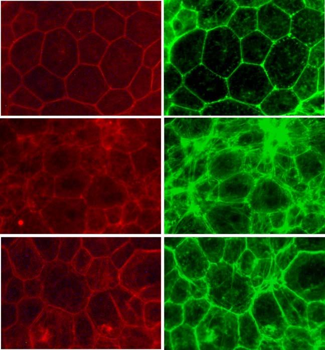
Fig. 3 Immunofluorescein occludin F-actin
staining of occludin (a, c, e)
and F-actin (b, d, f) of the RPE
cells 5 weeks after confluence.
a b
Occludin was stained with anti-
occludin antibody and TRITC-
conjugated second antibody.
F-actin filament was co-stained Control
with FITC-conjugated phalloidin.
a, b control cells, c, d H2O2
(1 mM)-treated cells, e, f TA
(10−5 M)+H2O2 (1 mM)-treated
cells: H2O2 –treated cells showed
less occuldin linear staining at the
cell border (c, arrow head) and c d
more in cytoplasm (c, asterisk),
and more actin stress fibers
(d, arrowhead) compared to the
control cells. TA reduced H2O2-
induced these changes of occlud- 1mM H2O 2
ing and actin fibers (e, f). Scale
bar=10 μm
*
e f
10-5M TA
+1mM H2O2
Discussion circumscribing the cell [48] which is known as the
perijunctional actomyosin ring. The retention of the
Oxidative stress can damage the cells in many degrees, from perijunctional actin ring results in the stable barrier
nonlethal to lethal. It attacks a variety of cellular component, function [49]. In our study, oxidative stress by H2O2
mainly DNA [43, 44] and cellular cytoskeleton [13, 45], resulted in disruption and internalization of occludin
and if the cells are not repaired properly, the accumulation protein along with reorganization of perijunctional actin
of this damage can lead to cell death [46]. In this study, we rings and the increase of actin stress fiber formation. Some
focused on the oxidant-induced junctional damage with previous studies demonstrated the mechanisms of
nonlethal oxidative stress. oxidative stress-induced actin reorganization in vascular
Tight junctions create a strong barrier to the movement endothelial cells [13, 50–52]. Regarding RPE cells,
of water, solutes and immune cells, and its dysfunction however, information about the mechanisms of this actin
leads to the increased paracellular permeability [47]. Tight reorganization is not available yet and further investigation is
junctions anchor physically into the apical actin cytoskeleton needed.
Table 1 The effects of triamcinolone acetonide (TA) and hydrogen peroxide (H2O2) on glutathione balance in cultured RPE cells
control TA H2O2 TA+H2O2
Total glutathione 119.0±7.7 128.7±9.6* 125.1±10.1* 140.5±7.7†
Oxidized (GSSG) 1.79±0.38 1.79±0.38 2.40±0.56* 2.17±0.45*
Reduced (GSH) 117.2±7.7 127.0±9.7* 122.7±9.9* 138.4±7.7†
GSH/GSSG ratio 67.6±14.9 73.2±16.8 52.6±10.7* 65.6±14,5
(pmol/106 cells)
†P646 Graefes Arch Clin Exp Ophthalmol (2009) 247:641–649
To our knowledge, time-course follow-up of RPE para- oxidative stress is of significant interest among researchers
cellular permeability after H2O2 stimulation has not been [62–66]. The protective effect of TA, however, has not yet
investigated before. been reported, even though TA is being widely used in
The present study demonstrates that the H2O2-induced clinical practice.
increase of paracellular permeability of RPE is reversible. The modulation of cellular tight junction protein expres-
This finding is supported by a previous study, in which the sion and paracellular permeability by corticosteroid has
survival of RPE cell after prolonged oxidative stress was been demonstrated in some cells, such as brain endothelial
investigated [12]. In our study, only 30-min pretreatment of cells [67], mouse mammary epithelial cells [68] and retinal
TA prevented the initial damage by H2O2, which can be vascular endothelial cells [69]. In cultured RPE cells in this
detected as occludin and actin dislocalization accompanied study, however, TA alone showed no significant effect on
by permeability increase, and resulted in an earlier recovery paracellular permeability. Thus we assume that, in RPE
of RPE junctional function than H2O2-treated cells. In the cells under oxidative stress, TA itself has no, or very little,
pathogenesis of AMD or diabetic retinopathy, lethal direct effect on junction proteins of RPE cells, but has the
damage is the last stage of degeneration, as seen in effect mainly to protect them from oxidative injury, and in
geographic atrophy of dry AMD. In most cases, nonlethal consequence, leading to the preservation of junctional
damage might be repeated and affect cell function gradu- integrity.
ally, which decreases the cellular recovery potential in the From previous studies, it has been shown that the
long run. Marin-Castaño reported that nonlethal oxidative mechanism of reducing macular edema by TA is considered
stress to human RPE cells causes cells membrane blebbing to be via its anti-inflammatory/angiogenic effects on
[53] and repetitive damage decreased extracellular matrix vascular endothelial cells [39] and down-regulation of
turnover and induced sub-RPE deposits [54]. We suspect VEGF expression [37]. Also in the treatment of AMD, it
that repetitive oxidative stress affects not only the turnover is thought that TA reduces the formation of choroidal
of extracellular matrix, but also many other functions of neovasucularization (CNV) by downregulating of VEGF
RPE cells, which have to be elucidated in future inves- [37, 70]. TA promoted RPE cell proliferation to enclose the
tigations. Therefore, cellular damage from oxidative stress areas of CNV [39, 71]. Our study shows an additional
should be minimized even if nonlethal and recoverable, and effect and suggests that stabilized outer BRB by TA might
the protective effect of TA shown in this study has a contribute to the healing process and the stabilization of
significant implication. visual acuity.
The redox status of the cells is of significance to In this study, the effectiveness of TA in protecting RPE
determine the cellular anti-oxidant defense. It is largely cellular junctional integrity under oxidative stress was
determined by reduced glutathione (GSH), which is a major proven. The results provide further important information
nonprotein cellular thiol in mammalian cells, and accounts in understanding the therapeutic effect of TA. Further
for more than about 98% of intracellular total glutathione in investigations are needed to elucidate more details, such
healthy cells [55–57]. GSH and its precursors have been as intracellular signaling pathways.
reported to protect cultured RPE cells from oxidant-induced
apoptosis [58–60]. The ratio of reduced glutathione (GSH) Acknowledgment We would like to thank Dr. Alexa Klettner for
helpful discussion.
to oxidized glutathione (GSSG) (GSH/GSSG ratio) has
been widely used as an indicator of cellular redox status
[61]. The preservation of cellular GSH/GSSG ratio means
less oxidative damage in the cell. Thus, for the pathogenesis References
and treatment of AMD and diabetic retinopathy, this redox
status is one of the important factors which may play crucial 1. Fukagawa NK, Li M, Liang P, Russell JC, Sobel BE, Absher PM
(1999) Aging and high concentrations of glucose potentiate injury
roles.
to mitochondrial DNA. Free Radic Biol Med 27:1437–1443,
H2O2 stimulation decreased cellular GSH/GSSG ratio doi:10.1016/S0891-5849(99)00189-6
significantly, with the increase of GSSG. Decreased GSH/ 2. Winkler BS, Boulton ME, Gottsch JD, Sternberg P (1999)
GSSG ratio indicates cellular oxidative stress and less Oxidative damage and age-related macular degeneration. Mol
Vis 5:32–43
antioxidant defense. Redox imbalance can induce the
3. Miceli MV, Liles MR, Newsome DA (1994) Evaluation of oxidative
oxidative damage of intracellular proteins and nucleotides, processes in human pigment epithelial cells associated with retinal
thus the functional damages, which can be lead to cell outer segment phagocytosis. Exp Cell Res 214:242–249,
death, either apoptosis or necrosis [61]. Our result suggests doi:10.1006/excr.1994.1254
4. Ruberti JW, Curcio CA, Millican CL, Menco BP, Huang JD, Johnson
that TA increases GHS synthesis to preserves the redox
M (2003) Quick-freeze/deep-etch visualization of age-related lipid
balance, thus maintains adequate cellular antioxidant accumulation in Bruch's membrane. Invest Ophthalmol Vis Sci
defenses. Recently, the protection of RPE cells from 44:1753–1759, doi:10.1167/iovs.02-0496Graefes Arch Clin Exp Ophthalmol (2009) 247:641–649 647
5. Tate DJ Jr, Miceli MV, Newsome DA (1995) Phagocytosis and photocoagulation for the treatment of macular edema in branch
H2O2 induce catalase and metallothionein gene expression in retinal vein occlusion. Ophthalmic Res 40:26–31, doi:10.1159/
human retinal pigment epithelial cells. Invest Ophthalmol Vis Sci 000111155
36:1271–1279 22. Obata R, Iriyama A, Inoue Y, Takahashi H, Tamaki Y, Yanagi Y
6. Ballinger SW, Van Houten B, Jin GF, Conklin CA, Godley BF (2007) Triamcinolone acetonide suppresses early proangiogenic
(1999) Hydrogen peroxide causes significant mitochondrial DNA response in retinal pigment epithelial cells after photodynamic
damage in human RPE cells. Exp Eye Res 68:765–772, therapy in vitro. Br J Ophthalmol 91:100–104, doi:10.1136/
doi:10.1006/exer.1998.0661 bjo.2006.098004
7. Wong CG, Lin NG (1989) Induction of stress proteins in cultured 23. Liggett PE, Colina J, Chaudhry NA, Tom D, Haffner G (2006)
human RPE-derived cells. Curr Eye Res 8:537–545, doi:10.3109/ Triple therapy of intravitreal triamcinolone, photodynamic therapy,
02713688908995751 and pegaptanib sodium for choroidal neovascularization. Am J
8. Kerendian J, Enomoto H, Wong CG (1992) Induction of stress Ophthalmol 142:1072–1074, doi:10.1016/j.ajo.2006.07.029
proteins in SV-40 transformed human RPE-derived cells by 24. Spaide RF, Sorenson J, Maranan L (2005) Combined photody-
organic oxidants. Curr Eye Res 11:385–396, doi:10.3109/ namic therapy and intravitreal triamcinolone for nonsubfoveal
02713689209001792 choroidal neovascularization. Retina 25:685–690, doi:10.1097/
9. Bailey TA, Kanuga N, Romero IA, Greenwood J, Luthert PJ, 00006982-200509000-00001
Cheetham ME (2004) Oxidative stress affects the junctional 25. Ito M, Okubo A, Sonoda Y, Yamakiri K, Sakamoto T (2006)
integrity of retinal pigment epithelial cells. Invest Ophthalmol Intravitreal triamcinolone acetonide for exudative age-related
Vis Sci 45:675–684, doi:10.1167/iovs.03-0351 macular degeneration among Japanese patients. Ophthalmologica
10. Hackett SF, Schoenfeld CL, Freund J, Gottsch JD, Bhargave S, 220:118–124, doi:10.1159/000090577
Campochiaro PA (1997) Neurotrophic factors, cytokines and 26. Karacorlu M, Karacorlu SA, Ozdemir H, Senturk F (2007)
stress increase expression of basic fibroblast growth factor in Intravitreal triamcinolone acetonide for treatment of serous
retinal pigmented epithelial cells. Exp Eye Res 64:865–873, macular detachment in central retinal vein occlusion. Retina
doi:10.1006/exer.1996.0256 27:1026–1030
11. Alizadeh M, Wada M, Gelfman CM, Handa JT, Hjelmeland LM 27. Sakamoto T, Miyazaki M, Hisatomi T, Nakamura T, Ueno A,
(2001) Downregulation of differentiation specific gene expression Itaya K, Ishibashi T (2002) Triamcinolone-assisted pars plana
by oxidative stress in ARPE-19 cells. Invest Ophthalmol Vis Sci vitrectomy improves the surgical procedures and decreases the
42:2706–2713 postoperative blood-ocular barrier breakdown. Graefes Arch
12. Strunnikova N, Zhang C, Teichberg D, Cousins SW, Baffi J, Clin Exp Ophthalmol 240:423–442, doi:10.1007/s00417-002-
Becker KG, Csaky KG (2004) Survival of retinal pigment 0454-2
epithelium after exposure to prolonged oxidative injury: a detailed 28. KangSW,ParkSC,ChoHY,KangJH(2007)Tripletherapyofvitrectomy,
gene expression and cellular analysis. Invest Ophthalmol Vis Sci intravitreal triamcinolone, and macular laser photocoagulation for
45:3767–3777, doi:10.1167/iovs.04-0311 intractable diabetic macular edema. Am J Ophthalmol 144:878–885,
13. Guay J, Lambert H, Gingras-Breton G, Lavoie JN, Huot J, Landry J doi:10.1016/j.ajo.2007.07.044
(1997) Regulation of actin filament dynamics by p38 map kinase- 29. Shimura M, Nakazawa T, Yasuda K, Shiono T, Iida T, Sakamoto
mediated phosphorylation of heat shock protein 27. J Cell Sci T, Nishida K (2008) Comparative Therapy Evaluation of Intra-
110:357–368 vitreal Bevacizumab and Triamcinolone Acetonide on Persistent
14. Forrest VJ, Kang YH, McClain DE, Robinson DH, Ramakrishnan N Diffuse Diabetic Macular Edema. Am J Ophthalmol 145:854–861,
(1994) Oxidative stress-induced appoptosi prevented by Trolox. Free doi:10.1016/j.ajo.2007.12.031
Radic Biol Med 16:675–684, doi:10.1016/0891-5849(94)90182-1 30. Roth DB, Realini T, Feuer WJ, Radhakrishnan R, Gloth J,
15. Geiger RC, Waters CM, Kamp DW, Glucksberg MR (2005) KGF Heimmel MR, Fechtner RD, Yarian DL, Green S (2008) Short-
prevents oxygen-mediated damage in ARPE-19 cells. Invest term complications of intravitreal injection of triamcinolone
Ophthalmol Vis Sci 46:3435–3442, doi:10.1167/iovs.04-1487 acetonide. Retina 28:66–70
16. Ho TC, Yang YC, Cheng HC, Wu AC, Chen SL, Tsao YP (2006) 31. Yamashita T, Uemura A, Kita H, Sakamoto T (2007) Intraocular
Pigment epithelium-derived factor protects retinal pigment pressure after intravitreal injection of triamcinolone acetonide
epithelium from oxidant-mediated barrier dysfunction. Biochem following vitrectomy for macular edema. J Glaucoma 16:220–
Bi o p h ys R e s Co m m u n 3 4 2: 3 7 2 –3 7 8 , d oi : 1 0. 1 0 16 / j . 224, doi:10.1097/IJG.0b013e31802d6e16
bbrc.2006.01.164 32. Bhavsar AR, Ip MS, Glassman AR, DRCRnet and the SCORE Study
17. Negi AK, Vernon SA, Lim CS, Owen-Armstrong K (2005) Groups (2007) The risk of endophthalmitis following intravitreal
Intravitreal triamcinolone improves vision in eyes with chronic triamcinolone injection in the DRCRnet and SCORE clinical trials.
diabetic macular oedema refractory to laser photocoagulation. Eye Am J Ophthalmol 144:454–456, doi:10.1016/j.ajo.2007.04.011
19:747–751, doi:10.1038/sj.eye.6701636 33. Chung H, Hwang JJ, Koh JY, Kim JG, Yoon YH (2007)
18. Toda J, Fukushima H, Kato S (2007) Injection of triamcinolone Triamcinolone acetonide-mediated oxidative injury in retinal cell
acetonide into the posterior sub-tenon capsule for treatment of culture: comparison with dexamethasone. Invest Ophthalmol Vis
diabetic macular edema. Retina 27:764–769, doi:10.1097/IAE.0- Sci 48:5742–5749, doi:10.1167/iovs.07-0566
b013e318030bfcd 34. Chang YS, Wu CL, Tseng SH, Kuo PY, Tseng SY (2007)
19. Jonas JB, Söfker A (2001) Intraocular injection of crystalline Cytotoxicity of triamcinolone acetonide on human retinal pigment
cortisone as adjunctive treatment of diabetic macular edema. epithelial cells. Invest Ophthalmol Vis Sci 48:2792–2798,
Am J Ophthalmol 132:425–427, doi:10.1016/S0002-9394(01) doi:10.1167/iovs.06-1146
01010-8 35. Albini TA, Abd-El-Barr MM, Carvounis PE, Iyer MN, Lakhanpal
20. Karacorlu M, Ozdemir H, Karacorlu S, Alacali N, Mudun B, RR, Pennesi ME, Chevez-Barrios P, Wu SM, Holz ER (2007)
Burumcek E (2005) Intravitreal triamcinolone as a primary Long-term retinal toxicity of intravitreal commercially available
therapy in diabetic macular oedema. Eye 19:382–386, preserved triamcinolone acetonide (Kenalog) in rabbit eyes. Invest
doi:10.1038/sj.eye.6701512 Ophthalmol Vis Sci 48:390–395, doi:10.1167/iovs.06-0145
21. Ozdek S, Deren YT, Gurelik G, Hasanreisoglu B (2008) Posterior 36. Yu SY, Damico FM, Viola F, D'Amico DJ, Young LH (2006)
subtenon triamcinolone, intravitreal triamcinolone and grid laser Retinal toxicity of intravitreal triamcinolone acetonide: a morpho-648 Graefes Arch Clin Exp Ophthalmol (2009) 247:641–649
logical study. Retina 26:531–536, doi:10.1097/00006982- 51. Pichon S, Bryckaert M, Berrou E (2004) Control of actin
200605000-00006 dynamics by p38 MAP kinase-Hsp27 distribution in the lamelli-
37. Penfold PL, Wen L, Madigan MC, Gillies MC, King NJ, podium of smooth muscle cells. J Cell Sci 117:2569–2577,
Provis JM (2000) Triamcinolone acetonide modulates perme- doi:10.1242/jcs.01110
ability and intercellular adhesion molecule-1 (ICAM-1) expres- 52. Nguyen A, Chen P, Cai H (2004) Role of CaMKII in hydrogen
sion of the ECV304 cell line: implications for maculr peroxide activation of ERK1/2, p38 MAPK, HSP27 and actin
degeneration. Clin Exp Immunol 121:458–465, doi:10.1046/ reorganization in endothelial cells. FEBS Lett 572:307–313,
j.1365-2249.2000.01316.x doi:10.1016/j.febslet.2004.06.061
38. Guzman J, Ruiz J, Eshraghi AA, Polak M, Garnham C, Balkany 53. Marin-Castaño ME, Csaky KG, Cousin SW (2005) Nonlethal
TJ, Van de Water TR (2006) Triamcinolone acetonide protects oxidant injury to human retinal pigment epithelium cells causes
auditory hair cells from 4-hydroxy-2,3-nonenal (HNE) ototoxicity cell membrane blebbing but decreased MMP-2 activity. Invest
in vitro. Acta Otolaryngol 126:685–690, doi:10.1080/ Ophthalmol Vis Sci 46:3331–3340, doi:10.1167/iovs.04-1224
00016480500492018 54. Marin-Castaño ME, Striker GE, Akcazar O, Catanuto P, Espinosa-
39. Matsuda S, Gomi F, Oshima Y, Tohyama M, Tano Y (2005) Heidmann DG, Cousins SW (2006) Repetitive nonlethal oxidant
Vascular endothelial growth factor reduced and connective tissue injury to retinal pigment epithelium decreased extracellular matrix
growth factor induced by triamcinolone in ARPE 19 cells under turnover in vitro and induced sub-RPE deposits in vivo. Invest
oxidative stress. Invest Ophthalmol Vis Sci 46:1062–1068, Ophthalmol Vis Sci 47:4098–4112, doi:10.1167/iovs.05-1230
doi:10.1167/iovs.04-0761 55. Meister A, Anderson ME (1983) Glutathione. Annu Rev Biochem
40. Zhang X, Bao S, Lai D, Rapkins RW, Gillies MC (2008) 52:711–760, doi:10.1146/annurev.bi.52.070183.003431
Intravitreal Triamcinolone acetonide inhibites breakdown of the 56. Kosower NS, Kosower EM (1978) The glutathione statue of the
blood-retinal barrier through differential regulation of VEGF-A cell. Int Rev Cytol 54:109–160, doi:10.1016/S0074-7696(08)
and its receptors in early diabetic retinas. Diabetes 57:1026–1033, 60166-7
doi:10.2337/db07-0982 57. Reed DJ (1990) Gluathione : Toxicological implications. Annu
41. Kim YH, Choi MY, Kim YS, Park CH, Lee JH, Chung IY, Yoo Rev Pharmacol Toxicol 30:603–631, doi:10.1146/annurev.
JM, Choi WS, Cho GJ, Kang SS (2007) Triamcinolone acetonide pa.30.040190.003131
protects the rat retina from STZ-induced acute inflammation and 58. Sternberg P Jr, Davidson PC, Jones DP, Hagen TM, Reed RL
early vascular leakage. Life Sci 81:1167–1173, doi:10.1016/j. (1993) Protection of retinal pigment epithelium from oxidative
lfs.2007.08.024 injury by glutathione and precursors. Invest Ophthalmol Vis Sci
42. Yanagihara N, Moriwaki M, Shiraki K, Miki T, Otani S (1996) 34:3661–3668
The involvement of polyamines in the proliferation of cultured 59. Nelson KC, Carlson JL, Newman ML, Sternberg P Jr, Jones
retinal pigment epithelial cells. Invest Ophthalmol Vis Sci DP, Kavanagh TJ, Diaz D, Cai J, Wu M (1999) Effect of
37:1975–1983 dietary inducer dimethylfumarate on glutathione in cultured
43. Ballinger SW, Van Houten B, Jin GF, Godley BF (1999) human retinal pigment epithelial cells. Invest Ophthalmol Vis
Hydrogen peroxide causes significant mitochondrial DNA dam- Sci 40:1927–1935
age in human RPE cells. Exp Eye Res 68:765–772, doi:10.1006/ 60. Wood JP, Pergande G, Osborne NN (1998) Prevention of
exer.1998.0661 glutathione depletion-induced apoptosis in cultured human RPE
44. Liang FQ, GOldey BF (2003) Oxidative stree-induced mitochon- cells by flupirtine. Restor Neurol Neurosci 12:119–125
drial DNA damage in human retinal pigment epithelial cells: a 61. Schafer FQ, Buettner GR (2001) Redox environment of the cell as
possible mechanism for RPE aging and age-related macular viewed through the redox state of the glutathione disulfide/
degeneration. Exp Eye Res 76:397–403, doi:10.1016/S0014- glutathione couple. Free Radic Biol Med 30:1191–1212,
4835(03)00023-X doi:10.1016/S0891-5849(01)00480-4
45. Dalle-Donne I, Rossi R, Milzani A, Di Simplicio P, Colombo R 62. Li X, Liu Z, Luo C, Jia H, Sun L, Hou B, Shen W, Packer L,
(2001) The actin cytoskelton response to oxidants: from small Cotman CW, Liu J (2008) Lipoamide protects retinal pigment
heat shock protein phosphorylation to changes in the redox state epithelial cells from oxidative stress and mitochondrial dysfunc-
of actin itself. Free Radic Biol Med 31:1624–1632, doi:10.1016/ tion. Free Radic Biol Med 44:1465–1474, doi:10.1016/j.free-
S0891-5849(01)00749-3 radbiomed.2008.01.004
46. Schraufstatter IU, Hinshaw DB, Hyslop PA, Spragg RG, 63. Chang JY, Bora PS, Bora NS (2008) Prevention of oxidative
Cochrane CG (1986) Oxidant injury of cells: DNA strand-breaks stress-induced retinal pigment epithelial cell death by the
activate polyadenosine diphosphate-ribose polymerase and lead to PPARgamma agonists, 15-Deoxy-Delta 12, 14-Prostaglandin J
depletion of nicotinamide adenine dinucleotide. J Clin Invest (2). PPAR Res :720163
77:1312–1320 64. Shamsi FA, Chaudhry IA, Boulton ME, Al-Rajhi AA (2007)
47. Powell DW (1981) Barrier function of epithelia. Am J Physiol L-carnitine protects human retinal pigment epithelial cells from
241:G275–G288 oxidative damage. Curr Eye Res 32:575–584, doi:10.1080/
48. Wittchen ES, Haskins J, Stevenson BR (1999) Protein interactions 02713680701363833
at the tight junctions. Actin has multiple binding partners, and 65. Ha KN, Chen Y, Cai J, Sternberg P Jr (2006) Increased
ZO-1 forms independent complexes with ZO-2 and ZO-3. J Biol glutathione synthesis through an ARE-Nrf2-dependent pathway
Chem 274:35179–35185, doi:10.1074/jbc.274.49.35179 by zinc in the RPE: implication for protection against oxidative
49. Madara JL, Barenberg D, Carlson S (1986) Effects of cytochalasin stress. Invest Ophthalmol Vis Sci 47:2709–2715, doi:10.1167/
D on Occludin Junctional of intestinal absorptive cells: further iovs.05-1322
evidence that the cytoskeleton may influence paracellular perme- 66. Tate DJ, Newsome DA (2006) A novel zinc compound (zinc
ability and junctional charge selectivity. J Cell Biol 102:2125– monocysteine) enhances the antioxidant capacity of human retinal
2136, doi:10.1083/jcb.102.6.2125 pigment epithelial cells. Curr Eye Res 31:675–683, doi:10.1080/
50. Huot J, Houle F, Marceau F, Landry J (1997) Oxidative stress- 02713680600801024
induced actin reorganization mediated by the p38 mitogen- 67. Romero IA, Radewicz K, Jubin E, Michel CC, Greenwood J,
activated protein kinase/heat shock protein 27 pathway in vascular Couraud PO, Adamson P (2003) Changes in cytoskeletal and
endothelial cells. Circ Res 80:383–392 tight junctional proteins correlate with decreased permeabilityGraefes Arch Clin Exp Ophthalmol (2009) 247:641–649 649
induced by dexamethasone in cultured rat brain endothelial phosphorylation of occludin. J Neurochem 80:667–677,
cells. Neurosci Lett 344:112–116, doi:10.1016/S0304-3940(03) doi:10.1046/j.0022-3042.2001.00740.x
00348-3 70. Ebrahem Q, Minamoto A, Hoppe G, Anand-Apte B, Sears JE
68. Zettl KS, Sjaastad PM, Riskin G, Parry G, Machen TE, Fierstone (2006) Triamcinolone acetonide inhibits IL-6 and VEGF-induced
GL (1992) Glucocorticoid-induced formation of tight junctions in angiogenesis downstream of the IL-6 and VEGF receptors. Invest
mouse mammary epithelial cells in vitro. Proc Natl Acad Sci USA Ophthalmol Vis Sci 47:4935–4941, doi:10.1167/iovs.05-1651
89:9069–9073, doi:10.1073/pnas.89.19.9069 71. Okada A, Wakabayashi T, Kojima E, Asano Y, Hida T (2004)
69. Antonetti DA, Wolpert EB, DeMaio L, Harhaj NS, Scaduto RC Trans-Tenon's retrobulbar triamcinolone infusion for small
(2002) Hydrocortisone decreases retinal endothelial cell water and choroidal neovascularisation. Br J Ophthalmol 88:1097–1098,
solute flux coincident with increased content and decreased doi:10.1136/bjo.2003.039719You can also read