Exosomal microRNAs in non-small cell lung cancer
←
→
Page content transcription
If your browser does not render page correctly, please read the page content below
Review Article on Clinic and Therapeutic Potential of Non-coding RNAs in Cancer

Exosomal microRNAs in non-small cell lung cancer
Elena Duréndez-Sáez1,2#, Susana Torres-Martinez1,2#, Silvia Calabuig-Fariñas1,2,3, Marina Meri-Abad4,
Macarena Ferrero-Gimeno1,2, Carlos Camps1,2,4,5
1
    Molecular Oncology Laboratory, Fundación Hospital General Universitario de Valencia, Valencia, Spain; 2CIBERONC, Valencia, Spain;
3
Department of Pathology, Universitat de València, Valencia, Spain; 4Department of Medical Oncology, Hospital General Universitario de Valencia,
Valencia, Spain; 5Department of Medicine, Universitat de València, Valencia, Spain
Contributions: (I) Conception and design: E Duréndez-Sáez, S Torres-Martinez, S Calabuig-Fariñas; (II) Administrative support: None; (III) Provision
of study materials or patients: None; (IV) Collection and assembly of data: E Duréndez-Sáez, S Torres-Martinez, S Calabuig-Fariñas; (V) Data
analysis and interpretation: E Duréndez-Sáez, S Torres-Martinez, S Calabuig-Fariñas; (VI) Manuscript writing: All authors; (VII) Final approval of
manuscript: All authors.
#
These authors contributed equally to this work.
Correspondence to: Silvia Calabuig-Fariñas. Molecular Oncology Laboratory, General University Hospital, Avda. Tres Cruces, Building B, 4th floor, 2
46014 Valencia, Spain. Email: calabuix_sil@gva.es.

                 Abstract: Lung cancer is one of the highest incidence cancer types worldwide and one with the lowest
                 5-year survival rate of all cancer types. Despite recent insights into lung cancer pathobiology, including
                 novel biomarker-targeted therapies and immunotherapies, most of lung patients are diagnosed at late stages
                 with limited and ineffective treatments. Therefore, more approaches are needed to eradicate lung cancer. In
                 the last years, small extracellular vesicles (EVs) secreted by tumor cells have been gaining relevance. These
                 intercellular signal mediators, called exosomes, contain a huge range of biological elements, including lipids,
                 nucleic acids and miRNAs, among others, that carry relevant information. The role of exosomes in cancer
                 progression is dependent on cancer type, molecular characteristics and stage. MicroRNAs molecules are a
                 big part of the content of exosomes cargo and probably the most studied ones. Due to the regulatory role
                 in gene expression, miRNAs may provide information of the molecular characteristics of the tumor and be
                 also able to reprogram distant target cells. Exosomal miRNAs can modulate different biological processes in
                 cancer such as growth, progression, invasion, angiogenesis, metastasis and drug resistance; playing a critical
                 role in modifying the microenvironment of non-small cell lung cancer (NSCLC). Therefore, they can act
                 by regulating tumor resistance and also be useful to monitoring the response/relapse to targeted therapies.
                 In this work, we summarize the relevant advances on the potential role of exosomal miRNAs in NSCLC
                 pathobiogenesis, highlighting the clinical utility of exosomal microRNAs as biomarkers for the NSCLC
                 diagnosis, prognosis, drug resistance and therapeutic strategies.

                 Keywords: Exosomes; miRNAs; liquid biopsy; biomarker; non-small cell lung cancer (NSCLC)

                 Submitted Aug 31, 2020. Accepted for publication Dec 10, 2020.
                 doi: 10.21037/tcr-20-2815
                 View this article at: https://dx.doi.org/10.21037/tcr-20-2815

Introduction                                                                 therapeutic options. Non-small cell lung cancer (NSCLC)
                                                                             is one of the most frequent diagnoses, representing 85% of
Lung cancer is the most frequent cause of death by cancer                    lung cancer cases, and it includes adenocarcinoma (ADC),
worldwide (1). The 5-year survival rate in lung cancer                       squamous cell carcinoma (SCC), and large-cell carcinoma
ranges from 73% (in stage IA) to 13% (in stage IV).                          (LCC) as the main NSCLC subtypes (2). Moreover, small
Nevertheless, approximately 85% of lung cancer diagnosis                     cell lung cancer (SCLC) represents 14–15% of all lung
is in advanced-disease (stage III or IV) with limited                        cancers. SCLC is classified as small cell carcinoma, when

© Translational Cancer Research. All rights reserved.             Transl Cancer Res 2021;10(6):3128-3139 | https://dx.doi.org/10.21037/tcr-20-2815
Translational Cancer Research, Vol 10, No 6 June 2021 3129
it presents a pure histology; in other cases, it may happen management of targeted therapies in lung cancer (9,10). In
that small cell carcinoma is combined with large cell patients with advanced disease, it is not feasible to obtain
carcinoma (3). biopsies of every metastatic location; however, body fluids
New insights into the molecular mechanisms of such as blood can include elements from the primary tumor
lung cancer have meant considerable progress in the and the metastases. Moreover, liquid biopsy samples can
development of new biomarkers for targeted therapies, be repeatedly obtained without the risk of comorbidities
such as therapies targeting the epidermal growth factor and used to monitor the disease as well as early detection of
receptor (EGFR), anaplastic lymphoma kinase (ALK), response, relapse or resistance to a determined therapy (11).
the proto-oncogene B-Raf (BRAF), Ret Proto-Oncogene For all the aforementioned reasons, the development and
(RET), hepatocyte growth factor receptor (MET) and implementation of more sensitive and minimally invasive
Neurotrophic Receptor Tyrosine Kinase 1 (NTRK), as techniques that allow the study of specific biomarkers in
well as for the most pioneering immunotherapies, based body fluids at low concentration, are necessary to carry out
in checkpoint inhibitors targeting the programmed cell a new approach in cancer management.
death 1 (PD-1) and programmed death-ligand 1 (PD-L1) Exosomes are very small vesicles, secreted by tumor cells,
pathways (4). capable of transferring their molecular cargo to distant cells.
Despite recent advances in cancer treatments and MicroRNAs are small non-coding RNAs often deregulated
screening methods, most lung cancer patients are still in cancer, which act as master regulators of the genome.
diagnosed in advanced stages, when the disease is already This type of small RNA can represent a large percentage
unresectable. Consequently, all treatment-determining of the exosome content, acting as valuable element in the
diagnoses must be made in small biopsies and/or cytology- development of lung cancer. In this review we focus on
type samples. Adding these facts to the lack of a variety the role of exosomal microRNAs in the pathobiogenesis of
in effective treatments, rare 5-year survival percentages NSCLC.
are produced (5). Taking the previous into account, the
identification of new diagnostic, prognostic and predictive
Exosomes as new source of biomarkers in
biomarkers is the utmost requirement to achieve effective
cancer
therapeutic response in NSCLC (6). Recently, liquid biopsy
has arisen as a new non-invasive approach that may provide Exosomes are small extracellular vesicles (EVs) with a
multiple indicators in cancer, including diagnosis, prognosis, diameter around 40–160 nm which are produced and
resistance to therapy and monitoring of response (7). secreted from most eukaryotic cells and are involved in
Liquid biopsy refers to the molecular analysis, with cell-to-cell communications. They are formed by a lipid
minimally invasive methods, used to study biomarkers in bilayer structure and contain inside a wide variety of cellular
different body fluids (peripheral blood, urine, saliva, pleural components such as proteins, mRNAs, small RNAs, DNAs
liquid), being a reliable alternative to conventional tissue and lipids, among others (12).
biopsies. The important biotechnological development in Exosomes are produced in a process that consists of
the field of detecting the components of liquid biopsy has a double invagination of the plasma membrane and the
allowed relevant clinical applications for cancer patients. formation of intracellular multivesicular bodies (MVBs),
The main components analysed in lung cancer liquid which contain intraluminal vesicles (ILVs). ILVs are finally
biopsies are circulating tumor cells (CTCs), circulating secreted as exosomes (with a maximum size 160 nm)
tumor DNA (ctDNA), exosomes, circulating microRNAs, through MVB fusion to the plasma membrane followed
circulating RNA, platelets or plasma/serum metabolites (8). by exocytosis. The amount of biogenesis of exosomes
Currently, liquid biopsy is not yet able to replace tissue between different cells depends on their physiological and
biopsy in the daily clinical practice, although it offers pathological states and can be found in the blood, urine,
additional data that cannot be obtained in any other way. semen and other body fluids (13). In last years, knowledge
For example, in cases in which patients cannot be biopsied about the role of exosomes in cancer has come a long way
or biopsies do not provide enough tissue for mutational compared to research into their activity in other non-
analysis, liquid biopsy is the only alternative. This novel neoplastic diseases.
tool is already being used in clinical practice through Different works have elucidated that exosomes are
the analysis of cfDNA-based tumor genotyping for the implied in many important biological processes such
© Translational Cancer Research. All rights reserved. Transl Cancer Res 2021;10(6):3128-3139 | https://dx.doi.org/10.21037/tcr-20-28153130 Duréndez-Sáez et al. NSCLC exosomal miRNAs
as intra/extracellular communication, immune system have shown that exosomal miRNAs stimulate angiogenesis.
modulation (14), cell growth and maintenance of energy Liu et al. revealed that exosomal miR-21 leads to STAT3
pathways (15). This type of EVs can have a pleiotropic activation, increasing VEGF levels and inducing malignant
role in cancer, being associated with several hallmarks of transformation of human bronchial epithelium (27).
this disease (16). Moreover, exosomes derived from cancer Other authors demonstrated that exosomal miR-9
cells influence tumor growth and metastasis (17), immune results in the activation of JAK/STAT pathway, promoting
evasion and drug resistance (18). angiogenesis and cell migration. Moreover, Hsu et al.
The role of exosomes in cancer progression is dynamic showed that exosomal miR-23a modulates tumor vasculature
and specific to cancer type, genetics, and stage. A great under normoxic and hypoxic conditions, demonstrating
number of relevant studies have been performed in that lung cancer cells transfer genetic information to distant
exosomes from NSCLC for improving the management cells (28).
and knowledge of the disease. Additionally, collected results Exosomal miRNAs can also be involved in epithelial-to-
show that cancer-derived exosomal miRNAs play a key role mesenchymal transition (EMT) and metastasis processes.
in the activation of molecules and reprogramming of the EMT is a process through which adherent epithelial cells
tumor environment (19). acquire the ability to migrate, losing cell-to-cell adhesion
and cellular polarity. Metastasis is a multifaceted process
by which cells separate from the primary tumor and invade
Exosome-derived microRNAs: a valuable tool in
adjacent or distant tissues (29).
NSCLC study
There seems to be different approaches of exosomal
MicroRNAs (miRNAs) are a short and endogenous type miRNA transfer during cancer metastasis. Firstly, exosomal
of non-coding RNAs. The majority, are transcribed from miRNAs are transferred from invasive cells to less invasive
DNA sequences into primary miRNAs, then transformed cells promoting the metastasis. For instance, ADC cell-
into precursors, and finally forming the mature miRNAs. In derived exosomal miR-494 and miR-524-3p showed
most cases, miRNAs interact with the 3′ untranslated region capacity to target non-transformed cells and modulate pre-
(3′ UTR) of target mRNAs to induce mRNA degradation metastatic organ cells (17). Another example, exosomes
and translational repression. However, the relationship of derived from ADC cells were able to transfer to osteoclast
miRNAs with other regions, including the 5′ UTRs, coding progenitor cells and target programmed cell death 4
sequences and promoters, have also been revealed (20-22). (PDCD4) to facilitate osteoclastogenesis through exosomal
MiRNAs may be secreted in extracellular fluids and miR-21. Secondly, exosomal miRNAs can be secreted
transported to target cells via protein binding or within by primary tumor cells to the tumor microenvironment,
microvesicles such as exosomes (23,24). These molecules contacting other cells. Fabbri et al. revealed that tumor-
of RNA are a considerable part of the content of exosomes secreted miR-21 and miR-29a could activate Toll-like
cargo and probably the most studied ones due to their receptors TLR7 and TLR8 in immune cells, triggering
involvement in regulation of gene expression at post- tumor growth and metastasis (30). Furthermore, lung
transcriptional/translational levels and modulation of cell cancer cells can influence metastatic bone colonization via
functions in several types of cancer (25). exosomal miR-192 (31). Thirdly, tumor cells can modify his
One of the most important aspects of exosomal miRNAs behaviour because of unique miRNAs released by normal
is that they represent a potential valuable tool for non- cells. For instance, exosomal miR-193a-3p, miR-210-3p
invasive early diagnosis, treatment selection and prediction and miR-5100 can be transferred from bone mesenchymal
of therapy resistance (26). stem cells (BMSCs) to lung cancers and activate STAT3
signalling, which leads to EMT and metastasis (32).
Exosomal miRNAs as biomarkers of relevant
tumor processes (angiogenesis, EMT and Potential role of exosomal miRNAs as
metastasis) in NSCLC biomarkers for clinical applications
The formation of new vasculature is essential for tumor Effective therapies and early detection of lung cancer ought
development, spread and metastasis. Tumors can modulate to be priorities and remain the most efficient approaches to
angiogenesis through exosomes. Concretely, some studies reduce the number of deaths from this disease. Exosomal
© Translational Cancer Research. All rights reserved. Transl Cancer Res 2021;10(6):3128-3139 | https://dx.doi.org/10.21037/tcr-20-2815Translational Cancer Research, Vol 10, No 6 June 2021 3131
microRNAs could be proposed as new non-invasive exosomal miRNAs (miR-200b-5p, miR-378a, miR-139-5p
biomarkers in neoplastic disease. In this section, we review and miR-379) distinguished nodules from non-nodules with
the new outcomes on exosomal miRNA involvement 96.0% of sensitivity and 60.0% of specificity (39). Zhou
as diagnostic, prognostic and predictive biomarkers for et al. also found a group of exosomal miRNAs (miR-19-3p,
NSCLC. miR-21-5p and miR-221-3p) for discriminating patients
with lung ADC from healthy subjects with sensitivity and
specificity rates of 67–73% and 66–80%, respectively (40).
Exosomal miRNA for early detection and
In terms of small cell lung carcinoma (SCLC) and
diagnosis of NSCLC
NSCLC histology, Poroyko et al. revealed that miRNAs
As previously mentioned, one of the main problems of lung exosomal cargo is different between patients with
cancer today is a late diagnosis due to the lack of efficient SCLC and NSCLC. In particular, 13 miRNAs were
screening methods. Currently, a common early screening suitable to differentiate the two histologies. Three
method is low-dose computed tomography. However, this of them (miR-331-5p, miR-451a, miR-363-3p) were
type of screening presents some limitations, which include capable of distinguishing SCLC and NSCLC cases
enhanced costs, high rate of benign nodule detection, over- with highest rates of specificity and sensitivity. What
diagnosis and radiation exposure (33). is more, miR-203, suitable for discerning NSCLC
In most cases, patients are diagnosed at advanced stages and SCLC cases (sensitivity 80%, specificity 100%),
when surgery is not an option. In addition, in many of these was previously reported by Rabinowits et al. in blood-
patients, there is not even an effective therapeutic option, derived exosomes as a biomarker for lung ADC (38).
which leads to a high mortality rate. Therefore, early These authors described 7 exosomal miRNAs (miR-451a,
detection and diagnosis are the most effective strategies for miR-486-5p, miR-363-3p, miR-660-5p, miR-15b-5p, miR-
prevention and treatment of lung cancer (34,35). For these 25-3p and miR-16-2-3p) specific to differentiate between
reasons, liquid biopsy takes on a key role in order to be a NSCLC patients and healthy subjects (41).
source of biomarkers. Concretely, exosomes have become Another study reported that exosomal miR-126 could
a valuable source of biomarkers, obtained in a minimally be a potential diagnostic biomarker for NSCLC (42).
invasive and effective way. The study of the extensive Furthermore, it has been described a group of 3 miRNAs
content of molecules present in these microvesicles makes (miR-21, miR-205 and miR-155) for the early detection of
elements such as miRNAs to be postulated as tools for the NSCLC (43).
early detection of this pathology (36). Focusing in the histologic heterogeneity of NSCLC, Jin
Interestingly, preceding studies about the diagnostic et al. performed a remarkable study which demonstrated
use of miRNAs associated with lung biopsies revealed that four exosomal miRNAs (miR-let-7b-5p, miR-let-7e-
the utility of 12 particular miRNAs: miR-17-3p, miR-21, 5p, miR-23a-3p and miR-486-5p) were potential diagnostic
miR-106a, miR-146, miR-155, miR-191, miR-192, miR- biomarkers of stage I NSCLC patients (sensitivity 80.5%
203, miR-205, miR-210, miR-212, and miR-214 (37). and specificity 92.31%). Besides, they also revealed that
Afterwards, Rabinowits et al., focused on these 12 miRNAs, miR-181-5p, miR-30a3p, miR-30e-3p, and miR-361-5p
demonstrated the resemblance of circulating exosomal were ADC-specific miRNAs, whereas miR-10b-5p, miR-
miRNA and tumor-derived miRNAs profiling, suggesting 15b-5p and miR-320b were squamous-specific with areas
that exosomal miRNAs could be used as a screening tool for under the curve (AUC) of 0.936 and 0.911, respectively (44).
detection of lung ADC (38). Previously, Aushev et al. revealed that the levels of a specific
It has also been demonstrated that miR-378a, miR- panel of exosomal miRNAs (miR-205, miR-19a, miR-19b,
379, miR-139-5p, and miR-200b-5p are useful for the miR-30b and miR-20a) decreased in the blood of patients
discrimination of nodule (lung ADCs and granulomas) and after lung SCC surgery (45).
non-nodule (healthy ex-smokers) (97.5% sensitivity, 72.0% Exosomes are not only present in blood. Many studies
specificity). Furthermore, same study revealed that a set of have been focused on others fluids like bronchoalveolar
6 exosomal miRNAs derived from human plasma (miR- lavage (BAL) and pleural fluid to find new diagnostic
629, miR-30a-3p, miR-100, miR-200b-5p, miR-154-3p and biomarkers. A study confirmed that exosomal miRNA levels
miR-151a-5p) differentiated granulomas from lung ADC are increased in plasma and BAL samples from patients with
(96% sensitivity and 60% specificity), and a group of 4 NSCLC in contrast to non-tumor patients (46).
© Translational Cancer Research. All rights reserved. Transl Cancer Res 2021;10(6):3128-3139 | https://dx.doi.org/10.21037/tcr-20-28153132 Duréndez-Sáez et al. NSCLC exosomal miRNAs
Additionally, some authors identified 9 exosomal comparing with those with low recurrence (57).
miRNAs exosomal miRNAs in pleural fluid (miR-141- Moreover, low level of exosomal let-7a-5p was
3p, miR-200a-3p, miR-200b-3p, miR-200c-3p, miR- recognized as poor survival biomarker in lung ADC.
205-5p, miR-375, miR-483-5p, miR-429, miR-203a-3p), Interestingly, downregulation of this let-7a-5p miRNA in
which were predominantly present in lung cancer over workers who are exposed to dust plays an important role in
tuberculous and other benign lesions (47,48). Furthermore, lung cancer development (58) (Table 2).
exosomal miR-200 was also described in pleural effusions to
distinguish ADC from patients with benign effusions (49).
Exosomal miRNAs as predictive biomarkers in
More recently, it has been revealed expression of miR-182
NSCLC treatments
and miR-210 in malignant pleural fluid from ADC over
benign pleural effusions (50) (Table 1). Despite substantial advances in chemotherapy, radiotherapy,
targeted therapies and immunotherapy, many patients
remain refractory to these therapies or acquire resistances
Exosomal miRNAs as prognosis biomarkers in
during treatment. Therefore, the identification of
NSCLC
patients who will derive benefit is a priority for treatment
TNM stage is the most important determinant for optimization. Looking for new biomarkers to guide patient
predicting recurrence rates and survival times in NSCLC. selection and provide indications of efficacy and/or toxicity
Most of NSCLC patients are diagnosed at advanced stages is essential. Exosomal miRNAs derived from peripheral
leading to unfavourable prognosis (51). blood or plasma samples might represent reliable predictive
An accurate prognosis prediction might reduce mortality biomarkers for optimal treatment selection of patients with
and improve patient’s survival time. In resected early-stage NSCLC.
cases, exosomal miRNAs might be useful for detecting Regarding radiotherapy treatment, exosomal miR-29a-
minimal residual disease, allowing patients to take advantage 3p and miR-150-5p were recognized as biomarkers of dose
of adjuvant treatments. To accomplish this, it would be exposure, that could be used as predictive biomarkers of
necessary the clinical validation of exosomal miRNAs on the response and/or toxicity (59). Moreover, it has been shown
prognosis of early-stage disease. Up to date, some exosomal that the presence in serum of exosomal miR-208a can affect
circulating miRNAs have been discovered as reliable the radiosensitivity of lung tumor cells by targeting p21 (60).
biomarkers for NSCLC prognosis. Zhang et al. showed With respect to chemotherapy treatments, Yuwen et al.
that high levels of serum exosomal miR-378 correlated revealed that serum exosomal miR-146a-5p could be used
with positive lymph node metastasis and advanced TNM as a biomarker for improving predictions of the efficacy
stage. In consequence, serum exosomal miR-378 might be of Cisplatin in NSCLC and monitoring of resistance to
recognized as a biomarker of poor survival in NSCLC (52). therapy (57). Some authors have also demonstrated exosomal
Another group demonstrated that high serum exosomal miR-425-3p as a potential biomarker for predicting the
miRNA-214 was associated with advanced disease and, clinical response to platinum-based-chemotherapy in
therefore, it could be used as a poor survival indicator in patients with NSCLC (61). Furthermore, plasma levels
NSCLC patients (53). of miR-32 are correlated with the efficacy of platinum-
It has also been revealed that plasma exosomal miR- based-chemotherapy and prognosis in NSCLC (62).
10b-5p, miR-23b-3p and miR-21-5p were independent Recently, serum levels of exosomal miR-222-3p have
prognostic biomarkers in NSCLC (54). Moreover, the been suggested to have a prognostic value for gemcitabine
prognosis of NSCLC can also be affected by miR-195 (55). sensitivity in NSCLC (63).
Additionally, it has been demonstrated that the In reference to targeted-therapies, exosomal plasma
expression of exosomal miR-21 and miR-4257 was levels of miR-221-3p and miR-222-3p were linked to
significantly increased during recurrence of NSCLC the response to Osimertinib in EGFR-mutated NSCLC
patients. Subsequently, the prognostic biomarker value of patients (64).
these miRNAs was confirmed in an extensive cohort of More studies are needed to clarify the role of exosomal
patients (56). miRNAs in the regulation of tumors treated with TKIs-
Furthermore, in advanced NSCLC, patients with high targeted therapies.
recurrence show lower levels of exosomal miR-146a-5p As for the most recent treatment strategies, plasma
© Translational Cancer Research. All rights reserved. Transl Cancer Res 2021;10(6):3128-3139 | https://dx.doi.org/10.21037/tcr-20-2815Translational Cancer Research, Vol 10, No 6 June 2021 3133
Table 1 Main studies focused on exosomal miRNAs used as diagnostic biomarkers in NSCLC
Exosomal miRNAs Source Clinical value Reference
miR-378a, miR-379, miR-139-5p, Plasma Diagnostic biomarkers to differentiate adenocarcinoma and (39)
and miR-200b-5p healthy subjects
miR-19-3p, miR-21-5p and miR-221-3p Plasma Diagnostic biomarkers for lung adenocarcinoma (40)
miR-331-5p, miR-451a, miR-363-3p Plasma Diagnostic biomarkers for distinguishing SCLC from NSCLC (41)
miR-451a, miR-486-5p, miR-363-3p, Plasma Diagnostic biomarkers to differentiate adenocarcinoma from (41)
miR-660-5p, miR-15b-5p, miR-25-3p and healthy subjects
miR-16-2-3p
miR-126 Plasma Diagnostic biomarker for NSCLC (42)
miR-21, miR-205 and miR-155 Plasma Diagnostic biomarkers for early-stage NSCLC (43)
miR-let-7b-5p, miR-let-7e-5p, miR-23a-3p and Plasma Diagnostic biomarkers of stage I NSCLC patients (44)
miR-486-5p
miR-181-5p, miR-30a3p, miR-30e-3p and Plasma Diagnostic biomarkers for lung adenocarcinoma (44)
miR-361-5p
miR-10b-5p, miR-15b-5p and miR-320b Plasma Diagnostic biomarkers for SCC (44)
miR-205, miR-19a, miR-19b, miR-30b and Plasma Diagnostic biomarkers for SCC after surgery (45)
miR-20a
miR-205-5p, miR-483-5p, miR-375, Pleural fluid Diagnostic biomarkers for discriminating lung adenocarcinoma (47,48)
miR-200c-3p, miR-429, miR-200b-3p, over tuberculosis and other benign lesions
miR-200a-3p, miR-203a-3p and miR-141-3p
miR-200 Pleural fluid Diagnostic biomarker for discriminating lung adenocarcinoma (49)
from benign effusions
miR-182 and miR-210 Pleural fluid Diagnostic biomarkers for distinguishing lung adenocarcinoma (50)
from benign effusions
exosomal miR-320d, miR-320c and miR-320b have been microRNAs will help to improve cancer treatments.
identified as potential biomarkers for predicting the efficacy Wu et al. reported a significant higher expression of
of immunotherapy in advanced NSCLC. Furthermore, exosomal miR-96 in lung cancer compared to control lung
in cases when the T-cell suppressor miR-125b-5p is tissues. This microRNA modifies the chemotherapeutic
downregulated during the treatment, patients achieve sensitivity by downregulating the drug resistance-related
increased T-cell function and show an excellent response to gene lime domain only protein 7 (LMO7). In consequence,
immunotherapy (65) (Figure 1, Table 2). by targeting LMO7, circulating exosomal microRNA-96
promotes cell proliferation, migration and drug
resistance (69).
Exosomal miRNAs as drug resistance
Yuwen et al. demonstrated that higher serum exosomal
biomarkers in NSCLC
levels of miR-146a-5p predicts increased therapeutic
The comprehension of drug resistance mechanisms is one effect of Cisplatin in NSCLC patients, possibly by
of the main objectives to reduce the poor prognosis in lung inhibiting autophagy-related protein 2 (ATG2) to suppress
cancer. Development of drug resistance to standardized autophagy (57). Additionally, exosomal miR-512 and miR-
chemotherapy as well as to targeted therapies, like tyrosine 373 have also been associated with high sensitivity to
kinase inhibitors, or immunotherapy is one of the main Cisplatin and suppression of tumor progression (70).
problems in cancer (66-68). Accordingly, the study of the On the other hand, exosomal miRNAs can also be
mechanisms of resistance potentially regulated by exosomal involved in resistance mechanisms to TKIs. In 2018, Jing
© Translational Cancer Research. All rights reserved. Transl Cancer Res 2021;10(6):3128-3139 | https://dx.doi.org/10.21037/tcr-20-28153134 Duréndez-Sáez et al. NSCLC exosomal miRNAs
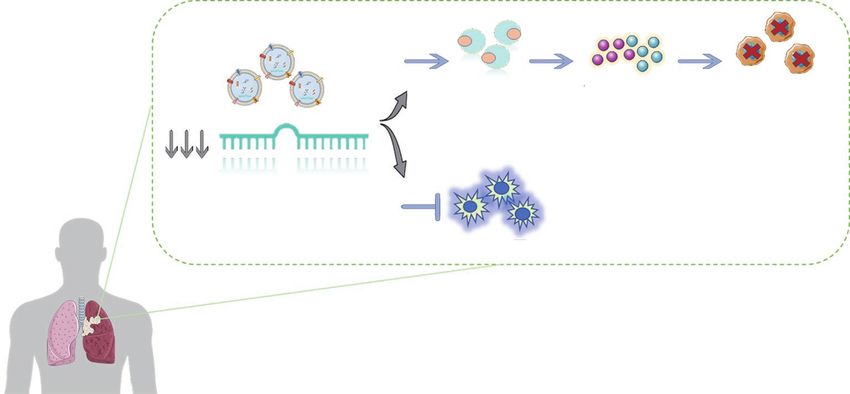
Tumor-derived exosomes
T-cell activation IFN-y and TNF-a Tumor cell death
secretion
MiR-125b-5p
T cell apoptosis
Figure 1 Diagram of the potential regulation process of miR-125-5p on T-cells.
Table 2 Main studies focused on exosomal miRNAs used as prognostic and predictive biomarkers in NSCLC
Exosomal miRNAs Source Clinical value Reference
miR-378 Serum Biomarker associated with poor survival in NSCLC (52)
miRNA-214 Serum Biomarker associated with advance disease and poor survival indicator in NSCLC (53)
patients
miR-23b-3p, miR-10b-5p and Plasma Independent prognostic biomarker of NSCLC (54)
miR-21-5p
miR-21 and miR-4257 Plasma Prognostic biomarkers in NSCLC (56)
miR-146a-5p Serum Predictive biomarker of the efficacy of Cisplatin in NSCLC and drug resistance (57)
let-7a-5p Serum Biomarker associated with poor survival in lung adenocarcinoma (58)
miR-29a-3p and miR-150-5p Plasma Biomarkers used to predict response or toxicity in radiotherapy treatment in NSCLC (59)
miR-208a Serum Predictive biomarker of radiosensitivity in lung cancer (60)
miR-425-3p Serum Predictive biomarker of the clinical response to platinum-based-chemotherapy in (61)
patients with NSCLC
miR-32 Plasma Predictive biomarker associated with the efficacy of platinum-based-chemotherapy (62)
and prognosis in NSCLC
miR-222-3p Serum Potential prognostic biomarker of Gemcitabine sensitivity in NSCLC (63)
miR-221-3p and miR- 222-3p Plasma Predictive biomarkers associated with the response to Osimertinib in EGFR-mutated (64)
NSCLC
miR-320d, miR-320c, Plasma Predictive biomarker of the efficacy of immunotherapy in advance NSCLC (65)
miR-320b and miR-125b-5p
et al. reported that exosomal transfer of miR-21 from higher expression of miR-214 in Gefitinib-resistant
Gefitinib-resistant H827R cells to Gefitinib-sensitive PC9GR cells and their derived exosomes than in Gefitinib-
HCC827 cells activated AKT signalling, leading to sensitive PC9 cells and exosomes. The authors reported
Gefitinib resistance (71). that sensitive cells acquired resistance to Gefitinib when
Same year, another group also revealed a significant exosomal miR-214 was transferred from PC9GR cells
© Translational Cancer Research. All rights reserved. Transl Cancer Res 2021;10(6):3128-3139 | https://dx.doi.org/10.21037/tcr-20-2815Translational Cancer Research, Vol 10, No 6 June 2021 3135
to PC9 cells. Nevertheless, when PC9GR cells were Conclusions
transfected with a miR-214 antagomir, cells became
The discovery of relevant functions of exosomes has
sensitive to Gefitinib (72).
been a milestone in the searching of biomarkers in liquid
Accordingly, we can conclude that exosomal miRNAs
biopsy. MicroRNAs, abundant elements in the exosome
can provide information on the molecular characteristics of
cargo, participate in the proliferation, metastasis, EMT,
the cells by which they were secreted and be endowed with
angiogenesis and immunomodulation processes in lung
the capacity to reprogram distant target cells. Moreover,
cancer. Furthermore, current researches have demonstrated
exosomal miRNAs can regulate tumor resistance and
the relevant use of exosomal miRNAs in the study of
represent a potential tool for monitoring the response/
lung cancer, providing a significant advance in the clinical
relapse of anti-neoplastic therapies.
management. Due to their great potential, exosomal
miRNAs can be used as an excellent non-invasive tool for
Exosomal miRNAs as immune-oncology early diagnosis, prognosis and prediction of treatment
biomarkers in NSCLC microenvironment success or drug resistances in this pathology.
However, there are still challenges to be achieved. Firstly,
The delivering of exosomal miRNAs by tumor and stromal
because of the heterogeneity in size (different subtypes of
cells in the microenvironment has been confirmed to be
vesicles), standardized methodologies must be established
associated with tumor initiation, relapse, progression and
for isolation, characterization and study of exosomes
metastasis. Exosomes can promote tumor progression by
cargo. Secondly, a complete comprehension of exosomal
transferring immunosuppressive factors such as exosomal
miRNA-mediated immunomodulation or drug resistance
miRNAs, which can modulate the function of immune cells
mechanisms is needed. Therefore, further studies are
such as T-lymphocytes, dendritic cells (DCs) and natural
required to determine specific components and mechanisms
killer (NK) cells (73).
of the exosomal miRNA to understand their role in cancer
Some exosomal miRNAs, such as miR-183, are TGF-
pathogenesis.
β-inducible and can silence tumor-associated NK cells by
In conclusion, exosomal miRNAs have a considerable
targeting and suppressing other activating proteins (74).
potential for the optimal management of NSCLC patients.
Furthermore, the susceptibility to the lysis by cytotoxic
Regardless multiple and recent technical and scientific
T cells is regulated by hypoxia inducible miR-210. Hypoxic
developments in the field of liquid biopsy, the ultimate goal
tumor-derived exosomes can negatively regulate NK cell
is to implement a useful and standardized methodology with
function by a mechanism which involves TGF-β and
high sensitivity/specificity for the isolation and analysis of
miR-23a (75).
exosomes and their content from minimally invasive samples,
Another example of this immunomodulation in lung
which might allow improvements in the prognosis, prediction
cancer cells is exosomal mir-21/29a, that is able to initiate
and monitoring of the disease in the clinical practice.
tumor growth and metastasis through activation of TLR7
and TLR8 on immune cells (30). Yin et al. also confirmed
communication between lung cancer cells and CD4+ Acknowledgments
lymphocytes through exosomal mir-214, which efficiently
Funding: This work is supported by Instituto de Salud
decreased PTEN expression, promoting regulatory T cell
Carlos III (ISCIII), Fondo de Investigación Sanitaria:
(Treg) expansion and tumor growth (76).
CB16/12/00350 and PI18/00226. E Duréndez-Sáez has a
Additionally, lung cancer cell invasion has been described
predoctoral fellowship by Asociación Española Contra el
to be stimulated by exosomal transfer of miR-223 from
Cáncer, Valencia (AECC Valencia). S. Torres-Martínez is
platelets, through suppression of erythrocyte membrane
supported by the Generalitat Valenciana and Fondo Social
protein band 4.1 like 3 (EPB41L3) (77). Finally, exosomal
Europeo, fellowship ACIF/2018/275.
miR-193a-3p, miR-210-3p and miR-5100 from bone
marrow-derived mesenchymal stem cells (BMSCs) are also
Footnote
reported to promote lung tumor cell invasion via activation
of STAT3 signalling-induced EMT (32). Provenance and Peer Review: This article was commissioned
© Translational Cancer Research. All rights reserved. Transl Cancer Res 2021;10(6):3128-3139 | https://dx.doi.org/10.21037/tcr-20-28153136 Duréndez-Sáez et al. NSCLC exosomal miRNAs
by the Guest Editors (Alfons Navarro, Joan Josep distribution for patients with non-small cell lung cancer:
Castellano and Marina Díaz-Beyá) for the series “Clinic and A national cancer database survey. J Thorac Oncol
Therapeutic Potential of Non-coding RNAs in Cancer” 2010;5:29-33.
published in Translational Cancer Research. The article has 7. Mathai RA, Vidya R, Reddy B, et al. Potential Utility of
undergone external peer review. Liquid Biopsy as a Diagnostic and Prognostic Tool for the
Assessment of Solid Tumors: Implications in the Precision
Conflicts of Interest: All authors have completed the Oncology. J Clin Med 2019;8:373.
ICMJE uniform disclosure forms (available at https:// 8. Calabuig-Fariñas S, Jantus-Lewintre E, Herreros-Pomares
dx.doi.org/10.21037/tcr-20-2815). The series “Clinic and A, et al. Circulating tumor cells versus circulating tumor
Therapeutic Potential of Non-coding RNAs in Cancer” was DNA in lung cancer-which one will win? Transl Lung
commissioned by the editorial office without any funding or Cancer Res 2016;5:466-82.
sponsorship. The authors have no other conflicts of interest 9. Rolfo C, Russo A. Liquid biopsy for early stage lung cancer
to declare. moves ever closer. Nat Rev Clin Oncol 2020;17:523-4.
10. Rolfo C, Cardona AF, Cristofanilli M, et al. Corrigendum
Ethical Statement: The authors are accountable for all to "Challenges and opportunities of cfDNA analysis
aspects of the work in ensuring that questions related implementation in clinical practice: Perspective of the
to the accuracy or integrity of any part of the work are International Society of Liquid Biopsy (ISLB)" [Crit.
appropriately investigated and resolved. Rev. Oncol. Hematol. 151 (July) (2020) 102978]. Crit Rev
Oncol Hematol 2020;154:103058.
Open Access Statement: This is an Open Access article 11. Molina-Vila MA, Mayo-de-Las-Casas C, Giménez-
distributed in accordance with the Creative Commons Capitán A, et al. Liquid Biopsy in Non-Small Cell Lung
Attribution-NonCommercial-NoDerivs 4.0 International Cancer. Front Med (Lausanne) 2016;3:69.
License (CC BY-NC-ND 4.0), which permits the non- 12. Kalluri R, LeBleu VS. The biology, function,
commercial replication and distribution of the article with and biomedical applications of exosomes. Science
the strict proviso that no changes or edits are made and the 2020;367:eaau6977.
original work is properly cited (including links to both the 13. Pucci M, Taverna S, Reclusa P, et al. Exosomes in semen:
formal publication through the relevant DOI and the license). opportunities as a new tool in prostate cancer diagnosis.
See: https://creativecommons.org/licenses/by-nc-nd/4.0/. Transl Cancer Res 2017;6:S1331-8.
14. Muller L, Simms P, Hong CS, et al. Human tumor-
derived exosomes (TEX) regulate Treg functions via
References
cell surface signaling rather than uptake mechanisms.
1. Siegel RL, Miller KD, Jemal A. Cancer statistics, 2020. Oncoimmunology 2017;6:e1261243.
CA Cancer J Clin 2020;70:7-30. 15. Jella KK, Nasti TH, Li Z, et al. Exosomes, Their
2. Herbst RS, Morgensztern D, Boshoff C. The biology Biogenesis and Role in Inter-Cellular Communication,
and management of non-small cell lung cancer. Nature Tumor Microenvironment and Cancer Immunotherapy.
2018;553:446-54. Vaccines (Basel) 2018;6:69.
3. Travis WD. Update on small cell carcinoma and its 16. Meehan K, Vella LJ. The contribution of tumour-derived
differentiation from squamous cell carcinoma and other exosomes to the hallmarks of cancer. Crit Rev Clin Lab Sci
non-small cell carcinomas. Mod Pathol 2012;25:S18-30. 2016;53:121-31.
4. Torres S, González Á, Cunquero Tomas AJ, et al. A 17. Rana S, Malinowska K, Zöller M. Exosomal tumor
profile on cobas® EGFR Mutation Test v2 as companion microRNA modulates premetastatic organ cells. Neoplasia
diagnostic for first-line treatment of patients with non- 2013;15:281-95.
small cell lung cancer. Expert Rev Mol Diagn 2020; 18. Giallombardo M, Taverna S, Alessandro R, et al. Exosome-
20:575-82. mediated drug resistance in cancer: the near future is here.
5. Howlader N, Forjaz G, Mooradian MJ, et al. The Effect Ther Adv Med Oncol 2016;8:320-2.
of Advances in Lung-Cancer Treatment on Population 19. Pan Z, Tian Y, Niu G, et al. Role of microRNAs in
Mortality. N Engl J Med 2020;383:640-9. remodeling the tumor microenvironment (Review). Int J
6. Morgensztern D, Ng SH, Gao F, et al. Trends in stage Oncol 2020;56:407-16.
© Translational Cancer Research. All rights reserved. Transl Cancer Res 2021;10(6):3128-3139 | https://dx.doi.org/10.21037/tcr-20-2815Translational Cancer Research, Vol 10, No 6 June 2021 3137
20. Ha M, Kim VN. Regulation of microRNA biogenesis. Nat and Early Detection for NSCLC: Advances in Thoracic
Rev Mol Cell Biol 2014;15:509-24. Oncology 2018. J Thorac Oncol 2019;14:1513-27.
21. Broughton JP, Lovci MT, Huang JL, et al. Pairing beyond 36. Tamkovich SN, Tutanov OS, Laktionov PP. Exosomes:
the Seed Supports MicroRNA Targeting Specificity. Mol Generation, structure, transport, biological activity, and
Cell 2016;64:320-33. diagnostic application. Biochem (Mosc) Suppl Ser A
22. Gallach S, Calabuig-Fariñas S, Jantus-Lewintre E, et al. Membr Cell Biol 2016;10:163-73.
MicroRNAs: promising new antiangiogenic targets in 37. Yanaihara N, Caplen N, Bowman E, et al. Unique
cancer. Biomed Res Int 2014;2014:878450. microRNA molecular profiles in lung cancer diagnosis and
23. Chen X, Ba Y, Ma L, et al. Characterization of microRNAs prognosis. Cancer Cell 2006;9:189-98.
in serum: a novel class of biomarkers for diagnosis of 38. Rabinowits G, Gerçel-Taylor C, Day JM, et al. Exosomal
cancer and other diseases. Cell Res 2008;18:997-1006. microRNA: a diagnostic marker for lung cancer. Clin
24. Arroyo JD, Chevillet JR, Kroh EM, et al. Argonaute2 Lung Cancer 2009;10:42-6.
complexes carry a population of circulating microRNAs 39. Cazzoli R, Buttitta F, Di Nicola M, et al. MicroRNAs
independent of vesicles in human plasma. Proc Natl Acad derived from circulating exosomes as noninvasive
Sci U S A 2011;108:5003-8. biomarkers for screening and diagnosing lung cancer. J
25. Thind A, Wilson C. Exosomal miRNAs as cancer Thorac Oncol 2013;8:1156-62.
biomarkers and therapeutic targets. J Extracell Vesicles 40. Zhou X, Wen W, Shan X, et al. A six-microRNA panel
2016;5:31292. in plasma was identified as a potential biomarker for lung
26. Pucci M, Reclusa Asiáin P, Duréndez Sáez E, et al. adenocarcinoma diagnosis. Oncotarget 2017;8:6513-25.
Extracellular Vesicles As miRNA Nano-Shuttles: Dual 41. Poroyko V, Mirzapoiazova T, Nam A, et al. Exosomal
Role in Tumor Progression. Target Oncol 2018;13:175-87. miRNAs species in the blood of small cell and nonsmall
27. Liu Y, Luo F, Wang B, et al. STAT3-regulated exosomal cell lung cancer patients. Oncotarget 2018;9:19793-806.
miR-21 promotes angiogenesis and is involved in 42. Grimolizzi F, Monaco F, Leoni F, et al. Exosomal miR-126
neoplastic processes of transformed human bronchial as a circulating biomarker in non-small-cell lung cancer
epithelial cells. Cancer Lett 2016;370:125-35. regulating cancer progression. Sci Rep 2017;7:15277.
28. Hsu YL, Hung JY, Chang WA, et al. Hypoxic lung cancer- 43. Lai X, Friedman A. Exosomal miRs in lung cancer: A
secreted exosomal miR-23a increased angiogenesis and mathematical model. PLoS One 2016;11:e0167706.
vascular permeability by targeting prolyl hydroxylase and 44. Jin X, Chen Y, Chen H, et al. Evaluation of tumor-derived
tight junction protein ZO-1. Oncogene 2017;36:4929-42. exosomal miRNA as potential diagnostic biomarkers
29. Aiello NM, Kang Y. Context-dependent EMT programs for early-stage non-small cell lung cancer using next-
in cancer metastasis. J Exp Med 2019;216:1016-26. generation sequencing. Clin Cancer Res 2017;23:5311-9.
30. Fabbri M, Paone A, Calore F, et al. MicroRNAs bind to 45. Aushev VN, Zborovskaya IB, Laktionov KK, et al.
Toll-like receptors to induce prometastatic inflammatory Comparisons of microRNA Patterns in Plasma before and
response. Proc Natl Acad Sci U S A 2012;109:E2110-6. after Tumor Removal Reveal New Biomarkers of Lung
31. Valencia K, Luis-Ravelo D, Bovy N, et al. miRNA cargo Squamous Cell Carcinoma. PLoS One 2013;8:e78649.
within exosome-like vesicle transfer influences metastatic 46. Rodríguez M, Silva J, López-Alfonso A, et al. Different
bone colonization. Mol Oncol 2014;8:689-703. exosome cargo from plasma/bronchoalveolar lavage in
32. Zhang X, Sai B, Wang F, et al. Hypoxic BMSC-derived non-small-cell lung cancer. Genes Chromosomes Cancer
exosomal miRNAs promote metastasis of lung cancer cells 2014;53:713-24.
via STAT3-induced EMT. Mol Cancer 2019;18:40. 47. Wang Y, Xu YM, Zou YQ, et al. Identification of
33. Ono K, Hiraoka T, Ono A, et al. Low-dose CT scan differential expressed PE exosomal miRNA in lung
screening for lung cancer: comparison of images and adenocarcinoma, tuberculosis, and other benign lesions.
radiation doses between low-dose CT and follow-up Medicine (Baltimore) 2017;96:e8361.
standard diagnostic CT. Springerplus 2013;2:393. 48. Lin J, Wang Y, Zou YQ, et al. Differential miRNA
34. Varela G, Thomas PA. Surgical management of advanced expression in pleural effusions derived from extracellular
non-small cell lung cancer. J Thorac Dis 2014;6 Suppl vesicles of patients with lung cancer, pulmonary
2:S217-23. tuberculosis, or pneumonia. Tumour Biol 2016;
35. Balata H, Fong KM, Hendriks LE, et al. Prevention 37:15835-45.
© Translational Cancer Research. All rights reserved. Transl Cancer Res 2021;10(6):3128-3139 | https://dx.doi.org/10.21037/tcr-20-28153138 Duréndez-Sáez et al. NSCLC exosomal miRNAs
49. Hydbring P, De Petris L, Zhang Y, et al. Exosomal RNA- small cell lung cancer patients receiving platinum-based
profiling of pleural effusions identifies adenocarcinoma chemotherapy can predict the effectiveness and prognosis
patients through elevated miR-200 and LCN2 expression. of chemotherapy. Medicine (Baltimore) 2019;98:e17335.
Lung Cancer 2018;124:45-52. 63. Wei F, Ma C, Zhou T, et al. Exosomes derived from
50. Tamiya H, Mitani A, Saito A, et al. Exosomal MicroRNA gemcitabine-resistant cells transfer malignant phenotypic
expression profiling in patients with lung adenocarcinoma- traits via delivery of miRNA-222-3p. Mol Cancer
associated malignant pleural effusion. Anticancer Res 2017;16:132.
2018;38:6707-14. 64. Giallombardo M, Chacartegui JJ, Reclusa P, et al. Follow
51. Woodard GA, Jones KD, Jablons DM. Lung Cancer up analysis by exosomal miRNAs in EGFR mutated
Staging and Prognosis. Cancer Treat Res 2016;170:47-75. non-small cell lung cancer (NSCLC) patients during
52. Zhang Y, Xu H. Serum exosomal miR-378 upregulation osimertinib (AZD9291) treatment: A potential prognostic
is associated with poor prognosis in non–small-cell lung biomarker tool. J Clin Oncol 2016;34:suppl.e23035.
cancer patients. J Clin Lab Anal 2020;34:e23237. 65. Peng XX, Yu R, Wu X, et al. Correlation of plasma
53. Xiong H, Lu M, Wu F, et al. Exosomal microRNA-214 exosomal microRNAs with the efficacy of immunotherapy
expression and its prognostic significance in non-small cell in EGFR/ALK wild-type advanced non-small cell lung
lung cancer patients. J King Saud Univ Sci 2020; cancer. J Immunother Cancer 2020;8:e000376.
32:1060-4. 66. Lin JJ, Shaw AT. Recent Advances in Targeting ROS1 in
54. Liu Q, Yu Z, Yuan S, et al. Circulating exosomal Lung Cancer. J Thorac Oncol 2017;12:1611-25.
microRNAs as prognostic biomarkers for non-small-cell 67. Pottier C, Fresnais M, Gilon M, et al. Tyrosine Kinase
lung cancer. Oncotarget 2017;8:13048-58. Inhibitors in Cancer: Breakthrough and Challenges of
55. Li L, Feng T, Zhang W, et al. MicroRNA Biomarker hsa- Targeted Therapy. Cancers (Basel) 2020;12:731.
miR-195-5p for Detecting the Risk of Lung Cancer. Int J 68. Bai R, Chen N, Li L, et al. Mechanisms of Cancer
Genomics 2020;2020:7415909. Resistance to Immunotherapy. Front Oncol 2020;10:1290.
56. Dejima H, Iinuma H, Kanaoka R, et al. Exosomal 69. Wu H, Zhou J, Mei S, et al. Circulating exosomal
microRNA in plasma as a non-invasive biomarker for microRNA-96 promotes cell proliferation, migration
the recurrence of non-small cell lung cancer. Oncol Lett and drug resistance by targeting LMO7. J Cell Mol Med
2017;13:1256-63. 2017;21:1228-36.
57. Yuwen DL, Sheng BB, Liu J, et al. MiR-146a-5p level in 70. Adi Harel S, Bossel Ben-Moshe N, Aylon Y, et al.
serum exosomes predicts therapeutic effect of cisplatin in Reactivation of epigenetically silenced miR-512 and miR-
non-small cell lung cancer. Eur Rev Med Pharmacol Sci 373 sensitizes lung cancer cells to cisplatin and restricts
2017;21:2650-8. tumor growth. Cell Death Differ 2015;22:1328-40.
58. Zhang L, Hao C, Zhai R, et al. Downregulation of 71. Jing C, Cao H, Qin X, et al. Exosome-mediated gefitinib
exosomal let-7a-5p in dust exposed- workers contributes resistance in lung cancer HCC827 cells via delivery of
to lung cancer development. Respir Res 2018;19:235. miR-21. Oncol Lett 2018;15:9811-7.
59. Dinh TKT, Fendler W, Chałubińska-Fendler J, et al. 72. Zhang Y, Li M, Hu C. Exosomal transfer of miR-214
Circulating miR-29a and miR-150 correlate with delivered mediates gefitinib resistance in non-small cell lung cancer.
dose during thoracic radiation therapy for non-small cell Biochem Biophys Res Commun 2018;507:457-64.
lung cancer. Radiat Oncol 2016;11:61. 73. Greening DW, Gopal SK, Xu R, et al. Exosomes and their
60. Tang Y, Cui Y, Li Z, et al. Radiation-induced miR-208a roles in immune regulation and cancer. Semin Cell Dev
increases the proliferation and radioresistance by targeting Biol 2015;40:72-81.
p21 in human lung cancer cells. J Exp Clin Cancer Res 74. Donatelli SS, Zhou JM, Gilvary DL, et al. TGF-β-
2016;35:1-14. inducible microRNA-183 silences tumor-associated natural
61. Yuwen D, Ma Y, Wang D, et al. Prognostic role of killer cells. Proc Natl Acad Sci U S A 2014;111:4203-8.
circulating exosomal miR-425-3p for the response of 75. Noman MZ, Buart S, Romero P, et al. Hypoxia-inducible
NSCLC to platinum-based chemotherapy. Cancer miR-210 regulates the susceptibility of tumor cells to lysis
Epidemiol Biomarkers Prev 2019;28:163-73. by cytotoxic T cells. Cancer Res 2012;72:4629-41.
62. Xu S, Li J, Chen L, et al. Plasma miR-32 levels in non- 76. Yin Y, Cai X, Chen X, et al. Tumor-secreted miR-214
© Translational Cancer Research. All rights reserved. Transl Cancer Res 2021;10(6):3128-3139 | https://dx.doi.org/10.21037/tcr-20-2815Translational Cancer Research, Vol 10, No 6 June 2021 3139
induces regulatory T cells: A major link between immune platelet-derived microvesicles promotes lung cancer cell
evasion and tumor growth. Cell Res 2014;24:1164-80. invasion via targeting tumor suppressor EPB41L3. Mol
77. Liang H, Yan X, Pan Y, et al. MicroRNA-223 delivered by Cancer 2015;14:58.
Cite this article as: Duréndez-Sáez E, Torres-Martinez S,
Calabuig-Fariñas S, Meri-Abad M, Ferrero-Gimeno M, Camps
C. Exosomal microRNAs in non-small cell lung cancer. Transl
Cancer Res 2021;10(6):3128-3139. doi: 10.21037/tcr-20-2815
© Translational Cancer Research. All rights reserved. Transl Cancer Res 2021;10(6):3128-3139 | https://dx.doi.org/10.21037/tcr-20-2815You can also read


