ECHIDNA Protein Impacts on Male Fertility in Arabidopsis by Mediating trans-Golgi Network Secretory Trafficking during Anther and Pollen ...
←
→
Page content transcription
If your browser does not render page correctly, please read the page content below
ECHIDNA Protein Impacts on Male Fertility in
Arabidopsis by Mediating trans-Golgi Network
Secretory Trafficking during Anther and
Pollen Development1[C][W][OPEN]
Xinping Fan, Caiyun Yang, Doris Klisch, Alison Ferguson, Rishi P. Bhaellero,
Xiwu Niu, and Zoe A. Wilson*
College of Life Science, Shanxi University, Taiyuan, Shanxi 030006, China (X.F., X.N.); School of Biosciences,
University of Nottingham, Sutton Bonington Campus, Loughborough, Leicestershire LE12 5RD, United
Kingdom (X.F., C.Y., D.K., A.F., Z.A.W.); Pomology Institute, Shanxi Academy of Agricultural Science, Taigu,
Shanxi 030815, China (X.F.); and Umeå Plant Science Centre, Department of Forest Genetics and Plant
Physiology, Swedish University of Agricultural Sciences, S–901 83 Umea, Sweden (R.P.B.)

The trans-Golgi network (TGN) plays a central role in cellular secretion and has been implicated in sorting cargo destined for the
plasma membrane. Previously, the Arabidopsis (Arabidopsis thaliana) echidna (ech) mutant was shown to exhibit a dwarf phenotype
due to impaired cell expansion. However, ech also has a previously uncharacterized phenotype of reduced male fertility. This
semisterility is due to decreased anther size and reduced amounts of pollen but also to decreased pollen viability, impaired anther
opening, and pollen tube growth. An ECH translational fusion (ECHPro:ECH-YELLOW FLUORESCENT PROTEIN) revealed
developmentally regulated tissue-specific expression, with expression in the tapetum during early anther development and
microspore release and subsequent expression in the pollen, pollen tube, and stylar tissues. Pollen viability and production,
along with germination and pollen tube growth, were all impaired. The ech anther endothecium secondary wall thickening also
appeared reduced and disorganized, resulting in incomplete anther opening. This did not appear to be due to anther secondary
thickening regulatory genes but perhaps to altered secretion of wall materials through the TGN as a consequence of the absence of
the ECH protein. ECH expression is critical for a variety of aspects of male reproduction, including the production of functional
pollen grains, their effective release, germination, and tube formation. These stages of pollen development are fundamentally
influenced by TGN trafficking of hormones and wall components. Overall, this suggests that the fertility defect is multifaceted, with
the TGN trafficking playing a significant role in the process of both pollen formation and subsequent fertilization.

   Pollen production and release is a critical stage in plant                 (Scott et al., 2004; Ma, 2005; Wilson and Zhang, 2009;
development that typically involves gene expression                           Cui et al., 2012); however, there are multiple aspects of
from over half of the genome. The extent of genomic                           pollen formation that are still unclear, and many defects
involvement in pollen development is illustrated by                           result in uncharacterized effects of reduced fertility or
the high frequency of mutations that result in a failure                      complete sterility.
of male fertility; these can be a consequence of the failure of                  The ECHIDNA (ECH) gene was initially identified
pollen development or pollen release, dehiscence. Detailed                    from expression profiling of the vascular cambium in
analysis of male-sterile mutants in Arabidopsis (Arabi-                       poplar (Populus spp.) and associated with secondary
dopsis thaliana) and rice (Oryza sativa) has improved the                     xylem formation (Hertzberg et al., 2001). The Arabidopsis
basic understanding of pollen and anther development                          ech mutant was shown to have a bushy stature with
                                                                              defects in root and hypocotyl elongation, which was
                                                                              linked to defective cell expansion and elongation (Gendre
   1
     This work was supported by the Shanxi Scholarship Council of             et al., 2011). Analysis of roots in the ech mutant and
China, the Biotechnology and Biological Science Research Council,             complementation analyses in yeast showed that the
and Bioimprove, Bio4ENERGY, and Berzili.                                      ECH protein impacts on cell expansion by mediating
   * Address correspondence to zoe.wilson@nottingham.ac.uk.                   trans-Golgi network (TGN) secretory trafficking but does
   The author responsible for distribution of materials integral to the
                                                                              not affect endocytosis (Gendre et al., 2011). However, in
findings presented in this article in accordance with the policy de-
scribed in the Instructions for Authors (www.plantphysiol.org) is:
                                                                              addition to the defects associated with plant stature, the
Zoe A. Wilson (zoe.wilson@nottingham.ac.uk).                                  ech mutant also displays a previously unreported phe-
   [C]
       Some figures in this article are displayed in color online but in       notype of reduced fertility.
black and white in the print edition.                                            Pollen development occurs in a specialized organ, the
   [W]
       The online version of this article contains Web-only data.             stamen, which comprises anthers that hold the developing
   [OPEN]
          Articles can be viewed online without a subscription.               pollen supported by a filament containing the vasculature
   www.plantphysiol.org/cgi/doi/10.1104/pp.113.227769                         connections. Stamen primordia arise from divisions in the
1338     Plant PhysiologyÒ, March 2014, Vol. 164, pp. 1338–1349, www.plantphysiol.org Ó 2014 American Society of Plant Biologists. All Rights Reserved.

                                    Downloaded on March 16, 2021. - Published by https://plantphysiol.org
                                  Copyright (c) 2020 American Society of Plant Biologists. All rights reserved.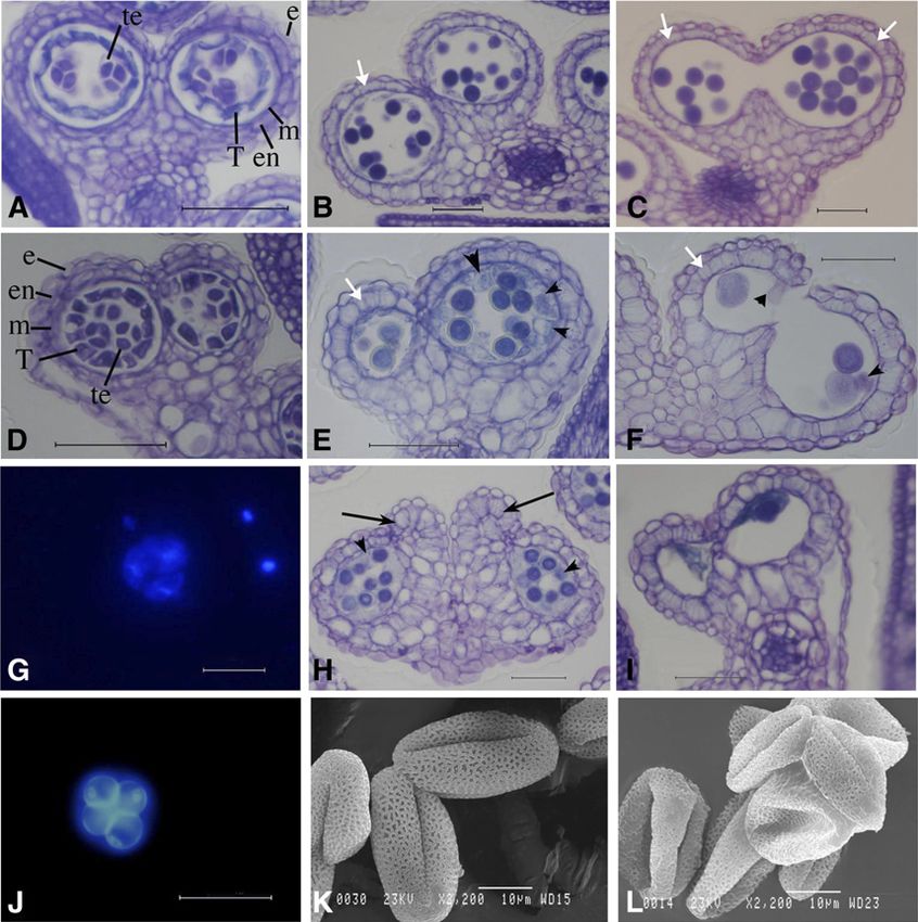
ECHIDNA-Mediated Trafficking Impacts on Plant Fertility

L1, L2, and L3 layers in the floral meristem. Divisions in                  release but was subsequently detected in the pollen,
the L2 layer result in four clusters of archesporial cells that            pollen tube, and stylar tissues. The reduced fertility
subsequently form the central sporogenous cells, which                     was linked to decreased anther size and pollen pro-
are surrounded by four maternal cell layers: the tapetum,                  duction but also to reductions in pollen viability, an-
middle cell layer, endothecium, and outer epidermis                        ther opening, and pollen tube growth. The anther
(Scott et al., 2004). The structure of the maternal anther                 wall thickening was reduced and disorganized in ech,
cell layers has been shown to be critical for the produc-                  possibly as a consequence of altered secretion of wall
tion and release of functional pollen, as demonstrated in                  materials through the TGN. The male-sterile myb26
a number of male-sterile mutants, which have defects in                    mutant has defects in anther endothecium wall thick-
cell division and early stages of differentiation of the ta-               ening resulting in a failure of dehiscence; the ech myb26
petum and sporogenous cells. For example, mutants of the                   double mutant exhibits the phenotypes of both mutants
Leu-rich repeat receptor kinase EXTRA SPOROGENOUS                          and fails to produce secondary thickening, indicating
CELLS (EXS)/EXCESS MICROSPOROCYTES1 (Canales                               that the ECH-mediated pathway is acting indepen-
et al., 2002; Zhao et al., 2002) and its ligand TAPETAL                    dently of or upstream through MYB26, possibly by
DETERMINANT1 (Jia et al., 2008) result in sterility due                    providing the components required for secondary cell
to the formation of additional male sporocytes and a lack                  wall thickening. The reduction in male fertility, there-
of tapetal cells.                                                          fore, is likely to be a consequence of multiple effects
   The tapetum has been shown to be critical for functional                due to altered secretion in the anther because of im-
pollen formation, with many of the characterized male-                     paired TGN transport in the ech mutant; the resulting
sterile mutants exhibiting abnormal tapetal development,                   defects are associated with tapetum and pollen wall
including DYSFUNCTIONAL TAPETUM1 (DYT1; Zhang                              development but also anther dehiscence and pollen
et al., 2006; Zhu et al., 2008), TAPETAL DEVELOPMENT                       tube formation.
AND FUNCTION1 (TDF1; Zhu et al., 2008), ABORTED
MICROSPORES (AMS; Sorensen et al., 2003; Xu et al.,
2010), and MALE STERILITY1 (MS1; Wilson et al.,
2001; Ito and Shinozaki, 2002). After differentiation,                     RESULTS
the tapetum layer becomes metabolically highly active                      ech Mutants Are Dwarf and Highly Branched and Show
and plays an essential role in the biosynthesis and se-                    Reduced Fertility
cretion of sporopollenin and other wall materials for the
developing pollen, prior to breakdown via programmed                          The homozygous ech mutant, as reported previously
cell death (Ariizumi and Toriyama, 2011). A frequently                     (Gendre et al., 2011), has a dwarf phenotype with a high
observed phenotype in male-sterile mutants is enlarged                     level of branching. However, the ech mutant also
tapetal cells that show defects in secretion and subse-                    exhibited significantly reduced fertility, although some
quent alterations in programmed cell death breakdown                       normal silique elongation and seed set still occurred,
(Wilson and Zhang, 2009). This indicates the important                     particularly late in development (Fig. 1). The ech mutant
role that the tapetum plays in the regulation of pollen                    is from the SAIL mutant population (Sessions et al.,
development and, in particular, the passage of materials                   2002), which was generated in the quartet (qrt) mutant
to the central locule for viable pollen production.                        background. The qrt mutation affects polygalacturase
   Male-sterile phenotypes have also been identified                        breakdown after pollen mother cell meiosis and thus
due to a failure of pollen release, dehiscence. Secondary                  enables tetrad analysis to be conducted; however, there
thickening occurs specifically in the endothecium layer of                  is no overall effect on fertility (Preuss et al., 1994). Thus,
the anther; this layer and the presence of selective                       the phenotype of incomplete tetrad separation in the ech
thickening within it are critical to generate the differ-                  qrt double mutant is likely to be due to the presence of
ential forces that are required for anther dehiscence                      the qrt mutation, while the reduction in fertility is likely
and pollen release (Wilson et al., 2011; Nelson et al.,                    to be principally associated with the lack of ECH ex-
2012). The importance of this secondary thickening is                      pression and not due to the effect of the qrt mutation.
demonstrated in the myb26 mutant (Yang et al., 2007) and                   Flowers from the ech mutant had a normal external
in the double NAC SECONDARY WALL THICKENING                                morphology with the typical organ number, except
PROMOTING FACTOR1 NAC SECONDARY WALL                                       that the sepals were slightly paler green than the wild
THICKENING PROMOTING FACTOR2 (nst1 nst2) mutant                            type. The reduction of fertility was associated with
(Mitsuda et al., 2005), which lack endothecium thickening                  male reproduction, since emasculated ech flowers had
and, as a result, fail to dehisce (Nelson et al., 2012).                   full seed set when pollinated with wild-type pollen,
   Previous investigations of the ech mutation indicated                   indicating that female fertility was not significantly
that it is impaired in TGN secretion, resulting in dwarf                   affected. Early stamen filament development appeared
plants with defects in root and hypocotyl cell elongation.                 to be generally normal; however, although filament ex-
The ech mutant also has an uncharacterized phenotype                       tension occurred, this was reduced compared with the
of impaired male fertility; therefore, a detailed analysis                 wild type; thus, at full maturity, the anthers did not
of reproduction in the ech mutant was conducted. ECH                       completely reach the top of the pistil (Fig. 1, D and E).
expression was seen in the anther tapetum during the                       The ech anthers also had a slightly distorted shape and
early stages of tapetal development and microspore                         were significantly smaller than the wild type. Pollen was
Plant Physiol. Vol. 164, 2014                                                                                                        1339

                                  Downloaded on March 16, 2021. - Published by https://plantphysiol.org
                                Copyright (c) 2020 American Society of Plant Biologists. All rights reserved.
Fan et al.
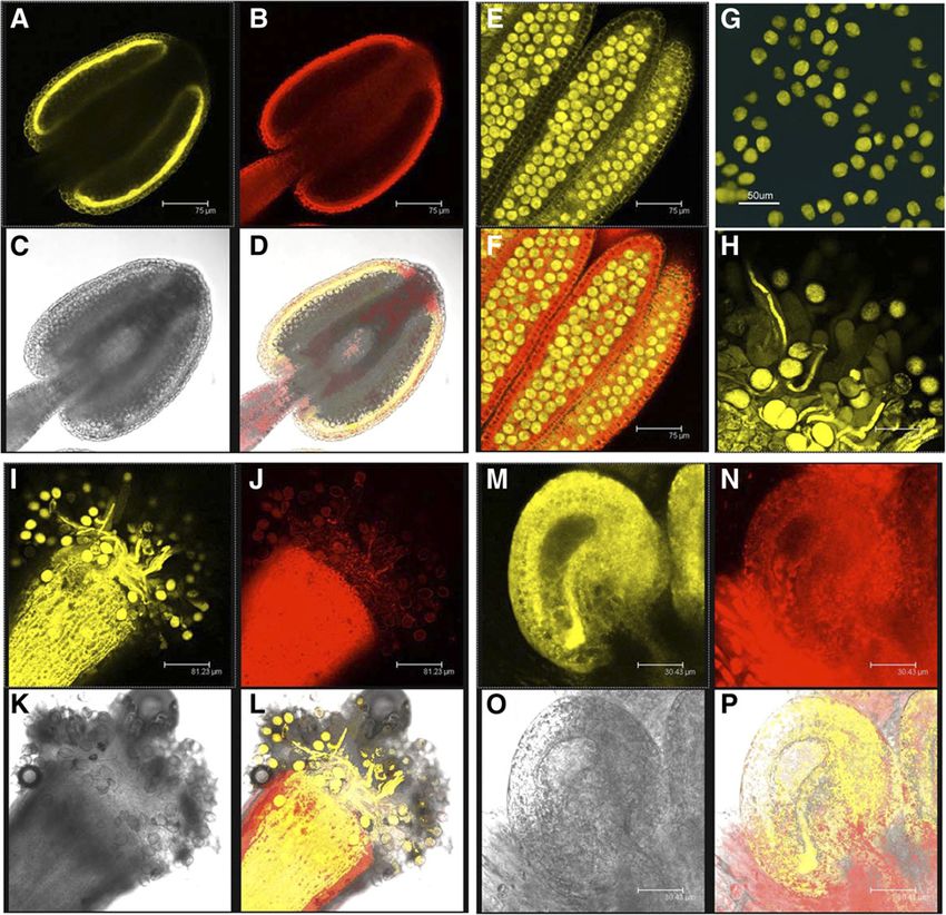
Pollen and Anther Development Is Impaired in
ech Mutants
The ech anthers were smaller and slightly malformed,
with a squat shape and individual locules that failed to
develop; however, some viable pollen was present
(Figs. 1 and 2). The early stages of ech anther and pollen
formation appeared generally normal, with the anthers
developing the correct number of cell layers and pollen
mother cell meiosis commencing as in the wild type
(Fig. 2, A and D); however, by the later stages of pollen
formation, a high proportion of pollen degeneration
had occurred (Fig. 2, E, F, H, and I) compared with the
wild type (Fig. 2, B and C). This could be seen as small,
misshapen, or broken microspores and immature pollen
in sectioned material or as inviable pollen by Alexander
staining of fresh anthers. Frequently in ech, individual
anther locules failed to develop, resulting in the anthers
having an abnormal, malformed shape (Fig. 2H;
Supplemental Fig. S1). During stages 10 and 11 of an-
ther development (Sanders et al., 2005), at the initiation
of tapetal degeneration and pollen mitosis I, the tapetum
and anther wall tissues in ech showed dense staining
with toluidine blue, with high levels of material in the
locule and tapetal tissues (Fig. 2, E and H); this may
reflect pollen wall materials that are not being secreted
correctly or are accumulating in the anther locule in the
ech mutant. In the corresponding stage in the wild type,
the tapetum was beginning to break down and much
lower amounts of materials were seen, presumably due
to their correct secretion for normal pollen development
(Fig. 2B). As pollen development proceeded, large
amounts of abnormal material could be observed in the
ech locule associated with degeneration of the tapetum
(Fig. 2, E and I), followed by subsequent degeneration
of some of the pollen. The ech anther appeared highly
constricted, with very prominent secondary thickening
in the endothecium layer (Fig. 2, E and F, white arrows),
compared with the wild type (Fig. 2, B and C). Varying
levels of severity of impaired pollen development were
observed, with some ech anthers showing limited pollen
degeneration (Fig. 2, E and F) while others failed to
develop normal locules (Fig. 2H, arrows) or had com-
pletely degenerated pollen (Fig. 2I).
Figure 1. Phenotypic analysis of floral development in the ech mutant. Nuclear 49,6-diamino-phenylindole (DAPI) staining
A, Wild-type plant showing silique development (arrows) and full indicated that both the wild type and the ech mutant
fertility. B and C, ech mutant showing increased branching, stunted
growth, and reduced fertility, as indicated by reduced elongation of
initiated meiosis and tetrad development; however, in
siliques (arrows). D, Wild-type flower showing complete dehiscence ech, abnormal nuclei were seen in the tetrads, with dif-
and pollen release. E, ech mutant flower showing reduced filament fuse staining of nuclear material suggesting nuclear
elongation and partial dehiscence (arrow) with associated reduced degeneration and in severe cases collapse of microspores
pollen release. F, Mature wild-type anther with abundant viable (Fig. 2, G and J). The ech mutant was generated in the
pollen, some of which has already been released from the anther. qrt1-1 mutant background, in which the microspores fail
G, Mature ech anther, which is stunted and reduced in size, containing to separate and the products of single meiotic events are
pollen that has reduced viability and limited release. Bars = 50 mm. kept together throughout pollen development (Preuss
et al., 1994). This allows the nature of the fertility defect,
whether gametophytic and/or sporophytic, to be de-
visible in the ech anthers; however, anther dehiscence termined (Johnson-Brousseau and McCormick, 2004).
was also impaired, with some anthers failing to dehisce Pollen was examined from anthers of open wild-type
while others only partially opened, with the pollen (qrt) and ech flowers. Viability staining (Alexander, 1969)
remaining predominantly inside (Fig. 1, D–G). revealed that almost all of the wild-type (qrt) pollen tetrads
1340 Plant Physiol. Vol. 164, 2014
Downloaded on March 16, 2021. - Published by https://plantphysiol.org
Copyright (c) 2020 American Society of Plant Biologists. All rights reserved.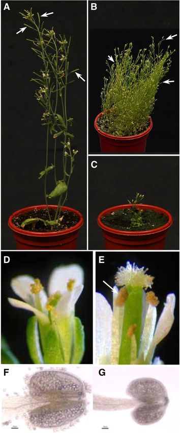
ECHIDNA-Mediated Trafficking Impacts on Plant Fertility

          Figure 2. Anther and pollen development in the ech mutant. A to C show the wild type, D and E show the mild ech mutant anther
          phenotype, and H and I show the severe ech mutant anther phenotype. A and D, Tetrad stage. B, E, and H, Tapetal degradation and
          microspore maturation. C, F, and I, Dehiscence. The early stages of pollen development, specification of cell layers and tetrad
          formation, and microspore release occurred normally in the ech mutant (D). However, post meiosis, abnormal tapetal develop-
          ment (E and H; arrowheads) and inviable pollen (F; arrowheads) and degeneration (I) were seen in ech. The secondary thickening
          that developed in the endothecium tissues (white arrows) appeared to be reduced and disorganized in ech (E, F, H, and I) compared
          with the wild type (B and C). Different levels of severity of phenotype were observed in the ech mutant; the most severe cases had
          completely malformed anther locules (H; arrows) and total pollen degeneration (I). G and J, DAPI-stained tetrads indicating the
          chromosomes and meiotic progression. J, Wild-type (qrt) pollen, showing typical incomplete separation of the tetraspores, as seen
          in the qrt background, but normal chromosome staining and meiotic progression. G, ech tetrads, showing incomplete microspore
          release associated with the qrt mutation but also occasional collapsed microspores with a lack of chromosomal staining. K, Wild-
          type (qrt) pollen with regular wall formation. L, ech pollen, showing some collapsed pollen. e, Epidermis; en, endothecium;
          m, middle cell layer; T, tapetum; te, tetrad. Bars = 50 mm (A–J) and 10 mm (K and L).

contained four viable pollen grains (84.5%; Table I).                         Alexander viability staining and by the ability of the
A lower number of pollen tetrads were observed in                             plants to self-fertilize and produce some seed. In vitro
the ech mutant, and these had a much higher fre-                              germination analysis indicated that ech pollen ger-
quency of abortion (Table I). Only 18% of the ech                             mination was much reduced compared with the wild
pollen tetrads contained four viable pollen grains,                           type, with only 1% of the ech pollen able to germinate
while 24% and 18% had one and two dead pollen                                 compared with 37% in the wild type (Table II). The
grains, respectively. Most had three inviable pollen                          length of the ech pollen tube was also reduced by
grains (34%); however, a very few (6%) showed com-                            approximately 20% compared with the wild type
pletely degenerated pollen (Table I). This frequency of                       (Table II). Despite the reduced quantity of viable ech
abortion does not equate to the typical 50% abortion                          pollen and its impaired rate of germination, a low
seen in gametophytic defects, suggesting that the ech                         level of self-fertilization was seen, with subsequent
defect is sporophytic in nature; this was further con-                        seed set indicating that some of the pollen is evidently
firmed by the 3:1 segregation of the ech phenotype and                         functional in planta. Scanning electron microscopy
as previously shown by segregation of the vegetative                          analysis showed that the surface of ech pollen was
phenotype (Gendre et al., 2011).                                              irregular compared with the wild type (qrt) and fre-
  The ech mutant produced a reduced amount of mature                          quently collapsed, although the overall exine pat-
pollen; however, some of this was viable, as indicated by                     terning appeared relatively normal (Fig. 2, K and L).
Plant Physiol. Vol. 164, 2014                                                                                                                  1341

                                  Downloaded on March 16, 2021. - Published by https://plantphysiol.org
                                Copyright (c) 2020 American Society of Plant Biologists. All rights reserved.
Fan et al.
Table I. Pollen viability in the ech mutant
Tetrad analysis is shown for pollen mother cell meiotic products in the wild type (qrt) and ech. In the qrt background, the tetraspores fail to
separate, enabling the viability of the meiotic products to be determined (Preuss et al., 1994). [See online article for color version of this table.]
Amount of Viable Pollen in Tetrads Containing Multiples of
Plant Total No.
4 3 2 1 0
% (n)
Wild type 84.5 (98) 11.2 (13) 4.3 (5) 0 (0) 0 (0) 116
ech 18.0 (9) 24.0 (12) 18.0 (9) 34.0 (17) 6.0 (3) 50
ech Anthers Have Reduced Cell Division and Expansion in the wild type (Fig. 1, D and E); this may be a conse-
with Altered Secondary Thickening quence of the smaller endothecium cells with decreased
amounts of irregular, disorganized secondary thickening
The length and width of the ech anthers were signif- that restrict anther opening.
icantly reduced, on average by 25.8% and 11.3%, re-
spectively (Fig. 3), compared with the wild type. Cell
numbers in the anther were analyzed to determine the Fertility in ech Was Not Rescued by Exogenous
nature of this reduced size; the numbers of endothe- Applications of GA, Auxin, or Jasmonic Acid
cium cells were counted from transverse sections taken In ech mutants, male fertility is reduced in part due to
prior to dehiscence of the anthers. There was a 23.2% re- pollen degeneration but also due to inefficient pollen
duction in endothecial cell number in ech anthers (Fig. 3). release and pollination, with the ech mutant filament
Although cells were counted at dehiscence, this re- significantly shorter than the wild type (Fig. 1, D and E).
duction in cell number was clearly evident in the ech Our observations prompted us to investigate whether
anther sections from the tetrad stage (stage 7) onward these defects might be due to hormonal concentration
(Fig. 2, B and E). These data suggest that the reduction effects during reproductive development in the ech
in anther size may be due to reduced levels of cell mutant. The ech mutant is known to have impaired
division as well as cell expansion. TGN trafficking, possibly affecting brassinosteroid (BR)
Lignocellulosic secondary thickening in the anther movement (Gendre et al., 2011), and has recently been
endothecium is an essential requirement for dehiscence shown to affect trafficking of the auxin influx carrier
and pollen release and, thus, is vital for male fertility. By AUX1 (Boutté et al., 2013). Previous work has shown
pollen mitosis (stage 11), both the wild type and the ech the significance of hormones during plant reproduction;
mutant showed endothecium expansion and the de- auxin has been shown to have a major effect on secondary
velopment of secondary thickening in the normal cell thickening and anther development (Nagpal et al., 2005;
layers; however, this appeared perturbed in ech (Fig. 2, Cecchetti et al., 2008, 2013), GA on male fertility (Plackett
B and E). The anther secondary thickening was ana- et al., 2011, 2012), and jasmonic acid (JA) on filament
lyzed by confocal microscopy using acridine orange/ extension (Ishiguro et al., 2001). In separate studies,
ethidium bromide stain, which differentially stain male-sterile mutants associated with hormone defi-
cellulose and lignin (Yang et al., 2007). As discussed ciencies have been overcome by hormone applications
previously, the ech endothecium had decreased num- (Wilson and Zhang, 2009). Therefore, the effects of
bers of cells compared with the wild type, with reduced
cell expansion. The ech endothecium cells showed ab-
normal secondary thickening patterning, which was
Table II. Pollen germination rates and tube growth in the ech mutant
reduced and disorganized as compared with the wild
type (Fig. 4, A–D). When analyzed under equivalent Pollen germination rates and pollen tube growth are shown for the
wild type (qrt) and ech. Columns 1 and 2 for pollen germination refer
intensity of excitation light and gain to the wild type,
to independent biological repeats
the ech mutant showed significantly less signal, suggesting
Pollen
a corresponding reduction in cell wall biosynthesis/altered Average Pollen Tube
Plant Germination
composition of the anther cell wall (Supplemental Fig. S2). Germination Length
1 2
Normal stomium and septum development was seen,
with lysis of the septum occurring apparently normally % mm
(Fig. 4, E and F). Nonetheless, dehiscence in the ech Wild type 36.9 37.2 37.0 0.22 6 0.12
mutant was frequently incomplete and less effective than ech 0.53 2.0 1.0 0.16 6 0.09
1342 Plant Physiol. Vol. 164, 2014
Downloaded on March 16, 2021. - Published by https://plantphysiol.org
Copyright (c) 2020 American Society of Plant Biologists. All rights reserved.ECHIDNA-Mediated Trafficking Impacts on Plant Fertility
Figure 3. Comparison of anther size in the ech
mutant and the wild type (wt). A, Average lon-
gitudinal and transverse diameters of wild-type
(qrt) and ech anthers. B, Average numbers of
endothecium cells in anther transverse sections
from wild-type (qrt) and ech anthers.
supplementing the ech mutant flowers with exogenous The ech Mutant Shows Reduced Expression of Genes
auxin, GA, and JA were evaluated. Plants were sprayed Associated with Anther Development
with hormones once the plants were in full flowering
and three flowers had formed; this was repeated after a RNA was isolated from closed buds from the wild
further 4 d. Male-sterile mutants typically have small, type and the ech mutant and used to determine the ex-
short siliques due to the failure of silique extension as a pression by quantitative reverse transcription (RT)-PCR
result of a lack of seed set. A quantitative assessment of of key genes previously linked to pollen development
fertility was made by measuring the length of four and anther dehiscence. These genes included those
siliques per plant, while checking also that seed for- associated with anther cell specification (SPOROCYTE-
mation was absent/reduced and corresponded to the LESS [SPL] and EXS), tapetal development (DYT1,
silique elongation, and also by assessing the total number RUPTURED POLLEN GRAIN1 [RPG1], MYB33, MYB65,
of fully developed seed-containing siliques per plant. In MYB108, and MYB99), genes involved in anther cuticle
all cases, silique length and associated seed development and pollen wall development (AMS, MS1, CYTO-
were reduced in the ech mutant as compared with the CHROME P450 [CYP703A2 and CYP704B1], DEFECTIVE
wild type, although a high level of variation was ob- IN EXINE PATTERNING1 [DEX1], MS2, and NO EXINE
served in silique length and the percentage of fertile FORMATION1 [NEF1]), and those linked to endothecium
siliques. The hormone treatments did not appear to secondary thickening and anther dehiscence (MYB26,
have any significant effect upon fertility or on the level NST1, and NST2). ECH had been previously linked to
of variation in the fertility observed (Fig. 5); seeds the TGN and the passage of materials destined for the
generated in the partially elongated siliques were fertile plasma membrane (Gendre et al., 2011) and specifically
and used to maintain the line in further generations. implicated in the sorting of cargo destined for the plasma
membrane or secretion, including the BR receptor kinase
BRASSINOSTEROID INSENSITIVE1 (BRI1; Gendre et al.,
ECH Is Differentially Expressed within the Developing 2011), and more recently the movement of pectin and
Anther, Pollen, and Pollen Tubes xyloglucan through the TGN (Gendre et al., 2013) and
the auxin influx carrier AUX1 (Boutté et al., 2013).
ECH expression was analyzed during anther and Therefore, the expression of genes associated with the BR
pollen development using the ECHPro:ECH-YELLOW pathway (TRANSIENT DEFECTIVE EXINE1 [TDE1],
FLUORESCENT PROTEIN (YFP) line that had been BRI1-EMS SUPRESSOR1 [BES1], and CELLULOSE
developed by Gendre et al. (2011); developmentally SYNTHASE-LIKE D1 [CSLD1]), including the receptor
regulated expression was seen in the anther, pollen, and kinase BRI1, was also investigated.
stigmatic tissues. ECH expression was initially detected The expression of all the genes tested that are known
in the tapetal tissue of the anther during the free mi- to be associated with the regulation of pollen devel-
crospore stage (pollen mitosis I-pollen mitosis II; Fig. 6, opment was down-regulated in ech buds (Fig. 7, A–C).
A–D). However, after tapetal degeneration, expression Expression still occurred, however, in most cases but
was only detected in the developing microspores and was reduced by about 50%, which may be a reflection
not in the maternal anther cells (Fig. 6, E and F). Ex- of the smaller anthers and impaired development of
pression was subsequently observed in the free pollen, the pollen in the ech mutant. Genes associated with the
developing pollen tubes, and stylar tissues (Fig. 6, G–L) regulation of anther secondary thickening, particularly
and also in the ovule integuments (Fig. 6, M–P). MYB26 and NST1, showed a more pronounced down-
Plant Physiol. Vol. 164, 2014 1343
Downloaded on March 16, 2021. - Published by https://plantphysiol.org
Copyright (c) 2020 American Society of Plant Biologists. All rights reserved.Fan et al.
during pollen tube growth (Wang et al., 2011). The ech
buds showed enhanced expression of CSLD1, which
may reflect the alteration of cellulose synthesis during
pollen development and pollen tube growth, possibly
due to the reorganization of wall biosynthesis as a con-
sequence of altered TGN trafficking.
ECH Affects Secondary Thickening in the Endothecium
But Is Not Directly Associated with the Regulation of
Secondary Thickening by MYB26
The ech mutant showed reduced endothecium cell
size and number, altered endothecium thickening, and
abnormal dehiscence. Since the MYB26 transcription
factor regulates anther endothecium cell expansion and
secondary thickening (Yang et al., 2007), the relation-
ship between these two genes was investigated. The
ECHPro:ECH-YFP construct was introduced into the
myb26 background to determine whether MYB26 had
any effect on ECH expression. The plants were selected
based upon their sterility and confirmed by PCR geno-
typing. Confocal microscopy indicated that ECH ex-
pression was not significantly affected by the myb26
mutation (Supplemental Fig. S3). The ECHPro:ECH-
YFP construct showed a similar pattern of expression
to that seen in the wild-type background, with ex-
pression observed in the tapetum during the free mi-
crospore stage (pollen mitosis I-pollen mitosis II), in
Figure 4. Anther endothecium cells and secondary thickening in the
ech mutant. A, C, and E, Wild type (qrt) anthers. B, D, and F, ech mutant
the pollen grains after tapetal degeneration, and then
anthers. Anthers stained with ethidium bromide/acridine orange and in the developing pollen tubes and the ovule integu-
visualized by confocal microscopy (ethidium bromide stains lignified ments (Supplemental Fig. S3).
cells [red fluorescence; 514-nm excitation; emission collection at 590 nm]
and the acridine orange stains lignified and nonlignified walls [green
fluorescence; 488-nm excitation; emission collection at 520 nm] and
merged images are shown. A and B, Confocal sections through an
anther showing thickening in the endothecium layer. C and D, Abaxial
surface of an anther. E and F, Anther opening. The ech mutant
exhibited decreased and disorganized thickening in the endothecium
(B and D) and partial anther dehiscence (F). Bars = 81.5 mm (A and C),
75 mm (B), 87.1 mm (D), and 18.8 mm (E and F).
regulation (Fig. 7C), which may be an indication of the
altered level of secondary thickening observed in the ech
mutant endothecium. This suggests that ech is generally
affecting multiple, key aspects of pollen and anther for-
mation, which are associated with tapetal and endothe-
cium function, as well as pollen wall development.
BRs have been shown to regulate a number of key
genes involved in pollen development, including SPL/
NOZZLE, TDF1, AMS, MS1, and MS2 (Ye et al., 2010),
and also to be involved in xylem differentiation
(Yamamoto et al., 2007). TDE1 has also been associated
with BR biosynthesis and pollen exine formation (Ariizumi
et al., 2008). The expression analysis of the BR-related
genes showed varied results, with BRI1 and TDE1
showing no significant changes in expression in ech
buds; however, BES1 was enhanced in ech (Fig. 7D). Figure 5. Effects of applications of GA, auxin, or JA on silique seed set in
CSLD1 has high sequence similarity to CESA proteins the ech mutant. A, Average silique length. B, Percentage of fertile siliques
and is thought to be involved in cellulose synthesis in ech after hormone treatment. IAA, Indole-3-acetic acid; wt, wild type.
1344 Plant Physiol. Vol. 164, 2014
Downloaded on March 16, 2021. - Published by https://plantphysiol.org
Copyright (c) 2020 American Society of Plant Biologists. All rights reserved.ECHIDNA-Mediated Trafficking Impacts on Plant Fertility
Figure 6. Expression of ECH in floral organs, as indicated by tissue-specific expression of an ECHPro:ECH-YFP construct.
A to D, Anther during the free microspore stage (pollen mitosis I-pollen mitosis II). E and F, Anther after tapetal degradation, prior to
dehiscence. G, Mature pollen. H, Mature pollen on stigma, forming pollen tubes. I to L, Mature stigma and stylar tissue showing
pollen germination and tube growth. M to P, Ovule. A, E, G, H, I, and M show the 497-nm excitation filter, exhibiting YFP expression.
B, J, and N show the 560-nm excitation filter, exhibiting chlorophyll autofluorescence. D, F, L, and P show superimposed YFP and
chlorophyll autofluorescence. C, K, and O show bright-field images. ECHPro:ECH-YFP expression is seen in the tapetal tissue of the
anther (A and D) during pollen mitosis I to pollen mitosis II, but no expression was seen in the free microspores. However, after tapetal
degradation during pollen mitosis II, ECHPro:ECH-YFP expression was no longer seen in the anther but was detected in the immature
pollen grains (E and F). This expression was maintained in the mature pollen grains after dehiscence (G and H), during pollination on
the stigmatic surface (H, I, and L), and in the pollen tube during subsequent growth (H, I, and L). Expression was also seen in the stylar
tissues (I and L) and in the ovule integuments (P). Bars = 75 mm (A–F), 50 mm (G), 42.4 mm (H), 81.2 mm (I–L), and 30.4 mm (M–P).
Abnormal secondary thickening was observed in the between the ECH and MYB26 pathways in their effects
ech mutant; therefore, a double mutant was generated on anther development, and in particular secondary
between myb26 and ech to determine the relationship thickening, despite the down-regulation of MYB26
between the two genes. The double mutant had the expression in the ech background (Fig. 7); however, the
phenotype of both mutants, with male sterility, re- lack of thickening in the double mutant indicates that
duced stature, and highly branched growth habit; ECH ECH acts upstream of MYB26, possibly by providing
expression appeared normal in the myb26 mutant pectin and hemicellulose molecules to the plasma
(Supplemental Fig. S3). The disorganized secondary membrane for secondary thickening in the anther.
thickening observed in ech was not apparent in the
double mutant ech myb26, which showed the complete
male sterility due to a lack of endothecial thickening DISCUSSION
and an associated failure of dehiscence, as seen in the ech Shows Reduced Male Fertility Due to Defects in Pollen
single myb26 mutant. This suggests that the effects on Formation and Release and Pollen Tube Growth
secondary thickening associated with ECH are up-
stream of the MYB26 secondary thickening pathway. The reductions in fertility observed in the ech
Therefore, it is likely that there is no direct connection mutant were principally associated with defects in
Plant Physiol. Vol. 164, 2014 1345
Downloaded on March 16, 2021. - Published by https://plantphysiol.org
Copyright (c) 2020 American Society of Plant Biologists. All rights reserved.Fan et al.
Figure 7. Quantitative RT-PCR expression analysis shows the down-regulation of key genes associated with pollen develop-
ment in the ech mutant. A, Early genes involved in establishing anther cell layers. B, Tapetum-specific genes involved in pollen
wall formation. C, Mid- to late-anther-expressed genes. D, BR and cellulase synthase genes. wt, Wild type.
male reproduction, with female fertility proceeding nor- ECH has been identified previously as a conserved
mally. A number of phenotypic changes were observed component of the TGN that is functioning in root and
that all impacted on effective male gamete production hypocotyl tissues and that is associated with the deliv-
and fertilization, including reduced pollen production, ery of cell wall components, such as xylans and pectins,
due to impaired development during and post meiosis, to the plasma membrane (Gendre et al., 2011, 2013). The
shorter stamen filaments, impaired dehiscence, and re- observed effects on anther development are consistent
duced pollen germination/tube growth. The ech anthers with the impaired delivery of cell wall components;
were significantly smaller than the wild type, due to a however, given the range of phenotypic changes and the
decrease in cell number and also reduced expansion of importance of hormones during anther development, it
these cells. The decrease in anther cell number was par- may also be that localized hormone levels are affected in
ticularly evident in the endothecium and epidermal cell the anther as a consequence of the impaired TGN traf-
layers from the tetrad stage onward. Previously, analysis ficking and that this is impacting on male reproduction.
of vegetative growth in the ech mutant indicated that the An overall reduction in microspore and pollen for-
reduction of growth was linked to decreased cell elon- mation was observed in ech; initiation of pollen meiosis
gation (Gendre et al., 2011); however, it may be that there appeared to be normal, although fewer initial tetrads
was also an associated reduction of cell number that were seen and degeneration of the meiotic products in
contributed to the overall dwarf phenotype. Anther de- the tetrads was observed, yet some functional pollen
hiscence was delayed compared with the wild type, and was seen, as indicated by self-fertilized seed set (Fig. 2).
the filament length in the ech mutant was reduced, This suggests a degree of redundancy in TGN traf-
resulting in dehiscence occurring below the pistil rather ficking in the anther and pollen, which is ensuring
than at the same level, as in the wild type. Similar effects sufficient trafficking for a threshold of materials to
had been reported previously in JA (e.g. dde1 [Sanders accumulate that enables some pollen development to
et al., 2000] and dad1 [Ishiguro et al., 2001]) and GA occur. Defects in pollen tube growth due to defective
mutants (Plackett et al., 2011). However, the defects in ech trafficking of components to the pollen tube tip have been
fertility could not be rescued by treatment with the hor- reported previously. For example, CSLD1 and CSLD4 are
mones indole-3-acetic acid, GA, and JA. In addition to the targeted to the Golgi apparatus and transported to the
smaller cell size in ech anthers, there was also frequent plasma membrane to participate in cellulose deposition at
abortion of some locules and malformed anther structure. the tip of the growing pollen tube; mutations in both
Similar aberrations have been observed in the DELLA genes cause a significant reduction in cellulose deposition
and GA mutants, which suggests that alteration of the and the organization of pollen tube wall layers (Wang
anther hormone levels may impact upon the synchrony et al., 2011). Glycosyltransferases have also been linked to
between the anther locules. pollen tube growth, with defects in the production of the
1346 Plant Physiol. Vol. 164, 2014
Downloaded on March 16, 2021. - Published by https://plantphysiol.org
Copyright (c) 2020 American Society of Plant Biologists. All rights reserved.ECHIDNA-Mediated Trafficking Impacts on Plant Fertility
Golgi-localized MALE GAMETOPHYTE DEFECTIVE4, treatment (Gendre et al., 2011). The TGN trafficking
which encodes a rhamnogalacturonan II xylosyltransferase, pathway has been associated with the transport of
resulting in impaired rhamnogalacturonan II synthesis cargo from the Golgi to the plasma membrane; these
and defective pollen tube and root growth (Liu et al., have typically been defined by the secreted GFP but
2011). The oscillatory increase in growth in lily (Lilium include BRI1 and cell wall components like xyloglucans
longiflorum) pollen tubes has also been shown to correlate and pectin. Therefore, it was speculated that hormone
with exocytosis and the increase of wall materials in the transport might also be affected in the ech mutant.
growing tube (McKenna et al., 2009). Therefore, the re- A number of hormones have been linked previously
duced rate of pollen germination and tube growth seen both to pollen production and dehiscence; auxin has
in ech is likely to be due, at least in part, to the defective been key for the production of early events in anther
TGN, possibly associated with pectin trafficking, during development and also the initiation of secondary thick-
pollen tube growth. ening processes (Cecchetti et al., 2008, 2013). BRs have
been shown to control many aspects of gene expres-
sion associated with pollen formation (Ye et al., 2010).
Pollen Wall Production Occurs in ech A number of mutants in the BR pathway have been
identified; these generally exhibit dwarfed stature and
Genes linked to pollen development showed down- frequently have fertility defects, and often a dark green
regulation in ech; this was seen in genes associated with appearance, which was not observed in ech. Most of the
the early events in anther differentiation but also those severe BR mutants show a reduction in pollen viability,
related to wall deposition, anther dehiscence, pollen partly due to a reduction in cell elongation and an as-
maturation, dehiscence, and pollen tube germination sociated short anther filament (Azpiroz et al., 1998),
(Fig. 7) and may be partly due to the impaired devel- although the BR biosynthetic mutant constitutive photo-
opment of the anther and abnormal breakdown of the morphogenesis and dwarfism had been linked previously
tapetum, resulting in a reduction of gene expression. to defects in pollen tube growth (Szekeres et al., 1996);
This is a common phenotype seen in many male-sterile however, this was not seen in a subsequent analysis (Ye
mutants, particularly those associated with biosynthesis et al., 2010). BES1, a key transcription factor associated
and secretion of the pollen wall materials (Vizcay- with BR signaling, has been shown to directly regulate
Barrena and Wilson, 2006; Zhang et al., 2006, 2008; Xu a number of genes linked to pollen formation (SPL/
et al., 2010). Nevertheless, some viable pollen was NOZZLE, TDF1, MS2, MYB103, MS1, and At3g23770;
formed, although this pollen was less effective in ger- Ye et al., 2010); however, these genes still showed some
mination and pollen tube growth. The mature ech pollen expression in the ech buds, suggesting that the defect in
exine and sporopollenin patterning appeared normal ech is reducing gene expression within the anther but
(Fig. 2, K and L); however, the pollen was frequently that this is not a complete block with sufficient devel-
collapsed and nonfunctional, indicating defective wall opment to enable partial fertility to be maintained. The
composition. ECH has been linked previously to con- BR mutants also showed distinctive enlargement and
trolling the balance between secretory trafficking and vacuolation of the tapetal cells (Ye et al., 2010); this was
vacuolar targeting (Gendre et al., 2011) and in particular not observed in ech, which suggests that although the
pectin and xyloglucan transport (Gendre et al., 2013). BR pathway may be affected, it is unlikely to be the
CSDL1 is a member of the cellulose synthase super- principal factor affecting male reproduction in ech.
family that has been shown to have a role in pollen tube Rescue of fertility was also not seen with any of the
growth (Wang et al., 2011); CSDL1 expression was sig- hormone treatments (JA, GA, and indole-3-acetic acid),
nificantly increased in the ech mutant. This altered ex- suggesting that the secretory defects are not down to
pression may reflect a compensation mechanism that is global changes in hormone levels.
acting to increase cellulose biosynthesis during pollen Endothecium thickening was reduced and disorga-
development in the absence of normal TGN-mediated nized in the ech mutant. The presence of thickening in
trafficking of pectin and other cell wall components. the endothecium is essential for effective dehiscence
These data suggest that Golgi trafficking is key to the (Dawson et al., 1999; Nelson et al., 2012). The disorga-
pollen wall synthesis and formation process; however, nized and decreased secondary thickening in ech may
the formation of some functional pollen indicates that explain why incomplete and delayed anther dehiscence
there is at least partial redundancy in the TGN traf- was seen. We have previously shown that MYB26
ficking to enable limited fertility to be maintained. regulates secondary thickening in the anther, with the
myb26 mutant failing to produce anther endothecium
thickening (Yang et al., 2007). However, the ech myb26
Altered TGN Trafficking in the ech Mutant Significantly double mutant suggests that the MYB26 pathway is
Affects Male Reproduction distinct from ECH, although the effects of TGN traf-
ficking clearly impact upon secondary thickening.
ECH has been localized to the TGN, with the ech mutant ECH-YFP expression was unaltered in the myb26 mu-
exhibiting defective secretory trafficking but normal tant, and there was no rescue of fertility in the double
endocytosis, with a similar phenotype observed by ech myb26 mutant, which showed the dwarf phenotype
blocking vacuolar H+-ATPases by concanamycin A of ech and a lack of endothecium secondary thickening.
Plant Physiol. Vol. 164, 2014 1347
Downloaded on March 16, 2021. - Published by https://plantphysiol.org
Copyright (c) 2020 American Society of Plant Biologists. All rights reserved.Fan et al.
This suggests that the regulation for secondary thickening observed using a Zeiss Axiophot microscope. For each plant line, a total of 300
pollen grains were counted. A pollen tube with a length greater than the grain
is acting via MYB26 in the anther and is not principally diameter was considered germinated.
determined by the secretory network; however, TGN
trafficking can have a significant reduction on the Confocal Microscopy of the Cell Wall
mobilization of the components required for such wall
formation. Anthers stained with ethidium bromide/acridine orange were visualized by
confocal microscopy according to Yang et al. (2007). The ethidium bromide
stains lignified cells (red fluorescence; 514-nm excitation; emission collection at
590 nm [570–620 nm]), and the acridine orange stains nonlignified walls (green
CONCLUSION fluorescence; 488-nm excitation; emission collection at 520 nm [510–530 nm]).
We have shown that ECH expression is critical for a Scanning Electron Microscopy
variety of aspects of male reproduction, which include
Wild-type and ech flowers were placed in a freeze drier (Christ Alpha 2-4
the production of functional pollen grains, their effective
LD) at 250°C and 100 millitorr for 3 h. Dehisced anthers from the wild type
release, and subsequent germination and pollen tube and manually opened anthers from the ech mutant were coated with
formation. These stages of pollen development are palladium-gold in a sputter coater (Hummer) and observed using a scanning
fundamentally influenced by TGN trafficking, and this electron microscope (ETEC) with an acceleration voltage of 10 kV.
is likely to affect both hormone and wall components
required for their function. Overall, this suggests that Quantitative RT-PCR
the fertility defects are likely to be multifaceted, with Total RNA was isolated from buds (RNeasy; Qiagen), and complementary DNA
TGN trafficking playing a significant role in the pro- was prepared using SuperScript II reverse transcriptase (Invitrogen) and an oligo
cess of pollen formation, release, and fertilization. (dT) primer (Invitrogen). Quantitative RT-PCR was performed according to Yang
et al. (2007) using gene-specific primers (Supplemental Table S1), Brilliant SYBR
Green qPCR Master Mix (Stratagene), and the LightCycler 480 Real-Time PCR Sys-
tem (Roche). Expression was normalized using the PROTEIN PHOSPHATASE2A-2
MATERIALS AND METHODS gene (Supplemental Table S1).
Plants and Growth Sequence data from this article can be found in the GenBank/EMBL data
libraries under accession numbers ECHIDNA (AT1G09330, NM_100803.3)
The ech transfer DNA insertion mutant line (SAIL_163_E09, Col0, Basta) in
and MYB26 (AT3G13890, NM_001084678.1).
the qrt mutant background and ECHPro:ECH-YFP lines were provided by
Dr. Rishi Bhalerao (Gendre et al., 2011); Arabidopsis (Arabidopsis thaliana) ecotype
Columbia wild type and the qrt mutant line were obtained from the Not-
tingham Arabidopsis Stock Centre. The myb26 mutant, myb26 3 ECHPro:YFP,
and myb26 3 ech lines were generated during this research. The ech mutant Supplemental Data
and the myb26 ech double mutant were selected by Basta spraying and then
The following materials are available in the online version of this article.
PCR confirmed using a transfer DNA insertion primer and ECH gene-specific
primers (Supplemental Table S1). For the myb26 ech double mutant, ms35 3 Supplemental Figure S1. Transverse sections through mature echidna and
ECHPro:ECH-YFP F2 seeds were screened on Murashige and Skoog medium + wild type anthers.
kanamycin (50 mg mL21), and plants were subsequently PCR genotyped for the
Supplemental Figure S2. Analysis of secondary wall thickening in wild
presence of the transgene (Supplemental Table S1).
type and echidna anthers.
Selected/screened plants were sown on Levington M3 (Scotts):vermiculite
(3:1; w/w) compost mix supplemented with 0.2 g L21 Intercept 70 WG (Scotts) Supplemental Figure S3. Expression of ECHpro:ECH-YFP in myb26 mutant
and grown under 22 h of light (150 mmol m22 s21) and 2 h of dark at 20°C 6 2°C. background.
Supplemental Table S1. Primers used in this study.
Tetraspore and Pollen Variability Staining
Anthers were taken from newly opened flowers of the ech and wild-type
(qrt) plants, and pollen viability was checked using Alexander viability stain ACKNOWLEDGMENTS
(Alexander, 1969). Young anthers at the meiosis stage were stained with DAPI
We thank Dr. Delphine Gendre for her comments on the manuscript and
(1 mg mL21) and observed using UV microscopy to analyze tetrad development.
Kim Simpson for technical support.
Received September 1, 2013; accepted January 14, 2014; published January 14,
Anther Sections 2014.
The main inflorescence was removed from wild-type (qrt) and ech plants
that showed three fertile or sterile siliques; flowers and buds were fixed with
4% (v/v) paraformaldehyde, embedded, and sectioned according to Vizcay-
LITERATURE CITED
Barrena and Wilson (2006). Sections were stained with 0.5% (w/v) toluidine Alexander MP (1969) Differential staining of aborted and nonaborted
blue in 0.1% (w/v) sodium carbonate. pollen. Stain Technol 44: 117–122
Ariizumi T, Kawanabe T, Hatakeyama K, Sato S, Kato T, Tabata S, Toriyama K
Pollen Germination Assay (2008) Ultrastructural characterization of exine development of the transient
defective exine 1 mutant suggests the existence of a factor involved in con-
Pollen germination assays were carried out in vitro on semisolid agarose structing reticulate exine architecture from sporopollenin aggregates. Plant Cell
medium [18% (w/v) Suc, 0.01% (w/v) boric acid, 1 mM CaCl2, 1 mM Ca(NO3)2, Physiol 49: 58–67
1 mM KCl, 0.01% (w/v) ferric ammonium citrate, 0.003% (w/v) casein hy- Ariizumi T, Toriyama K (2011) Genetic regulation of sporopollenin syn-
drolysate, 1 mg L21 polyvinylpyrrolidone, 1 mL L21 Heller basal microele- thesis and pollen exine development. Annu Rev Plant Biol 62: 437–460
ments solution, and 0.5% (w/v) agarose] on microscope slides. Pollen from Azpiroz R, Wu Y, LoCascio JC, Feldmann KA (1998) An Arabidopsis
freshly dehiscent anthers was placed on the agarose surface and maintained brassinosteroid-dependent mutant is blocked in cell elongation. Plant
vertically in a humid chamber for 3 h (dark; 24°C). Pollen germination was Cell 10: 219–230
1348 Plant Physiol. Vol. 164, 2014
Downloaded on March 16, 2021. - Published by https://plantphysiol.org
Copyright (c) 2020 American Society of Plant Biologists. All rights reserved.ECHIDNA-Mediated Trafficking Impacts on Plant Fertility
Boutté Y, Jonsson K, McFarlane HE, Johnson E, Gendre D, Swarup R, (2012) Analysis of the developmental roles of the Arabidopsis gibberellin
Friml J, Samuels L, Robert S, Bhalerao RP (2013) ECHIDNA-mediated 20-oxidases demonstrates that GA20ox1, -2, and -3 are the dominant
post-Golgi trafficking of auxin carriers for differential cell elongation. paralogs. Plant Cell 24: 941–960
Proc Natl Acad Sci USA 110: 16259–16264 Plackett AR, Thomas SG, Wilson ZA, Hedden P (2011) Gibberellin control
Canales C, Bhatt AM, Scott R, Dickinson H (2002) EXS, a putative LRR receptor of stamen development: a fertile field. Trends Plant Sci 16: 568–578
kinase, regulates male germline cell number and tapetal identity and pro- Preuss D, Rhee SY, Davis RW (1994) Tetrad analysis possible in Arabidopsis
motes seed development in Arabidopsis. Curr Biol 12: 1718–1727 with mutation of the QUARTET (QRT) genes. Science 264: 1458–1460
Cecchetti V, Altamura MM, Brunetti P, Petrocelli V, Falasca G, Ljung K, Sanders PM, Bui AQ, Le BH, Goldberg RB (2005) Differentiation and
Costantino P, Cardarelli M (2013) Auxin controls Arabidopsis anther degeneration of cells that play a major role in tobacco anther dehiscence.
dehiscence by regulating endothecium lignification and jasmonic acid Sex Plant Reprod 17: 219–241
biosynthesis. Plant J 74: 411–422 Sanders PM, Lee PY, Biesgen C, Boone JD, Beals TP, Weiler EW,
Cecchetti V, Altamura MM, Falasca G, Costantino P, Cardarelli M (2008) Goldberg RB (2000) The Arabidopsis DELAYED DEHISCENCE1 gene
Auxin regulates Arabidopsis anther dehiscence, pollen maturation, and encodes an enzyme in the jasmonic acid synthesis pathway. Plant Cell
filament elongation. Plant Cell 20: 1760–1774 12: 1041–1061
Cui X, Wang Q, Yin W, Xu H, Wilson ZA, Wei C, Pan S, Zhang D (2012) Scott RJ, Spielman M, Dickinson HG (2004) Stamen structure and func-
PMRD: a curated database for genes and mutants involved in plant male tion. Plant Cell (Suppl) 16: S46–S60
reproduction. BMC Plant Biol 12: 215–224 Sessions A, Burke E, Presting G, Aux G, McElver J, Patton D, Dietrich B,
Dawson J, Sözen E, Vizir I, Van Waeyenberge S, Wilson ZA, Mulligan BJ Ho P, Bacwaden J, Ko C, et al (2002) A high-throughput Arabidopsis
(1999) Characterization and genetic mapping of a mutation (ms35) reverse genetics system. Plant Cell 14: 2985–2994
which prevents anther dehiscence in Arabidopsis thaliana by affecting Sorensen AM, Kröber S, Unte US, Huijser P, Dekker K, Saedler H (2003)
secondary wall thickening in the endothecium. New Phytol 144: 213–222 The Arabidopsis ABORTED MICROSPORES (AMS) gene encodes a MYC
Gendre D, McFarlane HE, Johnson E, Mouille G, Sjödin A, Oh J, class transcription factor. Plant J 33: 413–423
Levesque-Tremblay G, Watanabe Y, Samuels L, Bhalerao RP (2013) Szekeres M, Németh K, Koncz-Kálmán Z, Mathur J, Kauschmann A,
Trans-Golgi network localized ECHIDNA/Ypt interacting protein Altmann T, Rédei GP, Nagy F, Schell J, Koncz C (1996) Brassinoste-
complex is required for the secretion of cell wall polysaccharides in roids rescue the deficiency of CYP90, a cytochrome P450, controlling cell
Arabidopsis. Plant Cell 25: 2633–2646 elongation and de-etiolation in Arabidopsis. Cell 85: 171–182
Gendre D, Oh J, Boutté Y, Best JG, Samuels L, Nilsson R, Uemura T, Vizcay-Barrena G, Wilson ZA (2006) Altered tapetal PCD and pollen wall
Marchant A, Bennett MJ, Grebe M, et al (2011) Conserved Arabidopsis development in the Arabidopsis ms1 mutant. J Exp Bot 57: 2709–2717
ECHIDNA protein mediates trans-Golgi-network trafficking and cell Wang W, Wang L, Chen C, Xiong G, Tan XY, Yang KZ, Wang ZC, Zhou Y,
elongation. Proc Natl Acad Sci USA 108: 8048–8053 Ye D, Chen LQ (2011) Arabidopsis CSLD1 and CSLD4 are required for
Hertzberg M, Aspeborg H, Schrader J, Andersson A, Erlandsson R, cellulose deposition and normal growth of pollen tubes. J Exp Bot 62:
Blomqvist K, Bhalerao R, Uhlén M, Teeri TT, Lundeberg J, et al (2001) 5161–5177
A transcriptional roadmap to wood formation. Proc Natl Acad Sci USA Wilson ZA, Morroll SM, Dawson J, Swarup R, Tighe PJ (2001) The Ara-
98: 14732–14737 bidopsis MALE STERILITY1 (MS1) gene is a transcriptional regulator of
Ishiguro S, Kawai-Oda A, Ueda J, Nishida I, Okada K (2001) The DEFECTIVE male gametogenesis, with homology to the PHD-finger family of tran-
IN ANTHER DEHISCIENCE gene encodes a novel phospholipase A1 cata- scription factors. Plant J 28: 27–39
lyzing the initial step of jasmonic acid biosynthesis, which synchronizes pollen Wilson ZA, Song J, Taylor B, Yang C (2011) The final split: the regulation
maturation, anther dehiscence, and flower opening in Arabidopsis. Plant Cell of anther dehiscence. J Exp Bot 62: 1633–1649
13: 2191–2209 Wilson ZA, Zhang DB (2009) From Arabidopsis to rice: pathways in pollen
Ito T, Shinozaki K (2002) The MALE STERILITY1 gene of Arabidopsis, encoding development. J Exp Bot 60: 1479–1492
a nuclear protein with a PHD-finger motif, is expressed in tapetal cells and is Xu J, Yang C, Yuan Z, Zhang D, Gondwe MY, Ding Z, Liang W, Zhang
required for pollen maturation. Plant Cell Physiol 43: 1285–1292 DB, Wilson ZA (2010) The ABORTED MICROSPORES regulatory net-
Jia G, Liu X, Owen HA, Zhao D (2008) Signaling of cell fate determination work is required for postmeiotic male reproductive development in
by the TPD1 small protein and EMS1 receptor kinase. Proc Natl Acad Sci Arabidopsis thaliana. Plant Cell 22: 91–107
USA 105: 2220–2225 Yamamoto R, Fujioka S, Iwamoto K, Demura T, Takatsuto S, Yoshida S,
Johnson-Brousseau SA, McCormick S (2004) A compendium of methods Fukuda H (2007) Co-regulation of brassinosteroid biosynthesis-related
useful for characterizing Arabidopsis pollen mutants and gametophytically- genes during xylem cell differentiation. Plant Cell Physiol 48: 74–83
expressed genes. Plant J 39: 761–775 Yang C, Xu Z, Song J, Conner K, Vizcay Barrena G, Wilson ZA (2007)
Liu XL, Liu L, Niu QK, Xia C, Yang KZ, Li R, Chen LQ, Zhang XQ, Zhou Arabidopsis MYB26/MALE STERILE35 regulates secondary thickening
Y, Ye D (2011) Male gametophyte defective 4 encodes a rhamnoga- in the endothecium and is essential for anther dehiscence. Plant Cell 19:
lacturonan II xylosyltransferase and is important for growth of pollen 534–548
tubes and roots in Arabidopsis. Plant J 65: 647–660 Ye Q, Zhu W, Li L, Zhang S, Yin Y, Ma H, Wang X (2010) Brassinosteroids
Ma H (2005) Molecular genetic analyses of microsporogenesis and micro- control male fertility by regulating the expression of key genes involved
gametogenesis in flowering plants. Annu Rev Plant Biol 56: 393–434 in Arabidopsis anther and pollen development. Proc Natl Acad Sci USA
McKenna ST, Kunkel JG, Bosch M, Rounds CM, Vidali L, Winship LJ, 107: 6100–6105
Hepler PK (2009) Exocytosis precedes and predicts the increase in Zhang DS, Liang WQ, Yuan Z, Li N, Shi J, Wang J, Liu YM, Yu WJ, Zhang
growth in oscillating pollen tubes. Plant Cell 21: 3026–3040 DB (2008) Tapetum degeneration retardation is critical for aliphatic
Mitsuda N, Seki M, Shinozaki K, Ohme-Takagi M (2005) The NAC metabolism and gene regulation during rice pollen development. Mol
transcription factors NST1 and NST2 of Arabidopsis regulate secondary Plant 1: 599–610
wall thickenings and are required for anther dehiscence. Plant Cell 17: Zhang W, Sun YL, Timofejeva L, Chen C, Grossniklaus U, Ma H (2006)
2993–3006 Regulation of Arabidopsis tapetum development and function by
Nagpal P, Ellis CM, Weber H, Ploense SE, Barkawi LS, Guilfoyle TJ, DYSFUNCTIONAL TAPETUM1 (DYT1) encoding a putative bHLH
Hagen G, Alonso JM, Cohen JD, Farmer EE, et al (2005) Auxin response transcription factor. Development 133: 3085–3095
factors ARF6 and ARF8 promote jasmonic acid production and flower Zhao DZ, Wang GF, Speal B, Ma H (2002) The excess microsporocytes1 gene
maturation. Development 132: 4107–4118 encodes a putative leucine-rich repeat receptor protein kinase that
Nelson MR, Band LR, Dyson RJ, Lessinnes T, Wells DM, Yang C, Everitt controls somatic and reproductive cell fates in the Arabidopsis anther.
NM, Jensen OE, Wilson ZA (2012) A biomechanical model of anther Genes Dev 16: 2021–2031
opening reveals the roles of dehydration and secondary thickening. Zhu J, Chen H, Li H, Gao JF, Jiang H, Wang C, Guan YF, Yang ZN (2008)
New Phytol 196: 1030–1037 Defective in tapetal development and function 1 is essential for anther
Plackett AR, Powers SJ, Fernandez-Garcia N, Urbanova T, Takebayashi development and tapetal function for microspore maturation in Arabi-
Y, Seo M, Jikumaru Y, Benlloch R, Nilsson O, Ruiz-Rivero O, et al dopsis. Plant J 55: 266–277
Plant Physiol. Vol. 164, 2014 1349
Downloaded on March 16, 2021. - Published by https://plantphysiol.org
Copyright (c) 2020 American Society of Plant Biologists. All rights reserved.You can also read