Lung Cancer Cell-Derived Exosomal let-7d-5p Down-Regulates OPRM1 to Promote Cancer-Induced Bone Pain
←
→
Page content transcription
If your browser does not render page correctly, please read the page content below
ORIGINAL RESEARCH
                                                                                                                                                 published: 26 May 2021
                                                                                                                                         doi: 10.3389/fcell.2021.666857

                                              Lung Cancer Cell-Derived Exosomal
                                              let-7d-5p Down-Regulates OPRM1 to
                                              Promote Cancer-Induced Bone Pain
                                              Xihan Li 1† , Yu Chen 1† , Jialun Wang 1 , Chengfei Jiang 1 and Ying Huang 2*
                                              1
                                               Department of Gastroenterology, Nanjing Drum Tower Hospital, The Affiliated Hospital of Nanjing University Medical School,
                                              Nanjing, China, 2 Department of Pain Medicine, Nanjing Drum Tower Hospital, The Affiliated Hospital of Nanjing University
                                              Medical School, Nanjing, China

                                              Cancer-induced bone pain (CIBP) is the pain caused by metastasis of malignant
                                              tumors to the bone, accounting for more than half of all chronic cancer pain, which
                                              seriously affects the quality of life among tumor patients. Up to 40% of patients
                                              with advanced lung cancer suffer from CIBP. MicroRNA (miRNA) transfers between
                                              cells through exosomes, mediates cell-to-cell communication, and performs various
                                              biological functions. Studies have shown that miRNAs secreted by cancer can modify
                              Edited by:
                                              the tumor microenvironment, but whether exosome-mediated miRNA transfer plays
                    Simone Patergnani,        a role in CIBP is still unknown. In this study, the expression levels of 15 miRNAs in
               University of Ferrara, Italy
                                              exosomes derived A549 cells and 18 miRNAs in exosomes derived NCI-H1299 cells
                        Reviewed by:
                                              were significantly up-regulated, and qRT-PCR further confirmed that the level of let-7d-
                      Anna Cieślińska,
     University of Warmia and Mazury          5p was increased most considerably. In vitro, exosomal let-7d-5p (EXO let-7d-5p) can
                    in Olsztyn, Poland        be taken up by dorsal root ganglion (DRG) neurons and inhibit the protein level of the
                      Francesca Orso,
                University of Turin, Italy
                                              target gene opioid receptor mu 1 (OPRM1). EXO let-7d-5p was further confirmed to
                   *Correspondence:
                                              be involved in the generation and maintenance of CIBP in vivo. Our findings clarify the
                          Ying Huang          molecular mechanism of CIBP caused by the inhibition of OPRM1 by EXO let-7d-5p,
               huangy0808@nju.edu.cn
                                              providing new clues and intervention targets for the prevention and treatment of CIBP.
     † These   authors have contributed
                    equally to this work      Keywords: exosome, miRNA, let-7d-5p, OPRM1, cancer-induced bone pain

                    Specialty section:
         This article was submitted to        INTRODUCTION
    Molecular and Cellular Oncology,
                a section of the journal      Lung cancer is one of the most aggressive tumors, and bone is the most common metastatic site
  Frontiers in Cell and Developmental         of lung cancer (Lee et al., 2008; Wang et al., 2010; Jemal et al., 2011). Up to 40% of patients with
                                Biology
                                              advanced lung cancer have bone metastases, which are the primary source of pain and disability
        Received: 11 February 2021            (Siegel et al., 2012). Patients with bone metastases have severe cancer-induced bone pain (CIBP),
           Accepted: 26 April 2021
                                              spinal cord compression, anemia, hypercalcemia or other nerve compression symptoms, which
           Published: 26 May 2021
                                              seriously affects the patients’ quality of life and reduces survival rate (Schneider et al., 2012; Ke et al.,
                              Citation:       2017). CIBP not only presents with persistent dull pain, but is often accompanied by unbearable
  Li X, Chen Y, Wang J, Jiang C and
                                              breakthrough pain (Zhou et al., 2015). At present, there is a lack of effective treatment for CIBP.
        Huang Y (2021) Lung Cancer
    Cell-Derived Exosomal let-7d-5p
                                              Abbreviations: CIBP, Cancer-induced bone pain; miRNA, MicroRNA; NSCLC, non-small cell lung cancer; EXO, exosome;
Down-Regulates OPRM1 to Promote               DRG, dorsal root ganglion; OPRM1, opioid receptor mu 1; TLR, Toll-like receptor; FCS, fetal calf serum; TBS, Tris-buffered
         Cancer-Induced Bone Pain.            saline; LSCM, laser scanning confocal microscope; TLDA, TaqMan low density array; SPF, specific pathogen free; IACUC,
      Front. Cell Dev. Biol. 9:666857.        Institutional Animal Care and Use Committee; PWT, paw withdrawal latency; NGF, nerve growth factor; NEDD4L, neural
     doi: 10.3389/fcell.2021.666857           precursor cell-expressed developmentally down-regulated gene 4-like.

Frontiers in Cell and Developmental Biology | www.frontiersin.org                    1                                             May 2021 | Volume 9 | Article 666857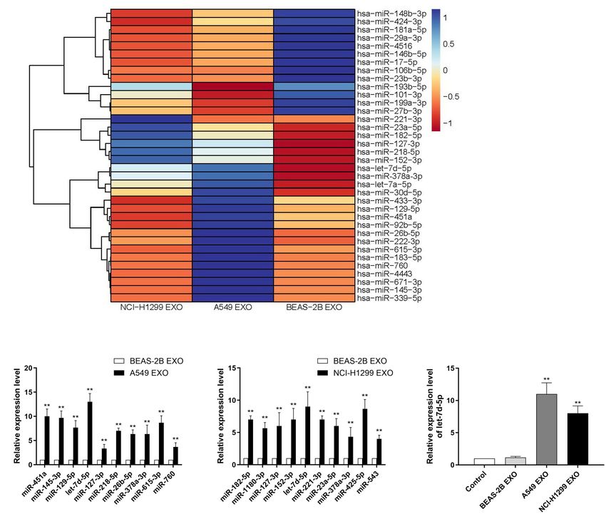
Li et al.                                                                                                                        Exosomal miRNA

Although opioids have been widely used in CIBP, which cannot               MATERIALS AND METHODS
be fully controlled, and some patients cannot tolerate the dose-
related side effects of opioids (respiratory depression, lethargy          Cell Lines
and constipation, etc.) (Christo and Mazloomdoost, 2008;                   The human non-small cell lung cancer (NSCLC) cell lines A549
Mantyh, 2013; Boland et al., 2015; Zhou et al., 2016). Therefore, it       and NCI-H1299 were obtained from ATCC (Manassas, VA,
is urgent to study the potential molecular mechanism of CIBP               United States). A549 cells were cultured in DMEM (GIBCO,
and develop new therapeutic methods with good efficacy and                 United States) supplemented with 10% fetal calf serum (FCS)
low side effects.                                                          (GIBCO, United States), 100 U/ml penicillin and 100 U/ml
   Exosomes are vesicle-like bodies with a diameter of 30–                 streptomycins (GIBCO, United States). NCI-H1299 cells were
120 nm that are actively secreted by cells (Singh et al.,                  cultured in RPMI1640 (GIBCO, United States) supplemented
2014; Hu et al., 2019). Exosomes are often cup-shaped when                 with 10% fetal calf serum (FCS) (GIBCO, United States),
viewed under an electron microscope, but they are usually                  100 U/ml penicillin and 100 U/ml streptomycin (GIBCO,
spherical in body fluids and can be released by different cells            United States). Cells were cultured at 37◦C and 5% CO2 .
such as dendritic cells, epithelial cells, mast cells, fibroblasts
and tumor cells (van Niel et al., 2006). Exosomes can be
detected in peripheral blood, urine, saliva, cerebrospinal fluid,          Exosomes Isolation
joint fluid and other parts, which play an important role in               The A549 and NCI-H1299 cells were cultured in FBS-
many physiological and pathological processes such as immune               supplemented culture media to deplete exosomes, and the cell
surveillance, inflammation and cancer development (Wahlgren                culture media were collected after 48-h cell cultures. Exosomes
et al., 2012). Tumor cells can actively secrete exosomes, which            were separated by sequential ultracentrifugation, the cell culture
can carry lipids, proteins, mRNA, microRNA and DNA to                      media were centrifuged at 300 × g for 10 min to remove cells,
participate in cell communication, cell migration, angiogenesis,           then the supernatant was centrifuged at 2,000 × g for 20 min
tumor cell growth and drug resistance (Mathivanan et al.,                  to collect exosomes. The supernatant was again centrifuged at
2010; Kharaziha et al., 2012). Exosomes are carriers of miRNAs             10,000 × g for 30 min, and the 0.22 µm filters were used
transfer between cells, which enable miRNA to avoid degradation            to remove dead cells and cell debris. The collected exosomes
in the transfer process and promote the effective uptake by                were centrifuged at 120,000 × g for 90 min to obtain pelleted
target cells (He et al., 2014). MiRNA is a non-coding small                exosomes. The exosomes were washed and resuspended with
RNA molecule regulating gene expression by targeting mRNA                  PBS, then centrifuged again at 100,000 × g for 90 min,
to inhibit its translation (Bartel, 2009). A variety of miRNAs             the pellets were resuspended in 1 ml cold PBS and stored
have been detected in exosomes from different tumor cells, so              at −80◦C.
the possible functions of miRNAs carried by exosomes in the
tumor microenvironment have also been gradually concerned                  Electron Microscopy
(Valadi et al., 2007; Taylor and Gercel-Taylor, 2008; Bartel,
                                                                           Exosomes were fixed with 2% paraformaldehyde and placed on a
2009; Mar-Aguilar et al., 2013). Studies have reported that
                                                                           Formvar-carbon-coated electron microscope grid. As a control,
changes in miRNA expression are related to the generation and
                                                                           the grids were negatively stained and embedded with 1%(w/v)
improvement of pain, which initially revealed the role of miRNA
                                                                           uranyl acetate, and incubated at 4◦C for 10 min. The excess
expression in pain research. Zhang ZJ et al. found that miR-21
                                                                           fluid was removed, the mesh was sucked dry by Whatman filter
acted on Toll-like receptors (TLRs) to regulate the maintenance
                                                                           paper, and imaged in JEM-200CX electron microscope (JEOL
of neuropathic pain (Zhang et al., 2018). Pan ZQ believed that
                                                                           Ltd., Tokyo, Japan). The Total protein contents of exosomes
miR-23a regulated neuropathic pain by directly targeting CXCR4
                                                                           were measured with the BCA protein detection kit (Abcam).
via TXNIP/NLRP3 inflammasome axis in spinal glial cells (Pan
                                                                           The size distribution of exosomes was tracked by Nanoparticle
et al., 2018). Fang BJ believed that miR-202 acted pivotal roles
                                                                           tracking analysis (NTA).
in the development of neuropathic pain partly through targeting
RAP1A gene (Fang et al., 2019).
   Our team injected exosomes from non-small cell lung cancer              Western Blotting
into nude mouse CIBP models and found that they developed                  Western blot was used to detect the presence of exosome-specific
mechanical hyperalgesia. However, we still know very little                protein markers on the isolated vesicles. Total proteins were
about how exosomes participate in tumor cells regulating bone              separated by 10%SDS polyacrylamide gel electrophoresis and
metastasis and induce CIBP (Yuan et al., 2021). In this study, we          transferred to nitrocellulose membranes (Millipore, Waltham,
revealed that EXO let-7d-5p secreted by non-small lung cancer              MA, United States). The membrane was washed with Tris-
cells could be delivered to spinal dorsal root neuron cells and            buffered saline (TBS) and incubated with 5% non-fat milk in
inhibit the expression of OPRM1, thereby participating in the              TBST (TBS, 0.1% Tween 20) to block for 2 h. The membrane
mechanism of CIBP production and maintenance. These data                   was then incubated with the primary antibody CD63 and
emphasize the role of exosomal miRNAs secreted by cancer                   CD9 overnight at 4◦ C, then incubated with the secondary
cells in regulating CIBP, help to further clarify the regulatory           antibody at room temperature for 1 h. The luminescence was
mechanism of CIBP, and provide new clues and potential                     observed using a chemiluminescence kit (Promega, Fitchburg,
intervention targets for the prevention and treatment of CIBP.             WI, United States).

Frontiers in Cell and Developmental Biology | www.frontiersin.org      2                                      May 2021 | Volume 9 | Article 666857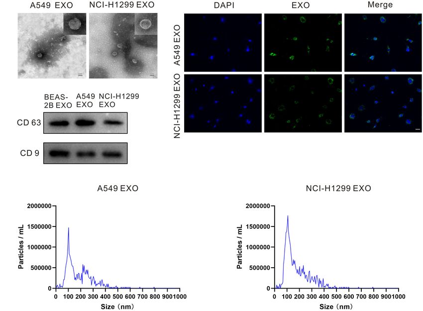
Li et al.                                                                                                                         Exosomal miRNA

Primary Culture of DRG Neurons                                            overall health of the mice was checked regularly and their weights
Take BALB/c mice, dorsal root ganglion (DRG) were collected               were measured every other day. All animal experiments were
and put in DMEM medium supplemented with 1ml pancreatin                   conducted by the animal experiment procedures approved by
and 0.1% collagenase type IV, digested at 150RPM for 40 min.              the Nanjing University Experimental Animal Ethics Committee.
The cell suspension was centrifuged at 500 RPM for 4 min, then            Animal handling follows the regulations of the Institutional
the cells were resuspended in DMEM medium containing 10%                  Animal Care and Use Committee (IACUC) of Nanjing
fetal bovine serum and cultured at 37◦ C for 24 h.                        University. The tumor cells were injected as described earlier.
                                                                          In the CIBP model, male mice were anesthetized with isoflurane
Exosome Uptake Analysis                                                   gas (2% isoflurane mixed with 100% oxygen). A distal femoral
Exosomes were fluorescently labeled with PKH67 membrane                   condyle arthrotomy was performed, and the patellar ligament
dye (Sigma-Aldrich, St. Louis, MO, United States). The Labeled            was cut with scissors to expose the distal femoral condyle. 20
exosomes were washed with 10ml PBS, collected by 100,000 × g              µL of cell suspension containing 106 A549 tumor cells was
ultracentrifugation, then resuspended in PBS. 50 µg/ml exosomes           injected into the intramedullary space of the femur. To prevent
were incubated with DRG neuron cells for 24 h at 37◦ C, then fixed        the cells from leaking out of the bone, the injection site was sealed
and stained with DAPI. The exosome uptake was observed by an              with dental amalgam.
LSCM (laser scanning confocal microscope; Zeiss LSM 710).
                                                                          Behavioral Analysis
                                                                          Pain-related behavioral tests were performed on mice before
RNA Extraction and Real-Time PCR
                                                                          and after tumor implantation. We used spontaneous flinches
Total RNAs from exosomes or cells were extracted using the
                                                                          to assess persistent pain, and paw withdrawal latency (PWT)
miRNeasy Mini Kit (Qiagen, Shanghai, China) according to the
                                                                          to assess mechanical pain. Before each experiment, the animals
manufacturer’s protocol. The RNA quality was quantified using a
                                                                          were placed in an experimental environment for 30 min to
NanoDrop 2000 spectrophotometer (Thermo Fisher Scientific).
                                                                          acclimatize. The number of spontaneous flinches was recorded
The expression profiles of miRNAs were analyzed by TaqMan
                                                                          during the 2-min observation period. Limb use was rated from
low density array (TLDA). The relative expression levels of
                                                                          0 to 4 as follows: 4, normal limb use; 3, insignificant limping;
miRNAs were evaluated by the 2−11Ct method using U6
                                                                          2, significant limping; 1, significant limping and lack of use of
snRNA as an internal reference. Quantitative RT-PCR (qRT-
                                                                          limbs; 0, complete lack of use of limbs. For PWT, the hind paws
PCR) analysis was performed using the LightCycler 96-well
                                                                          were stimulated with the von Frey single fiber. The mice were
block PCR system (Roche, Mannheim, Germany). The reverse
                                                                          placed in a transparent cage with a metal mesh, and von Frey
transcription reactions were completed using TaqMan miRNA
                                                                          single fibers were used to stimulate the hind paws in ascending
Reverse Transcription Kit and stem-loop primers for miRNAs.
                                                                          order. Each mouse was tested 5 times, the minimum stimulus
                                                                          intensity and the lowest von Frey fiber for inducing three or more
Prediction of miRNA Targets                                               positive reactions were considered to be the PWT. All the tests
With the help of bioinformatics analysis, miRNAs target                   were conducted by subjects who were unaware of the experiment.
genes prediction databases (TargetScan, miRWalk, miRDB and                The same assessment was repeated three times for all mice.
miranda) were used to predict the target genes of candidate
miRNAs. The potential downstream target genes of let-7d-5p                Intrathecal Injection
were predicted based on the intersections of four databases.              As mentioned earlier, miRNA mimics (agomir-let-7d-5p) or
                                                                          inhibitors (antagomir-let-7d-5p) were administered intrathecally
Luciferase Reporter Assay                                                 by implanting an intrathecal catheter. The PE10 catheter was
Human OPRM1 3 ’UTR fragment was amplified by PCR using                    inserted into the large pool for intrathecal implantation. 5 µl
human genomic DNA as a template. PCR products were cloned                 exosomes (1 mg/ml) or 5 µl miRNAs (20 µmol/L) were injected
to the SpeI and HindIII sites in the pMIR-reporter plasmid                into the intrathecal catheter using a microscope syringe. Starting
polyclonal region and sequenced to confirm successful insertion.          from the 15th day after tumor implantation, injections were
Firefly luciferase reporter gene system verified whether 30 -UTR          given every 24 h for 7 consecutive days. After injection, flush
of OPRM1 mRNA was targeted by let-7d-5p. Twenty-four hours                the tube with 5 µl normal saline after injections. At the end of
after transfection, cell analysis was performed using a luciferase        each experiment, laminectomy was performed to evaluate the
detection kit. All experiments were repeated three times.                 catheterization, indicating that the catheterizations were correct.

Animal Models                                                             Immunohistochemistry
BALB/c mice (male, 20–25 g, 6–8 weeks old) were purchased                 As described earlier, DRGs and spinal cord samples of mice were
from BK laboratory animals co., LTD., Shanghai, China, and                immunohistochemically analyzed. DRGs were continuously cut
raised under specific pathogen free (SPF) conditions with                 into 6 µm thick slices by a frozen microtome and mounted on
temperature control, including 12 h of the light-dark cycle. Forty-       glued slides. Specimens were placed in 3% BSA PBS solution,
two BALB/c mice were used in experiments (n = 6/group).                   and incubated at room temperature for 1 h. Anti-OPRM1 was
Mice can get food and water without restriction. The animals              diluted at 1:100 for OPRM1 antibody staining. After incubation
acclimated to the facility for a week before the study began. The         at room temperature for 3 h, slides were washed 3 times. The goat

Frontiers in Cell and Developmental Biology | www.frontiersin.org     3                                        May 2021 | Volume 9 | Article 666857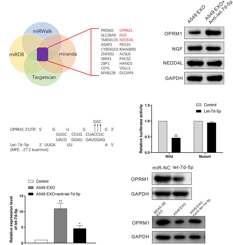
Li et al.                                                                                                                                       Exosomal miRNA

anti-rabbit IgG combined Alexa Fluor 555 were added, incubated                      the exosome-free medium. Exosomes in the culture medium
at room temperature for 2 h, and the stationary slides were                         were separated by ultracentrifugation, then exosome pellets
washed. Fluorescence images were obtained by laser scanning                         were resuspended in PBS, and the morphology of exosomes
confocal microscopy (LSCM) using a Zeiss LSM 710 fluorescence                       was examined by transmission electron microscopy (TEM).
microscope (Carl Zeiss Microscopy GmbH, Jena, Germany).                             Figure1A and Supplementary Figure 1 showed that exosomes
                                                                                    isolated from A549, NCI-H1299 and BEAS-2B cells had uniform
Statistical Analysis                                                                cup shapes and vesicle sizes of about 100 nm. Besides, western
All the analyses were performed using GraphPad Prism software.                      blot results showed that these microcapsules were positive for
The data were expressed as mean ± standard error of mean.                           exosome markers CD63 and CD9 (Figure 1B). In order to test
One-way ANOVA was used to compare the data of each group.                           the internalization of exosomes in DRG neuron cells, exosomes
P < 0.05 was considered statistically significant. All in vitro data                isolated from A549 and NCI-H1299 cells were labeled with
were analyzed from at least three separate experiments.                             PKH67 dye (green), washed thoroughly, and then added to DRG
                                                                                    neuron cells. The uptake of exosomes from the recipient cells was
                                                                                    observed under a confocal microscope. Almost all DRG neuron
RESULTS                                                                             cells showed green signals (Figure1C). These results indicated
                                                                                    that A549 and NCI-H1299 cell exosomes were internalized
Exosomes Characterization and                                                       by DRG neuron cells. NTA revealed that A549 exosomes are
Internalization in DRG Neuron Cells                                                 101.4 ± 42.4 nm in diameter and NCI-H1299 exosomes are
Exosomes can be actively released from a variety of cells including                 104.7 ± 39.1 nm in diameter (Figures 1D,E). BCA protein assay
cancer cells. We incubated NSCLC cell lines A549 and NCI-                           revealed that the concentration of proteins was 240 µg/ml in
H1299, normal human lung epithelial cell lines BEAS-2B in                           A549 exosomes and 280 µg/ml in NCI-H1299 exosomes.

  FIGURE 1 | Characterizations of exosome morphology and specificity. (A) Representative electron micrographs of exosomes isolated from A549 and NCI-H1299
  cells conditioned medium revealing the typical morphology and size. Scale bar represents 100 nm. (B) Western blot analysis showing abundant CD63 and CD9 in
  exosomes derived from the medium of A549 and NCI-H1299 cells. (C) The uptake of DRG cells after adding PKH67-labeled exosomes derived from A549 and
  NCI-H1299 cells. Images were taken 24 h after exosome addition by confocal microscope. Scale bar represents 50 µm. (D,E) Size distributions of A549 and
  NCI-H1299 exosomes were quantified using nanoparticle tracking analysis (NTA). All experiments were performed three times.

Frontiers in Cell and Developmental Biology | www.frontiersin.org               4                                            May 2021 | Volume 9 | Article 666857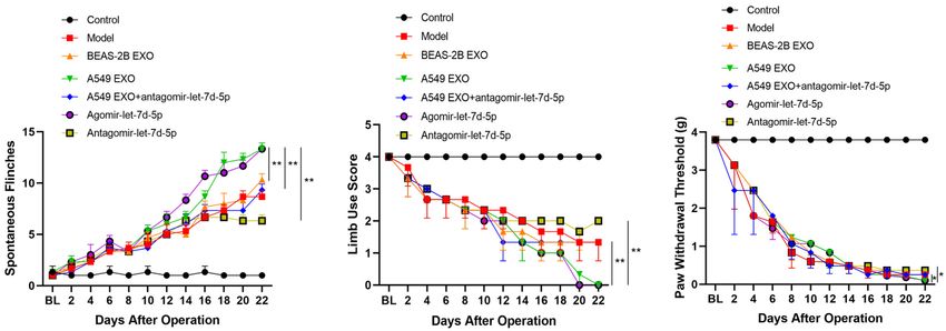
Li et al.                                                                                                                                           Exosomal miRNA

Identifying Exosomal miRNAs Markedly                                                  H1299 and BEAS-2B cell exosome miRNAs were also quite
Secreted by Lung Cancer Cell Lines                                                    different. There were 29 miRNAs with a fold difference of more
Exosomes contain multiple coding and non-coding RNAs. Total                           than 2 folds, of which 18 were up-regulated and 11 were down-
RNAs from A549, NCI-H1299 and BEAS-2B cell exosomes were                              regulated (Supplementary Table 2). We selected 10 miRNAs for
extracted and then analyzed using TLDA for exosomal miRNAs.                           qRT-PCR verification, the results also showed that the variation
We compared human NSCLC A549, NCI-H1299 and normal                                    trend was consistent with the microarray results (Figure 2C).
lung epithelial cells BEAS-2B, then quantitatively analyzed up to                     We found that the contents of let-7d-5p in A549 and NCI-
768 miRNAs (Figure 2A). Comprehensive microarray analysis                             H1299 cell exosomes were higher than other miRNAs. Compared
showed that compared with BEAS-2B cell exosomes, there were                           with the control BEAS-2B cell exosomes, let-7d-5p was were
24 miRNAs with a fold difference of more than 2 folds in A549                         up-regulated 13 times in A549 cell exosomes and 9 times in
cell exosomes, of which 15 were up-regulated and 9 were down-                         NCI-H1299 cell exosomes. We incubated DRG neuron cells with
regulated (Supplementary Table 1). We selected 10 miRNAs for                          exosomes extracted from A549, NCI-H1299 and BEAS-2B cells,
qRT-PCR verification, and the results showed that the variation                       and qRT-PCR detection also confirmed that let-7d-5p expression
trend was consistent with the microarray results (Figure 2B).                         was up-regulated in A549 and NCI-H1299 groups (Figure 2D).

  FIGURE 2 | Identifying exosomal microRNAs secreted by NSCLC lines. (A) The expression profile of miRNAs in exosomes secreted by NSCLC lines. Exosomes
  from non-cancerous cells (BEAS-2B) were used as a normalization control. Blue color denotes higher expression, and red color denotes lower expression relative to
  the control. (B) qRT–PCR analysis revealing the expression of miRNAs in A549 cell–derived exosomes. (C) qRT-PCR analysis revealing the expression of miRNAs in
  NCI-H1299 cell–derived exosomes. (D) The expression of let-7d-5p in A549 and NCI-H1299 groups was up-regulated after treated DRG neuron cells with
  exosomes extracted from A549, NCI-H1299 and BEAS-2B cells detected by qRT-PCR. **p < 0.01. All experiments were performed three times.

Frontiers in Cell and Developmental Biology | www.frontiersin.org                 5                                             May 2021 | Volume 9 | Article 666857
Li et al.                                                                                                                                              Exosomal miRNA

OPRM1 Is the Functional Target of                                                      detected the expression of OPRM1, NGF and NEDD4L in DRG
let-7d-5p in DRG Neuron Cells                                                          neuron cells by Western blot. The results showed that there was
In order to confirm the targets of let-7d-5p, we used four                             no significant difference in the expression of NGF and NEDD4L.
target prediction databases (Targetscan, miRWalk, miRDB and                            In contrast, the expression of OPRM1 was significantly up-
miranda) for computer analysis. The results showed that                                regulated (Figure 3B), suggesting that the expression of OPRM1
pain-related receptors OPRM1, NGF (nerve growth factor)                                may be regulated by let-7d-5p. Further computer analysis showed
and NEDD4L (neural precursor cell-expressed developmentally                            that the “UACCUC” site in the 3 ’UTR region of OPRM1 was
down-regulated gene 4-like) were overlapped in all databases                           complementary to the conserved sequence “GAGGUA” of let-7d-
(Figure 3A). We treated the DRG neuron cells with A549                                 5p (Figure 3C). We cloned the 3’UTR binding site of OPRM1
exosomes before infected with anti-let-7d-5p lentivirus, then                          into the luciferase gene and performed luciferase reporter gene

  FIGURE 3 | OPRM1 is the functional target of let-7d-5p in DRG neuron cells. (A) A diagram illustrating of target genes analysis. (B) Western blotting analysis of
  OPRM1, NGF and NEDD4L in DRG neuron cells with A549 exosomes plus anti-let-7d-5p lentivirus. GAPDH was used as loading control. (C) Predicted
  consequential binding sites of let-7d-5p and OPRM1 30 UTR. (D) Let-7d-5p suppressed the luciferase activity of the luciferase reporters carrying OPRM1 30 -UTR.
  Both wild-type (UTR-WT) or mutant (UTR-mut) reporters were introduced into 293T cells by transfection and then incubated with the let-7d-5p mimics. **P < 0.01.
  (E) The expression of let-7d-5p in A549 exosome plus anti-let-7d-5p lentivirus groups was down-regulated detected by qRT-PCR. *P < 0.05, **P < 0.01. (F) The
  relative expression levels of OPRM1 in DRG neuron cells infected with miR-NC and let-7d-5p lentivirus detected by Western blot. (G) The relative expression levels of
  OPRM1 in DRG neuron cells treated with BEAS-2B exosomes, A549 exosomes and A549 exosomes plus anti-let-7d-5p lentivirus detected by Western blot. All
  experiments were performed three times.

Frontiers in Cell and Developmental Biology | www.frontiersin.org                  6                                               May 2021 | Volume 9 | Article 666857
Li et al. Exosomal miRNA
analysis. The results confirmed that let-7d-5p can directly exosome and agomir-let-7d-5p groups showed lower limb use
complement the 30 UTR of the OPRM1, let-7d-5p inhibited the scores and PWT than the BEAS-2B exosome group (P < 0.05).
30 UTR luciferase activity of OPRM1, while let-7d-5p binding site Compared with the A549 exosome group, combined injection
mutations eliminated these inhibitory effects (Figure 3D). We of antagomir-let-7d-5p could improve the reduction of limb use
treated DRG neuron cells with exosomes extracted from A549, scores and PWT caused by A549 exosomes, while intrathecal
NCI-H1299 and BEAS-2B cells, and qRT-PCR confirmed that injection of antagomir-let-7d-5p alone enhanced the limb use
the expression of let-7d-5p was up-regulated in A549 and NCI- scores and PWT in CIBP mice (Figures 4B,C).
H1299 groups. We further infected DRG neuron cells with anti- We further studied the expression of OPRM1 in the DRG
let-7d-5p lentivirus, qRT-PCR confirmed that the expression of of mice. Seven days after intrathecal injection of exosomes and
let-7d-5p was down-regulated (Figure 3E). Western blot results miRNAs, western blot analysis showed that the expression of
showed that the overexpression of let-7d-5p significantly reduced OPRM1 protein in DRG in A549 exosome and agomir-let-7d-5p
the expression of OPRM1 in DRG neuron cells in let-7d-5p groups was significantly lower than that of the BEAS-2B exosome
lentivirus (Figure 3F) and A549 exosome groups, however, the and model groups (P < 0.01). In contrast, the expression of
expression of OPRM1 was not significantly inhibited in BEAS- OPRM1 protein in the A549 exosome combined antagomir-let-
2B exosome and A549 exosome plus anti-let-7d-5p lentivirus 7d-5p group did not decrease significantly. Moreover, OPRM1
groups (Figure 3G). protein was slightly increased in antagomir-let-7d-5p group
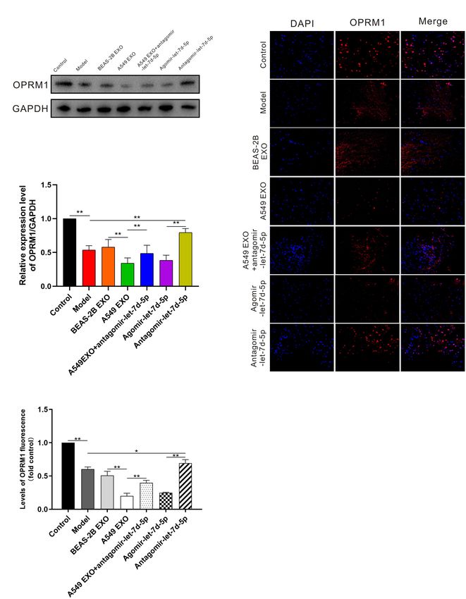
compared with that in the model group (Figures 5A,B). We
further studied the immunofluorescence staining of OPRM1
Effects of Exosomes From Lung Cancer in the DRG. In the DRG, OPRM1 immunoreactive fibers
Cell Lines on CIBP–Related Behaviors and neurons are mainly distributed in the superficial layer.
As described earlier, the pain behavior of CIBP mice was The immunoreactivity of OPRM1 in A549 exosome and
evaluated before and after femoral inoculation. On the 15th day agomir-let-7d-5p groups was significantly reduced (P < 0.01),
after femoral inoculation (Supplementary Figure 2), CIBP mice which was consistent with the quantitative results of western
were given the intrathecal injection of exosomes and miRNAs blot analysis. Compared with the A549 exosome group, the
every 24 h for 7 consecutive days. The results showed that intrathecal combined delivery of antagomir-let-7d-5 increased
intrathecal injection of A549 exosomes and agomir-let-7d-5p the expression of OPRM1 in the DRG. Besides, OPRM1 was
enhanced the pain behavior of CIBP mice, while antagomir- expressed at low levels in the DRG of CIBP mice, but antagomir-
let-7d-5p inhibited the pain enhancement caused by A549 let-7d-5p increased the expression of OPRM1 (Figures 5C,D).
exosomes. Also, intrathecal injection of antagomir-let-7d-5p
alone alleviated the pain of CIBP mice. CIBP mice in A549
exosome and agomir-let-7d-5p groups (n = 6) showed an DISCUSSION
increased number of spontaneous flinches (P < 0.01). Compared
with the A549 exosome group, combined injection of antagomir- Bone metastasis can cause severe pain, pathological fractures and
let-7d-5p inhibited the increase of spontaneous flinches caused spinal cord compression, which can seriously affect the quality of
by A549 exosomes, while intrathecal injection of antagomir-let- life of cancer patients (Siegel et al., 2012). Although some clinical
7d-5p alone reduced the spontaneous flinches of CIBP mice progress has been made, CIBP remains an important challenge
(Figure 4A). In terms of limb use scores, mice in the A549 for clinicians (Ke et al., 2017). There is an urgent need to
FIGURE 4 | Effect of lung cancer cell lines-derived exosomes on pain-related behaviors in CIBP mice. Spontaneous flinches (A), limb-use score (B) and paw
withdrawal threshold (C) were evaluated before preoperative or 2, 4, 6, 8, 10, 12, 14, 16, 18, 20, 22 days after femoral inoculation. A549 exosomes and
agomir-let-7d-5p significantly increased the number of flinches, reduced scores of limb use and paw withdrawal threshold. Data are presented as means ± SEM
(n = 6/group). *P < 0.05, **P < 0.01. All experiments were performed three times.
Frontiers in Cell and Developmental Biology | www.frontiersin.org 7 May 2021 | Volume 9 | Article 666857Li et al. Exosomal miRNA FIGURE 5 | Expression of OPRM1 in the dorsal root ganglion (DRG) of CIBP mice. (A,B) One week after intrathecal injection, western blot analysis showed that in the DRG, OPRM1 protein was significantly reduced in A549 exosome and agomir-let-7d-5p groups. (C,D) Immunofluorescent staining showed that OPRM1 immunoreactivity was significantly reduced in the DRG in A549 exosome and agomir-let-7d-5p groups. Levels of OPRM1 fluorescence in tissues were quantified using Image J software. Scale bar = 50 µm. *P < 0.05, **P < 0.01. Datas are presented as mean ± SEM (n = 6/group). All experiments were performed three times. further understand the underlying mechanism of CIBP, develop miRNAs and their functions in the occurrence and maintenance treatments based on specific mechanisms, and improve the of tumor-mediated chronic pain were analyzed in the CIBP patients’ quality of life. The mechanism of CIBP is complex, and model, to provide new clues and potential intervention targets may involve neuropathic pain and inflammatory pain (Schneider for the prevention and treatment of CIBP. et al., 2012). In this study, non-small cell lung cancer cell line In this study, A549 exosomes and NCI-H1299 exosomes exosome miRNAs were screened, and the expression of exosome labeled with PKH67 were added to DRG neuron cells for Frontiers in Cell and Developmental Biology | www.frontiersin.org 8 May 2021 | Volume 9 | Article 666857
Li et al.                                                                                                                        Exosomal miRNA

co-incubation. PKH67 fluorescence was observed in DRG                     exosomes and let-7d-5p. We demonstrated that exosomes from
neuron cells, indicating that A549 exosomes and NCI-H1299                 NSCLC and let-7d-5p were important pain-inducing factors of
exosomes could be absorbed by DRG neuron cells. We further                CIBP caused by NSCLC, which caused aggravation of pain in
injected A549 cells directly into the femoral bone marrow                 CIBP mice by inhibiting OPRM1 expression.
cavity of nude mice to construct CIBP models. On the 15th                     In conclusion, understanding the role and function of tumor-
day after inoculation, exosomes and miRNAs were injected                  derived exosomes is important for understanding CIBP. We
intrathecally for seven consecutive days, and the results showed          construct the human lung cancer CIBP model, in order to
that mechanical hyperalgesia in the A549 exosome group was                use these models to study the possible mechanism of human
significantly earlier than that in the BEAS-2B exosome group. The         lung cancer CIBP. This study provides evidence that let-7d-5p
above research results indicate that exosomes secreted by non-            transferred from exosomes derived from non-small cell lung
small cell lung cancer cells may be involved in the maintenance           cancer cells promotes the development of CIBP by targeting
of CIBP. To further understand how exosomes play a role in                OPRM1, but further studies are needed both preclinically and
the development of CIBP, we performed miRNA array analysis                clinically to develop a new potential therapy, which can alleviate
on A549 exosomes and NCI-H1299 exosomes. Compared with                    CIBP effectively, and increase the functional status and quality of
BEAS-2B exosomes, 15 up-regulated miRNAs and 9 down-                      life of CIBP patients.
regulated miRNAs were found in A549 exosomes (changes more
than 2 folds), 18 up-regulated miRNAs and 11 down-regulated
miRNAs were found in NCI-H1299 exosomes (changes more                     DATA AVAILABILITY STATEMENT
than 2 folds). In the subsequent RT-QPCR analysis, we confirmed
that the most significantly up-regulated miRNA was let-7d-                The original contributions presented in the study are included
5p. DRG neuron cells were transfected with A549 exosomes                  in the article/Supplementary Material, further inquiries can be
and NCI-H1299 exosomes, and the expression of let-7d-5p was               directed to the corresponding author/s.
also up-regulated.
   Let-7d-5p is associated with the development of breast cancer,
ovarian cancer and other tumors, but its relationship with pain           ETHICS STATEMENT
has not been reported (Hilly et al., 2016; Uhr et al., 2019; Zhang
et al., 2019). We predicted and analyzed the possible targeted            The animal study was reviewed and approved by Nanjing
genes of let-7d-5p, including OPRM1, NGF and NEDD4L. NGF,                 University Experimental Animal Ethics Committee.
a protein that induces nerve growth, was first discovered 60 years
ago by Rita Levi-Montalcini (Zeliadt, 2013). Studies have found
that NGF is also related to intractable pain (Hirose et al., 2016).       AUTHOR CONTRIBUTIONS
NEDD4L is an effective Nav s post-translational regulator. Down-
                                                                          XL and YH designed the experiment, drafted the manuscript and
regulation of NEDD4L can lead to hyperexcitability of DRG
                                                                          revised it. XL, YC, and JW carried the experiment. CJ analyzed
neurons and participate in pathological pain (Laedermann et al.,
                                                                          the data. All authors read and approved the final manuscript.
2013). We infected DRG neuron cells with antagomir-let-7d-5p
lentivirus, and western blot detected OPRM1, NGF and NEDD4L
in DRG cells. It was found that there was no significant difference
in the expression of NGF and NEDD4L, while OPRM1 was
                                                                          FUNDING
significantly up-regulated. Also, we added A549 exosomes and              This work was supported by grants from the National Natural
NCI-H1299 exosomes to DRG neuronal cells. The qRT-PCR                     Science Foundation of China (Nos. 81801100, 81972879),
results confirmed that let-7d-5p expression was up-regulated, and         the Natural Science Foundation of Jiangsu Province (No.
western blot confirmed that OPRM1 expression was significantly            BK20180130), Science and Technology Program of Nanjing
down-regulated. We further constructed a wild-type and mutant             (No. 201803037), Fundamental Research Funds for the Central
dual-luciferase reporter gene system at the binding site of let-7d-       Universities (No. 021414380449).
5p and OPRM1 3’UTR. The results also confirmed that let-7d-5p
could directly bind to the OPRM1 3’UTR complementary and
inhibit the expression of luciferase. The above results suggested         ACKNOWLEDGMENTS
that let-7d-5p in exosomes secreted by non-small cell lung cancer
cells could regulate the expression of OPRM1 in DRG cells.                We thank our colleagues in the Experimental Animal Center for
   We further studied the changes in pain after intrathecal               helpful discussions and valuable assistance.
injection of exosomes and miRNAs in CIBP mice, as well as
the changes in OPRM1 expression in the DRG. The results
showed that intrathecal injection of A549 exosomes and let-7d-            SUPPLEMENTARY MATERIAL
5p significantly enhanced the pain behavior of CIBP mice, while
antagomir-let-7d-5p inhibited the pain enhancement caused by              The Supplementary Material for this article can be found
A549 exosomes. And the expression of OPRM1 in the DRG was                 online at: https://www.frontiersin.org/articles/10.3389/fcell.2021.
consistent with the changes in hyperalgesia mediated by A549              666857/full#supplementary-material

Frontiers in Cell and Developmental Biology | www.frontiersin.org     9                                       May 2021 | Volume 9 | Article 666857
Li et al. Exosomal miRNA
REFERENCES Schneider, G., Voltz, R., and Gaertner, J. (2012). Cancer pain management and
bone metastases: an update for the clinician. Breast Care (Basel) 7, 113–120.
Bartel, D. P. (2009). MicroRNAs: target recognition and regulatory functions. Cell doi: 10.1159/000338579
136, 215–233. doi: 10.1016/j.cell.2009.01.002 Siegel, R., DeSantis, C., Virgo, K., Stein, K., Mariotto, A., Smith, T., et al. (2012).
Boland, J. W., Ziegler, L., Boland, E. G., McDermid, K., and Bennett, M. I. (2015). Cancer treatment and survivorship statistics, 2012. CA Cancer J. Clin. 62,
Is regular systemic opioid analgesia associated with shorter survival in adult 220–241.
patients with cancer? A systematic literature review. Pain 156, 2152–2163. doi: Singh, R., Pochampally, R., Watabe, K., Lu, Z., and Mo, Y. Y. (2014). Exosome-
10.1097/j.pain.0000000000000306 mediated transfer of miR-10b promotes cell invasion in breast cancer. Mol.
Christo, P. J., and Mazloomdoost, D. (2008). Interventional pain treatments for Cancer 13:256. doi: 10.1186/1476-4598-13-256
cancer pain. Ann. N. Y. Acad. Sci. 1138, 299–328. Taylor, D. D., and Gercel-Taylor, C. (2008). MicroRNA signatures of tumor-derived
Fang, B., Wei, L., Dong, K., Niu, X., Sui, X., and Zhang, H. (2019). miR-202 exosomes as diagnostic biomarkers of ovarian cancer. Gynecol. Oncol. 110,
modulates the progression of neuropathic pain through targeting RAP1A. J. Cell 13–21. doi: 10.1016/j.ygyno.2008.04.033
Biochem. 120, 2973–2982. doi: 10.1002/jcb.27025 Uhr, K., Prager-van, D. S. W., Heine, A., Ozturk, B., van Jaarsveld, M., Boersma,
He, W. A., Calore, F., Londhe, P., Canella, A., Guttridge, D. C., and Croce, A., et al. (2019). MicroRNAs as possible indicators of drug sensitivity in breast
C. M. (2014). Microvesicles containing miRNAs promote muscle cell death cancer cell lines. PLoS One 14:e216400.
in cancer cachexia via TLR7. Proc. Natl. Acad. Sci. U. S. A. 111, 4525–4529. Valadi, H., Ekstrom, K., Bossios, A., Sjostrand, M., Lee, J. J., and Lotvall, J. O. (2007).
doi: 10.1073/pnas.1402714111 Exosome-mediated transfer of mRNAs and microRNAs is a novel mechanism of
Hilly, O., Pillar, N., Stern, S., Strenov, Y., Bachar, G., Shomron, N., et al. (2016). genetic exchange between cells. Nat. Cell Biol. 9, 654–659. doi: 10.1038/ncb1596
Distinctive pattern of let-7 family microRNAs in aggressive carcinoma of the van Niel, G., Porto-Carreiro, I., Simoes, S., and Raposo, G. (2006). Exosomes:
oral tongue in young patients. Oncol. Lett. 12, 1729–1736. doi: 10.3892/ol.2016. a common pathway for a specialized function. J. Biochem. 140, 13–21. doi:
4892 10.1093/jb/mvj128
Hirose, M., Kuroda, Y., and Murata, E. (2016). NGF/TrkA signaling as a Wahlgren, J., De, L. K. T., Brisslert, M., Vaziri, S. F., Telemo, E., Sunnerhagen, P.,
therapeutic target for pain. Pain Pract. 16, 175–182. doi: 10.1111/papr.12342 et al. (2012). Plasma exosomes can deliver exogenous short interfering RNA
Hu, J. L., Wang, W., Lan, X. L., Zeng, Z. C., Liang, Y. S., Yan, Y. R., et al. (2019). to monocytes and lymphocytes. Nucleic Acids Res. 40:e130. doi: 10.1093/nar/
CAFs secreted exosomes promote metastasis and chemotherapy resistance by gks463
enhancing cell stemness and epithelial-mesenchymal transition in colorectal Wang, T., Nelson, R. A., Bogardus, A., and Grannis, F. J. (2010). Five-year lung
cancer. Mol. Cancer 18:91. cancer survival: which advanced stage nonsmall cell lung cancer patients attain
Jemal, A., Bray, F., Center, M. M., Ferlay, J., Ward, E., and Forman, D. (2011). long-term survival? Cancer 116, 1518–1525. doi: 10.1002/cncr.24871
Global cancer statistics. CA Cancer J. Clin. 61, 69–90. Yuan, X., Qian, N., Ling, S., Li, Y., Sun, W., Li, J., et al. (2021). Breast cancer
Ke, C., Gao, F., Tian, X., Li, C., Shi, D., He, W., et al. (2017). Slit2/Robo1 mediation exosomes contribute to pre-metastatic niche formation and promote bone
of synaptic plasticity contributes to bone cancer pain. Mol. Neurobiol. 54, metastasis of tumor cells. Theranostics 11, 1429–1445. doi: 10.7150/thno.45351
295–307. doi: 10.1007/s12035-015-9564-9 Zeliadt, N. (2013). Rita levi-montalcini: NGF, the prototypical growth factor. Proc.
Kharaziha, P., Ceder, S., Li, Q., and Panaretakis, T. (2012). Tumor cell-derived Natl. Acad. Sci. U. S. A. 110, 4873–4876. doi: 10.1073/pnas.1302413110
exosomes: a message in a bottle. Biochim. Biophys. Acta 1826, 103–111. doi: Zhang, H., Xu, S., and Liu, X. (2019). MicroRNA profiling of plasma exosomes from
10.1016/j.bbcan.2012.03.006 patients with ovarian cancer using high-throughput sequencing. Oncol. Lett. 17,
Laedermann, C. J., Cachemaille, M., Kirschmann, G., Pertin, M., Gosselin, R. D., 5601–5607.
Chang, I., et al. (2013). Dysregulation of voltage-gated sodium channels by Zhang, Z. J., Guo, J. S., Li, S. S., Wu, X. B., Cao, D. L., Jiang, B. C., et al. (2018). TLR8
ubiquitin ligase NEDD4-2 in neuropathic pain. J. Clin. Invest. 123, 3002–3013. and its endogenous ligand miR-21 contribute to neuropathic pain in murine
doi: 10.1172/jci68996 DRG. J. Exp. Med. 215, 3019–3037. doi: 10.1084/jem.20180800
Lee, E. S., Son, D. S., Kim, S. H., Lee, J., Jo, J., Han, J., et al. (2008). Zhou, Y. Q., Gao, H. Y., Guan, X. H., Yuan, X., Fang, G. G., Chen, Y.,
Prediction of recurrence-free survival in postoperative non-small cell lung et al. (2015). Chemokines and their receptors: potential therapeutic targets
cancer patients by using an integrated model of clinical information and for bone cancer pain. Curr. Pharm. Des. 21, 5029–5033. doi: 10.2174/
gene expression. Clin. Cancer Res. 14, 7397–7404. doi: 10.1158/1078-0432.ccr- 1381612821666150831141931
07-4937 Zhou, Y. Q., Liu, Z., Liu, H. Q., Liu, D. Q., Chen, S. P., Ye, D. W., et al. (2016).
Mantyh, P. (2013). Bone cancer pain: causes, consequences, and therapeutic Targeting glia for bone cancer pain. Expert Opin. Ther. Targets 20, 1365–1374.
opportunities. Pain 154(Suppl. 1), S54–S62.
Mar-Aguilar, F., Mendoza-Ramirez, J. A., Malagon-Santiago, I., Espino-Silva, Conflict of Interest: The authors declare that the research was conducted in the
P. K., Santuario-Facio, S. K., Ruiz-Flores, P., et al. (2013). Serum circulating absence of any commercial or financial relationships that could be construed as a
microRNA profiling for identification of potential breast cancer biomarkers. potential conflict of interest.
Dis. Markers 34, 163–169. doi: 10.1155/2013/259454
Mathivanan, S., Ji, H., and Simpson, R. J. (2010). Exosomes: extracellular organelles Copyright © 2021 Li, Chen, Wang, Jiang and Huang. This is an open-access article
important in intercellular communication. J. Proteomics 73, 1907–1920. doi: distributed under the terms of the Creative Commons Attribution License (CC BY).
10.1016/j.jprot.2010.06.006 The use, distribution or reproduction in other forums is permitted, provided the
Pan, Z., Shan, Q., Gu, P., Wang, X. M., Tai, L. W., Sun, M., et al. (2018). miRNA- original author(s) and the copyright owner(s) are credited and that the original
23a/CXCR4 regulates neuropathic pain via directly targeting TXNIP/NLRP3 publication in this journal is cited, in accordance with accepted academic practice. No
inflammasome axis. J. Neuroinflammation 15:29. use, distribution or reproduction is permitted which does not comply with these terms.
Frontiers in Cell and Developmental Biology | www.frontiersin.org 10 May 2021 | Volume 9 | Article 666857You can also read


