In vivo CT imaging of gold nanoparticle-labeled exosomes in a myocardial infarction mouse model
←
→
Page content transcription
If your browser does not render page correctly, please read the page content below
Original Article
                                                                                                                                        Page 1 of 10

In vivo CT imaging of gold nanoparticle-labeled exosomes in a
myocardial infarction mouse model
Lianggeng Gong1, Yingying Weng1, Wei Zhou1, Kunchi Zhang2, Wei Li2, Jia Jiang3, Jun Zhu4
1
    Department of Radiology, the Second Affiliated Hospital of Nanchang University, Nanchang, China; 2Shanghai Key Laboratory of Molecular
Imaging, Shanghai University of Medicine & Health Sciences, Shanghai, China; 3Department of Sports Medicine, Shanghai Jiaotong University
Affiliated Sixth People’s Hospital, Shanghai, China; 4Research Laboratory for Functional Nanomaterial, National Engineering Research Center for
Nanotechnology, Shanghai, China
Contributions: (I) Conception and design: L Gong, W Li; (II) Administrative support: J Jiang, J Zhu; (III) Provision of study materials or patients: L
Gong; (IV) Collection and assembly of data: Y Weng, W Zhou, K Zhang; (V) Data analysis and interpretation: L Gong; (VI) Manuscript writing: All
authors; (VII) Final approval of manuscript: All authors.
Correspondence to: Wei Li. Shanghai Key Laboratory of Molecular Imaging, Shanghai University of Medicine & Health Sciences, Shanghai, China.
Email: liwei860130@gmail.com; Jia Jiang. Department of Sports Medicine, Shanghai Jiaotong University Affiliated Sixth People’s Hospital,
Shanghai, China. Email: jessicajj19@hotmail.com; Jun Zhu. National Engineering Research Center for Nanotechnology, Shanghai, China.
Email: yzjzhu@alumni.sjtu.edu.cn.

                  Background: Acute myocardial infarction (MI) is the primary factor leading to cardiovascular diseases,
                  which are the main causes of morbidity and mortality in developed countries. Mesenchymal stem cell
                  (MSC)-derived exosomes have been reported to improve heart function after MI; however, the molecular
                  mechanisms responsible for this are unknown. In vivo imaging can reveal the trafficking process and in
                  vivo biodistribution of exosomes, which may provide an insight into the communication mechanisms and
                  pharmacokinetics of exosomes.
                  Methods: Glucose modified gold nanoparticles were used to label MSC-derived exosomes, aimed at
                  minimizing membrane damage and maintaining the integrity of the exosomes. After labeling, the exosomes
                  were visualized by in vivo computed tomography (CT) imaging to determine the biodistribution at 4 and
                  24 h after injection into a MI mouse model.
                  Results: MSC-derived exosomes were successfully labeled by glucose modified gold nanoparticles and CT
                  imaging of these labeled exosomes indicated that MSC-Exo remained in the MI area for up to 24 h after
                  intramyocardial injection. Additionally, few MSC-Exo were observed in some other organs, particularly the
                  liver, spleen, and kidney.
                  Conclusions: A gentle method was used for loading GNPs into exosomes, and their successful labeling
                  without causing aggregation was verified. In vivo CT imaging revealed the retention of MSC-Exo in the MI
                  area, indicating their usefulness for improving heart function after infarction.

                  Keywords: Myocardial infarction (MI); exosomes; mesenchymal stem cells (MSCs); in vivo imaging

                  Submitted Feb 03, 2021. Accepted for publication Mar 24, 2021.
                  doi: 10.21037/atm-21-981
                  View this article at: http://dx.doi.org/10.21037/atm-21-981

Introduction                                                                    $315.4 billion in 2010, and this number is projected to
                                                                                be $918 billion by 2030, as 43.9% of the population is
In developed countries, cardiovascular disease is the
leading cause of morbidity and mortality, resulting in                          predicted to suffer cardiovascular disease (1,2). Among
an extremely high economic burden (1). For example,                             cardiovascular diseases, acute myocardial infarction (MI),
the costs for cardiovascular disease-related care were                          which is defined pathologically as myocardial cell death due

© Annals of Translational Medicine. All rights reserved.                     Ann Transl Med 2021;9(6):504 | http://dx.doi.org/10.21037/atm-21-981
Page 2 of 10 Gong et al. Visualizing gold nanoparticle labeled exosomes in vivo
to prolonged ischemia, is the main contributor. Typically, therapeutic strategies for constructing natural drug delivery
after MI, the damaged and relatively non-regenerative vehicles loaded with a desired therapeutic molecule (27).
myocardium undergoes a degeneration and reconstruction Exosomes can deliver miRNA that might be useful to
process, causing heart failure and ultimately death. limit tissue injury, improve neovascularization, and prevent
Mesenchymal stem cells (MSCs) have been shown to subsequent negative cardiac remodeling that can lead to
repair infarcted myocardium by activating endogenous heart failure. However, few studies have directly compared
tissue repair through paracrine signaling as well as the therapeutic effects of MSCs and MSC-Exo so far.
exhibiting immunomodulatory properties, reducing Therefore, it is uncertain whether MSCs can be replaced
immune-mediated damage after MI (3,4). However, their by MSC-Exo for cardiac repair. Additionally, the molecular
safety and effectiveness have been widely concerning mechanisms behind MSC- and MSC- Exo-mediated cardiac
in clinical practice, with contamination related to cell repair are unknown. Moreover, the trafficking mechanisms,
transplantation, cell death, and immune rejection being in vivo biodistribution, and pharmacokinetics of exogenously
the main problems (5-7). Therefore, alternative treatment administered MSC-Exo must be revealed before it can
strategies must be developed (8,9). Numerous studies have be used as a therapeutic agent (28). In vivo imaging of the
shown that the therapeutic effect of MSCs is due to their exosomes can provide important information for explaining
paracrine effects, and MSC-derived exosomes (MSC- these issues (29-31). Among the imaging modalities, optical
Exo) can also improve heart function after infarction while imaging has been widely used for tracking and analyzing
avoiding the problems caused by MSCs (10-13). a series of biomolecules; however, the limited ability to
The term exosome [which differs from the exosome visualize within deep sites confines its application in living
complex involved in RNA degradation (14)] was initially animals (32,33). CT is a non-invasive imaging method that
used to describe vesicles secreted by various cell types that can take images of deep structures in the body and maybe
showed 5’n ucleotidase activity (15). Subsequently, the useful for exosome tracking in vivo. However, exosomes
term exosome was adopted for small (30–100 nm) vesicles cannot be detected by CT directly, and CT contrast
of endosomal origin that are released during reticulocyte agents must be added to label exosomes for visualization
differentiation (16). Exosomes are actually intraluminal by CT. Advances in nanotechnological techniques have
vesicles formed by the inward budding of the endosomal enabled the use of ultra-small gold nanoparticles (GNPs)
membrane during the maturation of multivesicular of several nanometers in size for labeling. And for their
endosomes (MVE), which are intermediates within the strong X-ray attenuation, a high degree of flexibility in
endosomal system, and are secreted after the fusion of MVE terms of functional groups for coating and targeting, and
with the cell surface (17,18). Exosomes were first found to be having been proved to be nontoxic and biocompatible in
vehicles for disposing of unwanted transferrin, and received vivo, GNPs are ideal contrast agents for in vivo CT imaging
little attention for many years (19). In the mid-1990s, it was and tracking of exosomes (34,35). Owing to their minute
reported that exosomes were secreted by B lymphocytes size, these nanoparticles can be easily applied to label
and dendritic cells with potential functions in connection nanovesicles such as exosomes (36).
with immune regulation, and were considered to be carriers For exosome labeling, an indirect strategy is typically
of anti-tumor immune responses (20,21). At present, employed, wherein GNPs are added into the parent
exosome secretion has been observed in most mammalian cell culture medium and are taken up by cells through
cells, and the significance of exosome vesicles lies in their endocytic pathways. Some of the contrast agents may
capacity to transfer information to other cells to modify load into the exosomes during their biogenesis, and the
the recipient cell’s function (22). Proteins, genetic material secreted exosomes are then isolated from the cell culture
(including, mRNAs, miRNAs, lncRNAs), metabolites, and supernatant (37,38). However, this indirect strategy results
lipids are selectively recruited and loaded into exosomes, in only a small fraction of GNPs loading into the exosomes
which deliver these cargos to recipient cells and thus (39,40). Some direct strategies are used to introduce GNPs,
act as vehicles of intercellular communication under generally by instantaneous destruction of the exosomal
both physiological and pathological conditions (23-26). membrane, however this may damage the integrity and
Furthermore, exosomes possess an intrinsic ability to functionality of the exosomes.
cross biological barriers. Based on this, researchers pay In the present study, we developed a gentle direct
considerable attention to developing exosome-based method for loading GNPs into isolated MSC-Exo without
© Annals of Translational Medicine. All rights reserved. Ann Transl Med 2021;9(6):504 | http://dx.doi.org/10.21037/atm-21-981Annals of Translational Medicine, Vol 9, No 6 March 2021 Page 3 of 10
damaging the exosomal membrane. A MI mouse model was pellets were resuspended in several milliliters of ice-cold
established by ischemia surgery, and GNP-labeled exosomes phosphate-buffered saline (PBS) in each tube. As for the
were injected for in vivo CT imaging of trafficking and in exosome washing process, all of the samples were combined
vivo distribution analyses in living mice. into one tube and the final volume was added to 65 mL
We present the following article in accordance with the with ice-cold PBS, and then centrifuged at 120,000 ×g for
ARRIVE reporting checklist (available at http://dx.doi. another 90 min at 4 ℃. The purified exosomes collected
org/10.21037/atm-21-981). into the pellet were resuspended in 200 µL of sterilized
PBS.
Methods
Exosome characterization
Cell culture
The size and concentration of exosomes were analyzed by
Mouse MSCs were purchased from Cyagen Biosciences
nanoparticle tracking analysis (NTA, Zetaview, Particle
(Guangzhou, China). The cells were maintained in a Matrix). For NTA measurement, 5 μL samples were diluted
culture medium in a humidified incubator with 5% carbon (1,000×) to a volume of 5 mL in PBS. Next, the diluted
dioxide (CO2) at 37 ℃, and routinely detected and verified sample was injected into the sample chamber using a
to be free of mycoplasma contamination. Before collecting disposable sterile syringe. Transmission electron microscopy
the cell culture supernatant for exosome isolation, fetal (TEM) was used to observe the morphology and size of
bovine serum (FBS, Biological Industries, Beit-Haemek, the exosomes. A 5 μL exosome suspension was dropped
Israel) was first depleted of exosomes. The exo-depleted on a 400-mesh copper grid and incubated for 20 min.
FBS was prepared by ultracentrifugation at 120,000 ×g After washing with PBS and ultrapure water several times,
for 18 h at 4 ℃, and then, the supernatant was filtered the sample was subjected to negative staining by adding
through a 0.22-µm filter unit. Complete culture medium 20 μL of 2% uranyl acetate, and incubated for 10 min. The
was prepared by supplementing 500 mL of Dulbecco’s exosome samples were observed by TEM using a JEM
modified eagle medium (DMEM) (Gibco, Grand Island, 2100F (JEOL, Tokyo, Japan).
NY, USA) with 50 mL of exo-depleted FBS, 5 mL of 100×
L-glutamine (Amresco, Dallas, TX, USA), and 2.5 mL of
100× Penicillin-Streptomycin Solution (Solarbio, Beijing, Surface modification of GNPs
China). Approximately 3×10⁶ C57BL/6 MSCs in 30 mL of GNPs were purchased from BBI Solutions (Crumlin, UK).
complete medium were seeded into each P150 tissue culture The glucose-modified GNPs were prepared as previously
plate. The cells were cultured for 72 h before supernatant described with some modifications (41-43). Briefly, 4 mL
collection. GNP solution was filtered through Amicon Ultra-15
100 kDa centrifugal filter devices (Millipore, Billerica, MA,
Exosome isolation USA), and ultrafiltration was carried out at 5,000 ×g for
10 min. The GNPs were then resuspended in 4 mL
Exosomes were isolated by collecting the cell culture deionized water. This process was repeated twice to remove
supernatant into 50 mL Falcon tubes and centrifugation the excess coating of polymers in the GNP solution. Next,
with 1,000 ×g at 10 ℃ for 5 min using a table-top centrifuge 200 μL 1 mM glucose-monofunctional polyethylene glycol-
(TDZ4-WS/TDZ4WS, Cence, Istanbul, Turkey). The thiol (glucose-mPEG-SH) was added to 1 mL 0.3 mM
supernatant was pipetted into ultracentrifuge tubes GNP solution. The mixture was stirred for 24 h at room
(65 mL/tube) and centrifuged at 15,000 ×g for 45 min temperature (25 ℃) and ultrafiltration was conducted as
at 4 ℃ to deplete apoptotic bodies, cell debris, and large described above to remove excess glucose-mPEG-SH.
microvesicles. A Type 45-Ti ultracentrifuge rotor (pre-
chilled at 4 ℃; Beckman Coulter, Brea, CA, USA) was
GNP characterization
used for this and the following process. The supernatant
was subsequently filtered through a 0.22-µm filter and TEM was used to evaluate the shape and size of the GNPs,
centrifuged at 120,000 ×g for 90 min at 4 ℃ to isolate the and these results were further verified by dynamic light
exosomes. After the supernatant was carefully discarded, the scattering. The average particle size and zeta potential of
© Annals of Translational Medicine. All rights reserved. Ann Transl Med 2021;9(6):504 | http://dx.doi.org/10.21037/atm-21-981Page 4 of 10 Gong et al. Visualizing gold nanoparticle labeled exosomes in vivo
the prepared GNPs were analyzed with a Zetasizernano their chest walls were shaved prior to the surgery. The
ZS90 (Malvern, Ltd., Malvern, UK). The size and zeta mice were placed in the supine position, and tracheal
potential were measured at room temperature, and the intubation was performed under a direct laryngoscopy. A
refractive index and viscosity of water were used to calculate small animal respirator was used to assist the breathing
the results. of the mice during the surgery. Correct intubation was
confirmed by observing the expansion and contraction
of the chest during assisted breathing. The ischemia
Exosomes labeled with glucose-coated GNPs
surgical operation was carried out by ligating the left
Since the exosomal membrane was similar to the parent coronary artery using a 10-cm 7-0 silk suture through
cell membrane in terms of topology and structural features, a 15-mm opening at the fourth intercostal space. A
it was possible to label exosomes directly rather than by plain knot was tied and the mice were left in situ for
employing indirect strategies through the parent cell taken- 10 min. Accomplishment of ischemia was verified by
up process (44). 100 μL exosomes suspended in PBS were the appearance of discoloration of the heart surface. For
mixed and then incubated with 100 μL 0.3 mM glucose- intramyocardial injection, 20 μL of labeled exosomes
coated GNPs for 3 or 10 h at 37 ℃. The separation of with a total number of approximately 2.8×10 9 were
exosomes from unlabeled GNPs was achieved by density administered slowly at three different sites around the
gradient ultracentrifugation. discolored area. The chest cavity was carefully closed
in layers, and the mice were gradually weaned from the
respirator. The endotracheal tube was removed once
Density gradient ultracentrifugation
spontaneous respiration resumed, and the mice were
Density gradient ultracentrifugation was conducted to then recovered under a heating lamp. The mice were
purify the exosomes and remove excess GNPs that failed maintained in a supervised setting until completely
to enter the exosomes. The density of exosomes was conscious, and were then provided with free access to
1.15–1.19 g/mL, and the density of GNPs was estimated to standard chow and water after returning to their cages.
be 2.4 g/mL. Three sucrose solutions (10 mL each), with Experiments were performed under a project license
concentrations of 25%, 45%, and 60% were prepared at (No. SYXK(HU)2018-0029) granted by Laboratory Animal
densities of 1.1036, 1.2025, and 1.2865, respectively. Ultra- Welfare Ethics Committee of Shanghai University of
clean centrifuge tubes with a volume of 4 mL were used. Medical and Health, in compliance with Chinese national
First, 1 mL 25% sucrose solution was injected into the or institutional guidelines for the care and use of animals.
tube, and then 1 mL 45% sucrose solution was injected into
the bottom layer under the initial 25% solution with a long
CT imaging
syringe needle. Subsequently, the 60% sucrose solution
was similarly injected into the bottom layer under the 45% In vivo CT imaging of the whole body was performed using
solution. The samples were then placed on top of the 25% a micro-CT scanner (PINGSENG, NEMO® Micro CT,
solution. Ultracentrifugation was conducted at 150,000 ×g KunShan, China) with a nominal resolution of 7.5 μm,
for 4 h at 4 ℃ in an SW60 ultracentrifuge rotor (pre-chilled 1 mm aluminum filter, and a tube voltage of 70 kV. The
at 4 ℃; Beckman Coulter). mice were scanned at 4 h and 24 h after injection of the
exosomes. Reconstruction was conducted with an ordered
subset expectation maximization iteration algorithm
Ischemia surgical and exosome administration
accelerated by graphics processing unit (GPU). Ring artifact
C57 BL/6 male mice (8 weeks, 25–30 g) were conditioned reduction and scatter correction were applied.
in a 12:12 h light/dark cycle with free access to fodder
and water, under a fixed temperature (23 ℃) and humidity
Statistical analysis
(50%). The mouse model of MI was established as
previously described (45,46). Eight-week-old male mice Data are presented as mean ± SEM. All statistical analyses
were anesthetized via intraperitoneal injection using were performed with Statistical Product and Service
mixture of ketamine and pentobarbital sodium. The mice Solutions (SPSS) software (version 17.0, SPSS, USA), and
were then weighed and the weight was recorded, and PAnnals of Translational Medicine, Vol 9, No 6 March 2021 Page 5 of 10
A A
1.0
0.8
Concentration relative
0.6
0.4
0.2
0.0
10 100 1000
Diameter (mm) B
B
Figure 2 TEM images of the GNPs. TEM image of the original
GNPs (A). And TEM image of the glucose-modified GNPs
Figure 1 The characterization results of isolated exosomes. The
(B). TEM, transmission electron microscopy; GNPs, gold
NTA result of the exosomes (A). And the TEM image with a scale
nanoparticles.
bar of 100 nm of the exosomes (B). NTA, nanoparticle tracking
analysis; TEM, transmission electron microscopy.
Table 1 Average size and zeta potential of original and
glucose-modified GNPs
Results
Size (nm) Zeta potential
Characterization of exosomes Original GNPs 7.5±0.2 −23.4±2
The cell culture supernatant (180 mL) was collected and Glucose-modified GNPs 32.5±1 −6.2±0.4
subjected to ultracentrifugation to isolate the exosomes. GNPs, gold nanoparticles.
Following this process, some sediment was visible by
direct observation. The sample size was measured to be
97.7 nm (Figure 1A) by NTA, which was also confirmed by than the original coating polymers. In addition, TEM
TEM analysis (Figure 1B). The sample concentration was indicated that glucose-modified GNPs were prepared
approximately 2.3×10¹¹ particles/mL. without forming aggregates. The sizes and zeta-potentials
of the resulting GNPs were measured (Table 1). The average
size of the original GNPs was ~7.5 nm, and increased to
Surface modification of GNPs
~32.5 nm after surface modification, which concurred with
Glucose-modified GNPs were prepared by mixing glucose- the TEM results. The zeta potential results indicated that
mPEG-SH with GNPs. After reacting with the thiol group glucose-modified GNPs had a more neutral surface.
of glucose-mPEG-SH on the surface of GNPs, the original
coating polymers were washed off by ultrafiltration. TEM
GNPs labeling of exosomes
revealed the same gold core size for both the original and
glucose-modified GNPs, while the distance among the Exosomes were incubated with glucose-coated GNPs
GNPs increased after surface modification (Figure 2A,B), for 3 or 10 h at 37 ℃ to load GNPs into exosomes. The
indicating that the glucose-mPEG-SH had a longer chain particle ratio of GNPs and exosomes was approximately
© Annals of Translational Medicine. All rights reserved. Ann Transl Med 2021;9(6):504 | http://dx.doi.org/10.21037/atm-21-981Page 6 of 10 Gong et al. Visualizing gold nanoparticle labeled exosomes in vivo
A
1.0
Concentration relative
Collection 0.8
0.6
Excess GNPs Control 0.4
0.2
0.0
10 100 1000
Diameter (mm)
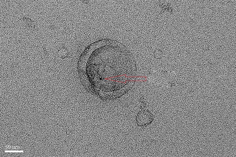
Figure 3 Exosome samples (left) and GNPs (right) after density
gradient ultracentrifugation. GNPs, gold nanoparticles. B
1.0
Concentration relative
0.8
200:1 during the incubation process, and GNPs that failed
0.6
to enter the exosomes were isolated by density gradient
ultracentrifugation. A sample of GNPs alone was used as a 0.4
control during density gradient ultracentrifugation. Excess
GNPs in the exosome sample were detected at the same 0.2
level as the control in the gradient, as shown in Figure 3.
0.0
GNPs were not detected at the bottom of the tube, possibly 10 100 1000
because the centrifugation time was too short. Diameter (mm)
After loading the GNPs into the exosomes, NTA was C
1.0
conducted for size measurement and aggregate detection.
The results showed that the curve shapes were very similar
Concentration relative
0.8
to each other, exhibiting only a slight shift, which indicated
an increase in size (Figure 4A,B,C). This size increase may 0.6
have been caused by internalization of the GNPs. No
0.4
obvious peaks indicating large-size particles were observed,
indicating that aggregation of the exosomes was limited. 0.2
The size and shape of the curves was consistent with those
0.0
reported previously (44). 10 100 1000
TEM imaging depicted an obvious dark spot, which Diameter (mm)
was confirmed as GNPs, in the same location as that of the
Figure 4 NTA results of exosomes with different incubation
exosomes (Figure 5). Thus, the GNPs appeared to have
conditions. NTA results of exosomes incubated without GNPs for
entered the exosomes.
3 h at 37 ℃ (A). NTA results of exosomes incubated with GNPs
for 3 h at 37 ℃ (B). And NTA results of exosomes incubated with
In vivo CT imaging GNPs for 10 h at 37 ℃ (C). NTA, nanoparticle tracking analysis;
GNPs, gold nanoparticles.
GNP-labeled exosomes were injected at three different points
around the discoloration area after ischemia surgery. In vivo
CT imaging was conducted at 4 h (Figure 6A) and 24 h (Figure
6B) after injection of the exosomes. GNPs were obvious in GNPs. However, few GNPs were observed in these organs,
the CT images (red area) both at 4 and 24 h, and the red demonstrating that most GNPs remained in the MI area.
area was found to have expanded after 24 h compared to at
4 h, indicating that MSC-Exo remained in the myocardium
Discussion
for up to 24 h. Additionally, some other organs, particularly
the liver, spleen, and kidney, were monitored to detect Due to the low efficiency of the indirect approach for loading
© Annals of Translational Medicine. All rights reserved. Ann Transl Med 2021;9(6):504 | http://dx.doi.org/10.21037/atm-21-981Annals of Translational Medicine, Vol 9, No 6 March 2021 Page 7 of 10
A
GNP
Figure 5 TEM image of exosomes incubated with GNPs for 10 h B
at 37 ℃. GNPs, gold nanoparticles.
the GNPs, several direct labeling methods have been
reported for loading contrast agents into purified exosomes
directly. For example, electroporation is an efficient method
of loading exosomes with drugs (47), RNA cargo (48-50),
or 5 nm superparamagnetic iron oxide nanoparticles (51). A
long electric pulse is used in electroporation to temporarily
disturb the phospholipid bilayer, and gives rise to temporary
pores in the membranes that permit molecules to pass Figure 6 In vivo CT imaging at different time after injection. In
into the exosomes (31). However, electroporation may vivo CT imaging at 4 h after injection (A). And In vivo CT imaging
increase exosome aggregation and damage the integrity of at 24 h after injection (B).
the exosomes. A gentle method was used in this study that
maintains the dispersant ability and integrity of exosomes
observed at 4 and 24 h after the injection, indicating
during the labeling process.
Exosomes were incubated with glucose-coated GNPs that the exosomes injected into the myocardium can
for 3 or 10 h at 37 ℃, as GNP internalization is known stay for enough time and may help repair the damaged
to be energy dependent and 37 ℃ is more suitable than a myocardium. Nonetheless, additional studies are necessary
low temperature (44). The internalization process is also to determine the exact mechanisms by which MSC-derived
associated with the glucose transporter (GLUT-1), and exosomes improve heart function. And the results also show
thus glucose modification of GNPs is necessary. Excess that the GNPs labeling can be used for studying the in
free glucose should be washed off to prevent occupation vivo biodistribution and pharmacokinetics of exogenously
and saturation of GLUT-1 at the exosome surface, which administered exosomes.
would prevent internalization (44). Ultrafiltration was
conducted to remove free glucose. Similarly, excess GNPs Conclusions
were removed by density gradient centrifugation after co-
incubating of GNPs and exosomes, avoiding the effect of In this study, a gentle method was used for loading GNPs
unlabeled gold nanoparticles on CT imaging results. into exosomes, and their successful labeling without causing
The NTA results of the exosomes before and after aggregation was verified. In vivo CT imaging revealed the
the GNPs labeling changed little, and no large particles retention of MSC-Exo in the MI area, indicating their
appeared after labeling, indicating that the labeling process usefulness for improving heart function after infarction.
did not cause the agglomeration of exosomes. The TEM The ability to track exosomes in the hearts of living bodies
images confirmed this result, meanwhile, the colocalization using conventional CT approaches significantly advances
of GNPs and exosomes was also observed, indicating the translational exosome science. Thus, it may be possible
successful labeling of GNPs. to conduct high-resolution, sensitive, and non-invasive
The retention of exosomes in the myocardium was tracking of exosomal nanomedicines for MI applications.
© Annals of Translational Medicine. All rights reserved. Ann Transl Med 2021;9(6):504 | http://dx.doi.org/10.21037/atm-21-981Page 8 of 10 Gong et al. Visualizing gold nanoparticle labeled exosomes in vivo
Acknowledgments References
Funding: The work was supported by the Shanghai 1. Go AS, Mozaffarian D, Roger VL, et al. Executive
Municipal Education Commission-Plateau Disciplinary summary: heart disease and stroke statistics--2014 update:
Program for Medical Technology of SUMHS, 2018-2020 a report from the American Heart Association. Circulation
(China), the National Natural Science Foundation of China 2014;129:399-410.
(No. 81660284 and 81860316), and the Key R & D projects 2. Gray WD, French KM, Ghosh-Choudhary S, et al.
of Jiangxi Province (No. 20192BBGL70035). Identification of therapeutic covariant microRNA clusters
in hypoxia-treated cardiac progenitor cell exosomes using
systems biology. Circ Res 2015;116:255-63.
Footnote
3. Uemura R, Xu M, Ahmad N, et al. Bonemarrow stem
Reporting Checklist: The authors have completed the cells prevent left ventricular remodeling of ischemic heart
ARRIVE reporting checklist. Available at http://dx.doi. through paracrine signaling. Circ Res 2006;98:1414-21.
org/10.21037/atm-21-981 4. Rosenberg M, Lutz M, Kuhl C, et al. Coculture with
hematopoietic stem cells protects cardiomyocytes against
Data Sharing Statement: Available at http://dx.doi. apoptosis via paracrine activation of AKT. J Transl Med
org/10.21037/atm-21-981 2012;10:115.
5. Hare JM, Traverse JH, Henry TD, et al. Dose-Escalation
Conflicts of Interest: All authors have completed the ICMJE Study of Intravenous Adult Human Mesenchymal Stem
uniform disclosure form (available at http://dx.doi. Cells (Prochymal) After Acute Myocardial Infarction. J Am
org/10.21037/atm-21-981). All authors report funding Coll Cardiol 2009;54:2277-86.
from Shanghai Municipal Education Commission- 6. Sanganalmath SK, Bolli R. Cell therapy for heart failure:
Plateau Disciplinary Program for Medical Technology of a comprehensive overview ofexperimental and clinical
SUMHS, 2018-2020 (China), the National Natural Science studies, current challenges, and future directions. Circ Res
Foundation of China (No. 81660284 and 81860316), 2013;113:810-34.
and the Key R & D projects of Jiangxi Province (No. 7. Golpanian S, DiFede DL, Pujoletal MV. Rationale and
20192BBGL70035). The authors have no other conflicts of design of the allogeneiC human mesenchymal stem cells
interest to declare. (hMSC) in patients with aging fRAilTy via intravenoUS
delivery (CRATUS) study: a phase I/II, randomized,
Ethical Statement: The authors are accountable for all blinded and placebo controlled trial to evaluate the Safety
aspects of the work in ensuring that questions related and potential efficacy of allogeneic human mesenchymal
to the accuracy or integrity of any part of the work are stem cell infusion in patients with aging frailty. Oncotarget
appropriately investigated and resolved. Experiments 2016;7:11899-912.
were performed under a project license (No.: 8. Boon RA, Dimmeler S. MicroRNAs in myocardial
SYXK(HU)2018-0029) granted by Laboratory Animal infarction. Nat Rev Cardiol 2015;12:135-42.
Welfare Ethics Committee of Shanghai University of 9. Li Y, Shen Z, Yu XY. Transport of microRNAs via
Medical and Health, in compliance with Chinese national exosomes. Nat Rev Cardiol 2015;12:198.
or institutional guidelines for the care and use of animals. 10. Teng X, Chen L, Chen W, et al. Mesenchymal stem
cell-derived exosomes improve the microenvironment
Open Access Statement: This is an Open Access article ofinfarctedmyocardium contributing to angiogenesis
distributed in accordance with the Creative Commons and anti-inflammation. Cell. Physiol. Biochem
Attribution-NonCommercial-NoDerivs 4.0 International 2015;37:2415-24.
License (CC BY-NC-ND 4.0), which permits the non- 11. Lai RC, Arslan F, Lee MM, et al. Exosome secreted by
commercial replication and distribution of the article with MSC reduces myocardial ischemia/reperfusion injury.
the strict proviso that no changes or edits are made and the Stem Cell Res 2010;4:214-22.
original work is properly cited (including links to both the 12. Bian S, Zhang L, Duan L, et al. Extracellular vesicles
formal publication through the relevant DOI and the license). derived from human bone marrow mesenchymal stem cells
See: https://creativecommons.org/licenses/by-nc-nd/4.0/. promote angiogenesis in a rat myocardial infarction model.
© Annals of Translational Medicine. All rights reserved. Ann Transl Med 2021;9(6):504 | http://dx.doi.org/10.21037/atm-21-981Annals of Translational Medicine, Vol 9, No 6 March 2021 Page 9 of 10
J Mol Med 2014;92:387-97. distinct nanoparticles and subsets of extracellular vesicles
13. Shao L, Zhang Y, Lan B, et al. MiRNA-Sequence Indicates by asymmetric flow field-flow fractionation. Nat Cell Biol
That Mesenchymal Stem Cells and Exosomes Have 2018;20:332-43.
Similar Mechanism to Enhance Cardiac Repair. BioMed 28. Takahashi Y, Nishikawa M, Shinotsuka H, et al.
Res Int 2017;2017:4150705. Visualization and in vivo tracking of the exosomes of
14. Wasmuth EV, Januszyk K, Lima CD. Structure of an murine melanoma B16-BL6 cells in mice after intravenous
Rrp6-RNA exosome complex bound to poly(A) RNA. injection. J Biotechnol 2013;165:77-84.
Nature 2014;511:435-9. 29. Johnsen KB, Gudbergsson JM, Skov MN, et al. A
15. Trams EG, Lauter CJ, Salem NJ, et al. Exfoliation of Comprehensive Overview of Exosomes as Drug Delivery
membrane ecto-enzymes in the form of micro-vesicles. Vehicles - Endogenous Nanocarriers for Targeted Cancer
Biochim Biophys Acta 1981;645:63-70. Therapy. Biochim Biophys Acta 2014;1846:75-87.
16. Johnstone RM, Adam M, Hammond JR, et al. Vesicle 30. Lai RC, Yeo RWY, Tan KH, et al. Exosomes for Drug
formation during reticulocyte maturation. Association Delivery - A Novel Application for the Mesenchymal Stem
of plasma membrane activities with released vesicles Cell. Biotechnol Adv 2013;31:543-51.
(exosomes). J Biol Chem 1987;262:9412-20. 31. Busato A, Bonafede R, Bontempi P, et al. Magnetic
17. Harding C, Heuser J, and Stahl P. Endocytosis and resonance imaging of ultrasmall superparamagnetic
intracellular processing of transferrin and colloidalgold iron oxide-labeled exosomes from stem cells: a new
transferrin in rat reticulocytes: demonstration of a pathway method to obtain labeled exosomes. Int J Nanomedicine
for receptor shedding. Eur J Cell Biol 1984;35:256-63. 2016;11:2481-90.
18. Pan BT, Teng K, Wu C, et al. Johnstone, Electron 32. Yang T, Martin P, Fogarty B, et al. Exosome Delivered
microscopic evidence for externalization of the transferrin Anticancer Drugs across the Blood-Brain Barrier for
receptor in vesicular form in sheep reticulocytes. J Cell Brain Cancer Therapy in Danio Rerio. Pharm. Res
Biol 1985;101:942-8 2015;32:2003-14.
19. Pan BT, Johnstone RM. Fate of the transferrin receptor 33. Pasternak O, Kubicki M, Shenton ME. In Vivo Imaging
during maturation of sheep reticulocytes in vitro: selective of Neuroinflammation in Schizophrenia. Schizophr Res
externalization of the receptor. Cell 1983;33:967-78. 2016;173:200-12.
20. Raposo G, Nijman HW, Stoorvogel W, et al. B 34. Arvizo R. Gold Nanoparticles: Opportunities and
lymphocytes secrete antigen-presenting vesicles. J Exp Challenges in Nanomedicine. Expert Opin Drug Deliv
Med 1996;183:1161-72. 2010;7:753-63.
21. Zitvogel L, Regnault A, Lozier A, et al. Eradication of 35. Reuveni T, Motiei M, Romman Z, et al. Targeted Gold
established murine tumors using a novel cell-free vaccine: Nanoparticles Enable Molecular CT Imaging of Cancer:
dendritic cell-derived exosomes. Nat Med 1998;4:594-600. An in Vivo Study. Int J Nanomedicine 2011;6:2859-64.
22. Yáñez-Mó M, Siljander PR, Andreu Z, et al. Biological 36. Busato A, Bonafede R, Bontempi P, et al. Labeling and
properties of extracellular vesicles and their physiological Magnetic Resonance Imaging of Exosomes Isolated
functions. J. Extracell. Vesicles 2015;4:27066. from Adipose Stem Cells. Curr Protoc Cell Biol
23. Balaj L, Lessard R, Dai L, et al. Tumour microvesicles 2017;75:3.44.1-15.
contain retrotransposon elements and amplified oncogene 37. Sun D, Zhuang X, Xiang X, et al. A Novel Nanoparticle
sequences. Nat Commun 2011;2:180. Drug Delivery System: The Anti-Inflammatory Activity of
24. Choi DS, Kim DK, Kim YK, et al. Proteomics, Curcumin Is Enhanced When Encapsulated in Exosomes.
transcriptomics and lipidomics of exosomes and ectosomes. Mol Ther 2010;18:1606-14.
Proteomics 2013;13:1554-71. 38. Haney MJ, Klyachko NL, Zhao Y, et al. Exosomes as
25. Thakur BK, Zhang H, Becker A, et al. Double-stranded Drug Delivery Vehicles for Parkinson’s Disease Therapy. J
DNA in exosomes: a novel biomarker in cancer detection. Control Release 2015;207:18-30.
Cell Res 2014;24:766-9. 39. Alhasan AH, Patel PC, Choi CHJ, et al. Exosome
26. Tetta C, Ghigo E, Silengo L, et al. Extracellular vesicles Encased Spherical Nucleic Acid Gold Nanoparticle
as an emerging mechanism of cell-to-cell communication. Conjugates as Potent microRNA Regulation Agents.
Endocrine 2013;44:11-9. Small 2014;10:186-92.
27. Zhang H, Freitas D, Kim HS, et al. Identification of 40. Roma-Rodrigues C, Pereira F, Alves de Matos AP, et al.
© Annals of Translational Medicine. All rights reserved. Ann Transl Med 2021;9(6):504 | http://dx.doi.org/10.21037/atm-21-981Page 10 of 10 Gong et al. Visualizing gold nanoparticle labeled exosomes in vivo
Smuggling Gold Nanoparticles across Cell Types - a New regenerative exosomes to myocardial infarction using
Role for Exosomes in Gene Silencing. Nanomedicine cardiac homing peptide. Theranostics 2018;8:1869-78.
2017;13:1389-98. 47. Tian Y, Li S, Song J, et al. A doxorubicin delivery platform
41. Niidome T, Yamagata M, Okamoto Y, et al. PEG- using engineered natural membrane vesicle exosomes for
modified gold nanorods with a stealth character for in vivo targeted tumor therapy. Biomaterials 2014;35:2383-90.
applications. J Control Release 2006;114:343-7. 48. Alvarez-Erviti L, Seow Y, Yin H, et al. Delivery of siRNA
42. Eghtedari M, Liopo AV, Copland JA, et al. Engineering of to the mouse brain by systemic injection of targeted
hetero-functional gold nanorods for the in vivo molecular exosomes. Nat Biotechnol 2011;29:341-5.
targeting of breast cancer cells. Nano Lett 2009;9:287-91. 49. Wahlgren J, De L Karlson T, Brisslert M, et al. Plasma
43. Jansen ED, Liopo A, Thomas RJ, et al. Photothermal exosomes can deliver exogenous short interfering RNA
therapy of acute leukemia cells in the near-infrared region to monocytes and lymphocytes. Nucleic Acids Res
using gold nanorods CD-33 conjugates. Proc SPIE 2012;40:e130.
- The International Society for Optical Engineering 50. Kooijmans SA, Stremersch S, Braeckmans K, et al.
2011;7897:789710. Electroporation-induced siRNA precipitation obscures the
44. Betzer O, Perets N, Angel A, et al. In Vivo Neuroimaging efficiency of siRNA loading into extracellular vesicles. J
of Exosomes Using Gold Nanoparticles. ACS Nano Control Release 2013;172:229-38.
2017;11:10883-93. 51. Hu L, Wickline SA, Hood JL. Magnetic resonance
45. Patten RD, Aronovitz MJ, Deras-Mejia L, et al. imaging of melanoma exosomes in lymph nodes. Magn
Ventricular remodeling in a mouse model of myocardial Reson Med 2015;74:266-71.
infarction. Am J Physiol 1998;274:H1812-20.
46. Vandergriff A, Huang K, Shen D, et al. Targeting (English Language Editor: A. Kassem)
Cite this article as: Gong L, Weng Y, Zhou W, Zhang K, Li W,
Jiang J, Zhu J. In vivo CT imaging of gold nanoparticle-labeled
exosomes in a myocardial infarction mouse model. Ann Transl
Med 2021;9(6):504. doi: 10.21037/atm-21-981
© Annals of Translational Medicine. All rights reserved. Ann Transl Med 2021;9(6):504 | http://dx.doi.org/10.21037/atm-21-981You can also read