Mature DIABLO/Smac Is Produced by the IMP Protease Complex on the Mitochondrial Inner Membrane
←
→
Page content transcription
If your browser does not render page correctly, please read the page content below
Molecular Biology of the Cell
Vol. 16, 2926 –2933, June 2005

Mature DIABLO/Smac Is Produced by the IMP Protease
Complex on the Mitochondrial Inner Membrane
Lena Burri,* Yvan Strahm,† Christine J. Hawkins,‡ Ian E. Gentle,*
Michelle A. Puryer,‡ Anne Verhagen,§ Bernard Callus,§ David Vaux,§ and
Trevor Lithgow*储

*Russell Grimwade School of Biochemistry and Molecular Biology, University of Melbourne, Parkville, VIC
3010, Australia; †Victorian Bioinformatics Consortium, Monash University, Clayton, VIC 3800, Australia;
‡
  Children’s Cancer Centre, Murdoch Children’s Research Institute, Royal Children’s Hospital, Parkville, VIC
3052, Australia; §The Walter and Eliza Hall Institute of Medical Research, Parkville, VIC 3050, Australia; and
储
 Bio21 Molecular Science and Biotechnology Institute, Parkville, VIC 3010, Australia

Submitted December 17, 2004; Revised March 14, 2005; Accepted March 24, 2005
Monitoring Editor: Donald Newmeyer

DIABLO/Smac is a mitochondrial protein that can promote apoptosis by promoting the release and activation of caspases.
To do so, DIABLO/Smac must first be processed by a mitochondrial protease and then released into the cytosol, and we
show this in an intact cellular system. We propose that the precursor form of DIABLO/Smac enters the mitochondria
through a stop-transfer pathway and is processed to its active form by the inner membrane peptidase (IMP) complex.
Catalytic subunits of the mammalian IMP complex were identified based on sequence conservation and functional
complementation, and the novel sequence motif RX5P in Imp1 and NX5S in Imp2 distinguish the two catalytic subunits.
DIABLO/Smac is one of only a few specific proteins identified as substrates for the IMP complex in the mitochondrial
intermembrane space.

INTRODUCTION                                                          caspase activity provides a layer of regulation over the cell
                                                                      death-promoting activities of this family of effector proteases.
Programmed cell death is a means whereby metazoans can                   In mammalian cells, signals for cell death can lead to
remove unwanted cells, with failure of programmed cell                rupture of the mitochondrial outer membrane, and under
death enabling cancer and autoimmune disease, and inap-               these conditions the inhibition of caspases can be antago-
propriate cell death contributing to neurodegenerative dis-           nized by the mitochondrial proteins DIABLO/Smac (Du et
ease. Several of the proteins that regulate cell death, includ-       al., 2000; Verhagen et al., 2000) and HtrA2/Omi (Suzuki et
ing Bcl-2 family members, signal-transduction receptors,              al., 2001; Martins et al., 2002; Verhagen et al., 2002). Structural
effector proteases and DIABLO/Smac, are specifically en-              studies (Chai et al., 2000; Liu et al., 2000; Wu et al., 2000) have
closed within subcellular structures allowing precise control         shown that purified DIABLO is a homodimer (Chai et al.,
over the commitment of a cell to die. Understanding where             2000), and each DIABLO dimer can bind to the BIR domains
and how these key regulators are localized is crucial in              of inhibitor proteins via contacts made to the N-terminal
understanding their normal biological function and their              residues of DIABLO (Liu et al., 2000; Wu et al., 2000). The
role in the pathogenesis of malignant diseases.                       avid interaction DIABLO makes with the inhibitor MIHA
   A family of intracellular proteases called caspases imple-         competes the inhibitor away from active caspase 9, freeing
ment programmed cell death (Ekert et al., 1999). The activity         caspase 9 to proteolytically activate downstream caspases
of caspases is regulated by a family of inhibitor of apoptosis        (Ekert et al., 2001).
proteins (IAPs) that bind and neutralize active caspases                 Mouse DIABLO is translated as a 237-residue precursor
(Deveraux and Reed, 1999). For example, the inhibitor                 protein (preDIABLO) with an N-terminal presequence that
MIHA/XIAP/hILP/BIRC4 can bind and inhibit processed                   must be cleaved to generate the mature form with the ami-
caspases 3, 7, and 9 (Duckett et al., 1996; Liston et al., 1996;      no-terminal sequence A54VPI (Du et al., 2000; Verhagen et al.,
Uren et al., 1996; Deveraux et al., 1997, 1998). This damping of      2000). Here, we provide evidence that the N-terminal pre-
                                                                      sequence of DIABLO precursor is a bipartite, stop-transfer
                                                                      type targeting signal. We suggest that the precursor form of
                                                                      DIABLO is recognized and translocated through the mito-
This article was published online ahead of print in MBC in Press      chondrial outer membrane via the translocase in the outer
(http://www.molbiolcell.org/cgi/doi/10.1091/mbc.E04 –12–1086)         mitochondrial membrane (TOM) complex and in the inter-
on April 6, 2005.                                                     membrane space is transferred to the translocase in the inner
Address correspondence to: Trevor Lithgow (t.lithgow@unimelb.         mitochondrial membrane (TIM23) complex. The stop-trans-
edu.au).                                                              fer sequence is cleaved by the IMP complex, an oligomeric
Abbreviations used: IAP, inhibitor of apoptosis protein; IMP, inner   inner membrane peptidase. We have identified metazoan
membrane peptidase; TIM23, translocase of the inner mitochondrial     homologues of the catalytically active subunits of the IMP
membrane; TOM, translocase of the outer mitochondrial membrane.       complex, hitherto only identified in yeast, and show the

2926                                                                                   © 2005 by The American Society for Cell Biology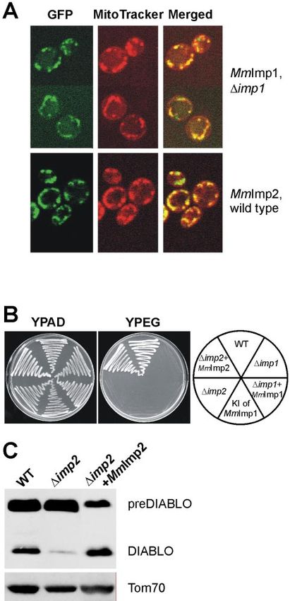
Maturation of DIABLO/Smac by the IMP Complex
mammalian orthologues function to process, and thereby
activate, DIABLO/Smac in mitochondria.
MATERIALS AND METHODS
Plasmids, Yeast Strains, and Media
DNA fragments corresponding to MmImp1 (AK015978) and MmImp2
(AF359564) were amplified by PCR by using primers that generated in-frame
restriction sites. PCR products were subcloned in front of green fluorescent
protein (GFP)-S65T under the control of the MET25 promoter (George et al.,
1998) for analysis by confocal microscopy or into pYADE4 under the control
of the ADH2 promoter (Brunelli and Pall, 1993) for the complementation
assays. FLAG-tagged MmImp1 and MmImp2 cDNAs were generated by
introducing an in frame XbaI restriction site into the 3⬘ end of the coding
regions by PCR, effectively removing the stop codon of each cDNA.
To generate yeast mutants lacking the IMP1 gene or the SOM1 gene,
PCR-mediated gene disruption (Wach et al., 1997) was used with the plasmid
pFA6a-HIS3MX6 as template. For the knockin strain, MmImp1 was first
cloned into a vector in front of a HIS3 gene marker. This vector was then used
for PCR-mediated gene disruption. Yeast plasmids Caspase-3-LacZ, pADH-
(TRP1)-MIHA, and pGALL-(HIS3)-Diablo54-237 and empty vector controls Figure 1. DIABLO is targeted to mitochondria in yeast. Yeast cells
have been described previously (Hawkins et al., 2001). pGALL-(HIS3)-Dia-
expressing preDIABLO [DIABLO(1-237)GFP], DIABLO(1-53)GFP,
blo1-237 was generated by subcloning full-length mouse DIABLO with XhoI
and NotI into pGALL-(HIS3). or DIABLO(54-237)GFP were costained with the fluorescent dye
Strains of Saccharomyces cerevisiae were grown at 30°C on YPAD [2% (wt/ MitoTracker Red and viewed by confocal microscopy. Filters selec-
vol) glucose, 1%(wt/vol) yeast extract, and 2% (wt/vol) peptone supple- tive for the green fluorescence of GFP (left) or the red fluorescence
mented with adenine sulfate)], grown until late log phase, and harvested by of MitoTracker Red (middle) were used. Green and red fluorescence
centrifugation; or grown on solid media containing 2% agar in YPAD or pictures merged are shown (right) (V, cell vacuole).
YPEG [2% (vol/vol) ethanol, 2% (wt/vol) glycerol, 1%(wt/vol) yeast extract,
and 2% (wt/vol) peptone) or YPGal (2% (wt/vol) galactose, 1%(wt/vol) yeast
extract, and 2% (wt/vol) peptone supplemented with adenine sulfate].
To determine expression levels of the DIABLO constructs, cytosolic extracts
of transformed yeast were prepared according to Cartwright et al. (1997) and Fluorescence Microscopy
analyzed by SDS-PAGE and immunoblotting. For fluorescence microscopy, cells were visualized directly or after staining
To rupture the mitochondrial outer membrane in vivo, yeast cells were with MitoTracker (MitoTracker Red CM-H2 ⫻ Ros) according to the standard
cultured transiently on media containing 120 mM acetic acid. Acetic acid is protocol from Molecular Probes (Eugene, OR). All fluorescence images were
not metabolized by glucose-repressed yeast cells, enters cells in the proton- captured using an MRC1024 confocal scanning laser microscope (Bio-Rad,
ated form, but, if the extracellular pH is lower than the intracellular pH, Hercules, CA) mounted on an Axioskop (Carl Zeiss, Jena, Germany).
deprotonation leads to transient acidification of the cytosol and some rupture
of the relatively fragile mitochondrial outer membrane (Ludovico et al., 2002).
RESULTS
Functional IAP Antagonism Assay The precursor Form of DIABLO Is Targeted to
Transformations were performed as described previously (Hawkins et al., Mitochondria by Its Amino Terminus
2000) and grown in selective minimal media with glucose overnight, recov-
ered, and washed three times in 10 mM Tris-HCl, pH 8.0, EDTA 1 mM (TE), Sequence analysis suggested that the previously reported
and the cell suspensions were standardized (as determined from OD600). mitochondrial localization of DIABLO in mammalian cells
After incubation for 8 h at room temperature in selective minimal media was due to the presence of a mitochondrial targeting se-
containing galactose, pH 3.0, lacking or containing 20 mM acetic acid, yeast was
recovered, washed once with TE, and resuspended in TE. The yeast suspensions
quence at the amino terminus of the protein. This would
were equalized, and 5-l drops of serial fivefold dilutions were spotted onto imply that DIABLO also should be targeted to mitochondria
selective minimal media containing either glucose (to repress expression of in nonmammalian cells. To determine whether this was true,
caspase 3 and DIABLO) or galactose (to induce their expression). three GFP reporter proteins were constructed for expression
in yeast (Figure 1). GFP was fused to the full preDIABLO
Preparation of Mitochondria and Protease Sensitivity sequence [DIABLO(1-237)GFP], to the 53 N-terminal resi-
Mitochondria were isolated according to Daum et al. (1982). Osmotic shock dues of DIABLO [DIABLO(1-53)GFP], or to DIABLO from
treatment, to produce rupture the outer membrane in purified mitochondria, which the first 53 amino acids had been removed [DIA-
was as described by Glick et al. (1992b), and trypsin treatments were per- BLO(54-237)GFP]. Analysis of yeast cells costained with Mi-
formed as described previously (Beilharz et al., 1998). Samples of mitochon-
drial protein (100 g) were separated by Tris-glycine SDS-PAGE, and West-
toTracker Red revealed that preDIABLO and DIABLO(1-
ern blots were carried out according to published methods (Lithgow et al., 53)GFP were exclusively localized to mitochondria, whereas
1994; Beilharz et al., 1998; Sambrook and Russell, 2001). DIABLO(54-237)GFP was distributed throughout the cytosol
(Figure 1). This indicates that the 53-amino acid presequence
Prediction of the Mitochondrial Targeting Sequence in of DIABLO is necessary and sufficient for targeting of pro-
DIABLO teins to mitochondria in yeast.
The mitochondrial targeting sequences of hundreds of proteins are now
Isolation of mitochondria from yeast cells expressing pre-
known and are usually rich in positively charged residues and with tenden- DIABLO showed that much of the precursor form of DIA-
cies to form two to three turns of a helix with amphipathic character (von BLO had been processed (Figure 2). As in mammalian cells
Heijne, 1986). Predotar (http://www.inra.fr/predotar/) is a neural network expressing DIABLO (Du et al., 2000; Verhagen et al., 2000),
predictor trained to find such extensions and predicts a high score (0.984) for
the likelihood that the amino terminus of DIABLO is a mitochondrial (matrix)
some unprocessed preDIABLO was still partially exposed
targeting sequence. MitoProtII (ftp://ftp.ens.fr/pub/molbio) calculates a on the mitochondrial surface, because it was degraded when
probability for a protein being mitochondrial based on physicochemical prop- mitochondria were incubated with trypsin. The processed
erties, including a mesohydrophobicity score (Claros et al., 1995; Claros and form, however, was protected from trypsin cleavage within
Vincens, 1996). Mitoprot scores DIABLO as 98.99% likely to be a mitochon-
drial protein. We recently showed that a combined prediction from both
the organelle, as is an apparent processing intermediate.
Predotar and MitoprotII is an excellent indicator for mitochondrial location Consistent with this conclusion, when the outer membrane
(Lucattini et al., 2004). was ruptured by osmotic shock, processed DIABLO was
Vol. 16, June 2005 2927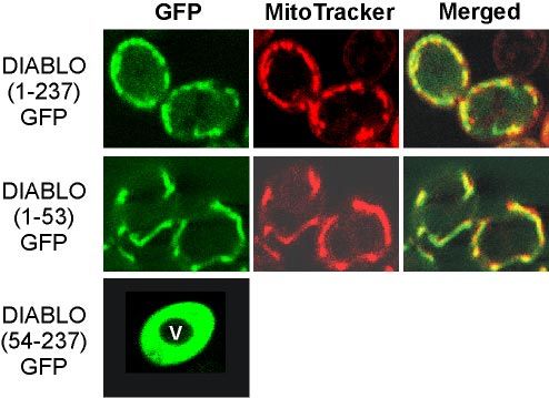
L. Burri et al.
packing in the small pocket in BIR3 that accommodates the
small alanine residue, and the presence of a glycine will
eliminate all the favorable packing that was contributed by
the large isoleucine residue in the large pocket of the IAP. In
addition, the mutant sequence IVPG at the N terminus of
DIABLO should favor the formation of a type-II beta-turn at
VPGA58, which should further destabilize the interaction
with IAP. These otherwise conservative mutations, A54L and
L57G, do not effect the expression of the mutant protein as
judged by Western blotting of cytosolic extracts (our unpub-
lished data), but the mutant DIABLO (IVPG) is ineffective at
promoting the caspase-mediated death of yeast cells (Figure
3B, lane 6).
Yeast cells expressing the precursor form of DIABLO,
which is targeted to mitochondria (Figure 1), together with
autoactivating caspase 3 and MIHA survived (Figure 3B,
lane 9) because the matured, active form is compartmental-
Figure 2. DIABLO is localized to the intermembrane space of ized (within the mitochondrial intermembrane space) from
mitochondria. Mitochondria (100 g of protein) were isolated from MIHA and caspase 3. Nevertheless, rupturing the outer
yeast cells expressing preDIABLO and incubated in the presence membrane of mitochondria by treatment of intact cells with
(⫹) or absence (⫺) of 1.5 g of trypsin with (⫹) and without (⫺) first 120 mM acetic acid (Ludovico et al., 2002) was able to an-
rupturing the outer membrane by osmotic shock. After separation tagonize the antiapoptotic effect of MIHA (Figure 3B, lanes
by SDS-PAGE, samples were analyzed by immunoblotting with 11 and 12). Because inhibition of MIHA requires the cor-
antibodies recognizing DIABLO, the intermembrane space protein
rectly processed AVPI amino terminus of DIABLO (Figure
Cytb2 and the matrix-located mtHSP70.
3A, lane 6), preDIABLO must have been cleaved, in vivo, at
the same processing site in yeast as it is in mammalian cells.
degraded by trypsin, as was the intermembrane space pro-
tein cytochrome (Cyt) b2. Matrix-located proteins, such as preDIABLO Is Processed by the IMP Complex
the mitochondrial 70-kDa heat-shock protein (mtHSP70) re- In yeast, few proteins have been identified that are proteo-
mained protected from trypsin by the inner mitochondrial lytically processed for release into the intermembrane space.
membrane, even after the outer membrane was ruptured The three best characterized, Cytb2, Cytc1, and Mcr1 (Gakh et
(Figure 2). The small fraction of DIABLO that is not de- al., 2002), each first dock with the TIM23 complex and are
graded in this sample remains resistant to protease even in then processed by the IMP complex. The IMP complex is a
the presence of Triton X-100 (our unpublished data) that hetero-oligomer integrated in the mitochondrial inner mem-
solubilizes the inner membrane, suggesting this fraction of brane, and deletion of any one subunit leads to destabiliza-
the protein is aggregated into a protease inaccessible form. tion and loss of the other subunits (Nunnari et al., 1993; Gakh
These experiments show that DIABLO is targeted by its et al., 2002). Two catalytic subunits, Imp1 and Imp2, are
amino terminal targeting sequence to the mitochondria, similar in sequence but seem to have nonoverlapping sub-
where the targeting peptide is removed liberating DIABLO strate specificities (Nunnari et al., 1993; Gakh et al., 2002). A
in the intermembrane space. third noncatalytic subunit, Som1 has been shown to assist
substrate recognition by the catalytic Imp1 subunit (Esser et
Processing of preDIABLO Is Required for Antagonism of
al., 1996; Jan et al., 2000). The oligomeric IMP complex pro-
IAP Function In Vivo
cesses mitochondrial presequences releasing matured pro-
Mature DIABLO can antagonize the caspase inhibitory teins to the intermembrane space. To determine whether
properties of MIHA in yeast (Hawkins et al., 2001), and we DIABLO is a substrate of the IMP complex, purified mito-
exploited this cellular system to explore the impact of the chondria of yeast strains expressing preDIABLO, but lacking
amino terminal region of DIABLO on its ability to antago-
one of the three subunits of the Imp complex, were assayed
nize IAP activity. Yeast tolerated expression of either the
in Western blots for the presence or absence of the mature
precursor or mature forms of DIABLO (Figure 3A, lanes 1
form of DIABLO (Figure 4).
and 2). Expression of autoactivating caspase 3 was toxic
(lane 3), unless inhibited by coexpression of MIHA (lane 4). The model substrate Cytb2 is processed in two steps:
Coexpression of the mature form of DIABLO [correspond- initially by the matrix peptidase MPP to generate an inter-
ing to DIABLO(54-237)] killed the cells due to liberation of mediate (i-Cytb2; Figure 4), and this intermediate is then
active caspase 3 from MIHA (Figure 3A, lane 5). substrate for the IMP complex. In wild-type or in ⌬som1
To be certain that the death of yeast cells is directly a mutants, processing to the mature form of Cytb2 occurred.
result of the DIABLO–IAP interaction and requires the cor- The Imp1 subunit is primarily responsible for Cytb2 process-
rectly processed N terminus of DIABLO, we designed and ing (Nunnari et al., 1993), and Figure 4 shows processing of
tested a mutant form (IVPG) of DIABLO. In the crystal Cytb2 is blocked completely in ⌬imp1 cells. Because yeast
structure of the DIABLO–IAP complex (PDB accession 1G73; ⌬imp2 mutants express decreased levels of the Imp1 subunit
the N-terminal residues are A54, V55, P56, and I57), residues (Nunnari et al., 1993), Cytb2 processing also is also blocked in
A54 and I57 are buried at the interface with the IAP BIR3 ⌬imp2 cells. Analogously, processing of preDIABLO was
domain, in small and large pockets, respectively. The V55 partially compromised in ⌬imp2 mutants and failed to occur
and P56 side chains largely project up and out of the DIAB- at all in either the ⌬imp1 and ⌬som1 mutants. We conclude
LO–IAP interface. The mutations A54L and L57G were made the IMP complex processes preDIABLO to its mature form,
on the basis that the leucine residue would have difficulty and it is likely the Imp1 catalytic site at which this occurs.
2928 Molecular Biology of the CellMaturation of DIABLO/Smac by the IMP Complex
Figure 4. Yeast mutants lacking Imp1 cannot process preDIABLO.
Mitochondria (100 g of protein) isolated from wild-type cells ex-
pressing preDIABLO, or the indicated yeast mutants expressing
preDIABLO, were compared after SDS-PAGE separation of pro-
teins. Western blot replicas of the samples were probed with anti-
sera recognizing DIABLO, the voltage-dependent anion channel
(VDAC), and Cytb2. Position of i-Cytb2 and mature (Cytb2) forms of
cytochrome b2 (Glick et al., 1992b; Lithgow et al., 1994) are shown.
Identification of Mouse Homologues for Yeast Imp1 and
Imp2 Proteases
An IMP complex has not yet been functionally characterized
in organisms other than S. cerevisiae. However, iterative
BLAST analyses revealed genes predicted to encode proteins
similar to Imp1 and Imp2 in all animals and fungi for which
substantial genome sequence data exist (Figure 5). The cat-
alytic serine and lysine residues and the structurally impor-
tant arginine and aspartate residues from the yeast Imp1
and Imp2 (Chen et al., 1999) are conserved across all species
(Figure 5, stars). In addition, we found a sequence motif that
distinguishes Imp1(RX5P) from Imp2(NX5S). The sequence
motif defining the Imp1 and Imp2 subunits sits close by
structurally important aspartate residues thought to stabi-
lize the shape of the substrate-binding cleft in the bacterial
leader peptidase (Paetzel et al., 1998).
MmImp1 and MmImp2 Function as Catalytic Subunits of
an IMP Complex
The cDNAs encoding the putative mouse proteases were
cloned and expressed in yeast cells to determine their intra-
cellular localization. Both MmImp1 (MmImp1 refers to the
Mus musculus form of Imp1 according to the nomenclature
of Pfanner et al. (1996) and MmImp2 localize to mitochondria
(Figure 6A). Like yeast Imp1, MmImp1 could be stably ex-
pressed in ⌬imp1 yeast (Figure 6A) but not in wild-type (i.e.,
Imp1⫹) yeast cells (our unpublished data). The cytochrome
substrates of the IMP complex are key components of the
mitochondrial electron transport chain, so that neither ⌬imp1
mutants nor ⌬imp2 yeast mutants can grow on nonferment-
Figure 3. DIABLO expressed in yeast inhibits the antiapoptotic able carbon sources. We exploited this phenotype to test for
effect of MIHA/XIAP. Yeast was transformed with the plasmids complementation, which would imply functional homology
coding for the indicated proteins or with the appropriate control by the proposed mouse counterparts of the yeast IMP genes.
vectors. Where indicated, cultures of transformed cells were incubated The MmImp2 subunit restored activity in the ⌬imp2 strain,
with or without acetic acid for 8 h at room temperature. Serial dilutions allowing it to process cytochromes and grow on the nonfer-
were made from samples with equivalent cell numbers, and 5 l of
mentable carbon sources ethanol and glycerol (Figure 6B).
each dilution was spotted onto expression-inducing (galactose) or -re-
pressing (glucose) solid media. The top-most row in each panel corre- Based on sequence similarities, correct mitochondrial loca-
sponds to the most concentrated yeast suspension and the serial dilu- tion and functional complementation, we suggest an IMP
tions were spotted vertically down the plate. Colony size indicates complex exists in mammalian cells for the processing and
growth rate and colony number cell viability. activation of proteins such as preDIABLO. That MmImp1
Vol. 16, June 2005 2929L. Burri et al.
Figure 5. Multiple sequence alignment of the Imp family of proteases. Iterative BLAST analysis was undertaken with the Imp2 sequence
from S. cerevisiae. The Imp1 and Imp2 sequences were aligned with ClustalW, and a representative selection is shown (Sc, S. cerevisiae; Sp,
Schizosaccharomyces pombe, Ce, Caenorhabditis elegans; Dm, Drosophila melanogaster; Hs, Homo sapiens; Mm, M. musculus). Asterisks designate a
possible C-terminal extension to CeImp1. Amino acid residues conserved across at least six species are highlighted, and the total number of
residues shown. Imp1-specific residues (RX5P) are colored pink and Imp2-specific residues (NX5S) are colored blue. Catalytic residues in
Imp1 and Imp2 (analogous to Ser90 and K145 of E. coli leader peptidase) are designated with stars.
does not complement the growth defect of ⌬imp1 yeast cells by the IMP complex, making its import pathway into mito-
(Figure 6B) suggests additional factors such as a mammalian chondria (Figure 7A) analogous to the stop-transfer path-
homolog of Som1 might be needed for full activity of the way traveled by Cytb2 and Cytc1 (Glick et al., 1992a,b).
MmImp1 subunit. Imp1 and Imp2 are members of the signal peptidase fam-
The loss of Imp2 destabilizes the IMP complex so that ily of proteases. In Escherichia coli, a signal peptidase re-
⌬imp2 yeast cells process preDIABLO only poorly. How- moves the presequence of proteins translocated across the
ever, this impairment was reversed by expression of bacterial membrane (Paetzel et al., 2002). Two proteins re-
MmImp2 in the ⌬imp2 yeast (Figure 6C). We conclude that lated to the IMP subunits are present in all animals and
preDIABLO is proteolytically activated to DIABLO by the fungi where complete genome information is available, and
IMP complex and that the mammalian Imp1 and Imp2 se- a diptych of amino acid residues corresponding to the motif
quences represent functional homologues of the yeast IMP RX5P in Imp1 and NX5S in Imp2 seems to be diagnostic for
complex subunits. Imp1 or Imp2. The RX5P motif of Imp1 and its surrounding
residues are conserved in the E. coli leader peptidase (Figure
5), and as part of a thorough examination of the structure of
DISCUSSION crystallized leader peptidase, Paetzel et al. (1998, 2002) sug-
In healthy cells, mature DIABLO is encapsulated within gested that arginine282 (in the RX5P motif) is important in
mitochondria, where it cannot interact with IAPs. Here, we stabilizing the active site region and positioning of sur-
have shown that after preDIABLO is translated in the cy- rounding amino acids). We note from their data that the
tosol and transported across the mitochondrial outer mem- proline288 residue seems to be involved in another structur-
brane, it is processed by the IMP complex in the intermem- ally important position, in a turn between two beta-sheets
brane space to produce the potent, mature form. Activated (Strahm and Lithgow, unpublished observations). The dis-
DIABLO then remains in the intermembrane space until an tinguishing motif in Imp2(NX5S) and the context provided
apoptotic signal is received (Figure 7A). by surrounding residues might influence the structure
Two well-studied mitochondrial proteins, Cytb2 and around the active site enough to broaden the range of sub-
Cytc1, have bipartite targeting sequences consisting of a strates that can be processed by the IMP complex. That the
short region of basic, amphipathic helix followed by a pro- RX5P and NX5S sequence motifs are each so widely con-
cessing site for the matrix processing peptidase and an ad- served through evolution shows they are fundamentally
ditional sorting signal. The sorting signal dictates arrest of important for IMP complex activity and makes them the
newly imported Cytb2 and Cytc1 in the inner membrane diagnostic motif in distinguishing Imp1 from Imp2.
TIM23 complex, providing access to the IMP complex for
proteolytic release of Cytb2 and Cytc1 from their prese- Other Substrates of the Mammalian IMP Complex
quences (Glick et al., 1992a,b; van Loon and Schatz, 1987). A number of proteins that might potentiate apoptosis are
The presequences of Cytb2 and Cytc1 resemble those in the released from mitochondria (van Gurp et al., 2003; Saelens et
targeting sequence of preDIABLO. A short region at the N al., 2004). None of these have the hallmarks that would
terminus of preDIABLO is predicted to form a basic amphi- suggest activation by the IMP complex. A candidate sub-
pathic helix that might direct the protein to the TIM23 com- strate, AIF is a peripheral component of the mitochondrial
plex (Figure 7B; see Materials and Methods). We have dem- inner membrane (Arnoult et al., 2002) made as a precursor
onstrated that the final processing of DIABLO is carried out protein, with a presequence of 101 residues processed after
2930 Molecular Biology of the CellMaturation of DIABLO/Smac by the IMP Complex
Figure 7. A stop-transfer pathway for the import and activation of
DIABLO by mitochondria. (A) The precursor form of DIABLO
(black) is synthesized in the cytosol and the N-terminal presequence
recognized by the TOM complex and thereby transferred across the
outer membrane. Engaged in the TIM23 complex, DIABLO might be
processed by the matrix-located peptidase, such as mitochondrial
processing peptidase, and is processed by the IMP complex com-
posed of Imp1, Imp2, and Som1. Release of processed DIABLO into
the intermembrane space allows for its assembly into a dimer and
its availability for release into the cytosol only if the outer mem-
brane is ruptured. (B) The presequences of DIABLO (top schematic)
and Cytb2 (bottom schematic) show similar regions corresponding
to a basic amphipathic helix with the matrix targeting information
(gray) and the intermembrane sorting sequence, containing a core of
hydrophobic residues (dark gray) preceded by a cluster of three
positively charged residues (black) important for recognition of the
sorting sequence. For DIABLO the IMP1 processing site is desig-
nated between C53 and A54 and for Cytb2 between N80 and E81.
import into mitochondria (Susin et al., 1999). It is not clear
yet which protease is responsible for AIF processing, but the
presequence of AIF shows no obvious similarity to a stop-
transfer sequence, and the processing site has none of the
residues suggested to be important for recognition by the
IMP complex (Gakh et al., 2002).
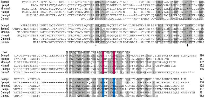
Figure 6. MmImp1 and MmImp2 are mouse subunits of the IMP Cytochrome c is imported into the intermembrane space
complex. (A) GFP was fused to the C terminus of MmImp1 and via the TOM complex without the participation of the TIM
MmImp2 and the fusion proteins expressed in ⌬imp1 or wild-type complex and without any proteolytic processing (Diekert et
yeast cells. Cells were costained with the fluorescent dye Mito-
Tracker Red and visualized by confocal microscopy. Z-sections
al., 2001; Wiedemann et al., 2003). HtrA2/Omi is targeted to
through representative cells are shown. Green fluorescence of GFP the intermembrane space of mitochondria, but it is pro-
in groups of cells (left), red fluorescence (middle), and the merged cessed autocatalytically in mammalian cells (Seong et al.,
pictures of green and red fluorescence (right). (B) Wild-type yeast 2004) and in yeast (Verhagen and Silke, unpublished re-
cells and the ⌬imp1 or ⌬imp2 mutants were transformed with the sults), and its role in apoptosis might be secondary to its role
respective mouse homologue to be tested for complementation of as a molecular chaperone for other mitochondrial proteins
the growth defect on the nonfermentable carbon sources glycerol (Vaux and Silke, 2003).
and ethanol (YPEG). In addition, the open reading frame in the Endonuclease G (EndoG) is a mitochondrial protein, re-
yeast gene encoding Imp1 (YMR150c) was directly replaced with the leased during cell death, that contributes to nuclear DNA
MmImp1 open-reading frame through homologous recombination
(KIMmImp1). (C) Mitochondria expressing preDIABLO isolated
fragmentation in the terminal stages of apoptosis (Li et al.,
from wild type, yeast mutant lacking Imp2, and yeast mutant lack- 2001). EndoG is encoded in the nucleus, translated as an
ing Imp2 but expressing MmImp2 were compared after SDS-PAGE ⬃33-kDa precursor in the cytosol, translocated across the
separation of proteins. Amounts of DIABLO and the control protein mitochondrial membranes with the presequence and then
Tom70 were compared by Western blot analysis. cleaved to yield the ⬃28-kDa mature nuclease (Schafer et al.,
2004). EndoG had been tentatively suggested to be located in
Vol. 16, June 2005 2931L. Burri et al.
the intermembrane space (Ohsato et al., 2002), but EndoG Daum, G., Gasser, S. M., and Schatz, G. (1982). Import of proteins into
must be located in the mitochondrial matrix for it to gener- mitochondria. Energy-dependent, two-step processing of the intermembrane
space enzyme cytochrome b2 by isolated yeast mitochondria. J. Biol. Chem.
ate the primers needed for mitochondrial DNA replication 257, 13075–13080.
(Cote and Ruiz-Carrillo, 1993) and would therefore be pro-
Deveraux, Q. L., and Reed, J. C. (1999). IAP family proteins–suppressors of
cessed by matrix-located proteases (Gakh et al., 2002). apoptosis. Genes Dev. 13, 239 –252.
Discrete defects in mitochondrial protein import and sort-
Deveraux, Q. L., Roy, N., Stennicke, H. R., Van Arsdale, T., Zhou, Q., Srini-
ing can lead to human disease. Mohr–Tranenberg syn- vasula, S. M., Alnemri, E. S., Salvesen, G. S., and Reed, J. C. (1998). IAPs block
drome, a deafness-dystonia disorder, results through muta- apoptotic events induced by caspase-8 and cytochrome c by direct inhibition
tions that effect assembly of the Tim8/Tim13 complex, of distinct caspases. EMBO J. 17, 2215–2223.
thereby inhibiting protein sorting to the TIM22 complex Deveraux, Q. L., Takahashi, R., Salvesen, G. S., and Reed, J. C. (1997). X-linked
(Roesch et al., 2002; Binder et al., 2003). Chromosomal map- IAP is a direct inhibitor of cell-death proteases. Nature 388, 300 –304.
ping of patients suffering from Gilles de la Tourette syn- Diekert, K., de Kroon, A. I., Ahting, U., Niggemeyer, B., Neupert, W., de
drome, another neurological condition, has revealed that Kruijff, B., and Lill, R. (2001). Apocytochrome c requires the TOM complex for
one of the genes located at a breakpoint region (7q31) that is translocation across the mitochondrial outer membrane. EMBO J. 20, 5626 –
associated with symptom development encodes the human 5635.
ortholog of the protein we designate here as MmImp2 (Petek Du, C., Fang, M., Li, Y., Li, L., and Wang, X. (2000). Smac, a mitochondrial
et al., 2001; Gakh et al., 2002). Speculation, based on sequence protein that promotes cytochrome c-dependent caspase activation by elimi-
similarity the authors noted to the yeast Imp2, suggested nating IAP inhibition. Cell 102, 33– 42.
that defects in respiratory chain complexes might impact on Duckett, C. S., Nava, V. E., Gedrich, R. W., Clem, R. J., Van Dongen, J. L.,
the etiology of Tourette syndrome and other neuropsychi- Gilfillan, M. C., Shiels, H., Hardwick, J. M., and Thompson, C. B. (1996). A
conserved family of cellular genes related to the baculovirus iap gene and
atric disorders (Petek et al., 2001; Gakh et al., 2002). Our encoding apoptosis inhibitors. EMBO J. 15, 2685–2694.
findings on the role of the IMP complex in processing non-
Ekert, P. G., Silke, J., Hawkins, C. J., Verhagen, A. M., and Vaux, D. L. (2001).
cytochrome substrates such as DIABLO, suggest that if de- DIABLO promotes apoptosis by removing MIHA/XIAP from processed
fects in the IMP complex contribute to Tourette syndrome caspase 9. J. Cell Biol. 152, 483– 490.
and other conditions, it could be through downstream in-
Ekert, P. G., Silke, J., and Vaux, D. L. (1999). Caspase inhibitors. Cell Death
fluences on cell development and function. Differ. 6, 1081–1086.
Esser, K., Pratje, E., and Michaelis, G. (1996). SOM 1, a small new gene
ACKNOWLEDGMENTS required for mitochondrial inner membrane peptidase function in Saccharo-
myces cerevisiae. Mol. Gen. Genet. 252, 437– 445.
We thank Miha Pakusch and Katherine Vascotto for technical assistance, and
Andrew Perry, Terry Mulhern, Paul Gooley, and James Whisstock for critical Gakh, O., Cavadini, P., and Isaya, G. (2002). Mitochondrial processing pepti-
discussions. This work was supported by a fellowship from the Swiss Na- dases. Biochim. Biophys. Acta 1592, 63–77.
tional Science Foundation (to L. B.), an early career research grant from the George, R., Beddoe, T., Landl, K., and Lithgow, T. (1998). The yeast nascent
University of Melbourne (to L. B.), a Center Grant from the Leukemia and polypeptide-associated complex initiates protein targeting to mitochondria in
Lymphoma Society (to D.L.V.), and a grant from the Australian Research vivo. Proc. Natl. Acad. Sci. USA 95, 2296 –2301.
Council (to T. L.).
Glick, B. S., Beasley, E. M., and Schatz, G. (1992a). Protein sorting in mito-
chondria. Trends Biochem. Sci. 17, 453– 459.
REFERENCES Glick, B. S., Brandt, A., Cunningham, K., Muller, S., Hallberg, R. L., and
Arnoult, D., Parone, P., Martinou, J. C., Antonsson, B., Estaquier, J., and Schatz, G. (1992b). Cytochromes c1 and b2 are sorted to the intermembrane
Ameisen, J. C. (2002). Mitochondrial release of apoptosis-inducing factor space of yeast mitochondria by a stop-transfer mechanism. Cell 69, 809 – 822.
occurs downstream of cytochrome c release in response to several proapop- Hawkins, C. J., Silke, J., Verhagen, A. M., Foster, R., Ekert, P. G., and Ashley,
totic stimuli. J. Cell Biol. 159, 923–929. D. M. (2001). Analysis of candidate antagonists of IAP-mediated caspase
Beilharz, T., Suzuki, C. K., and Lithgow, T. (1998). A toxic fusion protein inhibition using yeast reconstituted with the mammalian Apaf-1-activated
accumulating between the mitochondrial membranes inhibits protein assem- apoptosis mechanism. Apoptosis 6, 331–338.
bly in vivo. J. Biol. Chem. 273, 35268 –35272. Hawkins, C. J., Wang, S. L., and Hay, B. A. (2000). Monitoring activity of
Binder, J., Hofmann, S., Kreisel, S., Wohrle, J. C., Bazner, H., Krauss, J. K., caspases and their regulators in yeast Saccharomyces cerevisiae. Methods En-
Hennerici, M. G., and Bauer, M. F. (2003). Clinical and molecular findings in zymol. 322, 162–174.
a patient with a novel mutation in the deafness-dystonia peptide (DDP1) Jan, P. S., Esser, K., Pratje, E., and Michaelis, G. (2000). Som1, a third compo-
gene. Brain 126, 1814 –1820. nent of the yeast mitochondrial inner membrane peptidase complex that
Brunelli, J. P., and Pall, M. L. (1993). A series of yeast shuttle vectors for contains Imp1 and Imp2. Mol. Gen. Genet. 263, 483– 491.
expression of cDNAs and other DNA sequences. Yeast 9, 1299 –1308.
Li, L. Y., Luo, X., and Wang, X. (2001). Endonuclease G is an apoptotic DNase
Cartwright, P., Beilharz, T., Hansen, P., Garrett, J., and T. Lithgow. (1997). when released from mitochondria. Nature 412, 95–99.
Mft52, an acid-bristle protein in the cytosol that delivers precursor proteins to
yeast mitochondria. J. Biol. Chem. 272, 5320 –5325. Liston, P., et al. (1996). Suppression of apoptosis in mammalian cells by NAIP
and a related family of IAP genes. Nature. 379, 349 –353.
Chai, J., Du, C., Wu, J. W., Kyin, S., Wang, X., and Shi, Y. (2000). Structural and
biochemical basis of apoptotic activation by Smac/DIABLO. Nature 406, Lithgow, T., Junne, T., Wachter, C., and Schatz, G. (1994). Yeast mitochondria
855– 862. lacking the two import receptors Mas20p and Mas70p can efficiently and
specifically import precursor proteins. J. Biol. Chem. 269, 15325–15330.
Chen, X., Van Valkenburgh, C., Fang, H., and Green, N. (1999). Signal pep-
tides having standard and nonstandard cleavage sites can be processed by Liu, Z., Sun, C., Olejniczak, E. T., Meadows, R. P., Betz, S. F., Oost, T.,
Imp1p of the mitochondrial inner membrane protease. J. Biol. Chem. 274, Herrmann, J., Wu, J. C., and Fesik, S. W. (2000). Structural basis for binding of
37750 –37754. Smac/DIABLO to the XIAP BIR3 domain. Nature 408, 1004 –1008.
Claros, M. G., Perea, J., Shu, Y., Samatey, F. A., Popot, J. L., and Jacq, C. (1995). Lucattini, R., Likic, V. A., and Lithgow, T. (2004). Bacterial proteins predis-
Limitations to in vivo import of hydrophobic proteins into yeast mitochon- posed for targeting to mitochondria. Mol. Biol. Evol. 21, 652– 658.
dria. The case of a cytoplasmically synthesized apocytochrome b. Eur. J. Bio- Ludovico, P., Rodrigues, F., Almeida, A., Silva, M. T., Barrientos, A., and
chem. 228, 762–771. Corte-Real, M. (2002). Cytochrome c release and mitochondria involvement in
Claros, M. G., and Vincens, P. (1996). Computational method to predict programmed cell death induced by acetic acid in Saccharomyces cerevisiae. Mol.
mitochondrially imported proteins and their targeting sequences. Eur. J. Bio- Biol. Cell 13, 2598 –2606.
chem. 241, 779 –786.
Martins, L. M., et al. (2002). The serine protease Omi/HtrA2 regulates apo-
Cote, J., and Ruiz-Carrillo, A. (1993). Primers for mitochondrial DNA repli- ptosis by binding XIAP through a reaper-like motif. J. Biol. Chem. 277,
cation generated by endonuclease G. Science 261, 765–769. 439 – 444.
2932 Molecular Biology of the CellMaturation of DIABLO/Smac by the IMP Complex
Nunnari, J., Fox, T. D., and Walter, P. (1993). A mitochondrial protease with Susin, S. A., et al. (1999). Molecular characterization of mitochondrial apop-
two catalytic subunits of nonoverlapping specificities. Science 262, 1997–2004. tosis-inducing factor. Nature 397, 441– 446.
Ohsato, T., Ishihara, N., Muta, T., Umeda, S., Ikeda, S., Mihara, K., Hamasaki, Suzuki, Y., Imai, Y., Nakayama, H., Takahashi, K., Takio, K., and Takahashi,
N., and Kang, D. (2002). Mammalian mitochondrial endonuclease G. Diges- R. (2001). A serine protease, HtrA2, is released from the mitochondria and
tion of R-loops and localization in intermembrane space. Eur. J. Biochem. 269, interacts with XIAP, inducing cell death. Mol. Cell. 8, 613– 621.
5765–5770.
Uren, A. G., Pakusch, M., Hawkins, C. J., Puls, K. L., and Vaux, D. L. (1996).
Paetzel, M., Dalbey, R .E., and Strynadka, N. C. (1998). Crystal structure of a Cloning and expression of apoptosis inhibitory protein homologs that func-
bacterial signal peptidase in complex with a beta-lactam inhibitor. Nature 396, tion to inhibit apoptosis and/or bind tumor necrosis factor receptor-associ-
186 –190. ated factors. Proc. Natl. Acad. Sci. USA 93, 4974 – 4978.
Paetzel, M., Karla, A., Strynadka, N. C., and Dalbey, R. E. (2002). Signal van Gurp, M., Festjens, N., van Loo, G., Saelens, X., and Vandenabeele, P.
peptidases. Chem. Rev. 102, 4549 – 4580. (2003). Mitochondrial intermembrane proteins in cell death. Biochem. Bio-
phys. Res. Commun. 304, 487– 497.
Petek, E., Windpassinger, C., Vincent, J. B., Cheung, J., Boright, A. P., Scherer,
S. W., Kroisel, P. M., and Wagner, K. (2001). Disruption of a novel gene van Loon, A. P., and Schatz, G. (1987). Transport of proteins to the mitochon-
(IMMP2L) by a breakpoint in 7q31 associated with Tourette syndrome. Am. J. drial intermembrane space: the ‘sorting’ domain of the cytochrome c1 prese-
Hum. Genet. 68, 848 – 858. quence is a stop-transfer sequence specific for the mitochondrial inner mem-
brane. EMBO J. 6, 2441–2448.
Pfanner, N., Douglas, M. G., Endo, T., Hoogenraad, N. J., Jensen, R. E., Meijer,
M., Neupert, W., Schatz, G., Schmitz, U. K., and Shore, G. C. (1996). Uniform Vaux, D. L., and Silke, J. (2003). HtrA2/Omi, a sheep in wolf’s clothing. Cell
nomenclature for the protein transport machinery of the mitochondrial mem- 115, 251–253.
branes. Trends Biochem. Sci. 21, 51–52. Verhagen, A. M., Ekert, P. G., Pakusch, M., Silke, J., Connolly, L. M., Reid,
Roesch, K., Curran, S. P., Tranebjaerg, L., and Koehler, C. M. (2002). Human G. E., Moritz, R. L., Simpson, R. J., and Vaux, D. L. (2000). Identification of
deafness dystonia syndrome is caused by a defect in assembly of the DDP1/ DIABLO, a mammalian protein that promotes apoptosis by binding to and
TIMM8a-TIMM13 complex. Hum. Mol. Genet. 11, 477– 486. antagonizing IAP proteins. Cell 102, 43–53.
Saelens, X., Festjens, N., Walle, L. V., van Gurp, M., van Loo, G., and Verhagen, A. M., et al. (2002). HtrA2 promotes cell death through its serine
Vandenabeele, P. (2004). Toxic proteins released from mitochondria in cell protease activity and its ability to antagonize inhibitor of apoptosis proteins.
death. Oncogene 23, 2861–2874. J. Biol. Chem. 277, 445– 454.
Sambrook, J., and Russell, D. W. (2001). Molecular Cloning. A Laboratory von Heijne, G. (1986). Mitochondrial targeting sequences may form am-
Manual, 3rd ed., Cold Spring Harbor, NY: Cold Harbor Laboratory Press. phiphilic helices. EMBO J. 5, 1335–1342.
Wach, A., Brachat, A., Alberti-Segui, C., Rebischung, C., and Philippsen, P.
Schafer, P., Scholz, S. R., Gimadutdinow, O., Cymerman, I. A., Bujnicki, J. M.,
(1997). Heterologous HIS3 marker and GFP reporter modules for PCR-target-
Ruiz-Carrillo, A., Pingoud, A., and Meiss, G. (2004). Structural and functional
ing in Saccharomyces cerevisiae. Yeast 13, 1065–1075.
characterization of mitochondrial EndoG, a sugar non-specific nuclease which
plays an important role during apoptosis. J. Mol. Biol. 338, 217–228. Wiedemann, N., Kozjak, V., Prinz, T., Ryan, M. T., Meisinger, C., Pfanner, N.,
and Truscott, K. N. (2003). Biogenesis of yeast mitochondrial cytochrome c: a
Seong, Y. M., Choi, J. Y., Park, H. J., Kim, K. J., Ahn, S. G., Seong, G. H., Kim,
unique relationship to the TOM machinery. J. Mol. Biol. 327, 465– 474.
I. K., Kang, S., and Rhim, H. (2004). Autocatalytic processing of HtrA2/Omi
is essential for induction of caspase-dependent cell death through antagoniz- Wu, G., Chai, J., Suber, T. L., Wu, J. W., Du, C., Wang, X., and Shi, Y. (2000).
ing XIAP. J. Biol. Chem. 279, 37588 –37596. Structural basis of IAP recognition by Smac/DIABLO. Nature 408, 1008 –1012.
Vol. 16, June 2005 2933You can also read