The scaffold-protein IQGAP1 enhances and spatially restricts the actin-nucleating activity of Diaphanous-related formin 1 (DIAPH1)
←
→
Page content transcription
If your browser does not render page correctly, please read the page content below
JBC Papers in Press. Published on January 31, 2020 as Manuscript RA119.010476
      The latest version is at https://www.jbc.org/cgi/doi/10.1074/jbc.RA119.010476

  The scaffold-protein IQGAP1 enhances and spatially restricts the actin-nucleating activity of
                          Diaphanous-related formin 1 (DIAPH1)

        Anan Chen1, Pam D. Arora2, Christine C. Lai1, John W. Copeland3, Trevor F. Moraes1,
        Christopher A. McCulloch2, Brigitte D. Lavoie4 and Andrew Wilde1,4*

From the 1Department of Biochemistry, University of Toronto, 661 University Ave, Toronto, Ontario,
Canada, M5G 1M1; 2Faculty of Dentistry, University of Toronto, Toronto, Ontario, Canada; 3Department
of Cellular and Molecular Medicine, Faculty of Medicine, University of Ottawa, Ottawa, ON K1H 8M5,
Canada; 4Department Molecular Genetics, University of Toronto, 661 University Ave, Toronto, Ontario,
Canada, M5G 1M1

Running title: IQGAP1 enhances DIAPH1 activity

To whom correspondence should be addressed: Andrew Wilde: Department Molecular Genetics,
University of Toronto, 661 University Ave, Toronto, Ontario, Canada, M5G 1M1;
andrew.wilde@utoronto.ca; Tel. (416) 946-7714; FAX. (416) 978-6885

                                                                                                                     Downloaded from http://www.jbc.org/ by guest on November 3, 2020
Keywords: IQ motif containing GTPase activating protein-1 (IQGAP1), Diaphanous-related formin 1
(DIAPH1), Flightless-I (Fli-I), actin nucleation, RhoA, formins, cytoskeleton, cell signalling.

ABSTRACT                                                    actin nucleation to stimulate robust and localized
         The actin cytoskeleton is a dynamic array          actin filament production in vivo.
of filaments that undergoes rapid remodeling to
drive many cellular processes. An essential feature
of filament remodeling is the spatio-temporal                        The actin cytoskeleton is a dynamic
regulation of actin filament nucleation. One family         filamentous network that is crucially involved in
of actin filament nucleators, the Diaphanous-               directing cell migration, regulating cell shape and
related formins, is activated by the binding of small       enabling cell division (1,2). Actin filaments are
G-proteins such as RhoA. However, RhoA only                 generated by the polymerization of actin
partially activates formins, suggesting that                monomers. In vitro, polymerization of actin into
additional factors are required to fully activate the       filaments occurs at high concentrations of actin
formin. Here we identify one such factor, IQ motif          monomers; however, the lower concentration of
containing GTPase activating protein-1 (IQGAP1),            free cytoplasmic actin monomers in vivo requires
that enhances RhoA-mediated activation of the               nucleators and polymerases for filament assembly
Diaphanous-related formin, DIAPH1 and targets               within the cell (1,2). Tight regulation of these actin
DIAPH1 to the plasma membrane. We find that the             effectors enables cells to spatio-temporally control
inhibitory intramolecular interaction within                actin filament networks and consequently to
DIAPH1 is disrupted by the sequential binding of            sequester a particular cellular process at a specific
RhoA and IQGAP1. Binding of RhoA and IQGAP1                 intracellular site (1-3).
robustly stimulates DIAPH1-mediated actin                            The formin family of proteins comprises
filament nucleation in vitro. In contrast, the actin        one critically important class of actin filament
capping protein Flightless-I, in conjunction with           nucleators and elongators (3). In mammals, there
RhoA, only weakly stimulates DIAPH1 activity.               are 15 formins that can be subdivided further into
IQGAP1, but not Flightless-I, is required to recruit        families, of which the Diaphanous-related formins
DIAPH1 to the plasma membrane where actin                   (DRFs) is the largest (3,4). The DRFs are
filaments are generated. These results indicate that        autoinhibited through an intramolecular interaction
IQGAP1 enhances RhoA-mediated activation of                 between a C-terminally located Diaphanous
DIAPH1 in vivo. Collectively these data support a           autoregulatory domain (DAD) and an N-terminally
model where the combined action of RhoA and an              located DAD interacting domain (DID). In the
enhancer ensures the spatio-temporal regulation of          autoinhibited state, the N-terminus sterically

                                                        1
hinders actin binding to the formin homology                         We have previously shown that the
domain 2 (FH2), thereby preventing actin filament           DIAPH3 DID binding factor anillin enhanced the
formation (5-7). Binding of a small G-protein of the        G-protein mediated activation of DIAPH3 (13). In
Rac/Rho/Cdc42 family to the formin relieves the             this study, we sought to determine if, in principle,
autoinhibited state, enabling the formin to nucleate        this activation pathway was applicable to other
actin filaments (5,8). Structural studies reveal that       formins. Furthermore, we assessed whether both
Rho-GTP binding to DIAPH1 (also called mDia1,               DID and DAD-dependent formin activation
DRF1) elicits a conformational change that                  mechanisms are applicable to a single formin.
displaces the DAD from the DID (9,10); however,             Using DIAPH1 as a model formin, we examined
this binding causes only partial activation of the          the role of Fli-I, which interacts with the DAD, and
formin (8,11,12). Therefore, other regulatory steps         IQGAP1, which interacts with the DID and targets
must be involved.                                           DIAPH1 to the leading edge of migrating cells (18),
         We previously showed that anillin, a               on formin-dependent actin polymerization activity.
protein essential for cytokinesis, enhances the
RhoA-mediated activation of the formin DIAPH3               RESULTS
(also called mDia2, DRF3) by binding to the DID             RhoA stimulates IQGAP1 binding to DIAPH1
domain of DIAPH3 (13). Furthermore, anillin is                       In the absence of an activating interaction,

                                                                                                                    Downloaded from http://www.jbc.org/ by guest on November 3, 2020
required to target DIAPH3 to the cytokinetic furrow         Diaphanous related formins (DRFs) exist in an
(13,14) where DIAPH3 generates actin filaments              autoinhibited state due to the intramolecular
required for successful cytokinesis (13). These data        interaction between its Diaphanous Auto-
suggest that the targeting and full activation of           regulatory Domain (DAD) and DAD Interacting
formins may be linked to a common factor.                   Domain (DID) (Fig. 1A). Activation of the actin
         The mechanisms involved in the activation          filament nucleation activity of DIAPH1 and other
of other formins are less clear. The actin binding          formins requires the binding of a small G-protein,
protein Flightless-I (Fli-I) has been implicated in         often Rho, to partially release the formin from this
the activation of some formins based on in vitro            autoinhibited state (8). Previously, we showed that
experiments (12). Fli-I is a member of the gelsolin         the related formin DIAPH3 (also called mDia2,
family of actin binding proteins that has the               DRF3) requires the additional binding of anillin to
capacity to remodel the actin cytoskeleton by               the DID for the full activation of DIAPH3 and
capping and severing actin filaments (15,16) and            robust actin filament production in vitro and in vivo
binds to a conserved region within the DAD of               (13). Due to the degree of conservation of the amino
DRFs (12). In the case of the formins DAAM1 and             acid sequences between DIAPH1 and 3, we sought
DIAPH1, Fli-I binding enhances RhoA-mediated                to determine if DIAPH1 was activated through an
activation of the actin filament nucleation activity        analogous mechanism.
in vitro (12). Although Fli-I can bind and activate                  We first confirmed that the Diaphanous
DIAPH1 in vitro, a role in targeting DIAPH1 to              Binding Region (DBR) domain of IQGAP1 binds
sub-cellular locations remains to be determined.            to the DID of DIAPH1 (18) using binding assays
Previous studies discovered that the N-terminus of          and Bio-Layer Interferometry, BLI (Fig. 1 & 1S). A
DIAPH1 is sufficient to target DIAPH1 to the                recombinant IQGAP1-DBR domain fused to
plasma membrane (17,18). Furthermore, the                   glutathione S-transferase (GST-IQGAP1-DBR)
scaffold protein IQGAP1, a DID interacting                  was added to purified DIAPH1 N- or C-terminal
partner, was required to target DIAPH1 to the               fragments fused to 6X histidine or maltose binding
plasma membrane (18). Taken together, these data            protein (MBP), respectively, and interactions were
suggest that DID and DAD interactors have the               determined by pull down assays and western
potential to regulate small G-protein-mediated              blotting (Fig. 1). The N-terminal (NT) half of
activation and targeting of formins. Currently, it is       DIAPH1 (amino acids 1-575) contains the Rho
not known whether both mechanisms further                   binding domain and the DID, while the C-terminal
enhance the activation of formins in vitro and in           (CT) half (amino acids 580-1272) includes the
vivo and whether these activation mechanisms are            formin homology domains (FH1 & FH2) and the
widespread and generally applicable.                        DAD region (refer to Fig. 1A). As expected, the
                                                            GST-IQGAP1-DBR bound to the 6xHis-DIAPH1-

                                                        2
NT (Fig. 1B), with a Kd of 81.3+9.2nM, roughly 10-         autoinhibitory state. Based on this, we proposed
fold less than that observed for the binding between       that RhoA requires an enhancer to fully activate a
GST-RhoA and the 6xHis-DIAPH1-NT (Kd =                     formin. We therefore assessed if IQGAP1 similarly
8.3+3.3nM; Fig. S1). In contrast, no detectable            enhances the RhoA-mediated release of DIAPH1
interaction was observed between IQGAP1 and the            from its autoinhibited state. The autoinhibited
reconstituted autoinhibited formin generated by            DIAPH1 NT-CT complex was formed by pre-
pre-incubating the 6xHis-DIAPH1-NT and MBP-                binding 6xHis-DIAPH1-NT to MBP-DIAPH1-CT.
DIAPH1-CT (Fig. 1C), indicating that IQGAP1 is             We then assessed the degree of MBP-DIAPH1-CT
insufficient to displace the DIAPH1-DID from the           released from the 6xHis-DIAPH1-NT in the
DIAPH1-DAD under these conditions. Consistent              presence of up to equimolar concentrations of GST-
with these results, only a weak interaction between        RhoA, GST-IQGAP1-DBR, or both. While the
IQGAP1 and the reconstituted DIAPH1 NT-CT                  incubation of GST or GST-IQGAP1-DBR alone
complex could be observed using BLI (Kd =                  had little effect on the interaction of 6xHis-
2.1+0.145µM; Fig. 1S), suggesting that IQGAP1              DIAPH1-NT with MBP-DIAPH1-CT, the presence
has a strongly reduced (38-fold) affinity for the          of GST-RhoA caused a partial release of the MBP-
autoinhibited formin versus the isolated 6xHis-            DIAPH1-CT fragment from 6xHis-DIAPH1-NT
DIAPH1-NT fragment. In contrast, the formin                (Fig. 1E, F). Consistent with our model, we

                                                                                                                   Downloaded from http://www.jbc.org/ by guest on November 3, 2020
activator RhoA binds to the DIAPH1 NT-CT                   observed an increased release of MBP-DIAPH1-
complex both by pulldown assay and by BLI (Kd              CT from 6xHis-DIAPH1-NT in the presence of
=113+22.1nM, Fig S1), suggesting that the                  equimolar concentrations of GST-RhoA and GST-
activation of DIAPH1 is likely to be initiated             IQGAP1-DBR (Fig. 1E, F), indicating that
through the binding by RhoA.                               IQGAP1 enhances the RhoA-mediated release of
         To determine if RhoA facilitates the              DIAPH1 from its autoinhibited state at the
binding of IQGAP1 to DIAPH1, we again re-                  equimolar conditions used in these experiments.
constituted autoinhibited DIAPH1 and assessed the                   We next determined if individual formins
ability of the GST-IQGAP1-DBR to bind to 6xHis-            with conserved DID regions require specific
DIAPH1-NT in the presence and absence of GST-              enhancers for their activation by asking if IQGAP1
RhoA. In the presence of GST-RhoA, the GST-                and anillin can indiscriminately promote the
IQGAP1-DBR now co-purified with 6xHis-                     activation of different Diaphanous family formins.
DIAPH1-NT in the pulldown assay with a Kd of               We did observe specificity: IQGAP1 only bound to
93.6+27.8nM, a 22-fold increase in affinity versus         DIAPH1, while conversely, anillin only bound to
in the absence of GST-RhoA. Interestingly, in the          DIAPH3 (Fig. S2). These data suggest that while a
presence of GST-IQGAP1-DBR, there was also a               two-step formin-activation mechanism is likely to
2-fold increase in the amount of GST-RhoA co-              be generalizable to other formins, the specificity of
purifying with 6xHis-DIAPH1-NT (Fig. 1D),                  formin activation relies on the enhancer rather than
suggesting that the interaction of IQGAP1 to               on the Rho-GTPase.
DIAPH1 may stabilize RhoA binding to DIAPH1.
As GST-IQGAP1-DBR only bound to the 6xHis-                 IQGAP1 enhances DIAPH1 actin nucleation
DIAPH1-NT in the presence of GST-RhoA (Fig.                activity.
1C), these data suggest that RhoA must first bind to                 Our data shows that IQGAP1 enhances
the autoinhibited DIAPH1 to facilitate subsequent          RhoA-mediated release of DIAPH1 from the
IQGAP1 binding.                                            autoinhibited state, suggesting that RhoA and
                                                           IQGAP1 act together to increase the actin filament
IQGAP1 enhances RhoA mediated release of                   nucleation activity of DIAPH1. To test this directly,
DIAPH1 from the autoinhibited state                        we performed in vitro pyrene actin nucleation
        The dependence of IQGAP1 on RhoA for               assays. We confirmed that the constitutively active
binding to DIAPH1 is reminiscent of our previous           MBP-DIAPH1-CT fragment, which contains the
work where we showed that anillin binding to               FH2 domain, nucleates actin filaments in vitro (Fig.
DIAPH3 was dependent on RhoA (13). Our study               2A) (8,12). When MBP-DIAPH1-CT was pre-
also demonstrated that anillin enhanced the RhoA-          bound to 6xHis-DIAPH1-NT, the resulting
dependent release of DIAPH3 from its                       complex exhibited no detectable actin nucleation

                                                       3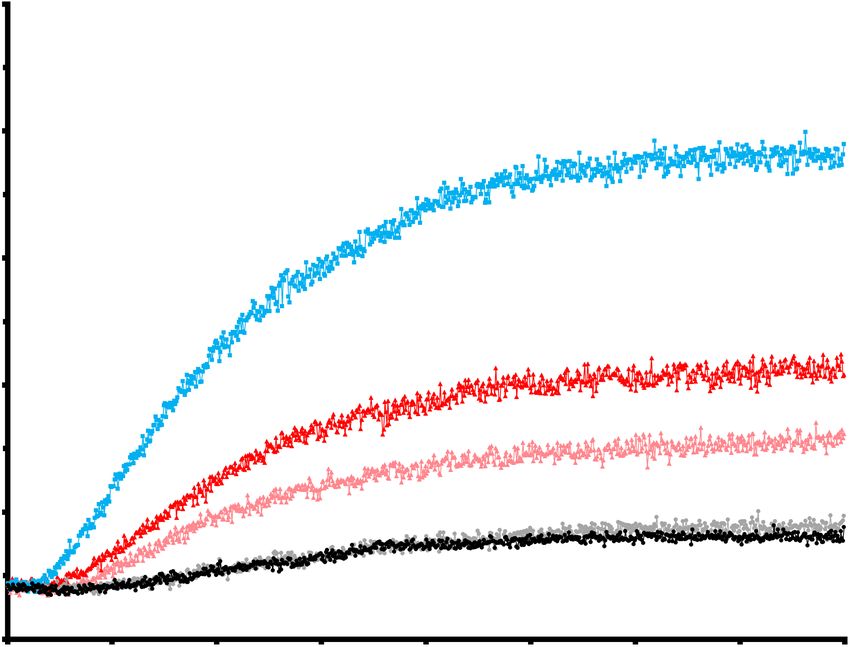
activity (Fig. 2B). Consistent with previous studies                    Our data suggest that DIAPH1 function is
(8,12), GST-RhoA only weakly stimulated the actin             regulated through the interaction of its DID region
nucleating activity of the DIAPH1 NT-CT complex               with IQGAP1. In a previous study, the regulation of
(Fig. 2C), while GST-IQGAP1-DBR alone did not                 DIAPH1 and DAAM1 was proposed to occur
stimulate the actin filament nucleation activity of           through an interaction between the formin DAD
the DIAPH1 NT-CT complex (Fig. 2D). In contrast,              regions and the gelsolin-like domains (GLD) of
in the presence of both GST-RhoA and GST-                     Flightless I (Fli-I). Fli-I binding to the DAD of
IQGAP1-DBR, actin nucleation activity was                     either DAAM1 or DIAPH1 enhanced RhoA-
dramatically increased, approaching the levels                mediated activation of the formins' actin nucleation
observed with the constitutively active MBP-                  activity (12). We confirmed this using a fragment
DIAPH1-CT alone (Fig. 2E).                                    of Fli-I (amino acids 398-1271) containing the
         The partial activation of DIAPH1 by RhoA             GLDs. GST-Fli-I-GLD bound to MBP-DIAPH1-
could reflect that RhoA only weakly or transiently            CT in the pulldown assay (Fig 3A) with a Kd of
promotes the dissociation of the formin DID and               139.3+7.6nM (Fig S1). However, there was no
DAD regions, or alternatively, could reflect that             detectable interaction with 6xHis-DIAPH1-NT
only a subset of DIAPH1 molecules are released                (Fig. 3A). Likewise, there was no detectable
from the autoinhibitory state, as our previous                interaction between GST-Fli-I-GLD and pre-

                                                                                                                       Downloaded from http://www.jbc.org/ by guest on November 3, 2020
binding data suggested (Fig. 1). To address this, we          formed DIAPH1 NT+CT complexes in pull down
determined the stoichiometry of complexes formed              assays (Fig. 3B), consistent with the weak
in the different reactions. Equimolar amounts of              interaction detected by BLI (Kd=3.93+0.18µM)
RhoA were added to preformed DIAPH1 NT-CT                     (Fig S1). However, in the presence of GST-RhoA,
complexes, then the 6xHis-DIAPH1-NT was re-                   GST-Fli-I-GLD bound to MBP-DIAPH1-CT in the
isolated and the amounts of co-purifying MBP-                 pulldown assay (Fig. 3B) with a Kd of
DIAPH1-CT and GST-RhoA were determined by                     166.5+47.5nM. These data suggest that RhoA binds
western blotting. When all the reaction components            to the DIAPH1 NT-CT complex to facilitate the
were present in equimolar amounts, the molar ratio            subsequent binding to Fli-I to the DAD region of
of       the        RhoA:DIAPH1-NT:DIAPH1-CT                  the DIAPH1-CT.
components in the re-isolated complex was 1: 2.3:                       To determine whether Fli-I binding could
1.2 (Fig. S3), suggesting that a single RhoA                  release DIAPH1 from its auto-inhibited state, we
releases a single DIAPH1-CT from a formin dimer.              next pre-formed the DIAPH1 NT-CT complex and
We interpret this to mean that only a subset of the           incubated the complex with either GST, GST-
DIAPH1 molecules are activated in the presence of             RhoA, GST-IQGAP1-DBR, GST-Fli-I-GLD, or a
RhoA and thus only partial actin nucleation activity          combination of these reagents (Fig. 3C, D). Fli-1
is observed. In contrast, when IQGAP1 was added               alone did not release the MBP-DIAPH1-CT from
to the reaction in equimolar amounts, the re-isolated         the 6xHis-DIAPH1-NT. However, GST-Fli-I-GLD
complexes contained no DIAPH1-CT, with a                      did enhance the GST-RhoA-mediated release of
RhoA:       IQGAP1:       DIAPH1-NT:DIAPH1-CT                 MBP-DIAPH1-CT from the 6xHis-DIAPH1-NT to
stoichiometric ratio of near 1:1:1:0 (1 : 0.95 : 1.15 :       a level comparable to IQGAP1. Consistent with
0.08 ; Fig. S3), suggesting that nearly every                 this, in vitro pyrene actin filament nucleation assays
DIAPH1 molecule is released from the                          showed that Fli-1 enhances RhoA-mediated
autoinhibited state, leading to the maximal                   activation of the actin filament nucleation activity
expected increase in actin polymerization in the              of the DIAPH1 NT-CT complex (Fig. 4). Fli-I was
pyrene actin assays (Fig. 2). Thus, IQGAP1                    however a weaker enhancer of DIAPH1 activity
enhances the RhoA-mediated activation of                      relative to IQGAP1. In the presence of GST-RhoA
DIAPH1 by enhancing the release of DIAPH1 from                and GST-Fli-I-GLD, MBP-DIAPH1-CT exhibited
its autoinhibited state and leading to robust actin           a 35% reduction in the actin polymerization rate (t
filament nucleation activity.                                 1/2) relative to that observed in the presence of GST-
                                                              RhoA and GST-IQGAP1-DBR (Fig. 4C, D). In the
Fli-I enhances RhoA-mediated release                of        presence of both GST-IQGAP1-DBR and GST-Fli-
DIAPH1 from the autoinhibited state                           I-GLD, we observed an increase in MBP-DIAPH1-
                                                              CT release from the 6xHis-DIAPH1-NT; however,

                                                          4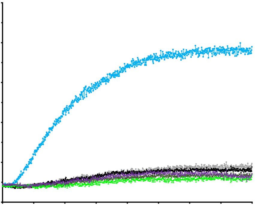
this did not increase the DIAPH1 actin nucleation            microscopy data suggest that in the cell lines
activity (Fig. 3C, D), suggesting that either the            examined, IQGAP1 enhances RhoA mediated
DAD region plays additional roles in actin                   activation of DIAPH1 actin nucleation activity at
polymerization or that Fli-I recruitment is itself           the plasma membrane while Fli-1 exerts little effect
inhibitory to actin filament production.                     in the cell lines and conditions examined.

IQGAP1 but not Fli-I targets DIAPH1 within the               DISCUSSION
cell                                                                  The precise control of when and where
         We previously showed that anillin both              actin filaments are generated in response to
enhances DIAPH3 actin nucleation activity, and               different stimuli is a fundamental aspect of
targets DIAPH3 to the cytokinetic furrow (13). As            cytoskeletal regulation. Small G-proteins are
Fli-I and IQGAP1 similarly enhance DIAPH1                    instrumental in activating formins, but in vitro,
activity, we sought to assess their ability to target        small G-proteins only partially overcome formin
DIAPH1 activity to the plasma membrane. In                   auto-inhibition, and do so indiscriminately (5,8,11-
MDA-MD-231 cells, DIAPH1 predominantly                       13). We previously demonstrated that the
localizes to the plasma membrane (Fig. 5). IQGAP1            cytokinetic factor anillin enhances RhoA-mediated
colocalizes with DIAPH1 at the plasma membrane               activation of the formin DIAPH3 by binding to the

                                                                                                                       Downloaded from http://www.jbc.org/ by guest on November 3, 2020
(Manders Overlapping Coefficient, MOC, of 0.819              Diaphanous Inhibitory Domain (DID) and directs
+ 0.012 and a Pearson Correlation Coefficient,               actin filament production to the cytokinetic furrow
PCC, of 0.565 + 0.018, Fig. 5A, C). In contrast, Fli-        (13). Anillin is therefore a bi-functional regulator of
I did not co-localize with DIAPH1 at the plasma              DIAPH3, ensuring that actin nucleation is both
membrane (MOC and PCC to 0.522 + 0.020 and                   enhanced and targeted to a specific sub-cellular
0.320 + 0.026 respectively, Fig. 5B, D). Consistent          location, the cytokinetic furrow. In this study we
with these results, depletion of IQGAP1 by siRNA             extend this principle of coupling formin activation
reduced DIAPH1 targeting to the plasma membrane              with sub-cellular localization to a second formin,
(Fig. 5A), whereas depletion of Fli-I did not (Fig.          DIAPH1. In this work, we show that RhoA-
5B). Similar observations were made in human                 mediated activation is enhanced by the binding of
gingival fibroblast (HGF) cells (Fig. S4).                   IQGAP1 to the DID of DIAPH1, which both targets
         To determine the role of DIAPH1,                    the formin to its sub-cellular localization and
IQGAP1, and Fli-I in actin filament production, we           promotes its full activation. A previous study
analyzed actin accumulation at the leading-edge of           demonstrated that IQGAP1 recruits DIAPH1 to the
MDA-MB-231 cells using rhodamine-phalloidin                  leading edge of migrating cells and to the
staining in the presence and absence of DIAPH1,              phagocytic cup, but found no role for IQGAP1 in
IQGAP1 and Fli-I (Fig. 6). In control siRNA treated          DIAPH1 activation (12), possibly because the
cells, we observed a strong actin signal at the              authors reported maximal activation of DIAPH1 in
leading edge that co-localized with IQGAP1 (MOC              the presence of RhoA. In contrast, other studies,
= 0.809 + 0.019, PCC = 0.533 + 0.018, Fig. 6B, D,            including our own, report only the partial activation
E) and also in part with DIAPH1, reflecting that a           of DIAPH1 by RhoA (including but not limited to
pool of DIAPH1 also localized to actin-poor peri-            references (5,8,11,12)), suggesting that in vivo actin
nuclear locations (MOC = 0.481 + 0.031, PCC =                nucleation is modulated by additional factors that
0.45 + 0.030, Fig. 6A, D, E). Fli-I localized                control both formin localization and function. By
primarily to actin-poor peri-nuclear regions and             exploiting a two-factor activation mechanism in the
thus did not co-localize with actin at the leading           control of formin activity, more precise spatio-
edge of cells (MOC = 0.256 + 0.023, PCC = 0.059              temporal regulation can be exerted as both factors
+ 0.013, Fig. 6C-E), suggesting that it is unlikely to       must be present in one location for maximal
play a role in regulating actin dynamics at the              activation.
plasma membrane. Consistent with these                                There are clear parallels between the
observations, depletion of either IQGAP1 or                  activation mechanisms of DIAPH1 and DIAPH3:
DIAPH1 reduced the actin staining at the leading             both formins exist in an auto-inhibited state that is
edge (Fig. 6A, B), while depletion of Fli-I did not          mediated by an intramolecular interaction between
(Fig. 6C). Taken together our biochemical and                the DID and DAD regions, and both formins exploit

                                                         5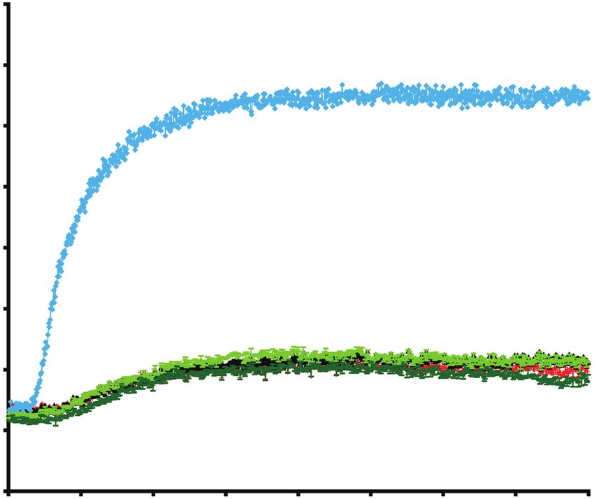
a DID-binding factor to augment RhoA-mediated                of actin with DIAPH1. Interestingly the mutations
activation. In both cases the enhancer has a dual            are directly adjacent to mutations that disrupt the
function, to both fully activate and target actin            formin-Fli-I interaction (12), suggesting that both
nucleating activity to a specific sub-cellular region.       actin and Fli-I may interact with formins at the
We propose that this general activation mechanism            same site. Consequently, Fli-I binding could
is likely to be a common feature of other                    attenuate formin-mediated actin nucleation and
Diaphanous-related formins (Fig. 7).                         elongation activities.
          Additional regulatory pathways are likely                   While we propose that the activation of
to contribute to the correct spatio-temporal                 Diaphanous family formins is a two-step process,
regulation of formin activity. Fli-I, a protein with         we do not envisage that indiscriminate protein
actin capping and filament severing activities (19),         binding to the DID necessarily confers a formin
binds to the DAD of DIAPH1 and DAAM1 at a site               activating function. For instance, the binding of
conserved across DRFs. Our work, and that of                 Liprin-a3 to the DID of DIAPH1 has an inhibitory
others (12), demonstrated that Fli-I binding to              effect, as it prevents the correct targeting of
DIAPH1 released DIAPH1 from its auto-inhibited               DIAPH1 to the membrane (21,22). Liprin-a3 has
state and enhanced RhoA dependent activation of              been proposed to displace IQGAP1 from DIAPH1
DIAPH1 in vitro. We extend these results to show             (21), suggesting that the control of formin activity

                                                                                                                       Downloaded from http://www.jbc.org/ by guest on November 3, 2020
that in our assays Fli-I is a less potent enhancer of        can occur through both positive and negative
DIAPH1 activation than IQGAP1.                               effectors, and providing yet greater scope for the
          The mechanism underlying the different             multi-layered regulation of the actin cytoskeleton.
formin enhancing activities of IQGAP1 and Fli-I is                    Our previous work demonstrated that
unclear. Full activation of formins must require             during cytokinesis, anillin interacts with DIAPH3,
structural changes to the formin that re-orient the          recruiting the formin to the cytokinetic furrow and
N- and C-terminal regions such that the N-terminus           enhancing its RhoA-dependent activation (13).
of the formin no-longer sterically blocks the                Here we find that IQGAP1 performs an analogous
interaction of actin monomers with the actin                 role in regulating DIAPH1: IQGAP1 targets
nucleating FH2 domain located in the formin C-               DIAPH1 to the plasma membrane and there
terminus (5). If DID binding factors such as                 enhances DIAPH1 actin nucleation activity. In
IQGAP1 and anillin more efficiently facilitate or            contrast, Fli-I was not required for DIAPH1
stabilize this “open” form of the formin than DAD            targeting in the cell lines tested (MDA-MB-231,
interacting factors such as Fli-I, then IQGAP1 and           Fig. 5, 6 and HGF, Fig. S4). It still remains possible
anillin would be expected to have a greater                  that in specialized cell lines, Fli-I could be involved
stimulatory effect on formin actin nucleation                in regulating DIAPH1 activity. Alternatively, Fli-I
activity.                                                    activity may be preferentially directed toward other
          Fli-I is a modulator of the actin                  formins in vivo, as the Fli-I interaction site on
cytoskeleton in its own right, and it may be that            DIAPH1 and DAAM1 is conserved in other
these activities account for its weaker enhancement          Diaphanous related formins.
of DIAPH1 activation in comparison to IQGAP1.                         Our studies define one regulatory
The actin capping and severing activities of Fli-I           mechanism for the spatio-temporal control of
may counter actin filament elongation by DIAPH1.             formin activity. However, in vivo formin regulation
At much higher concentrations of the Fli-I fragment          is likely to be more complex, as formins can
that contains all six gelsolin-like-domains, we              undergo post-translational modifications that may
observed such activities over extended periods (19).         modulate their activity (23-30). Taken together,
These data raise the interesting possibility that            these observations suggest that formins are subject
formin activity could be fine-tuned by recruiting            to a rich array of regulatory mechanisms, allowing
capping/severing proteins depending on the needs             for the dynamic control of distinct actin
of the cell. Alternatively, Fli-I could interfere with       superstructures within the cell.
actin binding to the formin. The DAD is required
for full DIAPH1 actin nucleating activity in vitro           EXPERIMENTAL PROCEDURES
through an as yet undefined interaction with actin
(20). Mutations in the DAD perturb the interaction           cDNA cloning

                                                         6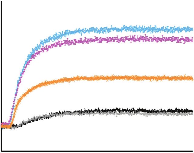
pGEX-6P-2-DIAPH1-CT        (580-1272aa)                 To purify MBP fusion proteins, ER2523 E.
and pET30a-DIAPH1-NT(1-575aa) plasmids were             coli cells were harvested, lysed as above. The
previously generated by J. C. The IQGAP1 full           lysates were cleared by centrifugation as above then
length cDNA was a synthetic gene corresponding          supernatant applied to amylose resin (New England
to NP 003861 a gift from Drs Y. Tong and C.             Biolabs). The resin was washed with 10 column
Arrowsmith (Structural Genomics Consortium,             volumes of CB and the MBP fusion proteins were
Toronto, Ontario, Canada). pGEX-4T-2-Fli-I-GLD          eluted in CB containing 10 mM maltose.
(1-6) plasmid was previously described (16).                     To purify 6× His fusion proteins, BL21 E.
Complementary DNA (cDNAs) fragments of                  coli cells were re-suspended in 25 mM HEPES, pH
human DIAPH1-CT (583-1272aa), human                     7.5, 500 mM NaCl, 5% (v/v) glycerol, 5 mM
IQGAP1-DBR (1500-1657aa) were amplified by              imidazole, 0.5 mM β-mercaptoethanol, 1 mM
PCR using the i-Max II DNA polymerase                   PMSF, and lysed by sonication. The lysates were
(Froggalab) using oligonucleotide primers               cleared by centrifugation as above then supernatant
(Integrated DNA Technologies) listed in                 applied to nickel-Sepharose beads (Amersham
Supplementary Table 1. PCR fragments of                 Biosciences). The beads were washed with 10
IQGAP1-DBR were cloned into pDEST15                     column volumes of 6× His column buffer (HCB)
destination vector (Life Technologies) using In-        containing 25 mM HEPES, pH 7.5, 500 mM NaCl,

                                                                                                                 Downloaded from http://www.jbc.org/ by guest on November 3, 2020
Fusion Cloning Kit (Clontech) to generate GST           5% (v/v) glycerol, 5 mM imidazole, 0.5 mM β-
fusion proteins. PCR fragments of DIAPH1-CT             mercaptoethanol, 1 mM PMSF, 0.1% (v/v) Triton
were cloned using the TOPO Gateway system (Life         X-100. The 6× His fusion proteins were eluted in
Technologies) being first cloned into the entry         HCB containing 500 mM imidazole.
plasmid vector pCR8/GW/TOPO, then moved into                     Eluted proteins were dialyzed against 10
the destination vectors pKM596 (Addgene plasmid         mM HEPES, pH 7.6, 100 mM KCl, 2 mM MgCl2,
8837) to generate MBP fusion proteins.                  50 mM sucrose for 16h at 4 °C and concentrated
                                                        using Millipore Ultrafree spin columns (Milipore,
Protein expression and purification                     Ireland) with a 10-kDa cutoff. Proteins were then
         Recombinant proteins were purified from        aliquoted, flash-frozen in N2(l), and stored at −80°C.
BL21 E. coli cells transformed with plasmids
containing 6x His or GST fusion proteins, or            Nucleotide loading of GST-RhoA
ER2523 E. coli (New England Biolabs) with                       To generate GTP- or GDP-loaded GST-
plasmids containing MBP fusion proteins. Cells          RhoA (previously described in (13,31) for the in
were grown in LB media at 37 °C to an optical           vitro binding and pyrene-actin polymerization
density of 0.6 at A600. Recombinant protein             assays, purified GST-RhoA fusion proteins were
expression was induced by the addition of 1 mM          added to 25 mM EDTA, 1 mM DTT, and GTP or
isopropyl β-D-1-thiogalactopyranoside (IPTG) and        GDP added to a 100× molar excess of the proteins.
further incubated at 16 °C overnight. Cells were        The reactions were incubated on ice for 40 min
harvested by centrifugation and stored in −80°C.        before adding MgCl2 to a final concentration of 50
         To purify GST fusion proteins, BL21 E.         mM. GTP- or GDP-loaded proteins were then
coli cells were re-suspended in 25 mM HEPES, pH         dialyzed, concentrated, and stored as described
7.5, 250 mM NaCl, 100 mM KCl, 0.5 mM β-                 above.
mercaptoethanol, 1 mM PMSF, and lysed by
sonication. The lysates were cleared by                 In vitro binding assays
centrifugation at 10,000×g for 30 min at 4 °C and                To determine if IQGAP1 binds to
supernatant applied to glutathione beads                DIAPH1-NT, 0.05nmol 6×His-DIAPH1-NT was
(Invitrogen). The glutathione beads were washed         immobilized onto 25µl nickel-Sepharose beads in
with 10 column volumes of column buffer (CB)            100µl of 6×His incubation buffer (HIB) containing
containing 25 mM HEPES, pH 7.5, 250 mM NaCl,            25mM HEPES, pH 7.5, 120mM NaCl, 1mM
100 mM KCl, 0.5 mM β- mercaptoethanol, 1 mM             EGTA, 0.3% (v/v) Triton X-100, 1mM β-
PMSF, 0.1% (v/v) Triton X-100. The GST fusion           mercaptoethanol and incubated for 1h at 4 °C. The
proteins were eluted in CB containing 10 mM             beads were washed in HIB (all subsequent 6×His
glutathione.                                            protein washes were done in this buffer unless

                                                    7
otherwise noted) and blocked with 3% (w/v) BSA          protocol above was used except analysis was
for 20min. The beads were then washed and mixed         carried out with an anti-IQGAP1 polyclonal
with 0.05nmol of GST-IQGAP1-DBR and                     antibody (ab109292, Abcam, 1:250 dilution) to
incubated for 2h at 4 °C. Unbound protein was           detect co-purifying IQGAP1 or an anti-RhoA
removed by washing the beads in HIB. The beads          monoclonal antibody (ab86297, Abcam, 1:200
were re-isolated by centrifugation and boiled in        dilution) to detect co-purifying RhoA. To
SDS sample buffer then analyzed by Western              determine if Fli-I or RhoA bound to the DIAPH1
blotting using an anti-IQGAP1 polyclonal antibody       CT-NT complex first, the same protocol above was
(ab109292, Abcam, 1:250 dilution) to detect co-         used except analysis was carried out with the
purifying IQGAP1. To determine if IQGAP1 binds          homemade anti-GST polyclonal antibody to detect
to DIAPH1-CT, 0.05nmol MBP-DIAPH1-CT was                co-purifying Fli-I or an anti-RhoA monoclonal
immobilized onto 25µl amylose resin in 100µl of         antibody (ab86297, Abcam, 1:200 dilution) to
incubation buffer (IB) containing 50 mM Hepes pH        detect co-purifying RhoA.
7.5, 50 mM NaCl, 1 mM EDTA, 5 mM MgCl2,
0.3% (v/v) Triton X-100, 1 mM β-mercaptoethanol         Biolayer interferometry assays
and incubated for 1h at 4°C. The beads were                      Dissociation constants between different
blocked, incubated with GST-IQGAP1-DBR, re-             proteins were determined by the Octet RED96

                                                                                                               Downloaded from http://www.jbc.org/ by guest on November 3, 2020
isolated and analyzed by Western blotting as            system (FortéBio), which measures association
described above to detect co-purifying IQGAP1.          onto and dissociation from a sensor surface using
         To assess the roles of IQGAP1 and RhoA         Biolayer interferometry (BLI) as previously
in regulating the interaction between DIAPH1-NT         described (32,33). Briefly, purified GST-tagged
and DIAPH1-CT, 0.05nmol of 6×His-DIAPH1-NT              ligands (RhoA, IQGAP1, Fli-I, DIAPH1-CT or
was immobilized onto 25µl nickel-Sepharose beads        preformed DIAPH1-NT-CT complex) were
in 100µl HIB as described above and incubated for       equilibrated into kinetics buffer (KB) which
1h at 4 °C. The beads were washed to removed            contains phosphate-buffered saline (PBS), 0.002%
unbound protein, then blocked with 3% (w/v) BSA         Tween-20, and 0.1mg/ml BSA, to a concentration
for 45min and further washed. 0.05nmol MBP-             of 25µg/ml. The GST biosensors (FortéBio) were
DIAPH1-CT was then incubated with the beads for         first equilibrated in KB for 60s and then followed
2h at 4 °C, followed by a washing step. GST-            by incubation with GST ligands in KB for 300s.
IQGAP1-DBR or GST-RhoA were then added to               Sensors were then rinsed in KB for 250s to obtain a
the beads, incubated for 2h then washed in HIB to       baseline of the levels of GST ligands loading onto
remove unbound protein. The beads were re-              the biosensors. Binding assays were then performed
isolated by centrifugation, boiled in SDS sample        in a series of increased concentration of analyte
buffer then analyzed by Western blotting using an       (DIAPH1-NT, GST tag-removed RhoA or
anti-MBP monoclonal antibody (E8032, New                IQGAP1 or DIAPH1-CT) from 0.5nM to 5µM,
England Biolabs, 1:2500 dilution) to detect co-         from the lowest concentration to the highest. Each
purifying MBP-DIAPH1-CT.                                binding sequence started with a baseline incubation
         To assess the roles of Fli-I and RhoA in       in KB (150s), followed by the association with the
regulating the interaction between DIAPH1-NT            analyte (300s), the dissociation in KB (250s), and a
and DIAPH1-CT, DIAPH1 CT-NT complex was                 regeneration step (200s) where sensors were rinsed
reconstituted as described above. GST-Fli-I-GLD         in regeneration buffer (100mM sodium citrate
or GST-RhoA were then added to the beads,               pH=4.5, 50mM EDTA, and 150mM NaCl), to
incubated for 2h then washed in HIB to remove           remove analyte still bound to GST ligands.
unbound protein. The beads were re-isolated by          Reference sensors that were loaded with GST
centrifugation, boiled in SDS sample buffer then        ligands but only assayed in a series of pure KB, or
analyzed by Western blotting using an anti-MBP          a series of increasing concentration of MBP were
monoclonal antibody (E8032, New England                 also measured as controls. All incubation steps
Biolabs, 1:2500 dilution) to detect co-purifying        were performed at 30°C with a shaking speed of the
MBP-DIAPH1-CT.                                          plates at 1000rpm.
         To determine if IQGAP1 or RhoA bound to                 Data analysis was performed using Octet
the DIAPH1 CT-NT complex first, the same                Software (FortéBio) and GraphPad Prism (8.2.0).

                                                    8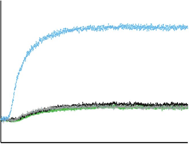
The control signals measured by the reference               unlabeled actin in a 15%: 85% ratio to make a final
sensors were subtracted from the signals measured           concentration of 2 µM total actin. Freshly purified
by analyte-bound sensors. Steady state analysis was         proteins were added to actin at a concentration of
performed using FortéBio Data Analysis 9 software           2.5 nM and incubated for 5 min at room
to obtain the dissociation constant (Kd) from the           temperature. The reaction was initiated by adding
equilibrium response. Each binding sequence was             polymerization buffer (25 mM Tris, pH 7.0, 50 mM
repeated >4 times. The resulted Kd values and               KCl, 2 mM MgCl2, 0.1 mM ATP). The increase in
averages were plotted in GraphPad Prism, with               fluorescence intensity was monitored in a PTI
error bars representing ±s.d.                               fluorimeter with excitation at 365 nm and emission
                                                            at 386 nm (34). The initial time point was set to
Quantification of Western blots                             when the polymerization buffer was added and the
         The PVDF membrane of Western blots was             reaction was monitored for 800s. The t(1/2) was
developed by chemiluminescent solution (Life                defined as the time point when half amount of total
Technologies) for 5 min at room temperature and             actin monomers polymerized to filaments (35).
visualized using a BioRad MP Imager (Bio-Rad,               Actin polymerization rates at t(1/2) were calculated
Canada). The intensities of individual bands on the         using the same methods as previously described
blots were measured using ImageLab software                 (35). Briefly, 10 data points at the minimum and the

                                                                                                                            Downloaded from http://www.jbc.org/ by guest on November 3, 2020
(Bio-Rad Inc.). To determine the relative binding in        maximum intensities were chosen and used to
the in vitro competition assays (Fig. 3B), a control        calculate Imin (the average minimum intensity) and
pulldown reaction of wild-type “bait” protein that          Imax (the average maximum intensity). The data
bound to beads and prey protein was performed and           point with intensities between 0.48 × (Imax−Imin) +
run on each gel. The intensity of the of bait protein       Imin and 0.52 × (Imax−Imin) + Imin was chosen, fitted
band that bound to beads and the band of prey               to a linear line and where the slope = m (1/2) and the
protein that was pulldown were set as the control           intercept = b (1/2). Subsequently the t (1/2) (time point
standard to 1. In the comparative reactions, where          when half amount of total actin monomers
potential competitor proteins were added or                 polymerize to filaments) was calculated as: t (1/2) =
changing concentrations of proteins were added,             (0.5 × (Imax−Imin) + Imin + b (1/2))/ m (1/2), the AP t (1/2)
the band intensity of the bait protein in each              (actin polymerization rate at t (1/2)) as APt (1/2) = 1.88
reaction was first compared and the differences             × m (1/2)/ (Imax−Imin).
used to adjust the band intensity of the amount of
co-purifying prey protein to allow comparison. The          siRNA knock down assays
adjusted prey band intensities were then compared                     MDA-MB-231 cells (a gift from Dr L.
to the intensity of the prey in the control reaction.       Attisano, University of Toronto) and human
Each binding assay was repeated at least three              gingival fibroblasts (HGF) cells were transfected
times. Analogous strategies were used to compare            with 40nM double-strand IQGAP1 or Fli-I siRNA
direct in vitro binding assays in Fig. 1E, F and Fig.       using Lipofectamine 2000 Reagent (Invitrogen)
3. The relative intensities of different bands were         and      DIAPH1      localization   analyzed    by
compared using a Student’s t test to calculate the p-       immunofluorescence. Alternatively, MDA-MB-
value. Error bars indicate +s.d.                            231 cells were transfected with 40nM double-strand
                                                            DIAPH1 siRNA as described above and IQGAP1
In-vitro pyrene-actin polymerization assays                 or       Fli-I    localization     analyzed     by
        To determine the actin polymerization               immunofluorescence. siRNAs were obtained from
activity of DIAPH1, 1 mg lyophilized pyrene-                Integrated DNA Technologies. The negative
labeled or unlabeled actin (Cytoskeleton Inc) was           control siRNA was previously used and validated
re-suspended in 50 µl H2O at 4 °C, then 150 µl G-           (31). All siRNA duplexes used in this study were
buffer (monomer actin buffer: 2 mM Tris pH 8.0,             listed in Supplementary Table 1.
0.2 mM CaCl2, 0.2 mM ATP, 0.5 mM β-
mercaptoethanol) was added to make an actin stock           Immunofluorescence and microscopy analysis
(58 µM) and incubated on ice for 2 h. For individual                 To analyze DIAPH1 and IQGAP1
assays, the actin stock was further diluted in G-           localization in MDA-MB-231 cells, with 2.5% PFA
buffer and the pyrene-labeled actin was mixed with          at room temperature for 15min, permeabilized for

                                                        9
10 min with 0.1% Triton X-100 in PBS then Quantification and statistical analysis
blocked by 3% (w/v) BSA in PBS for 1h at room The quantification of the band intensities in
temperature. Cells were stained by anti-DIAPH1 the Western blots (Fig. 1, 3) was performed using
(BD Transduction Laboratories, 1:100 dilution) or ImageLab software (Bio-Rad Inc.), as detailed in
anti-IQGAP1 (Abcam, 1:200 dilution) antibodies. the section “Quantification of Western blots”. Each
Subsequently cells were washed three times with competitive binding assay was repeated at least 3
PBS; a secondary goat anti-mouse antibody times with the numerical values of band intensities
conjugated to Alexa 594 (Life Technologies, analyzed in Excel (Fig. 1F, 3C). All results
1:1000 dilution) (against anti-DIAPH1 antibody) or presented in graphs indicate the mean ± s.d. A
goat anti-rabbit antibody conjugated to Alexa 488 Student’s t test was used to calculate p-values.
(Life Technologies, 1:1000 dilution) (against anti- The quantification of the actin
IQGAP1 antibody) were used to visualize the polymerization rate (APt(1/2)) (Fig. 2F, 4D) in the in-
cellular DIAPH1 or IQGAP1 localization. Cells vitro actin polymerization assays was performed
were then stained with 4’,6-diamidino-2- using GraphPad Prism (v8.1.2). Each actin
phenylindole (DAPI) to visualize DNA. polymerization assay was independently repeated
To analyze DIAPH1 and Fli-I localization three times and the average actin polymerization
in MDA-MB-231 cells, cells were fixed, rate of actin alone (2µM) with polymerization
Downloaded from http://www.jbc.org/ by guest on November 3, 2020
permeabilized and blocked as described above. buffer was set to 1.0. Dots presented in graphs
Cells were stained by anti-DIAPH1 (BD indicate individual data points. Red bars indicate
Transduction Laboratories, 1:100 dilution) or anti- the average value of the data points in each
Fli-I (Santa Cruz Biotechnology, 1:200 dilution) experimental group. The quantification of co-
antibodies. Subsequently cells were washed three localization analysis of DIAPH1, IQGAP1, Fli-I or
times with PBS; a secondary goat anti-mouse IgG1 actin in MDA-MB-231 cells were performed using
conjugated to Alex 594 (Life Technologies, 1:1000 the Coloc 2 plugin of ImageJ (Fiji package, v 2.0.0-
dilution) (against anti-DIAPH1 antibody) or goat rc-69). A total of 30 sets of images in each co-
anti-mouse IgG2a conjugated to Alex 488 (Life staining group were analyzed to calculate both the
Technologies, 1:1000) (against anti-Fli-I antibody) auto-threshold Mander’s overlapping coefficient
were used to visualize the cellular DIAPH1 or (tM) or Pearson’s correlation coefficient (PCC,
IQGAP1 localization. Alternatively, cells were above threshold). For the analysis of Mander’s
stained with rhodamine-labeled phalloidin overlapping coefficient or Pearson’s correlation
(Invitrogen, 1:1000 dilution) to visualize the total coefficient of the whole image, two channels of a
actin cytoskeleton. co-staining set were first subjected to background
Coverslips were mounted on glass slides subtraction with a rolling ball algorithm (radius =
using Mowiol (Polyvinyl alcohol 4-88, Fluka). 50.0) in ImageJ. The two channels were then
Cells were visualized using a Perkin Elmer analyzed by Coloc 2 plugin using Costes method
UltraView spinning disk con- focal scanner threshold regression (37). The point spread function
mounted on a Nikon TE2000-E with a ×60/1.4 NA (PSF) was set to 10, with Costes randomization set
oil-immersion objective lens and 1.515 immersion to 100. The M1 channel was assigned as DIAPH1
oil at room temperature. Images were acquired (Figure 5C, D) or phalloidin staining (Figure 6D,
using METAMORPH software (Molecular E), while M2 channel assigned as the co-staining
Devices) driving an electron multiplying charge- protein in each group as indicated in the graph. The
coupled device (CCD) camera (ImagEM, values of tM1, PCC (above threshold) were
Hammamatsu) (36). Z sections (0.2 µm apart) were exported for statistical analysis. The control groups
acquired to produce a stack that was then imported were set up to compare a DIAPH1 (or phalloidin)
into AUTO-QUANT X2 (Media Cybernetics) for single channel image with the exact same image (0°
deconvolution (10 iterations). Maximum rotation, positive control) or rotating it 90°
projections and cross sections were performed (negative control). For the analysis of tM or PCC at
using METAMORPH. Images were overlaid in the leading edge, a region of interest (ROI) was first
PHOTOSHOP (Adobe), involving adjustments in drawn using the Freehand tools of ImageJ to
brightness and contrast of images. generate the leading edge segment of both channels
of a co-staining set. The images were then analyzed
10by Coloc2 to calculate the tM1 or PCC using the Conflicts of Interest
same methods described above. All graphs and The authors declare that they have no conflicts of
statistical tests in co-localization analysis were interest with the contents of the article
generated by GraphPad Prism (v8.1.2). The non-
parametric unpaired Mann Whitney t test (n=30)
was used to calculate p- values. No statistical
method was used to predetermine sample size.
Acknowledgements
AW is supported by CIHR grant PJT 148575, CAM
is supported by a Tier 1 Canada Research Chair and
CIHR grant MOP36332, TFM is supported by
NSERC grant RGPIN-2018-06546 and JWC is
supported by grant 24154 from the Cancer
Research Society.
Downloaded from http://www.jbc.org/ by guest on November 3, 2020
REFERENCES
1. Skau, C. T., and Waterman, C. M. (2015) Specification of Architecture and Function of
Actin Structures by Actin Nucleation Factors. Annu Rev Biophys 44, 285-310
2. Blanchoin, L., Boujemaa-Paterski, R., Sykes, C., and Plastino, J. (2014) Actin dynamics,
architecture, and mechanics in cell motility. Physiol Rev 94, 235-263
3. Kuhn, S., and Geyer, M. (2014) Formins as effector proteins of Rho GTPases. Small
GTPases 5, e29513
4. Schonichen, A., and Geyer, M. (2010) Fifteen formins for an actin filament: a molecular
view on the regulation of human formins. Biochim Biophys Acta 1803, 152-163
5. Maiti, S., Michelot, A., Gould, C., Blanchoin, L., Sokolova, O., and Goode, B. L. (2012)
Structure and activity of full-length formin mDia1. Cytoskeleton (Hoboken) 69, 393-405
6. Otomo, T., Tomchick, D. R., Otomo, C., Panchal, S. C., Machius, M., and Rosen, M. K.
(2005) Structural basis of actin filament nucleation and processive capping by a formin
homology 2 domain. Nature 433, 488-494
7. Nezami, A., Poy, F., Toms, A., Zheng, W., and Eck, M. J. (2010) Crystal structure of a
complex between amino and carboxy terminal fragments of mDia1: insights into
autoinhibition of diaphanous-related formins. PloS one 5
8. Li, F., and Higgs, H. N. (2003) The mouse formin mDia1 is a potent actin nucleation
factors regulated by autoinhibition. Current biology : CB 13, 1335-1340
9. Rose, R., Weyand, M., Lammers, M., Ishizaki, T., Ahmadian, M. R., and Wittinghofer,
A. (2005) Structural and mechanistic insights into the interaction between Rho and
mammalian Dia. Nature 435, 513-518
10. Lammers, M., Rose, R., Scrima, A., and Wittinghofer, A. (2005) The regulation of
mDia1 by autoinhibition and its release by Rho*GTP. The EMBO journal 24, 4176-4187
11. Li, F., and Higgs, H. N. (2005) Dissecting requirements for auto-inhibition of actin
nucleation by the formin, mDia1. The Journal of biological chemistry 280, 6986-6992
12. Higashi, T., Ikeda, T., Murakami, T., Shirakawa, R., Kawato, M., Okawa, K., Furuse, M.,
Kimura, T., Kita, T., and Horiuchi, H. (2010) Flightless-I (Fli-I) regulates the actin
assembly activity of diaphanous-related formins (DRFs) Daam1 and mDia1 in
11cooperation with active Rho GTPase. The Journal of biological chemistry 285, 16231-
16238
13. Chen, A., Arora, P. D., McCulloch, C. A., and Wilde, A. (2017) Cytokinesis requires
localized beta-actin filament production by an actin isoform specific nucleator. Nat
Commun 8, 1530
14. Watanabe, S., Okawa, K., Miki, T., Sakamoto, S., Morinaga, T., Segawa, K., Arakawa,
T., Kinoshita, M., Ishizaki, T., and Narumiya, S. (2010) Rho and anillin-dependent
control of mDia2 localization and function in cytokinesis. Molecular biology of the cell
21, 3193-3204
15. Arora, P. D., Di Gregorio, M., He, P., and McCulloch, C. A. (2017) TRPV4 mediates the
Ca(2+) influx required for the interaction between flightless-1 and non-muscle myosin,
and collagen remodeling. Journal of cell science 130, 2196-2208
16. Arora, P. D., Wang, Y., Bresnick, A., Janmey, P. A., and McCulloch, C. A. (2015)
Flightless I interacts with NMMIIA to promote cell extension formation, which enables
collagen remodeling. Molecular biology of the cell 26, 2279-2297
17. Copeland, S. J., Green, B. J., Burchat, S., Papalia, G. A., Banner, D., and Copeland, J. W.
Downloaded from http://www.jbc.org/ by guest on November 3, 2020
(2007) The diaphanous inhibitory domain/diaphanous autoregulatory domain interaction
is able to mediate heterodimerization between mDia1 and mDia2. The Journal of
biological chemistry 282, 30120-30130
18. Brandt, D. T., Marion, S., Griffiths, G., Watanabe, T., Kaibuchi, K., and Grosse, R.
(2007) Dia1 and IQGAP1 interact in cell migration and phagocytic cup formation. The
Journal of cell biology 178, 193-200
19. Mohammad, I., Arora, P. D., Naghibzadeh, Y., Wang, Y., Li, J., Mascarenhas, W.,
Janmey, P. A., Dawson, J. F., and McCulloch, C. A. (2012) Flightless I is a focal
adhesion-associated actin-capping protein that regulates cell migration. FASEB J 26,
3260-3272
20. Gould, C. J., Maiti, S., Michelot, A., Graziano, B. R., Blanchoin, L., and Goode, B. L.
(2011) The formin DAD domain plays dual roles in autoinhibition and actin nucleation.
Current biology : CB 21, 384-390
21. Brenig, J., de Boor, S., Knyphausen, P., Kuhlmann, N., Wroblowski, S., Baldus, L.,
Scislowski, L., Artz, O., Trauschies, P., Baumann, U., Neundorf, I., and Lammers, M.
(2015) Structural and Biochemical Basis for the Inhibitory Effect of Liprin-alpha3 on
Mouse Diaphanous 1 (mDia1) Function. The Journal of biological chemistry 290, 14314-
14327
22. Sakamoto, S., Ishizaki, T., Okawa, K., Watanabe, S., Arakawa, T., Watanabe, N., and
Narumiya, S. (2012) Liprin-alpha controls stress fiber formation by binding to mDia and
regulating its membrane localization. Journal of cell science 125, 108-120
23. Floyd, S., Whiffin, N., Gavilan, M. P., Kutscheidt, S., De Luca, M., Marcozzi, C., Min,
M., Watkins, J., Chung, K., Fackler, O. T., and Lindon, C. (2013) Spatiotemporal
organization of Aurora-B by APC/CCdh1 after mitosis coordinates cell spreading through
FHOD1. Journal of cell science 126, 2845-2856
24. Iskratsch, T., Reijntjes, S., Dwyer, J., Toselli, P., Degano, I. R., Dominguez, I., and Ehler,
E. (2013) Two distinct phosphorylation events govern the function of muscle FHOD3.
Cellular and molecular life sciences : CMLS 70, 893-908
1225. Staus, D. P., Taylor, J. M., and Mack, C. P. (2011) Enhancement of mDia2 activity by
Rho-kinase-dependent phosphorylation of the diaphanous autoregulatory domain. The
Biochemical journal 439, 57-65
26. Cheng, L., Zhang, J., Ahmad, S., Rozier, L., Yu, H., Deng, H., and Mao, Y. (2011)
Aurora B regulates formin mDia3 in achieving metaphase chromosome alignment.
Developmental cell 20, 342-352
27. Wang, Y., El-Zaru, M. R., Surks, H. K., and Mendelsohn, M. E. (2004) Formin
homology domain protein (FHOD1) is a cyclic GMP-dependent protein kinase I-binding
protein and substrate in vascular smooth muscle cells. The Journal of biological
chemistry 279, 24420-24426
28. Vogt, T. F., Jackson-Grusby, L., Rush, J., and Leder, P. (1993) Formins: phosphoprotein
isoforms encoded by the mouse limb deformity locus. Proceedings of the National
Academy of Sciences of the United States of America 90, 5554-5558
29. Zhou, Q., Wei, S. S., Wang, H., Wang, Q., Li, W., Li, G., Hou, J. W., Chen, X. M., Chen,
J., Xu, W. P., Li, Y. G., and Wang, Y. P. (2017) Crucial Role of ROCK2-Mediated
Phosphorylation and Upregulation of FHOD3 in the Pathogenesis of Angiotensin II-
Downloaded from http://www.jbc.org/ by guest on November 3, 2020
Induced Cardiac Hypertrophy. Hypertension 69, 1070-1083
30. Greseth, M. D., Carter, D. C., Terhune, S. S., and Traktman, P. (2017) Proteomic Screen
for Cellular Targets of the Vaccinia Virus F10 Protein Kinase Reveals that
Phosphorylation of mDia Regulates Stress Fiber Formation. Mol Cell Proteomics 16,
S124-S143
31. Liu, J., Fairn, G. D., Ceccarelli, D. F., Sicheri, F., and Wilde, A. (2012) Cleavage furrow
organization requires PIP(2)-mediated recruitment of anillin. Current biology : CB 22,
64-69
32. Pogoutse, A. K., Lai, C. C., Ostan, N., Yu, R. H., Schryvers, A. B., and Moraes, T. F.
(2016) A method for measuring binding constants using unpurified in vivo biotinylated
ligands. Anal Biochem 501, 35-43
33. Abdiche, Y., Malashock, D., Pinkerton, A., and Pons, J. (2009) Exploring blocking
assays using Octet, ProteOn, and Biacore biosensors. Anal. Biochem 386, 172-180
34. Arora, P. D., and McCulloch, C. A. (1996) Dependence of fibroblast migration on actin
severing activity of gelsolin. The Journal of biological chemistry 271, 20516-20523
35. Doolittle, L. K., Rosen, M. K., and Padrick, S. B. (2013) Measurement and analysis of in
vitro actin polymerization. Methods Mol Biol 1046, 273-293
36. Renshaw, M. J., Liu, J., Lavoie, B. D., and Wilde, A. (2014) Anillin-dependent
organization of septin filaments promotes intercellular bridge elongation and Chmp4B
targeting to the abscission site. Open Biol 4, 130190
37. Costes, S. V., Daelemans, D., Cho, E. H., Dobbin, Z., Pavlakis, G., and Lockett, S.
(2004) Automatic and quantitative measurement of protein-protein colocalization in live
cells. Biophys J 86, 3993-4003
13FIGURE LEGENDS
Figure 1. IQGAP1 enhances RhoA-dependent release of DIAPH1-NT from DIAPH1-CT. A.
DIAPH1 and IQGAP1 domain organization and the fragments used in this study. The domain boundaries
are denoted as the amino acid number in the sequence. B. In vitro binding assay between either
bacterially expressed 6xHis-DIAPH1-NT and GST-IQGAP1-DBR or MBP-DIAPH1-CT and GST-
IQGAP1-DBR. C and D. Preformed DIAPH1 NT-CT complexes (0.05nmol) were incubated with
increasing concentrations of GST-RhoA, GST-IQGAP1-DBR or GST-RhoA plus GST-IQGAP1-DBR,
up to an equimolar ratio. 6xHis-DIAPH1-NT was re-isolated and co-purifying GST-IQGAP1-DBR (C) or
GST-RhoA (D) were detected by immunoblotting. E. Preformed DIAPH1 NT-CT complexes (0.05nmol)
were incubated with increasing concentrations of either GST, GST-RhoA, GST-IQGAP1-DBR or GST-
RhoA and GST-IQGAP1-DBR, up to an equimolar ratio. 6xHis-DIAPH1-NT was then re-isolated and co-
purifying DIAPH1-CT detected by immunoblotting. F. Quantification of the western blots in (E) to
determine the relative amount of MBP-DIAPH1-CT that co-purifies with 6xHis-DIAPH1-NT in the
Downloaded from http://www.jbc.org/ by guest on November 3, 2020
absence (-) or presence of purified GST, GST-tagged RhoA, IQGAP-DBR or both, as indicated (n=3,
error bars indicate + s.d).
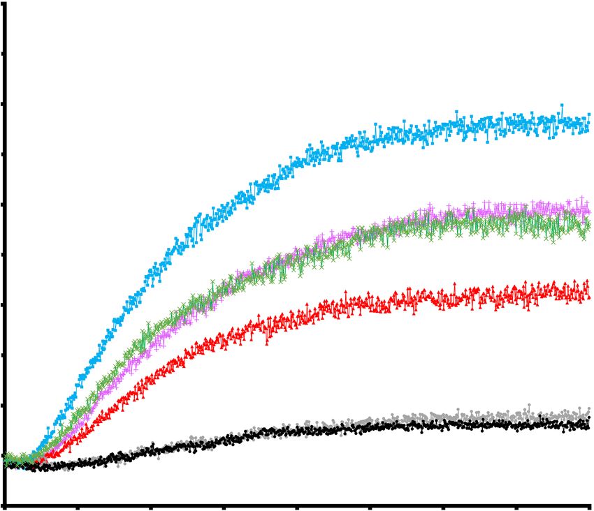
Figure 2. IQGAP1 enhances RhoA-dependent activation of DIAPH1 actin nucleation activity.
A-E, pyrene actin nucleation assays performed in the presence of recombinant proteins and 2µM pyrene
labelled actin. Fluorescence intensity measure in arbitrary units (A.U.). The fluorescence of pyrene-
labelled actin is much higher after actin filament polymerization. CT = MBP-DIAPH1-CT, NT = 6xHis-
DIAPH1-NT, RhoA = GST-RhoA, IQGAP1 = GST-IQGAP1-DBR. F, Normalized actin polymerization
rates at t1/2 in the different conditions shown in A-E. Abbreviations as A-E except IQ = GST-IQGAP1-
DBR. (n=3, error bars indicate + s.d).
Figure 3. In vitro Fli-I enhances RhoA-dependent release of DIAPH1-NT from DIAPH1-CT.
A. Domain organization of Flightless-I and the recombinant fragment used in this study. The domain
boundaries are denoted as the amino acid number in the sequence. B, In vitro binding assay between
either bacterially expressed 6xHis DIAPH1-NT and GST-Fli-I-GLD or MBP-DIAPH1-CT and GST-Fli-
I-GLD. C, Preformed 6xHis DIAPH1-NT plus MBP-DIAPH1-CT complexes (0.05nmol) were
immobilized on amylose beads and incubated with increasing concentrations of GST-Fli-I-GLD +/- GST-
RhoA (upper panel) or immobilized on nickel-Sepharose and incubated with increasing amounts of GST-
RhoA +/- GST-Fli-I-GLD (lower panel). The DIAPH-loaded beads were then isolated and analyzed by
immunoblotting to detect co-purifying GST-Fli-I-GLD or GST-RhoA as indicated. D. Preformed
DIAPH1 NT-CT complexes as above were immobilized on Ni-Sepharose, which binds 6His-DIAPH1-
NT, and incubated with GST, GST-RhoA, GST-IQGAP-DBR, GST-Fli-1-GLD or combinations thereof
in an equimolar ratio. The Ni-Sepharose beads were then washed and analyzed by immunoblotting for the
co-purifying MBP-DIAPH1-CT. Quantitation of the immunoblots indicates the ratio of co-purifying
DIAPH1-CT recovered under each condition relative to that found in the input lane with the DIAPH1
NT+CT complex (leftmost lane).
Figure 4. Fli-I is a weaker enhancer of RhoA-mediated DIAPH1 activation compared to
IQGAP1. A-C, pyrene actin nucleation assays performed in the presence of different recombinant
proteins and 2µM pyrene labelled actin. Fluorescence intensity measure is in arbitrary units (A.U.). CT =
MBP-DIAPH1-CT, NT = 6x His-DIAPH1-NT, RhoA = GST-RhoA, IQGAP1 = GST-IQGAP1-DBR, Fli-
I = GST-Fli-1-GLD. D, Normalized actin polymerization rates at t1/2 in the different conditions shown in
A-C (n=3, error bars indicate + s.d.)
14You can also read