Radotinib enhances cytarabine (Ara-C)- induced acute myeloid leukemia cell death - BMC ...
←
→
Page content transcription
If your browser does not render page correctly, please read the page content below
Heo et al. BMC Cancer      (2020) 20:1193
https://doi.org/10.1186/s12885-020-07701-8

 RESEARCH ARTICLE                                                                                                                                    Open Access

Radotinib enhances cytarabine (Ara-C)-
induced acute myeloid leukemia cell death
Sook-Kyoung Heo1, Eui-Kyu Noh2, Ho-Min Yu1, Do Kyoung Kim1, Hye Jin Seo1, Yoo Jin Lee2, Jaekyung Cheon2,
Su Jin Koh2, Young Joo Min2, Yunsuk Choi2* and Jae-Cheol Jo1,2*

  Abstract
  Background: Acute myeloid leukemia (AML) is a heterogeneous disease that frequently relapses after standard
  chemotherapy. Therefore, there is a need for the development of novel chemotherapeutic agents that could treat
  AML effectively. Radotinib, an oral BCR-ABL tyrosine kinase inhibitor, was developed as a drug for the treatment of
  chronic myeloid leukemia. Previously, we reported that radotinib exerts increased cytotoxic effects towards AML
  cells. However, little is known about the effects of combining radotinib with Ara-C, a conventional
  chemotherapeutic agent for AML, with respect to cell death in AML cells. Therefore, we investigated combination
  effects of radotinib and Ara-C on AML in this study.
  Methods: Synergistic anti-cancer effects of radotinib and Ara-C in AML cells including HL60, HEL92.1.7, THP-1 and
  bone marrow cells from AML patients have been examined. Diverse cell biological assays such as cell viability assay,
  Annexin V-positive cells, caspase-3 activity, cell cycle distribution, and related signaling pathway have been
  performed.
  Results: The combination of radotinib and Ara-C was found to induce AML cell apoptosis, which involved the
  mitochondrial pathway. In brief, combined radotinib and Ara-C significantly induced Annexin V-positive cells,
  cytosolic cytochrome C, and the pro-apoptotic protein Bax in AML cells including HL60, HEL92.1.7, and THP-1. In
  addition, mitochondrial membrane potential and Bcl-xl protein were markedly decreased by radotinib and Ara-C.
  Moreover, this combination induced caspase-3 activity. Cleaved caspase-3, 7, and 9 levels were also increased by
  combined radotinib and Ara-C. Additionally, radotinib and Ara-C co-treatment induced G0/G1 arrest via the
  induction of CDKIs such as p21 and p27 and the inhibition of CDK2 and cyclin E. Thus, radotinib/Ara-C induces
  mitochondrial-dependent apoptosis and G0/G1 arrest via the regulation of the CDKI–CDK–cyclin cascade in AML
  cells. In addition, our results showed that combined treatment with radotinib and Ara-C inhibits AML cell growth,
  including tumor volumes and weights in vivo. Also, the combination of radotinib and Ara-C can sensitize cells to
  chemotherapeutic agents such as daunorubicin or idarubicin in AML cells.
  Conclusions: Therefore, our results can be concluded that radotinib in combination with Ara-C possesses a strong
  anti-AML activity.
  Keywords: Radotinib, Acute myeloid leukemia, Cytarabine, Ara-C, Anti-leukemic activity

* Correspondence: choiysmd@gmail.com; jcjo97@hanmail.net
2
 Department of Hematology and Oncology, Ulsan University Hospital,
University of Ulsan College of Medicine, 877 Bangeojinsunhwan-doro,
Dong-gu, Ulsan 44033, Republic of Korea
1
 Biomedical Research Center, Ulsan University Hospital, University of Ulsan
College of Medicine, Ulsan 44033, Republic of Korea

                                         © The Author(s). 2020 Open Access This article is licensed under a Creative Commons Attribution 4.0 International License,
                                         which permits use, sharing, adaptation, distribution and reproduction in any medium or format, as long as you give
                                         appropriate credit to the original author(s) and the source, provide a link to the Creative Commons licence, and indicate if
                                         changes were made. The images or other third party material in this article are included in the article's Creative Commons
                                         licence, unless indicated otherwise in a credit line to the material. If material is not included in the article's Creative Commons
                                         licence and your intended use is not permitted by statutory regulation or exceeds the permitted use, you will need to obtain
                                         permission directly from the copyright holder. To view a copy of this licence, visit http://creativecommons.org/licenses/by/4.0/.
                                         The Creative Commons Public Domain Dedication waiver (http://creativecommons.org/publicdomain/zero/1.0/) applies to the
                                         data made available in this article, unless otherwise stated in a credit line to the data.
Heo et al. BMC Cancer (2020) 20:1193 Page 2 of 15 Background the mitochondrial- and caspase-dependent pathway. Acute myeloid leukemia (AML) is characterized by the Further, we attempt to show that radotinib treatment rapid growth of abnormal white blood cells that with Ara-C-based regimens could be clinically evaluated accumulate in the bone marrow and/or peripheral blood for AML patients. [1, 2]. It is a heterogeneous disease, characterized by nu- merous cytogenetic and molecular alterations [3]. There- Methods fore, AML is still one of the most difficult malignancies Reagents to cure. Moreover, many patients with AML die from Radotinib was generously gifted by Ilyang Pharmaceut- disease recurrence and therapy options in both the ical Co., Ltd., (Seoul, South Korea). Its purity was found relapsed and refractory settings of this disease are re- to be 99.9% by HPLC analysis [16]. The cell culture stricted [4]. In addition, there are unmet needs in AML plates were obtained from SPL Life Sciences (Pocheon, treatment because the current standard treatment is South Korea). All reagents were obtained from Sigma- based on old chemotherapeutic regimens. Therefore, the Aldrich (St. Louis, MO, USA) unless otherwise indicated. development of novel chemotherapeutic agents that can The Apoptosis Detection Kit I was purchased from BD treat AML effectively is required. Biosciences (San Jose, CA, USA). The CellTiter 96 Cytarabine (1-β-D-arabinofuranosylcytosine, cytosine AQueous One Solution Cell Proliferation Assay was pur- arabinoside, Ara-C) has been used as a mainstream ther- chased from Promega (Madison, WI, USA). NE-PER apy for AML for over 40 years [5, 6]. It slows down Nuclear and Cytoplasmic Extraction Reagents were DNA synthesis in the S-phase of the cell cycle, and its obtained from Thermo Scientific (Rockford, IL, USA). action is mainly related to DNA fragmentation and All antibodies for western blot were purchased from Cell chain termination [7, 8]. Generally, it is prescribed alone Signaling Technology (Beverly, MA, USA). or in combination with other drugs. Induction therapy consisting of Ara-C in combination with anthracyclines Patient samples leads to responses in approximately 60% of adult AML All patients were newly diagnosed with AML (n = 5) at patients [9]. However, the development of resistance and Ulsan University Hospital, Ulsan, South Korea, as de- high rates of relapse is a significant obstacle to the suc- scribed in Supplementary Table 1. Bone marrow samples cessful treatment of AML [10]. Therefore, it is essential were collected before administering the first round of to develop potential therapeutic regimens that include chemotherapy. new drugs that can maximize the effectiveness of Ara-C. Radotinib, an oral BCR-ABL tyrosine kinase inhibitor, Isolation of patient cells and culture was developed as a drug for the management of chronic The patient cells were isolated by the density gradient phase-chronic myeloid leukemia (CML-CP) in South method, as previously described [18]. In brief, bone Korea [11, 12]. It is an effective inhibitor of naive and marrow cells (BMCs) were isolated via density gradient kinase-domain mutant BCR-ABL1 [12]. More recently, a centrifugation at 400×g using Lymphoprep (Axis-Shield, phase III clinical trial on the efficacy and safety of rado- Oslo, Norway). They were washed with phosphate- tinib showed the generation of complete cytogenetic buffered saline (PBS) and cultured in RPMI1640 with responses and major molecular responses in patients 10% FBS and 1% penicillin-streptomycin in a 5% CO2 newly evaluated with Philadelphia chromosome-positive humidified atmosphere at 37 °C. CML-CP [13]. Radotinib also shows neuroprotective ef- fects in a Parkinson’s disease mouse model [14]. More re- Cell culture cently, radotinib was found to activate NK cell cytotoxicity The human AML cell lines HL60, HEL92.1.7, and THP- against various Fas-expressing solid cancer cells [15]. 1 in this study were grown as suspension cultures in We previously demonstrated that radotinib exhibits RPMI-1640 medium with 10% FBS and a 1% penicillin- increased cytotoxicity against diverse AML cells [16]. In streptomycin solution (final concentration: 100 units/ml addition, it inhibits AML cell proliferation by inducing and 100 μg/ml, respectively) in a 5% CO2 humidified CDK inhibitors including p21 and p27 [17]. Moreover, atmosphere at 37 °C, as previously described [16]. In radotinib induces apoptosis in differentiated cells from addition, the human small cell lung cancer (SCLC) cell AML blasts [16]. Furthermore, targeting c-KIT (CD117) line H209 were cultured as described previous herein. with radotinib promotes cell death in c-KIT-positive AML [18, 19]. However, little is known about the effects Cell viability assay of a combination of radotinib and Ara-C with respect to The effect of each drug on cell growth both as a single cell death and cell cycle distribution in AML cells. Here, agent and in combination was determined by cell viabil- we show that combination therapy, comprising radotinib ity assay. Cells were seeded (density, 2 × 10⁴ cells/well) and Ara-C, induces AML cell apoptosis, which involves in 96-well plates containing 200 μl medium per well and
Heo et al. BMC Cancer   (2020) 20:1193                                                                        Page 3 of 15

were incubated with 5 μM radotinib and/or 50 nM Ara-          Preparation of cytosolic extractions for cytochrome C
C for 48 h at 37 °C. CellTiter 96 solution (20 μl; Pro-       analysis
mega, Madison, WI, USA) was added directly to each            HEL92.1.7 cells were treated with 5 μM radotinib and/or
well, and the plates were incubated for 4 h in a humidi-      50 nM Ara-C for 48 h at 37 °C. Cells were washed with
fied atmosphere of 5% CO2 at 37 °C. Absorbance was            ice-cold PBS, resuspended in cold lysis buffer, and incu-
measured at 490 nm using a SpectraMax iD3 Microplate          bated on ice for 30 min. Next, the cytosolic fractions of
Reader (Molecular Devices, San Jose, CA, USA). Results        cells were separated using the NE-PER Nuclear and
are expressed as percent change from baseline condi-          Cytoplasmic Extraction Reagents according to the man-
tions determined using four to five culture wells for each    ufacturer’s instructions (Thermo Fisher Scientific, MA,
experimental condition. The following equation was            USA). The release of cytochrome C was analyzed by im-
used: death (% of control) = 100 − cell viability [(OD tar-   munoblotting with an anti-cytochrome C mAb.
get group / OD of 0 μM radotinib group) × 100]. In some
experiments HL60 cells were treated with various con-         Western blotting analysis
centrations of radotinib (0, 10, 30, 40 and 50 μM) and        Cells were incubated with each drug and their combin-
Ara-C (0, 40, 80, 120 and 160 nM) for 48 h. Additionally,     ation for 48 h at 37 °C. They were then washed three
cells were treated with a combined low dosage of idaru-       times with ice-cold PBS and harvested. Western blotting
bicin and daunorubicin.                                       was performed as previously described [17, 18].

Detection of Annexin V-positive cells                         Xenograft animal model
                                         5
HL60 and HEL92.1.7 cells (1 × 10 cells/ml) were seeded        Specific-pathogen-free five-week-old athymic nude male
in 24-well plates and treated with 5 μM radotinib and/or      mice were purchased from Koatech (Pyeongtaek, Korea)
50 nM Ara-C for 48 h at 37 °C. The cells were harvested       and kept in a clean environment of the Ulsan University
and washed twice with FACS buffer (PBS containing             of Korea (Korea, Ulsan). All mice were housed in stand-
0.2% bovine serum albumin and 0.1% NaN3). Then, the           ard conditions (12-h light/dark cycle) under constant
cells were stained with Annexin V-FITC from the Apop-         temperature (22–24 °C) and humidity (50–60%), given
tosis Detection Kit I according to the manufacturer’s in-     free access to food and water, and handled in accordance
structions. Cells were analyzed using the FACSCalibur         with the Institutional Animal Care and Use Committee
flow cytometer and CellQuest Pro software.                    (IACUC) of the University of Ulsan (Ulsan, Korea,
                                                              Approval No. 0117–07). For anesthesia, mice were
                                                              injected intraperitoneally with tribromoethanol (250
Measurement of caspase-3 activity
                                                              mg/kg). Mice were sacrificed using carbon dioxide
Cells were examined using the CaspGLOW™ Fluorescein
                                                              (CO2) gas per IACUC protocol.
Active Caspase-3 Staining Kit according to the manufac-
                                                                 All mice were naïve to previous experimental manipu-
turer’s instructions (Thermo Fisher Scientific, MA, USA).
                                                              lations. Each mouse was considered as one experimental
                                                              unit, and mice were housed in 3–5 mice per cage. To
Cell cycle analysis                                           minimize experimental bias, mice were randomized into
HL60, HEL92.1.7 and THP-1 cells were treated with             all prospective treatment cages for in vivo preclinical ex-
5 μM radotinib and/or 50 nM Ara-C for 48 h at 37 °C.          periments. The inoculations of tumor cells ex vivo were
They were then washed twice with PBS and fixed with           also blinded. The number of cohorts/mice used in each
70% ethanol overnight at − 20 °C, followed by washing         experiment is described in Supplementary Table 2. The
again with PBS and incubation with 0.5 ml PI/RNase            xenograft animal model was generated as previously de-
stain buffer for 15 min at room temperature. The samples      scribed [18]. Briefly, HEL92.1.7 tumors were established
were then analyzed using a FACSCalibur flow cytometer         by subcutaneous injection of 1 × 10⁷ cells into the right
and CellQuest Pro software (BD Biosciences).                  flank of five-week-old athymic nude male mice (n = 5
                                                              per group). To aid precise inoculations, mice were
Analysis of mitochondrial membrane potential                  anaesthetized. Once tumors were established, mice were
HL60 and HEL92.1.7 cells were incubated with 5 μM             treated with vehicle (0.5% carboxymethylcellulose/DW),
radotinib and/or 50 nM Ara-C for 48 h at 37 °C, har-          50 mg/kg po radotinib daily, 50 mg/kg ip Ara-C daily
vested, and washed twice with PBS buffer. Mitochondrial       every 5/7 day or their combination for up to 24 day. The
membrane potential (MMP, ΔΨm) was evaluated by                maximal length and width of the tumor were measured
staining the cells with DiOC6(3) for 30 min. After incu-      once per week using digital calipers, and the tumor vol-
bation, the cells were harvested and washed. Percentages      ume (V) was calculated using the following formula: V =
of DiOC6(3)-positive cells were determined using a flow       (length × width2) × 0.5. The mice were sacrificed on days
cytometer and CellQuest Pro software.                         30–34 following tumor cell implantation. If the size of
Heo et al. BMC Cancer (2020) 20:1193 Page 4 of 15
the subcutaneous tumor exceeded 1000 mm in volume, Ara-C produced a significant inhibitory effect of cell via-
the animals were excluded from the study and the stand- bility at 48 h and 72 h (42% at 48 h, 36% at 72 h; see Sup-
ard was established for euthanasia before a predeter- plementary Figure 1C and Fig. 1a). Combination effect
mined time point. All tumors met the criteria, and there was better at 48 h than 72 h. Accordingly, we used these
were no exclusions. The body weights of the tumor- conditions for the remainder of the experiments.
bearing mice did not change significantly during the dur- We examined the cell viability of diverse AML cells in-
ation of study. The tumors were excised and weighed, and cluding HL60, HEL 92.1.7, THP-1, and BMCs from
each tumor tissue was homogenized for the preparation of AML patients (n = 5). Combined treatment with radoti-
cell samples for several analyses including western blotting nib and Ara-C exerted synergistic effects on AML cell
for specific molecular markers. death (Fig. 1a-d). In addition, the human SCLC cell line
H209 were tested for cell viability after radotinib and
TUNEL assay for measurement of DNA double-strand Ara-C treatment. Here, these agents did not have a syn-
breaks in tumor tissue ergistic effect on cell viability, as expected (Fig. 1e).
Tumors were frozen in optimal cutting temperature Therefore, radotinib enhances Ara-C-induced AML, but
(OCT) compound, and stored at − 80 °C until use. The not MM and SCLC, cell death (Fig. 1).
frozen tissue samples were sectioned by a microtome-
cryostat (CM1950, Leica Biosystems, IL, USA). Samples Combined treatment with radotinib and Ara-C has
were fixed in 4% paraformaldehyde for 10 min, washed synergistic effects on HL60 and HEL92.1.7 cell apoptosis
in PBS and then treated with 0.1% Triton X-100 in PBS via activation of the mitochondrial- and caspase-
for 10 min. Then, tumor tissue samples were evaluated dependent apoptosis pathway
for apoptosis using the TUNEL Assay Kit according to We first observed that combined treatment with radoti-
the manufacturer’s instructions (Abcam, Cambridge, nib and Ara-C had a synergistic effect on AML cell
United Kingdom). Cells were analyzed with a Fluores- death. We then performed Annexin V staining to see
cence microscope (Olympus, NY, USA). what pathways regulate this cell death using HL60 and
HEL 92.1.7 cells. As a result, we showed that combined
Staining of proliferating cell nuclear antigen (PCNA) treatment with 5 μM radotinib and 50 nM Ara-C exerts
positive cells in tumor tissue a synergistic effect on HL60 and HEL92.1.7 cell apop-
Under the above experimental conditions, tumor tissue tosis (Fig. 2a-c).
was stained with anti-PCNA monoclonal antibody Next, we measured the effects of radotinib and Ara-C
(mAb) or isotype control mAb at 4 °C for 30 min. The on the mitochondrial-dependent apoptotic pathway.
samples were then analyzed with a Fluorescence micro- Cells were collected and MMP was measured by flow cy-
scope (Olympus, NY, USA). tometry using DiOC6(3) dye. As shown in Fig. 3a and b,
the DiOC6(3)-positive cells were markedly decreased by
Statistical analysis combined treatment with radotinib and Ara-C stimula-
Data are presented as means ± the standard error of the tion in AML cells including HL60 and HEL92.1.7 cells.
mean (SEM) based on at least three independent experi- Further, as shown in Fig. 3c, cytosolic cytochrome C was
ments. All values were evaluated by a one-way analysis drastically increased in radotinib and Ara-C-treated
of variance followed by Tukey post-hoc test, as imple- HEL92.1.7 cells. In addition, combined radotinib and
mented by GraphPad Prism 7.0 (GraphPad Software, Ara-C also increased the pro-apoptotic protein Bax in
Inc., La Jolla, USA). Differences were considered signifi- AML cells such as HL60, HEL92.1.7, and THP-1. As ex-
cant when P < 0.05. pected, this treatment also decreased Bcl-xl expression
in diverse AML cells (Fig. 3d). To inhibit MMP disrup-
Results tion, bongkrekic acid (BA, 50 μM) was added to HL60
Radotinib enhances Ara-C-induced AML cell death in AML cells for 2 h prior to combined treatment of radotinib
cell lines and primary patient samples and Ara-C. After 48 h, the cells were analyzed by im-
HL60 cells were treated with various concentrations of munoblotting using an anti-Bax mAb. The BA efficiently
radotinib (0, 10, 30, 40 and 50 μM) and Ara-C (0, 40, 80, blocked the radotinib/Ara-C-induced Bax expression in
120 and 160 nM) for 48 h. Radotinib and Ara-C signifi- HL60 cells (relative band density: with 5 μM radotinib
cantly inhibited the viability of the HL60 cells in a dose- and 50 nM Ara-C, 100%; with 50 μM BA preinculation,
dependent manner (Supplementary Figure 1A and 1B). 4.6%), as shown in Fig. 3e. Thus, these data indicate that
Interestingly, however, although 5 μM of radotinib and radotinib/Ara-C-induces cell death via a mitochondrial-
50 nM of Ara-C alone had little effect on the viability of dependent apoptosis pathway (Fig. 3).
these cells (over 89 and 81% cell viability, respectively), We further confirmed that caspase-3 activity was en-
in combination these concentrations of radotinib and hanced by treatment with radotinib and Ara-C in HL60Heo et al. BMC Cancer (2020) 20:1193 Page 5 of 15 Fig. 1 Combination of radotinib and Ara-C enhances acute myeloid leukemia (AML) cell death. Cells were treated with 5 μM radotinib and/or 50 nM Ara-C for 48 h. The cytotoxicity was then evaluated by an MTS assay. a HL60 cells. b HEL92.1.7 cells. c THP-1 cells. d Bone marrow cells (BMCs) from AML patients (n = 5). e H209 cells. Representative data are shown from at least three independent experiments. These data represent the means ± SEM. Significantly different from control (*) or combination of radotinib and Ara-C (#); ***, ###: P < 0.001. R, radotinib; A, Ara-C; R + A, combination of radotinib and Ara-C and HEL92.1.7 cells (Fig. 4a and b). At the same time, activity using the CaspGLOW™ Fluorescein Active we observed that cleaved caspase-3, 7, and 9 increased Caspase-3 Staining Kit. The Z-VAD-FMK efficiently with radotinib/Ara-C administration in both cell types blocked the radotinib/Ara-C-induced capase-3 activity in (Fig. 4c-e). To inhibit caspase activation, pan caspase in- HL60 cells (relative intensity: with 5 μM radotinib and hibitor, Z-VAD-FMK (10 μM) was added to HL60 cells 50 nM Ara-C, 100%; with 10 μM Z-VAD-FMK preincu- for 30 min prior to combined treatment of radotinib and lation, 4%), as shown in Fig. 4f. The cells were also col- Ara-C. After 48 h, the cells were analyzed by capase-3 lected and treated under the same conditions described
Heo et al. BMC Cancer       (2020) 20:1193                                                                                                   Page 6 of 15

 Fig. 2 Combination of radotinib and Ara-C increases Annexin V-positive HL60 and HEL92.1.7 cells. Cells were treated with 5 μM radotinib and/or 50
 nM Ara-C for 48 h. Cells were stained with annexin V-FITC followed by flow cytometric analysis. a Annexin V staining of HL60 cells. b Data show the
 percentage of Annexin V-positive cells (apoptotic cells) in (a). c Data show the percentage of Annexin V-positive HEL92.1.7 cells. Representative data
 are shown for at least three independent experiments. These data represent the means ± SEM. Significantly different from control (*) or combination
 of radotinib and Ara-C (#); *: P < 0.05; **: P < 0.01; ***, ###: P < 0.001. R, radotinib; A, Ara-C; R + A, combination of radotinib and Ara-C

in Fig. 4f. The cells were analyzed by immunoblotting                          Generally, it is well-known that the caspase-dependent
using an anti-cleaved PARP-1 (cPARP-1) mAb and an                              apoptosis pathway is downstream of the mitochondrial
anti-cleaved caspase-3 mAb. The Z-VAD-FMK effi-                                pathway [20]. Therefore, combined treatment with rado-
ciently reversed the radotinib/Ara-C-induced cPARP-1                           tinib and Ara-C has a synergistic effect on AML cell
and cleaved caspase-3 expression in HL60 cells (relative                       death via the activation of mitochondrial-dependent
band density of cPARP-1: with 5 μM radotinib and 50                            apoptosis.
nM Ara-C, 100%; with 10 μM Z-VAD-FMK preincula-
tion, 6%, and relative band density of cleaved caspase-3:                      Combined treatment with radotinib and Ara-C has a
with 5 μM radotinib and 50 nM Ara-C, 100%; with                                synergistic effect on G0/G1 phase arrest in HL60 and
10 μM Z-VAD-FMK preinculation, 2.7%), as shown in as                           HEL92.1.7 cells via the induction of p21 and p27
shown in Fig. 4g. Therefore, these data indicate that                          We also monitored the cell cycle distribution after rado-
radotinib/Ara C induces cell death via a caspase-                              tinib and Ara-C treatment in HL60, HEL92.1.7 and
dependent pathway. In addition, the bongkrekic acid                            THP-1 cells. First, combination treatment with radotinib
powerfully blocked the radotinib/Ara-C-induced cPARP-                          and Ara-C caused G0/G1 phase cell cycle arrest in AML
1 and cleaved caspase-3 expression in HL60 cells (rela-                        cells such as HL60 and HEL92.1.7 cells (Fig. 5a). More-
tive band density of cPARP-1: with 5 μM radotinib and                          over, we examined the effects of combined radotinib and
50 nM Ara-C, 100%; with 50 μM BA preinculation, 3.7%,                          Ara-C on cell cycle regulatory proteins including not
and relative band density of cleaved caspase-3: with                           only CDK2 and cyclin E but also p21 and p27. As a re-
5 μM radotinib and 50 nM Ara-C, 100%; with 50 μM BA                            sult, the expression of CDK2 and cyclin E was remark-
preinculation, 3.2%), as shown in as shown in Fig. 4h.                         ably decreased in the radotinib and Ara-C combination
Therefore, these data indicate that radotinib/Ara C in-                        group (Fig. 5b). Moreover, as shown in Fig. 5c, the ex-
duces cell death via a mitochondrial-dependent pathway.                        pression levels of p21 and p27 in cells co-treated with
Heo et al. BMC Cancer     (2020) 20:1193   Page 7 of 15

 Fig. 3 (See legend on next page.)
Heo et al. BMC Cancer      (2020) 20:1193                                                                                               Page 8 of 15

 (See figure on previous page.)
 Fig. 3 Radotinib/Ara-C-induced apoptosis involves the mitochondrial pathway. A combination of radotinib and Ara-C inhibited the mitochondrial
 membrane potential of HL60 (a) and HEL92.1.7 cells (b). The cells were also collected and treated under the same conditions described in Fig. 2.
 The mitochondrial membrane potential was measured by flow cytometry using DiOC6(3) dye. c The effects of radotinib/Ara-C on cytosolic
 cytochrome C. HEL92.1.7 cells were incubated with 5 μM radotinib and/or 50 nM Ara-C for 48 h. The cytosolic fractions were then separated, after
 which each sample was analyzed for the expression of cytochrome C. The membrane was stripped and re-probed with an anti-β-actin mAb to
 confirm equal loading. d The effects of radotinib/Ara-C on Bax and Bcl-xl expression. Diverse AML cells including HL60, HEL92.1.7, and THP-1 cells
 were treated with 5 μM radotinib and/or 50 nM Ara-C for 48 h at 37 °C. Then, cells were washed twice PBS buffer and stained with an anti-Bax
 and anti-Bcl-xl mAb. e HL60 cells were incubated with 5 μM radotinib and 50 nM Ara-C in the presence or absence of bongkrekic acid (BA,
 50 μM) for 48 h and then harvested. Cells were analyzed by immunoblotting using an anti-Bax mAb. The membrane was stripped and re-probed
 with an anti-β-actin mAb to confirm equal loading. These data represent the means ± SEM. Significantly different from control (*) or combination
 of radotinib and Ara-C (#); ***, ###: P < 0.001. R, radotinib; A, Ara-C; R + A, combination of radotinib and Ara-C; BA, bongkrekic acid

radotinib and Ara-C were higher than those in single                        AML cell death. It also indicates that mitochondrial-
treatment group, as expected. These results suggest that                    dependent apoptosis and G0/G1 phase cell cycle arrest
combined treatment with radotinib and Ara-C increases                       contribute to radotinib/Ara-C mediated anti-AML
expression of the inhibitory proteins p21 and p27 and                       activity.
decreases the expression of CDK2 and cyclin E in both
cell types, maintaining those cells in the G0/G1 phase.                     Combined treatment with radotinib and Ara-C inhibits
Therefore, the combination of radotinib and Ara-C in-                       AML cell growth in vivo
duces G0/G1 arrest in HL60 and HEL92.1.7 AML cells                          To examine the effect of combined treatment with
via regulation of the CDKI–CDK–cyclin cascade (Fig. 5).                     radotinib and Ara-C on AML cell death in vivo, we
Especially, the induction of CDK inhibitors, namely p21                     implanted HEL92.1.7 cells into nude mice. Through
and p27, contributed to the G0/G1 arrest of HL60 and                        previous preliminary studies, radotinib and Ara C were
HEL92.1.7 cells. Consequently, these results indicate that                  administered by concentration, and then the tumor vol-
the combination of radotinib and Ara-C has a synergistic                    ume was used in combination with each concentration
effect on G0/G1-phase arrest in HL60 and HEL92.1.7                          that decreased by 5–8% compared to the control group
cells via the induction of CDK inhibitors including p21                     (data not shown). As shown in Fig. 6a-b, combined
and p27.                                                                    treatment with radotinib and Ara-C inhibited AML cell
  Furthermore, we examined cell cycle markers in-                           growth, including tumor volume and weight in vivo. In
cluding p21 and p27 using pan caspase inhibitor, Z-                         addition, the body weights of the tumor-bearing mice
VAD-FMK and MMP disruption inhibitor, and                                   did not change significantly during the duration of study
bongkrekic acid to clarify the relationship between                         (Fig. 6c). The expression of Bcl-xl, Cyclin E, and PCNA
cell cycle and cell death. In briefly, the cells were also                  with combined radotinib and Ara-C treatment was
collected and treated under the same conditions de-                         significantly decreased in tumor tissues isolated from the
scribed in Fig. 4g and h. The cells were analyzed by                        mice, as shown in Fig. 6d-e. Further, the expression of
immunoblotting using an anti-p21 mAb and an anti-                           Bax and p21, as a well-known CDK inhibitor, was
p27 mAb. The Z-VAD-FMK significantly blocked the                            significantly increased in tumor tissues with combined
radotinib/Ara-C-induced p21 and p27 expression in                           treatment comprising radotinib and Ara-C. In addition,
HL60 cells (relative band density of p21: with 5 μM                         the expression of TUNEL-positive cells was significantly
radotinib and 50 nM Ara-C, 100%; with 10 μM Z-                              amplified, while the expression of PCNA-positive cells
VAD-FMK preinculation, 2.6%, and relative band                              was dramatically reduced in the tumor tissue with com-
density of p27: with 5 μM radotinib and 50 nM Ara-                          bined treatment of radotinib and Ara-C (Fig. 6f-g). In
C, 100%; with 10 μM Z-VAD-FMK preinculation,                                particular, the combination of the two drugs was shown
4.7%), as shown in as shown in Fig. 5d. In addition,                        to lead to the profound inhibition of acute myeloid
the bongkrekic acid strongly blocked the radotinib/                         leukemia tumor growth (Fig. 6).
Ara-C-induced (relative band density of p21: with 5 μM
radotinib and 50 nM Ara-C, 100%; with 50 μM BA prein-                       Radotinib and Ara-C sensitize AML cells to
culation, 11.4%, and relative band density of p27: with                     chemotherapeutic agents including daunorubicin or
5 μM radotinib and 50 nM Ara-C, 100%; with 50 μM BA                         idarubicin
preinculation, 1.2%), as shown in as shown in Fig. 5e.                      Finally, we performed cell viability tests to determine the
Thus, these data showed that radotinib/Ara C-induced                        efficacy of a combination of radotinib and various anti-
cell cycle markers, p21 and p27, were closely related to                    cancer drugs commonly used for AML therapy.
Heo et al. BMC Cancer     (2020) 20:1193   Page 9 of 15

 Fig. 4 (See legend on next page.)
Heo et al. BMC Cancer      (2020) 20:1193                                                                                               Page 10 of 15

 (See figure on previous page.)
 Fig. 4 Radotinib/Ara-C induces caspase-dependent apoptosis and activates caspase-3 activity in HL60 and HEL92.1.7 cells. The cells were also
 collected and treated under the same conditions described in Fig. 2. a Caspase-3 activity induced by radotinib and Ara-C stimulation in HL60
 cells. b Caspase-3 activity induced by radotinib and Ara-C stimulation in HEL92.1.7 cells. c-e The expression of cleaved caspase-3, cleaved caspase-
 7, and cleaved caspase-9 was analyzed by SDS-PAGE, followed by western blotting using specific antibodies. f HL60 cells were incubated with
 5 μM radotinib and 50 nM Ara-C in the presence or absence of Z-VAD-FMK (10 μM) for 48 h and then harvested. Cells were analyzed by capase-3
 activity. g HL60 cells were also collected and treated under the same conditions described in Fig. 4f. Cells were analyzed by immunoblotting
 using an anti-cPARP-1 mAb and an anti-cleaved caspase-3 mAb. h HL60 cells were also collected and treated under the same conditions
 described in Fig. 3e. Cells were analyzed by immunoblotting using an anti-cPARP-1 mAb and an anti-cleaved caspase-3 mAb. The experiments
 were repeated three times and the data show representative results. The membrane was stripped and re-probed with an anti-β-actin mAb to
 confirm equal loading. These data represent the means ± SEM. Significantly different from control (*) or combination of radotinib and Ara-C (#);
 ***, ###: P < 0.001. R, radotinib; A, Ara-C; R + A, combination of radotinib and Ara-C; BA, bongkrekic acid

Daunorubicin (DNR) is a chemotherapy medication used                         therapy resistance [27]. Therefore, understanding the
to treat cancer. It is specifically used for AML, acute                      molecular mechanisms that regulate caspase activation
lymphoblastic leukemia, CML [21]. Moreover, idarubi-                         in cancer cells is very important. Thus, the modulation
cin (Idar) is an anthracycline antileukemic drug. It                         of caspase activation including apoptosis represents a
inserts itself into DNA and prevents DNA unwinding                           promising approach for the development of new thera-
by interfering with the enzyme topoisomerase II [22].                        peutic opportunities to induce cancer cell death.
It is also an analog of daunorubicin. As shown in                              The combination of BCR-ABL inhibitors such as
Figs. 1, 2, 3, 4 and 5, radotinib enhanced sensitivity                       dasatinib and imatinib, with other used chemothera-
to the chemotherapeutic agent Ara-C in AML cells.                            peutic agents for AML is not new. According to Dos
Moreover, triple combination with radotinib, Ara-C,                          Santos et al., it has already been shown that the com-
and DNR or Idar significantly inhibited the viability                        bination of dasatinib with daunorubicin or Ara-C also
of AML cells (Supplementary Fig. 2). Therefore, these                        resulted in significantly increased AML CD34+ cell
results indicate that a combination of radotinib and                         death [29]. And chemotherapy plus dasatinib provided
Ara-C can sensitize cells to chemotherapeutic agents                         excellent outcomes for both younger and older patients
such as DNR or Idar in AML cells, as well as HL60                            with or without KIT mutation [30]. Also, the Ara-C
and HEL92.1.7 cells.                                                         and imatinib showed synergistic effects in vitro [31].
                                                                             According to a phase 1 study of imatinib mesylate in
Discussion                                                                   combination with cytarabine and daunorubicin, espe-
Generally, apoptosis is a form of programmed cell                            cially for c-kit positive recurrent acute myeloid
death that occurs in multicellular organisms. More-                          leukemia, cytotoxic therapy that includes imatinib mes-
over, it is a key regulator of physiological growth con-                     ylate for relapsed AML was effective [32]. In addition,
trol and the regulation of tissue homeostasis for the                        the combination of DNR and the novel BCR-ABL in-
elimination of damaged, old, or infected cells [23, 24].                     hibitor ponatinib showed the AML cell death [33]. We
It is well known that activating the apoptosis pathway                       have previously shown that radotinib significantly in-
in various cancers can make cancer treatments suc-                           creases cytotoxicity or apoptosis in AML and CML cells
cessful, whereas inhibiting the activation of apoptosis                      [17]. Radotinib also acts as a CDK inhibitor, which
leads to cancer resistance, which in turn makes can-                         strongly inhibits AML cell proliferation [17]. Alterna-
cer treatment difficult [23, 25]. Especially, targeting                      tively, it functions as an AURKA inhibitor, which sup-
apoptosis proteins in hematologic malignancies has                           presses the expression of AURKA and related proteins
been shown to be very useful [24, 26].                                       including Bora, polo-like kinase 1, and TPX2 [34].
  The induction of apoptosis is a cell suicide mechan-                       Moreover, radotinib induces apoptosis directly in cells
ism and an ideal way to treat cancer including AML                           differentiated from AML blasts [16]. In addition, we
[27]. Basically, killing tumor cells with anticancer                         previously confirmed that c-KIT (CD117) could be tar-
therapies commonly used for the treatment of cancer,                         geted by radotinib, acting as a c-KIT inhibitor or
such as chemotherapy, gamma-irradiation, and im-                             HSP90 inhibitor in c-KIT-positive AML cells [18, 19].
munotherapy, among others, is mainly mediated by                             Previous studies have shown that radotinib promotes
triggering apoptosis, wherein these modalities activate                      AML cell death through various cellular mechanisms.
the cell’s intrinsic cell death process [28]. The activa-                    However, little is known about the effects of the com-
tion of caspases is often impaired in human cancers                          bination of radotinib and Ara-C on cell death and cell
and contributes to cancer formation, progression, and                        cycle distribution in AML cells.
Heo et al. BMC Cancer (2020) 20:1193 Page 11 of 15 Fig. 5 (See legend on next page.)
Heo et al. BMC Cancer      (2020) 20:1193                                                                                              Page 12 of 15

 (See figure on previous page.)
 Fig. 5 Radotinib/Ara-C induces G0/G1 phase cell cycle arrest by regulating the CDKI–CDK–cyclin cascade in HL60, HEL92.1.7 and THP-1 cells. a Cell
 cycle distribution at 48 h after radotinib/Ara-C treatment. b The expression of CDK2 and cyclin E after radotinib/Ara-C treatment. c The expression
 of CDK inhibitors such as p21 and p27 by western blot analysis following radotinib/Ara-C treatment. d, e HL60 cells were also collected and
 treated under the same conditions described in Fig. 4f and g. Cells were analyzed by immunoblotting using an anti-p21 mAb and an anti-p27
 mAb. The experiments were repeated three times and the data show representative results. β-Actin was used to confirm equal loading. The
 results are representative of three independent experiments. R, radotinib; A, Ara-C; R + A, combination of radotinib and Ara-C; BA, bongkrekic acid

  AML is a heterogeneous group of rapidly growing                           index: 0.525, Supplementary Fig. 3). Further, radotinib
cancers of myeloid progenitor cells [2, 35]. Ara-C has                      and Ara-C significantly induced apoptosis in HL60
remained the backbone of chemotherapy for adult                             and HEL92.1.7 cells via the activation of the mito-
AML patients for decades [6]. Especially, high dose                         chondrial- and caspase-dependent apoptosis pathway
Ara-C is a part of an induction regimen as a first-line                     (Figs. 2, 3 and 4). Moreover, combined treatment with
therapy for AML [36]. However, the resistance of                            both drugs exerted a synergistic effect on G0/G1 phase
AML cells to Ara-C chemotherapy is one of the most                          arrest of AML cells via the induction of p21 and p27,
important reasons for relapse or chemo-refractoriness                       as shown in Fig. 5. Specifically, radotinib/Ara-C-in-
in AML patients [37]. For that reason, combination                          duced bax expression was blocked by bongkrekic acid
therapy has become the standard therapy for the                             (Fig. 3e), and capase-3 activity was blocked by Z-
treatment of several different cancers [38]. Therefore,                     VAD-FMK (Fig. 4f). Also we showed that Z-VAD-
it is of utmost importance to discover or develop                           FMK and bongkrekic acid reversed the radotinib/Ara-
drugs to maximize the effects of Ara-C. Clinicians, as                      C-induced effects including apoptotic proteins
well as scientists involved in basic research, interested                   (cleaved PARP-1 and cleaved caspase-3; Fig. 4g and h),
in the treatment of AML, have focused on developing                         cell cycle markers (p21 and p27; Fig. 5d and e). These
or discovering drugs that can be used in combination                        results showed mitochondrial-dependent apoptosis
with Ara-C. Much attention has been given to the                            and G0/G1 phase cell cycle arrest were contributed to
combination of two or more drugs, including Ara-C.                          radotinib/Ara-C mediated anti-AML activity (Figs. 4
Such examples include a combination of Ara-C and                            and 5). In addition, the suppressive effect of a combin-
differentiating agents, a combination of Ara-C and                          ation of radotinib and Ara-C, on AML cell growth,
DNA hypomethylating agents, and a combination of                            was also demonstrated using in vivo xenograft models
Ara-C and HSP90 inhibitors. Briefly, low-dose Ara-C                         (Fig. 6). In addition, radotinib and Ara-C sensitized
combined with differentiating drugs or DNA hypo-                            AML cells to daunorubicin- or idarubicin-induced cell
methylating agents comprises a potential regimen to                         death (Supplementary Fig. 2). It can, thus, be con-
treat AML patients who are unfit for high-intensity                         cluded that radotinib enhances Ara-C-induced AML
chemotherapy [39]. Phase I and pharmacological stud-                        cell death via mitochondrial-dependent apoptosis and
ies of Ara-C and tanespimycin, known as an HSP90                            that radotinib in combination with Ara-C possesses a
inhibitor, have been performed on relapsed and re-                          strong anti-AML activity. Moreover, these results sug-
fractory acute leukemia [40, 41], and in particular, for                    gest that a clinical evaluation of radotinib with Ara-C-
patients with AML (except M3) or acute lymphocytic                          based regimens for AML patients is warranted.
leukemia. Therefore, the combination of Ara-C and                             Cytarabine (Ara-C) has been used as a majority ther-
other drugs has been shown to be quite effective in                         apy for AML for a long time [5, 8]. However, the devel-
treating acute leukemia. In this regard, our results                        opment of resistance and high rates of relapse is a
pertaining to the combined use of Ara-C and radoti-                         significant obstacle to the successful treatment of AML
nib suggest the successful development of a potential                       [5, 42]. In particular, we understand that the failure of
novel chemotherapeutic method for the treatment of                          treatment of AML is very closely related to the resist-
AML. To our knowledge, this is the first discovery                          ance to the Ara-C. Given our past diverse results, there
and study of this particular combination of drugs for                       are a lot of potential for radotinib to play a role in Ara-
AML therapy.                                                                C-resistant AML cells. And we think research from this
  According to our results, radotinib enhanced Ara-C-                       point of view is a great idea. Therefore, we are also in
induced AML cell death in diverse cell lines and                            the process of starting a mechanism study of the Ara-C
BMCs from AML patients (Fig. 1). Moreover, com-                             resistance. And we plan to actively conduct research on
bined treatment with radotinib and Ara-C had a syn-                         the mechanism study of the Ara-C resistance this in the
ergistic effect on HL60 cell viability (combination                         future.
Heo et al. BMC Cancer (2020) 20:1193 Page 13 of 15
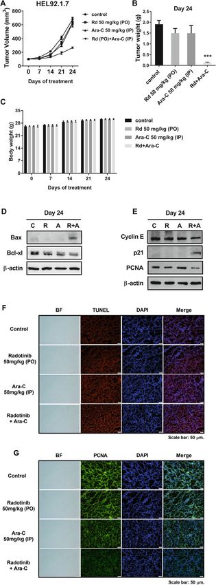
Fig. 6 Radotinib and Ara-C inhibit tumor growth in a xenograft
animal model using HEL92.1.7 cells. a Tumor volume (mm3) after
radotinib and Ara-C treatment (n = 5 for each group). When the
tumors were ~ 150 mm3 in size at ~ 7 days post-implantation, 0.2 ml
radotinib (50 mg/kg body weight, PO) and Ara-C (50 mg/kg body
weight, IP) were injected orally and intraperitoneally five times per
week. Tumor sizes were measured once per week using digital
calipers, and tumor volumes were calculated using the formula
(length × width2) × 0.5. b Tumor weight (g) on day 24. c The body
weight of mice. d, e The expression of diverse proteins including
Bcl-xl, Cyclin E, PCNA, Bax, and p21 in the tumor. f The expression of
TUNEL-positive cells in the tumor tissue. g The expression of PCNA-
positive cells in the tumor tissue. These data represent the means ±
SEM. Significantly different from the control (*); ***, P < 0.001
Conclusions
Therefore, our results can be concluded that radotinib
in combination with Ara-C possesses a strong anti-AML
activity. These results warrant the clinical evaluation of
radotinib with Ara-C-based regimens in AML patients.
Supplementary Information
The online version contains supplementary material available at https://doi.
org/10.1186/s12885-020-07701-8.
Additional file 1: Supplementary Table 1. Information of AML
patients.
Additional file 2: Supplementary Figure 1. Combination of radotinib
and Ara-C inhibits HL60 cell proliferation. Cells were stimulated with vari-
ous concentrations of 0, 10, 30, 40 and 50 μM radotinib and 0, 40, 80, 120
and 160 nM Ara-C for 48 h. The cytotoxicity was then evaluated by a cell
viability assay. (A) Dose-dependent responses of radotinib on cell viability.
(B) Dose-dependent responses of Ara-C on cell viability. (C) Treatment of
radotinib and/or Ara-C at 48 h. Representative data are shown for at least
three independent experiments. These data represent the means ± SEM.
Significantly different from the control (*) or combination of radotinib
and Ara-C (#); *: P, 0.05; ***, ###: P, 0.001. C: DMSO-control, R: radotinib, A:
Ara-C.
Additional file 3: Supplementary Figure 2. Radotinib and Ara-C
sensitize the chemotherapeutic agents including daunorubicin (DNR) or
idarubicin (Idar) in AML cells. (A) HL60 cells were cultured with 3 μM
radotinib, 10 nM Ara-C and 50 nM DNR for 48 h. The cell viability was
then evaluated by an MTS assay. Triple combination of radotinib, Ara-C
and DNR on cell viability is more potent. (B) HEL92.1.7 cells were cultured
with 5 μM radotinib, 10 nM Ara-C and 75 nM DNR for 48 h. Triple combin-
ation of radotinib, Ara-C and DNR on cell viability is more potent (C)
HEL92.1.7 cells were cultured with 5 μM radotinib, 10 nM Ara-C and 2 nM
idarubicin for 48 h. Triple combination of radotinib, Ara-C and idarubicin
on cell viability is more powerful. These data represent the means ± SEM.
Significantly different from control (*) or triple combination of radotinib,
Ara-C and DNR/or idarubicin (#); ***, ###: P < 0.001.
Additional file 4: Supplementary Figure 3. Isobologram analysis of
radotinib and Ara-C combination on AML cell death. Cell viability assay
by radotinib and Ara-C was analyzed in HL60 cells. Cells were seeded
(density, 2 × 10⁴ cells/well) in 96-well plates containing 200 μl medium
per well and were incubated with diverse concentration of radotinib
and/or Ara-C for 48 h at 37 °C. CellTiter 96 solution (20 μl; Promega, Madi-
son, WI, USA) was added directly to each well, and the plates were incu-
bated for 4 h in a humidified atmosphere of 5% CO2 at 37 °C. Absorbance
was measured at 490 nm by using SpectraMax iD3 Microplate Reader
(Molecular Devices, San Jose, CA, USA). We found the strong synergism
on radotinib and Ara-C combination on AML cell death. Fifty % of inhib-
ition concentration (IC50) on AML cell death in HL60 cells: Radotinib only,Heo et al. BMC Cancer (2020) 20:1193 Page 14 of 15
200 μM; Ara-C only, 140 nM; combination of radotinib and Ara-C = 5 μM + Received: 31 July 2020 Accepted: 30 November 2020
70 nM. Combination Index: 0.52).
Additional file 5: Supplementary Figure 4. Original western blots
used for Fig. 3c, d and e. The blots were developed using the
References
ChemiDoc™ Touch Imaging System, and analyzed with the Image Lab™
1. Kropf PL, Wang L, Zang Y, Redner RL, Johnson DE. Dasatinib promotes
Software. The red boxes indicate the cropped regions used in the
ATRA-induced differentiation of AML cells. Leukemia. 2010;24(3):663–5.
representative figures.
2. Hu X, Xu S, Chen Y, Gao Z, Li Y, Hu J, Huang X, Zhang Y, Jiang X, Li L, Yang C,
Additional file 6: Supplementary Figure 5. Original western blots Chen J, Gao N. Depletion of Ars2 inhibits cell proliferation and leukemogenesis
used for Fig. 4c, g and h. The blots were developed using the in acute myeloid leukemia by modulating the miR-6734-3p/p27 axis.
ChemiDoc™ Touch Imaging System, and analyzed with the Image Lab™ Leukemia. 2019;33(5):1090–101.
Software. The red boxes indicate the cropped regions used in the 3. Marcucci G, Haferlach T, Dohner H. Molecular genetics of adult acute
representative figures. myeloid leukemia: prognostic and therapeutic implications. J Clin Oncol.
Additional file 7: Supplementary Figure 6. Original western blots 2011;29(5):475–86.
used for Fig. 5b, c, d and e. The blots were developed using the 4. Mims AS, Blum W. Progress in the problem of relapsed or refractory acute
ChemiDoc™ Touch Imaging System, and analyzed with the Image Lab™ myeloid leukemia. Curr Opin Hematol. 2019;26(2):88–95.
Software. The red boxes indicate the cropped regions used in the 5. Lowenberg B, Downing JR, Burnett A. Acute myeloid leukemia. N Engl J
representative figures. Med. 1999;341(14):1051–62.
Additional file 8: Supplementary Figure 7. Original western blots 6. Malani D, Murumagi A, Yadav B, Kontro M, Eldfors S, Kumar A, Karjalainen R,
used for Fig. 6d and e. The blots were developed using the ChemiDoc™ Majumder MM, Ojamies P, Pemovska T, et al. Enhanced sensitivity to
Touch Imaging System, and analyzed with the Image Lab™ Software. The glucocorticoids in cytarabine-resistant AML. Leukemia. 2017;31(5):1187–95.
red boxes indicate the cropped regions used in the representative 7. Momparler RL. Optimization of cytarabine (ARA-C) therapy for acute
figures. myeloid leukemia. Exp Hematol Oncol. 2013;2:20.
8. Rechkoblit O, Choudhury JR, Buku A, Prakash L, Prakash S, Aggarwal AK.
Additional file 9: Supplementary Table 2. Supplementary Methods. Structural basis for polymerase eta-promoted resistance to the anticancer
nucleoside analog cytarabine. Sci Rep. 2018;8(1):12702.
Abbreviations 9. Tallman MS, Gilliland DG, Rowe JM. Drug therapy for acute myeloid
AML: Acute myeloid leukemia; CML-CP: Chronic phase-chronic myeloid leukemia. Blood. 2005;106(4):1154–63.
leukemia (CML-CP); BM: Bone marrow; BMCs: Bone marrow cells; 10. Matthews JP, Bishop JF, Young GA, Juneja SK, Lowenthal RM, Garson OM,
PCNA: Proliferating cell nuclear antigen; DNR: Daunorubicin; Idar, idarubicin Cobcroft RG, Dodds AJ, Enno A, Gillett EA, et al. Patterns of failure with
increasing intensification of induction chemotherapy for acute myeloid
Acknowledgments leukaemia. Br J Haematol. 2001;113(3):727–36.
The authors would like to thank Ilyang Pharmaceutical Co., Ltd., (Seoul, South 11. Kim SH, Menon H, Jootar S, Saikia T, Kwak JY, Sohn SK, Park JS, Jeong SH,
Korea) for providing the radotinib. Kim HJ, Kim YK, et al. Efficacy and safety of radotinib in chronic phase
chronic myeloid leukemia patients with resistance or intolerance to BCR-
Authors’ contributions ABL1 tyrosine kinase inhibitors. Haematologica. 2014;99(7):1191–6.
SKH, EKN and JCJ designed the study. SKH, EKN, HMY, DKK and HJS performed 12. Zabriskie MS, Vellore NA, Gantz KC, Deininger MW, O'Hare T. Radotinib is an
the experiments. SKH, EKN, YJL, YC and JC analyzed and interpreted the effective inhibitor of native and kinase domain-mutant BCR-ABL1. Leukemia.
experimental data. SKH, JC, SK, YC and YJM provided the discussion and 2015;29(9):1939–42.
suggestions to the experiments. SKH and JCJ wrote the manuscript with input 13. Kwak JY, Kim SH, Oh SJ, Zang DY, Kim H, Kim JA, Do YR, Kim HJ, Park JS,
from all authors. All authors read and approved the final manuscript. Choi CW, et al. Phase III clinical trial (RERISE study) results of efficacy and
safety of Radotinib compared with Imatinib in newly diagnosed chronic
Funding phase chronic myeloid leukemia. Clin Cancer Res. 2017;23(23):7180–8.
This study was supported by the Basic Science Research Program of the 14. Proia DA, Zhang C, Sequeira M, Jimenez JP, He S, Spector N, Shapiro GI,
National Research Foundation of Korea (NRF), funded by the Ministry of Tolaney S, Nagai M, Acquaviva J, et al. Preclinical activity profile and
Education, Science and Technology (2017R1A1A3A04069314). This research therapeutic efficacy of the HSP90 inhibitor ganetespib in triple-negative
was also supported by Basic Science Research Program of the Biomedical breast cancer. Clin Cancer Res. 2014;20(2):413–24.
Research Center, funded by the Ulsan University Hospital (UUHBRC-2016- 15. Acquaviva J, Smith DL, Jimenez JP, Zhang C, Sequeira M, He S, Sang J, Bates
001); and the Ulsan University Hospital Research Grant (UUH-2019-06). RC, Proia DA. Overcoming acquired BRAF inhibitor resistance in melanoma
via targeted inhibition of Hsp90 with ganetespib. Mol Cancer Ther. 2014;
Availability of data and materials 13(2):353–63.
The datasets used and/or analyzed during the current study are available 16. Heo SK, Noh EK, Yoon DJ, Jo JC, Choi Y, Koh S, Baek JH, Park JH, Min YJ, Kim
from the corresponding author on reasonable request. H. Radotinib induces apoptosis of CD11b+ cells differentiated from acute
myeloid leukemia cells. PLoS One. 2015;10(6):e0129853.
Ethics approval and consent to participate 17. Heo SK, Noh EK, Gwon GD, Kim JY, Jo JC, Choi Y, Koh S, Baek JH, Min YJ,
All human-related methods were carried out in accordance with relevant Kim H. Radotinib inhibits acute myeloid leukemia cell proliferation via
guidelines and regulations. All patients were given written informed consent induction of mitochondrial-dependent apoptosis and CDK inhibitors. Eur J
prior to study commencement, and written informed consent was obtained Pharmacol. 2016;789:280–90.
from all patients. The study protocol and patient consent form and information 18. Heo SK, Noh EK, Kim JY, Jeong YK, Jo JC, Choi Y, Koh S, Baek JH, Min YJ, Kim
were approved by the Ethics Committee and Institutional Review Board of the H. Targeting c-KIT (CD117) by dasatinib and radotinib promotes acute
Ulsan University Hospital (UUH-IRB-11-18). All procedures involving animals myeloid leukemia cell death. Sci Rep. 2017;7(1):15278.
were in accordance with the Laboratory Animals Welfare Act, the Guide for the 19. Heo SK, Noh EK, Kim JY, Jo JC, Choi Y, Koh S, Baek JH, Min YJ, Kim H.
Care and Use of Laboratory Animals, and the Guidelines and Policies for Rodent Radotinib induces high cytotoxicity in c-KIT positive acute myeloid leukemia
Experimentation provided by the Institutional Animal Care and Use Committee cells. Eur J Pharmacol. 2017;804:52.
of the Ulsan University of Korea (Approval No. 0117–07). 20. Elmore S. Apoptosis: a review of programmed cell death. Toxicol Pathol.
2007;35(4):495–516.
Consent for publication 21. Murphy T, Yee KWL. Cytarabine and daunorubicin for the treatment of
Not applicable. acute myeloid leukemia. Expert Opin Pharmacother. 2017;18(16):1765–80.
22. Buckley MM, Lamb HM. Oral idarubicin. A review of its pharmacological
Competing interests properties and clinical efficacy in the treatment of haematological
The authors declare that no competing interests exist. malignancies and advanced breast cancer. Drugs Aging. 1997;11(1):61–86.Heo et al. BMC Cancer (2020) 20:1193 Page 15 of 15
23. Fulda S, Debatin KM. Apoptosis signaling in tumor therapy. Ann N Y Acad
Sci. 2004;1028:150–6.
24. Zaman S, Wang R, Gandhi V. Targeting the apoptosis pathway in
hematologic malignancies. Leukemia Lymphoma. 2014;55(9):1980–92.
25. Proposal to change ASHP's name to the American Society of Health-
System Pharmacists. ASHP Board of Directors. Am J Hosp Pharm. 1994;
51(9):1208–11.
26. Droin N, Guery L, Benikhlef N, Solary E. Targeting apoptosis proteins in
hematological malignancies. Cancer Lett. 2013;332(2):325–34.
27. Ashkenazi A, Dixit VM. Death receptors: signaling and modulation. Science.
1998;281(5381):1305–8.
28. Fulda S, Debatin KM. Extrinsic versus intrinsic apoptosis pathways in
anticancer chemotherapy. Oncogene. 2006;25(34):4798–811.
29. Dos Santos C, McDonald T, Ho YW, Liu H, Lin A, Forman SJ, Kuo YH, Bhatia
R. The Src and c-kit kinase inhibitor dasatinib enhances p53-mediated
targeting of human acute myeloid leukemia stem cells by
chemotherapeutic agents. Blood. 2013;122(11):1900–13.
30. Marcucci G, Geyer S, Laumann K, Zhao W, Bucci D, Uy GL, Blum W, Eisfeld
AK, Pardee TS, Wang ES, et al. Combination of dasatinib with chemotherapy
in previously untreated core binding factor acute myeloid leukemia: CALGB
10801. Blood Adv. 2020;4(4):696–705.
31. Heidel F, Cortes J, Rucker FG, Aulitzky W, Letvak L, Kindler T, Huber C,
Dohner H, Kantarjian H, Fischer T. Results of a multicenter phase II trial for
older patients with c-kit-positive acute myeloid leukemia (AML) and high-
risk myelodysplastic syndrome (HR-MDS) using low-dose Ara-C and
Imatinib. Cancer. 2007;109(5):907–14.
32. Advani AS, Tiu R, Saunthararajah Y, Maciejewski J, Copelan EA, Sobecks R,
Sekeres MA, Bates J, Rush ML, Tripp B, et al. A phase 1 study of imatinib
mesylate in combination with cytarabine and daunorubicin for c-kit positive
relapsed acute myeloid leukemia. Leuk Res. 2010;34(12):1622–6.
33. Sen R, Natarajan K, Bhullar J, Shukla S, Fang HB, Cai L, Chen ZS, Ambudkar
SV, Baer MR. The novel BCR-ABL and FLT3 inhibitor ponatinib is a potent
inhibitor of the MDR-associated ATP-binding cassette transporter ABCG2.
Mol Cancer Ther. 2012;11(9):2033–44.
34. Heo SK, Noh EK, Jeong YK, Ju LJ, Sung JY, Yu HM, Cheon J, Koh S, Min YJ,
Choi Y, et al. Radotinib inhibits mitosis entry in acute myeloid leukemia cells
via suppression of Aurora kinase a expression. Tumour Biol. 2019;41(5):
1010428319848612.
35. Li L, Cui Y, Shen J, Dobson H, Sun G. Evidence for activated Lck protein
tyrosine kinase as the driver of proliferation in acute myeloid leukemia cell,
CTV-1. Leuk Res. 2019;78:12–20.
36. Rassidakis GZ, Herold N, Myrberg IH, Tsesmetzis N, Rudd SG, Henter JI,
Schaller T, Ng SB, Chng WJ, Yan B, et al. Low-level expression of SAMHD1 in
acute myeloid leukemia (AML) blasts correlates with improved outcome
upon consolidation chemotherapy with high-dose cytarabine-based
regimens. Blood Cancer J. 2018;8(11):98.
37. Chen K, Chen Y, Chen Z, Shi Y, He Z, Ding B, Wang C, Yu L. miR-134
increases the antitumor effects of cytarabine by targeting Mnks in acute
myeloid leukemia cells. OncoTargets Therapy. 2018;11:3141–7.
38. Mayer LD, Tardi P, Louie AC. CPX-351: a nanoscale liposomal co-formulation
of daunorubicin and cytarabine with unique biodistribution and tumor cell
uptake properties. Int J Nanomedicine. 2019;14:3819–30.
39. Guo H, Lin SY, Ren WX, Lei Q, Chen ZC, Zhang L, Li QB. Enhanced response
of acute Monocytic leukemia cells to low-dose Cytarabine by 1,25-
dihydroxyvitamin D3. Curr Med Sci. 2018;38(1):35–42.
40. Kaufmann SH, Karp JE, Litzow MR, Mesa RA, Hogan W, Steensma DP, Flatten
KS, Loegering DA, Schneider PA, Peterson KL, et al. Phase I and
pharmacological study of cytarabine and tanespimycin in relapsed and
refractory acute leukemia. Haematologica. 2011;96(11):1619–26.
41. Erlichman C. Tanespimycin: the opportunities and challenges of targeting
heat shock protein 90. Expert Opin Investig Drugs. 2009;18(6):861–8.
42. Bargal SA, Rafiee R, Crews KR, Wu H, Cao X, Rubnitz JE, Ribeiro RC, Downing
JR, Pounds SB, Lamba JK. Genome-wide association analysis identifies SNPs
predictive of in vitro leukemic cell sensitivity to cytarabine in pediatric AML.
Oncotarget. 2018;9(79):34859–75.
Publisher’s Note
Springer Nature remains neutral with regard to jurisdictional claims in
published maps and institutional affiliations.You can also read