7 Cell-Cell Interactions - Concept Outline
←
→
Page content transcription
If your browser does not render page correctly, please read the page content below
7
Cell-Cell Interactions
Concept Outline
7.1 Cells signal one another with chemicals.
Receptor Proteins and Signaling between Cells.
Receptor proteins embedded in the plasma membrane
change shape when they bind specific signal molecules,
triggering a chain of events within the cell.
Types of Cell Signaling. Cell signaling can occur between
adjacent cells, although chemical signals called hormones act
over long distances.
7.2 Proteins in the cell and on its surface receive
signals from other cells.
Intracellular Receptors. Some receptors are located
within the cell cytoplasm. These receptors respond to lipid-
soluble signals, such as steroid hormones.
Cell Surface Receptors. Many cell-to-cell signals are
water-soluble and cannot penetrate membranes. Instead, the
signals are received by transmembrane proteins protruding
out from the cell surface.
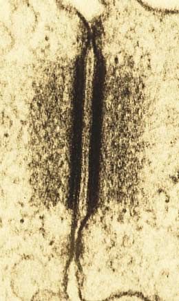
7.3 Follow the journey of information into the cell. FIGURE 7.1
Persimmon cells in close contact with one another. These
Initiating the Intracellular Signal. Cell surface receptors plant cells and all cells, no matter what their function, interact
often use “second messengers” to transmit a signal to the with their environment, including the cells around them.
cytoplasm.
Amplifying the Signal: Protein Kinase Cascades.
Surface receptors and second messengers amplify signals as
they travel into the cell, often toward the cell nucleus.
D id you know that each of the 100 trillion cells of your
body shares one key feature with the cells of tigers,
bumblebees, and persimmons (figure 7.1)—a feature that
7.4 Cell surface proteins mediate cell-cell interactions.
most bacteria and protists lack? Your cells touch and com-
The Expression of Cell Identity. Cells possess on their municate with one another. Sending and receiving a variety
surfaces a variety of tissue-specific identity markers that of chemical signals, they coordinate their behavior so that
identify both the tissue and the individual.
your body functions as an integrated whole, rather than as a
Intercellular Adhesion. Cells attach themselves to one
massive collection of individual cells acting independently.
another with protein links. Some of the links are very strong,
The ability of cells to communicate with one another is the
others more transient.
hallmark of multicellular organisms. In this chapter we will
Tight Junctions. Adjacent cells form a sheet when
connected by tight junctions, and molecules are encouraged look in detail at how the cells of multicellular organisms in-
to flow through the cells, not between them. teract with one another, first exploring how they signal one
Anchoring Junctions. The cytoskeleton of a cell is another with chemicals and then examining the ways in
connected by an anchoring junction to the cytoskeleton of which their cell surfaces interact to organize tissues and
another cell or to the extracellular matrix. body structures.
Communicating Junctions. Many adjacent cells have
direct passages that link their cytoplasms, permitting the
passage of ions and small molecules.
123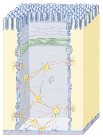
7.1      Cells signal one another with chemicals.

Receptor Proteins and                                     Signal
Signaling between Cells                                   molecules

Communication between cells is com-
mon in nature. Cell signaling occurs
in all multicellular organisms, provid-                                                         Extracellular
ing an indispensable mechanism for                                                              surface
cells to influence one another. The
cells of multicellular organisms use a
variety of molecules as signals, includ-
ing not only peptides, but also large
proteins, individual amino acids, nu-
cleotides, steroids and other lipids.
   Even dissolved gases are used as                                                             Cytoplasm
signals. Nitric oxide (NO) plays a role
in mediating male erections (Viagra
                                           FIGURE 7.2
functions by stimulating NO release).
                                           Cell surface receptors recognize only specific molecules. Signal molecules will bind
   Some of these molecules are at-         only to those cells displaying receptor proteins with a shape into which they can fit snugly.
tached to the surface of the signaling
cell; others are secreted through the
plasma membrane or released by
exocytosis.
                                                                         Monoclonal antibodies. The first method uses mono-
Cell Surface Receptors                                                   clonal antibodies. An antibody is an immune system pro-
                                                                         tein that, like a receptor, binds specifically to another
Any given cell of a multicellular organism is exposed to a
                                                                         molecule. Each individual immune system cell can make
constant stream of signals. At any time, hundreds of differ-
                                                                         only one particular type of antibody, which can bind to
ent chemical signals may be in the environment surround-
                                                                         only one specific target molecule. Thus, a cell-line de-
ing the cell. However, each cell responds only to certain
                                                                         rived from a single immune system cell (a clone) makes
signals and ignores the rest (figure 7.2), like a person fol-
                                                                         one specific antibody (a monoclonal antibody). Mono-
lowing the conversation of one or two individuals in a
                                                                         clonal antibodies that bind to particular receptor pro-
noisy, crowded room. How does a cell “choose” which sig-
                                                                         teins can be used to isolate those proteins from the
nals to respond to? Located on or within the cell are re-
                                                                         thousands of others in the cell.
ceptor proteins, each with a three-dimensional shape that
fits the shape of a specific signal molecule. When a signal              Gene analysis. The study of mutants and isolation of
molecule approaches a receptor protein of the right shape,               gene sequences has had a tremendous impact on the
the two can bind. This binding induces a change in the re-               field of receptor analysis. In chapter 19 we will present a
ceptor protein’s shape, ultimately producing a response in               detailed account of how this is done. These advances
the cell. Hence, a given cell responds to the signal mole-               make it feasible to identify and isolate the many genes
cules that fit the particular set of receptor proteins it pos-           that encode for various receptor proteins.
sesses, and ignores those for which it lacks receptors.                  Remarkably, these techniques have revealed that the
                                                                      enormous number of receptor proteins can be grouped into
The Hunt for Receptor Proteins                                        just a handful of “families” containing many related recep-
                                                                      tors. Later in this chapter we will meet some of the mem-
The characterization of receptor proteins has presented a             bers of these receptor families.
very difficult technical problem, because of their relative
scarcity in the cell. Because these proteins may constitute              Cells in a multicellular organism communicate with
less than 0.01% of the total mass of protein in a cell, puri-            others by releasing signal molecules that bind to
fying them is analogous to searching for a particular grain              receptor proteins on the surface of the other cells.
of sand in a sand dune! However, two recent techniques                   Recent advances in protein isolation have yielded a
                                                                         wealth of information about the structure and function
have enabled cell biologists to make rapid progress in this
                                                                         of these proteins.
area.

124    Part II   Biology of the Cell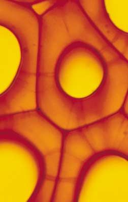
Types of Cell Signaling
                                                                                                                    Secretory cell
Cells communicate through any of four
basic mechanisms, depending primarily
on the distance between the signaling
and responding cells (figure 7.3). In ad-                                            Gap
dition to using these four basic mecha-                                              junction
nisms, some cells actually send signals
to themselves, secreting signals that                                                                                         Adjacent target cells

bind to specific receptors on their own
plasma membranes. This process,
called autocrine signaling, is thought                        (a) Direct contact                            (b) Paracrine signaling
to play an important role in reinforcing
developmental changes.
                                                                   Hormone secretion into
                                                                   blood by endocrine gland
                                                                                                                       Neurotransmitter
Direct Contact
As we saw in chapter 6, the surface of a                                                                                                  Target cell
eukaryotic cell is a thicket of proteins,
carbohydrates, and lipids attached to               Blood vessel
                                                                                                    Nerve cell
and extending outward from the                       Distant target cells                                                  Synaptic gap
plasma membrane. When cells are very
close to one another, some of the mol-                    (c) Endocrine signaling                                (d) Synaptic signaling
ecules on the cells’ plasma membranes
may bind together in specific ways.          FIGURE 7.3
Many of the important interactions           Four kinds of cell signaling. Cells communicate in several ways. (a) Two cells in direct
between cells in early development           contact with each other may send signals across gap junctions. (b) In paracrine signaling,
                                             secretions from one cell have an effect only on cells in the immediate area. (c) In endocrine
occur by means of direct contact be-
                                             signaling, hormones are released into the circulatory system, which carries them to the target
tween cell surfaces (figure 7.3a). We’ll
                                             cells. (d) Chemical synaptic signaling involves transmission of signal molecules, called
examine contact-dependent interac-           neurotransmitters, from a neuron over a small synaptic gap to the target cell.
tions more closely later in this chapter.

Paracrine Signaling                                                         Synaptic Signaling
Signal molecules released by cells can diffuse through the                  In animals, the cells of the nervous system provide rapid
extracellular fluid to other cells. If those molecules are                  communication with distant cells. Their signal molecules,
taken up by neighboring cells, destroyed by extracellular                   neurotransmitters, do not travel to the distant cells
enzymes, or quickly removed from the extracellular fluid in                 through the circulatory system like hormones do. Rather,
some other way, their influence is restricted to cells in the               the long, fiberlike extensions of nerve cells release neuro-
immediate vicinity of the releasing cell. Signals with such                 transmitters from their tips very close to the target cells
short-lived, local effects are called paracrine signals (figure             (figure 7.3d). The narrow gap between the two cells is
7.3b). Like direct contact, paracrine signaling plays an im-                called a chemical synapse. While paracrine signals move
portant role in early development, coordinating the activi-                 through the fluid between cells, neurotransmitters cross the
ties of clusters of neighboring cells.                                      synapse and persist only briefly. We will examine synaptic
                                                                            signaling more fully in chapter 54.
Endocrine Signaling
If a released signal molecule remains in the extracellular fluid,              Adjacent cells can signal others by direct contact, while
it may enter the organism’s circulatory system and travel                      nearby cells that are not touching can communicate
widely throughout the body. These longer lived signal mole-                    through paracrine signals. Two other systems mediate
cules, which may affect cells very distant from the releasing                  communication over longer distances: in endocrine
cell, are called hormones, and this type of intercellular com-                 signaling the blood carries hormones to distant cells,
                                                                               and in synaptic signaling nerve cells secrete
munication is known as endocrine signaling (figure 7.3c).
                                                                               neurotransmitters from long cellular extensions close to
Chapter 58 discusses endocrine signaling in detail. Both ani-                  the responding cells.
mals and plants use this signaling mechanism extensively.

                                                                                                  Chapter 7 Cell-Cell Interactions               125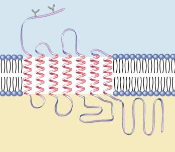
7.2      Proteins in the cell and on its surface receive signals from other cells.

Intracellular Receptors                                                                                                          Signal molecule

All cell signaling pathways share certain common elements,                                                                       Signal molecule-
including a chemical signal that passes from one cell to an-              Inhibitor                                              binding site
other and a receptor that receives the signal in or on the                protein
target cell. We’ve looked at the sorts of signals that pass
from one cell to another. Now let’s consider the nature of                                                                       Signal molecule-
                                                                                                                                 binding domain
the receptors that receive the signals. Table 7.1 summarizes
the types of receptors we will discuss in this chapter.
   Many cell signals are lipid-soluble or very small mole-                                                   DNA binding
                                                                                                             domain
cules that can readily pass across the plasma membrane of                   Transcription-activating
the target cell and into the cell, where they interact with a               domain
receptor. Some bind to protein receptors located in the cy-                FIGURE 7.4
toplasm; others pass across the nuclear membrane as well                   Basic structure of a gene-regulating intracellular receptor.
and bind to receptors within the nucleus. These intracel-                  These receptors are located within the cell and function in the
lular receptors (figure 7.4) may trigger a variety of re-                  reception of signals such as steroid hormones, vitamin D, and
sponses in the cell, depending on the receptor.                            thyroid hormone.

                                              Table 7.1   Cell Communicating Mechanisms
 Mechanism                           Structure                          Function                                  Example
 INTRACELLULAR RECEPTORS No extracellular signal-binding                Receives signals from lipid-soluble      Receptors for NO, steroid
                                     site                               or noncharged, nonpolar small            hormone, vitamin D, and
                                                                        molecules                                thyroid hormone

 CELL SURFACE RECEPTORS
 Chemically gated                    Multipass transmembrane            Molecular “gates” triggered              Neurons
 ion channels                        protein forming a central pore     chemically to open or close
 Enzymic receptors                   Single-pass transmembrane          Binds signal extracellularly,            Phosphorylation of protein
                                     protein                            catalyzes response intracellularly       kinases
 G-protein-linked                    Seven-pass transmembrane           Binding of signal to receptor causes     Peptide hormones, rod
 receptors                           protein with cytoplasmic           GTP to bind a G protein; G protein,      cells in the eyes
                                     binding site for G protein         with attached GTP, detaches to
                                                                        deliver the signal inside the cell

 PHYSICAL CONTACT WITH OTHER CELLS
 Surface markers                     Variable; integral proteins or     Identify the cell                        MHC complexes, blood
                                     glycolipids in cell membrane                                                groups, antibodies
 Tight junctions                     Tightly bound, leakproof,          Organizing junction: holds cells         Junctions between
                                     fibrous protein “belt” that        together such that material passes       epithelial cells in the gut
                                     surrounds cell                     through but not between the cells
 Desmosomes                          Intermediate filaments of          Anchoring junction: “buttons”            Epithelium
                                     cytoskeleton linked to adjoining   cells together
                                     cells through cadherins
 Adherens junctions                  Transmembrane fibrous              Anchoring junction: “roots”              Tissues with high
                                     proteins                           extracellular matrix to cytoskeleton     mechanical stress, such as
                                                                                                                 the skin
 Gap junctions                      Six transmembrane connexon          Communicating junction: allows           Excitable tissue such as
                                    proteins creating a “pipe”          passage of small molecules from          heart muscle
                                    that connects cells                 cell to cell in a tissue
 Plasmodesmata                      Cytoplasmic connections             Communicating junction between           Plant tissues
                                    between gaps in adjoining           plant cells
                                    plant cell walls

126    Part II     Biology of the Cell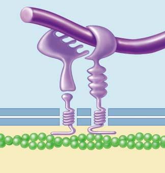
Receptors That Act as Gene Regulators                                                                                Cortisol
                                                                  Transcription
Some intracellular receptors act as regulators of gene            activating
                                                                                         Inhibitor
transcription. Among them are the receptors for steroid           domain
hormones, such as cortisol, estrogen, and progesterone, as
well as the receptors for a number of other small, lipid-                                                          Signal molecule-
                                                                                                                   binding domain
soluble signal molecules, such as vitamin D and thyroid
hormone. All of these receptors have similar structures;                             DNA binding
the genes that code for them may well be the evolutionary                            site blocked
descendants of a single ancestral gene. Because of their
structural similarities, they are all part of the intracellular
receptor superfamily.
    Each of these receptors has a binding site for DNA. In
its inactive state, the receptor typically cannot bind DNA
because an inhibitor protein occupies the binding site.
When the signal molecule binds to another site on the re-
ceptor, the inhibitor is released and the DNA binding site
is exposed (figure 7.5). The receptor then binds to a spe-
cific nucleotide sequence on the DNA, which activates (or,                           DNA binding
in a few instances, suppresses) a particular gene, usually lo-                       site exposed
cated adjacent to the regulatory site.
    The lipid-soluble signal molecules that intracellular re-
ceptors recognize tend to persist in the blood far longer
than water-soluble signals. Most water-soluble hormones
break down within minutes, and neurotransmitters within
seconds or even milliseconds. A steroid hormone like corti-
sol or estrogen, on the other hand, persists for hours.
    The target cell’s response to a lipid-soluble cell signal
can vary enormously, depending on the nature of the cell.
This is true even when different target cells have the
same intracellular receptor, for two reasons: First, the
binding site for the receptor on the target DNA differs           FIGURE 7.5
from one cell type to another, so that different genes are        How intracellular receptors regulate gene transcription. In
affected when the signal-receptor complex binds to the            this model, the binding of the steroid hormone cortisol to a DNA
                                                                  regulatory protein causes it to alter its shape. The inhibitor is
DNA, and second, most eukaryotic genes have complex
                                                                  released, exposing the DNA binding site of the regulatory
controls. We will discuss them in detail in chapter 16,
                                                                  protein. The DNA binds to the site, positioning a specific
but for now it is sufficient to note that several different       nucleotide sequence over the transcription activating domain of
regulatory proteins are usually involved in reading a eu-         the receptor and initiating transcription.
karyotic gene. Thus the intracellular receptor interacts
with different signals in different tissues. Depending on
the cell-specific controls operating in different tissues,
the effect the intracellular receptor produces when it
binds with DNA will vary.                                            NO has only recently been recognized as a signal mole-
                                                                  cule in vertebrates. Already, however, a wide variety of
                                                                  roles have been documented. For example, when the brain
Receptors That Act as Enzymes                                     sends a nerve signal relaxing the smooth muscle cells lining
Other intracellular receptors act as enzymes. A very inter-       the walls of vertebrate blood vessels, the signal molecule
esting example is the receptor for the signal molecule, ni-       acetylcholine released by the nerve near the muscle does
tric oxide (NO). A small gas molecule, NO diffuses readily        not interact with the muscle cell directly. Instead, it causes
out of the cells where it is produced and passes directly into    nearby epithelial cells to produce NO, which then causes
neighboring cells, where it binds to the enzyme guanylyl          the smooth muscle to relax, allowing the vessel to expand
cyclase. Binding of NO activates the enzyme, enabling it to       and thereby increase blood flow.
catalyze the synthesis of cyclic guanosine monophosphate
                                                                      Many target cells possess intracellular receptors, which
(GMP), an intracellular messenger molecule that produces
                                                                      are activated by substances that pass through the
cell-specific responses such as the relaxation of smooth
                                                                      plasma membrane.
muscle cells.

                                                                                         Chapter 7 Cell-Cell Interactions       127
Cell Surface Receptors                                                    across the plasma membrane several times. In the center of
                                                                          the protein is a pore that connects the extracellular fluid
Most signal molecules are water-soluble, including neuro-                 with the cytoplasm. The pore is big enough for ions to pass
transmitters, peptide hormones, and the many proteins                     through, so the protein functions as an ion channel. The
that multicellular organisms employ as “growth factors”                   channel is said to be chemically gated because it opens
during development. Water-soluble signals cannot diffuse                  when a chemical (the neurotransmitter) binds to it. The
through cell membranes. Therefore, to trigger responses                   type of ion (sodium, potassium, calcium, chloride, for ex-
in cells, they must bind to receptor proteins on the sur-                 ample) that flows across the membrane when a chemically
face of the cell. These cell surface receptors (figure 7.6)               gated ion channel opens depends on the specific three-
convert the extracellular signal to an intracellular one, re-             dimensional structure of the channel.
sponding to the binding of the signal molecule by produc-
ing a change within the cell’s cytoplasm. Most of a cell’s
                                                                          Enzymic Receptors
receptors are cell surface receptors, and almost all of them
belong to one of three receptor superfamilies: chemically                 Many cell surface receptors either act as enzymes or are di-
gated ion channels, enzymic receptors, and G-protein-                     rectly linked to enzymes (figure 7.6b). When a signal mole-
linked receptors.                                                         cule binds to the receptor, it activates the enzyme. In al-
                                                                          most all cases, these enzymes are protein kinases, enzymes
                                                                          that add phosphate groups to proteins. Most enzymic re-
Chemically Gated Ion Channels
                                                                          ceptors have the same general structure. Each is a single-
Chemically gated ion channels are receptor proteins that                  pass transmembrane protein (the amino acid chain passes
ions pass through. The receptor proteins that bind many                   through the plasma membrane only once); the portion that
neurotransmitters have the same basic structure (figure                   binds the signal molecule lies outside the cell, and the por-
7.6a). Each is a “multipass” transmembrane protein, mean-                 tion that carries out the enzyme activity is exposed to the
ing that the chain of amino acids threads back and forth                  cytoplasm.

                           Signal               Ions                                                                       Signal

                                                                                               Inactive                        Active
                                                                                               catalytic                       catalytic
                                                                                               domain                          domain
(a) Chemically gated ion channel                                   (b) Enzymic receptor

                                                                                          Enzyme or                          Activated
                                    G protein                    Activated                ion channel                        enzyme or
                                                                 G protein                                                   ion channel

(c) G-protein-linked receptor

FIGURE 7.6
Cell surface receptors. (a) Chemically gated ion channels are multipass transmembrane proteins that form a pore in the cell membrane.
This pore is opened or closed by chemical signals. (b) Enzymic receptors are single-pass transmembrane proteins that bind the signal on
the extracellular surface. A catalytic region on their cytoplasmic portion then initiates enzymatic activity inside the cell. (c) G-protein-
linked receptors bind to the signal outside the cell and to G proteins inside the cell. The G protein then activates an enzyme or ion
channel, mediating the passage of a signal from the cell’s surface to its interior.

128     Part II    Biology of the Cell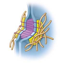
G-Protein-Linked Receptors
                                                                            NH2
A third class of cell surface receptors acts indirectly on                                   Protein signal-binding site
enzymes or ion channels in the plasma membrane with
the aid of an assisting protein, called a guanosine triphos-
phate (GTP)-binding protein, or G protein (figure 7.6c).
Receptors in this category use G proteins to mediate pas-
sage of the signal from the membrane surface into the
cell interior.

How G-Protein-Linked Receptors Work. G pro-
teins are mediators that initiate a diffusible signal in the
cytoplasm. They form a transient link between the recep-
tor on the cell surface and the signal pathway within the                                                 COOH
cytoplasm. Importantly, this signal has a relatively short
life span whose active age is determined by GTP. When                                                 G-protein-binding sites
a signal arrives, it finds the G protein nestled into the G-
protein-linked receptor on the cytoplasmic side of the
plasma membrane. Once the signal molecule binds to the
receptor, the G-protein-linked receptor changes shape.
                                                                FIGURE 7.7
This change in receptor shape twists the G protein, caus-
                                                                The G-protein-linked receptor is a seven-pass transmembrane
ing it to bind GTP. The G protein can now diffuse away          protein.
from the receptor. The “activated” complex of a G pro-
tein with attached GTP is then free to initiate a number
of events. However, this activation is short-lived, because     protein discussed earlier, and in many other sensory recep-
GTP has a relatively short life span (seconds to minutes).      tors. Vertebrate rhodopsin is in fact a G-protein-linked re-
This elegant arrangement allows the G proteins to acti-         ceptor and utilizes a G protein. Bacteriorhodopsin is not.
vate numerous pathways, but only in a transient manner.         The occurrence of the seven-pass structural motif in both,
In order for a pathway to “stay on,” there must be a con-       and in so many other G-protein-linked receptors, suggests
tinuous source of incoming extracellular signals. When          that this motif is a very ancient one, and that G-protein-
the rate of external signal drops off, the pathway shuts        linked receptors may have evolved from sensory receptors
down.                                                           of single-celled ancestors.

The Largest Family of Cell Surface Receptors. Sci-              Discovery of G Proteins. Martin Rodbell of the Na-
entists have identified more than 100 different G-              tional Institute of Environmental Health Sciences and
protein-linked receptors, more than any other kind of           Alfred Gilman of the University of Texas Southwestern
cell surface receptor. They mediate an incredible range         Medical Center received the 1994 Nobel Prize for Medi-
of cell signals, including peptide hormones, neurotrans-        cine or Physiology for their work on G proteins. Rodbell
mitters, fatty acids, and amino acids. Despite this great       and Gilman’s work has proven to have significant ramifi-
variation in specificity, however, all G-protein-linked re-     cations. G proteins are involved in the mechanism em-
ceptors whose amino acid sequences are known have a             ployed by over half of all medicines in use today. Study-
similar structure. They are almost certainly closely re-        ing G proteins will vastly expand our understanding of
lated in an evolutionary sense, arising from a single an-       how these medicines work. Furthermore, the investiga-
cestral sequence. Each of these G-protein-linked recep-         tion of G proteins should help elucidate how cells com-
tors is a seven-pass transmembrane protein (figure              municate in general and how they contribute to the over-
7.7)—a single polypeptide chain that threads back and           all physiology of organisms. As Gilman says, G proteins
forth across the lipid bilayer seven times, creating a chan-    are “involved in everything from sex in yeast to cognition
nel through the membrane.                                       in humans.”

Evolutionary Origin of G-Protein-Linked Receptors.                 Most receptors are located on the surface of the plasma
As research revealed the structure of G-protein-linked re-         membrane. Chemically gated ion channels open or
ceptors, an interesting pattern emerged: the same seven-           close when signal molecules bind to the channel,
pass structural motif is seen again and again, in sensory re-      allowing specific ions to diffuse through. Enzyme
ceptors such as the light-activated rhodopsin protein in the       receptors typically activate intracellular proteins by
                                                                   phosphorylation. G-protein-linked receptors activate an
vertebrate eye, in the light-activated bacteriorhodopsin
                                                                   intermediary protein, which then effects the
proton pump that plays a key role in bacterial photosynthe-
                                                                   intracellular change.
sis, in the receptor that recognizes the yeast mating factor

                                                                                     Chapter 7 Cell-Cell Interactions       129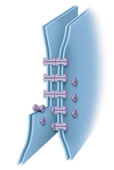
7.3      Follow the journey of information into the cell.

Initiating the                                   Oligosaccharide
                                                 unit                          NH3+
Intracellular Signal
Some enzymic receptors and most G-
protein-linked receptors carry the sig-                                                                         Extracellular
nal molecule’s message into the target
cell by utilizing other substances to
relay the message within the cyto-
plasm. These other substances, small
molecules or ions commonly called
second messengers or intracellular
mediators, alter the behavior of partic-
ular proteins by binding to them and
changing their shape. The two most
widely used second messengers are
cyclic adenosine monophosphate
(cAMP) and calcium.

cAMP                                            Intracellular
All animal cells studied thus far use
cAMP as a second messenger (chap-
ter 56 discusses cAMP in detail). To                                                                                              COO-
see how cAMP typically works as a
messenger, let’s examine what hap-
pens when the hormone epinephrine
binds to a particular type of G- FIGURE 7.8
protein-linked receptor called the β- Structure of the β-adrenergic receptor. The receptor is a G-protein-linked molecule
                                             which, when it binds to an extracellular signal molecule, stimulates voluminous production
adrenergic receptor (figure 7.8).
                                             of cAMP inside the cell, which then effects the cellular change.
When epinephrine binds with this re-
ceptor, it activates a G protein, which
then stimulates the enzyme adenylyl
cyclase to produce large amounts of cAMP within the                    activities. For example, the efflux of Ca++ from the endo-
cell (figure 7.9a). The cAMP then binds to and activates               plasmic reticulum causes skeletal muscle cells to contract
the enzyme α-kinase, which adds phosphates to specific                 and some endocrine cells to secrete hormones.
proteins in the cell. The effect this phosphorylation has                 The gated Ca++ channels are opened by a G-protein-
on cell function depends on the identity of the cell and               linked receptor. In response to signals from other cells,
the proteins that are phosphorylated. In muscle cells, for             the receptor activates its G protein, which in turn acti-
example, the α-kinase phosphorylates and thereby acti-                 vates the enzyme, phospholipase C. This enzyme catalyzes
vates enzymes that stimulate the breakdown of glycogen                 the production of inositol trisphosphate (IP3) from phospho-
into glucose and inhibit the synthesis of glycogen from                lipids in the plasma membrane. The IP3 molecules diffuse
glucose. Glucose is then more available to the muscle                  through the cytoplasm to the endoplasmic reticulum and
cells for metabolism.                                                  bind to the Ca++ channels. This opens the channels and
                                                                       allows Ca++ to flow from the endoplasmic reticulum into
                                                                       the cytoplasm (figure 7.9b).
Calcium
                                                                          Ca ++ initiates some cellular responses by binding to
Calcium (Ca++) ions serve even more widely than cAMP                   calmodulin, a 148-amino acid cytoplasmic protein that con-
as second messengers. Ca++ levels inside the cytoplasm of a            tains four binding sites for Ca++ (figure 7.10). When four
cell are normally very low (less than 107 M), while outside           Ca++ ions are bound to calmodulin, the calmodulin/Ca++
the cell and in the endoplasmic reticulum Ca++ levels are              complex binds to other proteins, and activates them.
quite high (about 103 M). Chemically gated calcium chan-
nels in the endoplasmic reticulum membrane act as                         Cyclic AMP and Ca++ often behave as second
                                                                          messengers,    intracellular substances that relay
switches; when they open, Ca++ rushes into the cytoplasm
                                                                          messages    from  receptors to target proteins.
and triggers proteins sensitive to Ca++ to initiate a variety of
130    Part II   Biology of the Cell
Signal molecule                                                  Signal molecule
                              Cell surface receptor                                          Cell surface receptor

                                                   Adenylyl cyclase                                                   Phospholipase C

                                                            cAMP                          G protein                   Inositol
                       G protein                                                                                      trisphosphate
                                           ATP                                     Endoplasmic                        intermediary
                                                                                   reticulum

                                                  Target protein                                      Ca++
                    Cytoplasm                                                       Cytoplasm                                Target
                                                                                                                             protein
                                     Nucleus                                                          Nucleus

                    Cell                                                             Cell
                    membrane                                                         membrane

            (a)                  cAMP pathway                                (b)                    Ca++ pathway

FIGURE 7.9
How second messengers work. (a) The cyclic AMP (cAMP) pathway. An extracellular receptor binds to a signal molecule and, through a
G protein, activates the membrane-bound enzyme, adenylyl cyclase. This enzyme catalyzes the synthesis of cAMP, which binds to the
target protein to initiate the cellular change. (b) The calcium (Ca++) pathway. An extracellular receptor binds to another signal molecule
and, through another G protein, activates the enzyme phospholipase C. This enzyme stimulates the production of inositol trisphosphate,
which binds to and opens calcium channels in the membrane of the endoplasmic reticulum. Ca++ is released into the cytoplasm, effecting a
change in the cell.

                                                                                                                      Ca++
                                                  Ca++
                             Ca++

                                                                                           Calmodulin                                    Inactive
                                                                                                                                         protein
                              Calmodulin
FIGURE 7.10
Calmodulin. (a)
Calmodulin is a protein
containing 148 amino acid
residues that mediates
Ca++ function. (b) When                                               Ca++
four Ca++ are bound to the
calmodulin molecule, it
undergoes a                                                                                                                    Active
conformational change                                        Ca++
                                                                                                                               protein
that allows it to bind to
other cytoplasmic proteins
and effect cellular                        (a)                                                                  (b)
responses.

                                                                                                       Chapter 7 Cell-Cell Interactions         131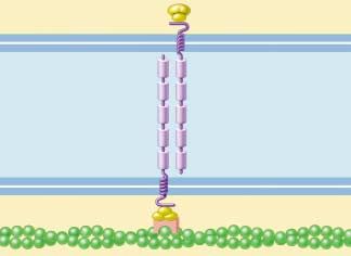
Amplifying the Signal: Protein                                               receptors use a chain of other protein messengers to am-
                                                                             plify the signal as it is being relayed to the nucleus.
Kinase Cascades                                                                 How is the signal amplified? Imagine a relay race where,
Both enzyme-linked and G-protein-linked receptors re-                        at the end of each stage, the finishing runner tags five new
ceive signals at the surface of the cell, but as we’ve seen, the             runners to start the next stage. The number of runners
target cell’s response rarely takes place there. In most cases               would increase dramatically as the race progresses: 1, then
the signals are relayed to the cytoplasm or the nucleus by                   5, 25, 125, and so on. The same sort of process takes place
second messengers, which influence the activity of one or                    as a signal is passed from the cell surface to the cytoplasm
more enzymes or genes and so alter the behavior of the                       or nucleus. First the receptor activates a stage-one protein,
cell. But most signaling molecules are found in such low                     almost always by phosphorylating it. The receptor either
concentrations that their diffusion across the cytoplasm                     adds a phosphate group directly, or, it activates a G protein
would take a great deal of time unless the signal is ampli-                  that goes on to activate a second protein that does the
fied. Therefore, most enzyme-linked and G-protein-linked                     phosphorylation. Once activated, each of these stage-one
                                                                             proteins in turn activates a large number of stage-two pro-

  Signal
 molecule         1                              Activated
                        Receptor              adenylyl cyclase
                         protein

   2    Amplification
                                                                                                                                  Not yet
                                                                                                                                 activated
                                                                                            4        Amplification

                                                                                                                              cAMP
                                          3
                                                                                       5
                                                                                                                              Protein kinase
                  GTP         G protein
                                                                               6     Amplification

                                                                                                                     Enzyme

                                                                 7   Amplification

                                                                                                Enzymatic product

FIGURE 7.11
Signal amplification. Amplification at many steps of the cell-signaling process can ultimately produce a large response by the cell. One
cell surface receptor (1), for example, may activate many G protein molecules (2), each of which activates a molecule of adenylyl cyclase
(3), yielding an enormous production of cAMP (4). Each cAMP molecule in turn will activate a protein kinase (5), which can
phosphorylate and thereby activate several copies of a specific enzyme (6). Each of those enzymes can then catalyze many chemical
reactions (7). Starting with 1010 M of signaling molecule, one cell surface receptor can trigger the production of 106 M of one of the
products, an amplification of four orders of magnitude.

132     Part II   Biology of the Cell
FIGURE 7.12 O
Cyclic GMP. Cyclic GMP is a N
guanosine monophosphate nucleotide N
molecule with the single phosphate
group attached to a sugar residue in
N N NH2
two places (this cyclic One rhodopsin molecule
part is shown in CH2 Guanine absorbs one photon, which
yellow). Cyclic GMP O O
is an important
second messenger
linking G proteins –
to signal O P O OH
transduction Sugar
O
pathways within
Phosphate
the cytoplasm. activates 500 transducin
molecules, which
teins; then each of them activates a large number of stage-
three proteins, and so on (figure 7.11). A single cell surface
receptor can thus stimulate a cascade of protein kinases to
amplify the signal.
The Vision Amplification Cascade activate 500 phosphodiesterase
molecules, which
Let’s trace a protein amplification cascade to see exactly
how one works. In vision, a single light-activated rhodopsin
(a G-protein-linked receptor) activates hundreds of mole-
cules of the G protein transducin in the first stage of the
relay. In the second stage, each transducin causes an en-
zyme to modify thousands of molecules of a special inside-
the-cell messenger called cyclic GMP (figure 7.12). (We
will discuss cyclic GMP in more detail later.) In about 1 hydrolyze 105 cyclic GMP
second, a single rhodopsin signal passing through this two- molecules, which
step cascade splits more than 105 (100,000) cyclic GMP
molecules (figure 7.13)! The rod cells of humans are suffi- Na+
Na+
ciently sensitive to detect brief flashes of 5 photons.
The Cell Division Amplification Cascade
The amplification of signals traveling from the plasma close 250 Na+ channels, preventing
membrane to the nucleus can be even more complex than 106–107 Na+ per second from entering Na +
the process we’ve just described. Cell division, for example, the cell for a period of 1 second, which
is controlled by a receptor that acts as a protein kinase. The
receptor responds to growth-promoting signals by phos-
phorylating an intracellular protein called ras, which then
activates a series of interacting phosphorylation cascades,
some with five or more stages. If the ras protein becomes
hyperactive for any reason, the cell acts as if it is being con-
stantly stimulated to divide. Ras proteins were first discov- hyperpolarizes the rod cell membrane
ered in cancer cells. A mutation of the gene that encodes by 1 mV, sending a visual signal to the brain.
ras had caused it to become hyperactive, resulting in unre-
strained cell proliferation. Almost one-third of human can- FIGURE 7.13
The role of signal amplification in vision. In this vertebrate rod
cers have such a mutation in a ras gene.
cell (the cells of the eye responsible for interpreting light and
dark), one single rhodopsin pigment molecule, when excited by a
A small number of surface receptors can generate a vast
photon, ultimately yields 100,000 split cGMP molecules, which
intracellular response, as each stage of the pathway
will then effect a change in the membrane of the rod cell, which
amplifies the next.
will be interpreted by the organism as a visual event.
Chapter 7 Cell-Cell Interactions 1337.4 Cell surface proteins mediate cell-cell interactions.
The Expression of Cell Identity MHC Proteins. The immune system uses other cell sur-
face markers to distinguish between “self” and “nonself”
With the exception of a few primitive types of organisms, cells. All of the cells of a given individual, for example, have
the hallmark of multicellular life is the development of the same “self” markers, called major histocompatibility com-
highly specialized groups of cells called tissues, such as plex (MHC) proteins. Because practically every individual
blood and muscle. Remarkably, each cell within a tissue makes a different set of MHC proteins, they serve as dis-
performs the functions of that tissue and no other, even tinctive identity tags for each individual. The MHC pro-
though all cells of the body are derived from a single fertil- teins and other self-identifying markers are single-pass pro-
ized cell and contain the same genetic information. How teins anchored in the plasma membrane, and many of them
do cells sense where they are, and how do they “know” are members of a large superfamily of receptors, the im-
which type of tissue they belong to? munoglobulins (figure 7.14). Cells of the immune system
continually inspect the other cells they encounter in the
body, triggering the destruction of cells that display foreign
Tissue-Specific Identity Markers
or “nonself” identity markers.
As it develops, each animal cell type acquires a unique set
of cell surface molecules. These molecules serve as markers The immune systems of vertebrates, described in detail in
proclaiming the cells’ tissue-specific identity. Other cells chapter 57, shows an exceptional ability to distinguish self
that make direct physical contact with them “read” the from nonself. However, other vertebrates and even some
markers. simple animals like sponges are able to make this distinc-
tion to some degree, even though they lack a complex im-
Glycolipids. Most tissue-specific cell surface markers are mune system.
glycolipids, lipids with carbohydrate heads. The glycolipids
on the surface of red blood cells are also responsible for the
Every cell contains a specific array of marker proteins
differences among A, B, and O blood types. As the cells in a
on its surface. These markers identify each type of cell
tissue divide and differentiate, the population of cell surface in a very precise way.
glycolipids changes dramatically.
Constant region
Variable region
s s
s s
S S Disulfide bond
s s
s s
s s
s s
s s
s s s s
Light chain s
s s
s Light chain
s s chain
chain
chain chain
s s chain
s s
ss ss Heavy s s
ss ss s s
s s
chains
s s -2
microglobulin
s s s s s s
s s s s s s s s
s s
s s
Cell
membrane
T Receptor B Receptor MHC-I MHC-II
FIGURE 7.14
Structure of the immunoglobulin family of cell surface marker proteins. T and B cell receptors help mediate the immune response in
organisms by recognizing and binding to foreign cell markers. MHC antigens label cells as “self,” so that the immune system attacks only
invading entities, such as bacteria, viruses, and usually even the cells of transplanted organs!
134 Part II Biology of the CellIntercellular Adhesion a tissue in large measure determines what the tissue is like.
Indeed, a tissue’s proper functioning often depends criti-
Not all physical contacts between cells in a multicellular cally upon how the individual cells are arranged within it.
organism are fleeting touches. In fact, most cells are in Just as a house cannot maintain its structure without nails
physical contact with other cells at all times, usually as and cement, so a tissue cannot maintain its characteristic
members of organized tissues such as those in the lungs, architecture without the appropriate cell junctions.
heart, or gut. These cells and the mass of other cells clus-
tered around them form long-lasting or permanent connec-
Cells attach themselves to one another with long-
tions with each other called cell junctions (figure 7.15).
lasting bonds called cell junctions.
The nature of the physical connections between the cells of
Microvilli
Tight junction
Adherens junction
(anchoring junction)
Desmosome
(anchoring junction)
Intermediate
filament
Gap junction
(communicating junction)
Hemidesmosome
(anchoring junction)
Basal lamina
FIGURE 7.15
A summary of cell junction types. Gut epithelial cells are used here to illustrate the comparative structures and locations of common
cell junctions.
Chapter 7 Cell-Cell Interactions 135Tight Junctions Intracellular Transmembrane Primary cell
wall
Plasma
membrane
attachment linking proteins
Cell junctions are divided into three proteins Plasma Middle
membranes lamella
categories, based upon the functions
they serve (figure 7.16): tight junc- Lumen
tions, anchoring junctions, and com-
municating junctions. Smooth
Sometimes called occluding junc- ER
tions, tight junctions connect the
Tight
plasma membranes of adjacent cells in junction
a sheet, preventing small molecules Cell Cell
from leaking between the cells and 1 2
through the sheet (figure 7.17). This Inter- Cytoskeletal
allows the sheet of cells to act as a cellular filament
space
wall within the organ, keeping mole- Central
cules on one side or the other. Cell Cell tubule
1 Cell 2 3 Cell Cell
Extracellular matrix 1 2
Creating Sheets of Cells (a) Tight junction (b) Anchoring junction (c) Communicating junction
The cells that line an animal’s diges-
FIGURE 7.16
tive tract are organized in a sheet The three types of cell junctions. These three models represent current thinking on how
only one cell thick. One surface of the structures of the three major types of cell junctions facilitate their function: (a) tight
the sheet faces the inside of the tract junction; (b) anchoring junction; (c) communicating junction.
and the other faces the extracellular
space where blood vessels are lo-
cated. Tight junctions encircle each
cell in the sheet, like a belt cinched
around a pair of pants. The junc-
tions between neighboring cells are so securely attached
that there is no space between them for leakage. Hence, Glucose
nutrients absorbed from the food in the digestive tract Apical surface
must pass directly through the cells in the sheet to enter
the blood. Lumen of gut
Partitioning the Sheet
The tight junctions between the cells lining the digestive
tract also partition the plasma membranes of these cells
into separate compartments. Transport proteins in the
membrane facing the inside of the tract carry nutrients Tight junction
from that side to the cytoplasm of the cells. Other proteins,
located in the membrane on the opposite side of the cells, Plasma membranes
of adjacent cells
transport those nutrients from the cytoplasm to the extra-
cellular fluid, where they can enter the blood. For the sheet
to absorb nutrients properly, these proteins must remain in Intercellular space
the correct locations within the fluid membrane. Tight
Cell Cell Cell
junctions effectively segregate the proteins on opposite 2
1 3
sides of the sheet, preventing them from drifting within the
membrane from one side of the sheet to the other. When Extracellular
fluid Blood
tight junctions are experimentally disrupted, just this sort
of migration occurs.
FIGURE 7.17
Tight junctions. Encircling the cell like a tight belt, these
Tight junctions connect the plasma membranes of
intercellular contacts ensure that materials move through the
adjacent cells into sheets.
cells rather than between them.
136 Part II Biology of the CellAnchoring Junctions single-pass transmembrane glycoproteins, create the criti-
cal link. A variety of attachment proteins link the short cy-
Anchoring junctions mechanically attach the cytoskele- toplasmic end of a cadherin to the intermediate filaments in
ton of a cell to the cytoskeletons of other cells or to the the cytoskeleton. The other end of the cadherin molecule
extracellular matrix. They are commonest in tissues sub- projects outward from the plasma membrane, joining di-
ject to mechanical stress, such as muscle and skin rectly with a cadherin protruding from an adjacent cell in a
epithelium. firm handshake binding the cells together.
Connections between proteins tethered to the interme-
diate filaments are much more secure than connections be-
Cadherin and Intermediate Filaments:
tween free-floating membrane proteins. Proteins are sus-
Desmosomes
pended within the membrane by relatively weak
Anchoring junctions called desmosomes connect the cy- interactions between the nonpolar portions of the protein
toskeletons of adjacent cells (figure 7.18), while and the membrane lipids. It would not take much force to
hemidesmosomes anchor epithelial cells to a basement pull an untethered protein completely out of the mem-
membrane. Proteins called cadherins, most of which are brane, as if pulling an unanchored raft out of the water.
Adjacent plasma Cytoplasmic protein
membranes plaque
Cadherin
Intercellular
space
Cytoskeletal filaments
anchored to
cytoplasmic plaque
(b)
FIGURE 7.18
Desmosomes. (a) Desmosomes anchor adjacent cells to each
other. (b) Cadherin proteins create the adhering link between
(a) 0.1 µm adjoining cells.
Chapter 7 Cell-Cell Interactions 137Cadherin and Actin Filaments
β
Cadherins can also connect the actin frame- Cytoplasm
works of cells in cadherin-mediated junc-
tions (figure 7.19). When they do, they form
NH2 Adjoining cell
less stable links between cells than when membrane
they connect intermediate filaments. Many
kinds of actin-linking cadherins occur in dif-
Extracellular
ferent tissues, as well as in the same tissue at
domains of
different times. During vertebrate develop- cadherin protein Cadherin of
ment, the migration of neurons in the em- adjoining cell
bryo is associated with changes in the type of
cadherin expressed on their plasma mem- Plasma
branes. This suggests that gene-controlled membrane
changes in cadherin expression may provide
the migrating cells with a “roadmap” to their
Cytoplasm COOH
destination.
α βγ
Intracellular
Actin attachment proteins
x
Integrin-Mediated Links
Anchoring junctions called adherens junc- 10 nm
tions are another type of junction that con-
nects the actin filaments of one cell with FIGURE 7.19
those of neighboring cells or with the extra- A cadherin-mediated junction. The cadherin molecule is anchored to actin in the
cytoskeleton and passes through the membrane to interact with the cadherin of an
cellular matrix (figure 7.20). The linking
adjoining cell.
proteins in these junctions are members of a
large superfamily of cell surface receptors
called integrins. Each integrin is a trans-
membrane protein composed of two differ-
ent glycoprotein subunits that extend out-
ward from the plasma membrane. Together, Extracellular
matrix protein
these subunits bind a protein component of
the extracellular matrix, like two hands
clasping a pole. There appear to be many
different kinds of integrin (cell biologists
have identified 20), each with a slightly dif-
ferent shaped “hand.” The exact component
of the matrix that a given cell binds to de-
pends on which combination of integrins
that cell has in its plasma membrane.
Integrin
Anchoring junctions attach the Integrin subunit
cytoskeleton of a cell to the matrix subunit
surrounding the cell, or to the S
cytoskeleton of another cell. Plasma
S
membrane
HOOC COOH
FIGURE 7.20
An integrin-mediated junction. These Actin
adherens junctions link the actin filaments 10 nm
Cytoplasm
inside cells to their neighbors and to the
extracellular matrix.
138 Part II Biology of the CellCommunicating Junctions Adjacent plasma
membranes
Many cells communicate with adjacent cells through direct
connections, called communicating junctions. In these
junctions, a chemical signal passes directly from one cell to
an adjacent one. Communicating junctions establish direct "Gap" of
physical connections that link the cytoplasms of two cells 2-4 nm
together, permitting small molecules or ions to pass from
one to the other. In animals, these direct communication Two adjacent connexons
forming an open channel
channels between cells are called gap junctions. In plants,
between cells
they are called plasmodesmata.
Gap Junctions in Animals
Communicating junctions called gap junctions are com- Channel
posed of structures called connexons, complexes of six (diameter 1.5 nm)
identical transmembrane proteins (figure 7.21). The pro-
teins in a connexon are arranged in a circle to create a Connexon
channel through the plasma membrane that protrudes sev-
eral nanometers from the cell surface. A gap junction forms
when the connexons of two cells align perfectly, creating an
open channel spanning the plasma membranes of both
cells. Gap junctions provide passageways large enough to
permit small substances, such as simple sugars and amino
acids, to pass from the cytoplasm of one cell to that of the
next, yet small enough to prevent the passage of larger Intercellular
molecules such as proteins. The connexons hold the plasma space
membranes of the paired cells about 4 nanometers apart, in
marked contrast to the more-or-less direct contact between FIGURE 7.21
the lipid bilayers in a tight junction. Gap junctions. Connexons in gap junctions create passageways
Gap junction channels are dynamic structures that can that connect the cytoplasms of adjoining cells. Gap junctions
open or close in response to a variety of factors, including readily allow the passage of small molecules and ions required for
Ca++ and H+ ions. This gating serves at least one important rapid communication (such as in heart tissue), but do not allow
function. When a cell is damaged, its plasma membrane the passage of larger molecules like proteins.
often becomes leaky. Ions in high concentrations outside
the cell, such as Ca++, flow into the damaged cell and shut
its gap junction channels. This isolates the cell and so pre- Vacuoles Nuclei
vents the damage from spreading to other cells.
Plasmodesmata in Plants
In plants, cell walls separate every cell from all others. Cell-
cell junctions occur only at holes or gaps in the walls, Cytoplasm
where the plasma membranes of adjacent cells can come
Primary cell wall
into contact with each other. Cytoplasmic connections that
form across the touching plasma membranes are called Plasmodesmata
plasmodesmata (figure 7.22). The majority of living cells
within a higher plant are connected with their neighbors by
these junctions. Plasmodesmata function much like gap
junctions in animal cells, although their structure is more
complex. Unlike gap junctions, plasmodesmata are lined
with plasma membrane and contain a central tubule that Middle lamella
connects the endoplasmic reticulum of the two cells.
FIGURE 7.22
Plasmodesmata. Plant cells can communicate through specialized
Communicating junctions permit the controlled
openings in their cell walls, called plasmodesmata, where the
passage of small molecules or ions between cells.
cytoplasms of adjoining cells are connected.
Chapter 7 Cell-Cell Interactions 139Chapter 7 http://www.mhhe.com/raven6e http://www.biocourse.com
Summary Questions Media Resources
7.1 Cells signal one another with chemicals.
• Cell signaling is accomplished through the 1. What determines which signal • Cell Interactions
recognition of signal molecules by target cells. molecules in the extracellular
environment a cell will respond
to?
2. How do paracrine, endocrine, • Student Research:
and synaptic signaling differ? Retrograde
Messengers between
Nerve Cells
• Student Research:
Vertebrate Limb
formation
7.2 Proteins in the cell and on its surface receive signals from other cells.
• The binding of a signal molecule to an intracellular 3. Describe two of the ways in
receptor usually initiates transcription of specific which intracellular receptors
regions of DNA, ultimately resulting in the control cell activities.
production of specific proteins. 4. What structural features are
• Cell surface receptors bind to specific molecules in characteristic of chemically
gated ion channels, and how are
the extracellular fluid. In some cases, this binding
these features related to the
causes the receptor to enzymatically alter other function of the channels?
(usually internal) proteins, typically through
5. What are G proteins? How
phosphorylation.
do they participate in cellular
• G proteins behave as intracellular shuttles, moving responses mediated by G-
from an activated receptor to other areas in the cell. protein-linked receptors?
7.3 Follow the journey of information into the cell.
• There are usually several amplifying steps between 6. How does the binding of a • Exploration: Cell-Cell
the binding of a signal molecule to a cell surface single signal molecule to a cell Interactions
receptor and the response of the cell. These steps surface receptor result in an
often involve phosphorylation by protein kinases. amplified response within the
target cell?
• Scientists on Science:
G Proteins
7.4 Cell surface proteins mediate cell-cell interactions.
• Tight junctions and desmosomes enable cells to 7. What are the functions of
adhere in tight, leakproof sheets, holding the cells tight junctions? What are the
together such that materials cannot pass between functions of desmosomes and
them. adherens junctions, and what
proteins are involved in these
• Gap junctions (in animals) and plasmodesmata (in junctions?
plants) permit small substances to pass directly from
8. What are the molecular
cell to cell through special passageways. components that make up gap
junctions? What sorts of
substances can pass through gap
junctions?
9. Where are plasmodesmata
found? What cellular
constituents are found in
plasmodesmata?
140 Part II Biology of the CellYou can also read