SCANNING & TRANSMISSION ELECTRON MICROSCOPY REVEALS GRAPHENE OXIDE IN COV-19 VACCINES
←
→
Page content transcription
If your browser does not render page correctly, please read the page content below
Scanning & Transmission
Electron Microscopy Reveals
Graphene Oxide in CoV-19
Vaccines
Phase Contrast Microscopy, Transmission
and Scanning Electron Microscopy and
Energy-Dispersive X-ray Spectroscopy
Reveal the Ingredients in the CoV-19
Vaccines!

[Germs Are Born In Us and From Us as an Outfection and NOT an Infection of the Body Cells.
In otherwords germs are symptoms of cellular and genetic disorganization and NOT the specific
cause of the cellular and genetic disorganization! The GERM is NOTHING and the TERRAIN is
EVERYTHING. Germs can only contribute to a state of toxic imbalance but NEVER cause
ANY specific sickness or disease! [55] - Dr. Robert O. Young]
Abstract
Currently there are four major pharmaceutical companies who
manufacture a SARS-CoV-2 now called SARS-CoV-19 vaccine.
These manufactures and their vaccine are Pfizer--BioNTech
mRNA Vaccine, the Moderna-Lonza mRNA-1273 Vaccine, the
Serum Institute Oxford Astrazeneca Vaccine and the Janssen
COVID -19 Vaccine, manufactured by Janssen Biotech Inc., a
Janssen Pharmaceutical Company of Johnson & Johnson, a
recombinant, replication-incompetent adenovirus type 26
expressing the SARS-CoV-2 spike protein. The intended
purpose of these vaccines are to provide immunity from the so-
called infectious novel coronavirus or SARS-CoV - 2 virus now
called the SARS-CoV - 19. These four pharmaceutical
companies have not provided complete FDA disclosure on their
vaccine box, insert fact sheet or label for many of the major
and/or minor ingredients contained within these so-called
vaccines. The purpose of this research article is to identify those
specific major and minor ingredients contained in the Pfizer
Vaccine, the Moderna Vaccine, the Astrazeneca Vaccine and the
Janssen Vaccine using various scientific anatomical,
physiological and functional testing for each SARS-COV-2-19
vaccine. As a human right, governed under World Law by the
Nuremberg Code of 1947, the vaccine specific ingredient
information is critical, required and necessary to know so that
any human from any country in the World can make an
informed decision whether or not to consent to the SAR-CoV-2-
19 inoculation. We have conducted the scientific testing on each
vaccine and have identified several ingredients or adjuvants that
have not been disclosed which are contained in these four
SARS-CoV - 2 -19 vaccines. Currently, these vaccines are being
administered to millions of humans around the World under an
Emergency Use Authorization (EUA) issued by each country
without full disclosure of all ingredients and in some cases
mandated by governments or employers in violation of
individual human rights under the Nuremberg Code of 1947.

Methodology       and Techniques
Four “vaccines” were analyzed which are the Pfizer-BioNtech,
Moderna-Lonza mRNA-1273 Vaccine, Vaxzevria by
Astrazeneca, Janssen by Johnson & Johnson, using different
instrumentation and protocols of preparation according to new
nano particulate technological approaches. The different
instrumentation includes Optical Microscopy, Bright-Field
Microscopy, pHase Contrast Microscopy, Dark-Field
Microscopy, UV absorbance and Fluorescence Spectroscopy,
Scanning Electron Microscopy, Transmission Electron
Microscopy, Energy Dispersive Spectroscopy, X-ray
Diffractometer, Nuclear Magnetic Resonance instruments were
used to verify the “vaccines” morphologies and contents. For the
high-technology measurements and the care of the investigation,
all the controls were activated and reference measurements
adopted in order to obtain validated results.
Live Blood Phase Contrast and Dark-Field Microscopy
Images of the aqueous fractions of the vaccines were
subsequently obtained to visually assess the possible presence of
carbon particulates or graphene.
The observations under optical microscopy revealed and
abundance of transparent 2D laminar objects that show great
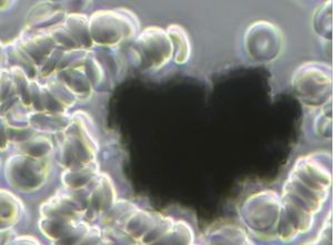
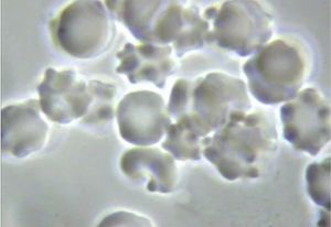
similarity with images from literature (Xu et al, 2019), and with
images obtained from rGO standard (SIGMA)(Figures 1, 2 and
3).
Images of big transparent sheets of variable size and shapes
were obtained, showing corrugated and flat, irregular. Smaller
sheets of polygonal shapes, also similar to flakes described in
literature (Xu et al, 2019) can be revealed with pHase Contrast
and Dark-Field microscopy (Figure 3).
All these laminar objects were widespread in the aqueous
fraction of the blood (Figure 1) or vaccine sample (Figures 2 and
3) and no component described by the registered patent can be
associated with these sheets.
In Figure 1 You Can See What A Cluster Bomb of Reduced
Graphene Oxide (rGO) Looks Like in the Live Unstained
Human Blood after a CoV-19 Inoculation Causing
Pathological Blood Coagulation![1][2][55][56][57]

[Figure 1 is a Micrograph of a Carbon Cluster of Reduced Graphene Oxide (rGO) Viewed in the
Live Unstained Human Blood with pHase Contrast Microscopy at 1500x. Note that the Red
Blood Cells are Clotting in and Around the rGO Crystal in a Condition Known as Rouleau! A
French Word Which Means to Chain.]

What Are the Non-Disclosed Ingredients Contained in CoV -
19 So-Called Pfizer, Moderna, Astrazeneca and Janssen
Vaccines?
To answer this question an aqueous fraction of the Pfizer,
Moderna, Astrazeneca and Janssen vaccines were taken from
each vile and then viewed separately under pHase Contrast
Microscopy at 100x, 600x up to1500x magnification showing
anatomical evidence of reduced Graphene Oxide (rGO)
particulates which were compared to micrographs of rGO from
Choucair et al, 2009 for identification and verification.[3]

Steps of Analysis of Vaccine Aqueous
Fractions
Refrigerated samples were processed under sterile conditions,
using laminar flow chamber and sterilized lab ware.
Steps for analyses were:
1. Dilution in 0.9% sterile physiological saline (0.45 ml + 1.2
ml)
2. Polarity fractionation: 1.2 ml hexane + 120 ul of RD1 sample
3. Extraction of hydrophilic aqueous pHase
4. UV absorbance and fluorescence spectroscopy scanning
5. Extraction and quantification of RNA in the sample
6. Electron and optical microscopy of aqueous pHase

The Pfizer "Vaccine" Non-disclosed
Ingredients
The micrographs in Figures 2 and 3 were obtained using 100X,
600X and 1500X pHase Contrast, Dark Field and Bright Field
Optical Microscopy.[3]
On the left of each micrograph you will view micrographs
obtained from the Pfizer vaccine aqueous fraction containing
rGO.
On the right of each micrograph ou will view a match from
known sources containing rGO for anatomical validation.
The observations under a pHase Contrast, Dark-Field, Bright-
Field microscopy, Transmission and Scanning Electron
microscopy of the vaccine product by Pfizer, including vaccine
products of Moderna, Astrazeneca and Janssen revealed some
entities that can be graphene strips as seen below in Figure 3.

[Figure 2 shows an aqueous fraction image from Pfizer vaccine sample (left) and from reduced
graphene oxide (rGO) standard (right) (Sigma-777684). Optical microscopy, 100X]

 [Figure 3 - Aqueous fraction images containing reduced graphene oxide from Pfizer vaccine
sample (left) and sonicated reduced graphene oxide (rGO) standard (right) (Sigma-777684).
Optical pHase contrast microscopy, 600X. In addition, the Muestra RD1, La Quinta Columna
Report, June 28, 2021; Graphene Oxide Detection in Aqueous Suspension; Delgado Martin,
Campra Madrid confirmed our findings.][4]
For a definitive identification of graphene by TEM, it is
necessary to complement the observation with the structural
characterization by obtaining a characteristic electron
diffraction standard sample (as the figure 'b' shown
below).[4]
The standard sample corresponding to graphite or graphene
has a hexagonal symmetry, and generally has several
concentric hexagons.

[Figure 4b Reveals X ray Diffraction Pattern of the Graphene Particles.] [4]

Using Transmission Electron Microscopy (TEM) we
observed an intricate matrix or mesh of folded translucent
flexible rGO sheets with a mixture of darker multilayer
agglomerations and lighter colored of unfolded monolayers
as seen in Figure 5. [3][4]
[Figure 4 shows the liposome Capsid containing rGO that Pfizer uses for its product to
vehiculate the graphene oxide by attaching the Liposome capsid to specific mRNA molecules for
driving the Liposome contents of fGO to specific organs, glands and tissues, namely the ovaries
and testes, bone marrow, heart and brain. The image was obtained by a SEM-Cryo preparation.]

Using Transmission Electron Microscopy (TEM) revealed an
intricate matrix or mesh of folded translucent flexible rGO
sheets with a mixture of darker multilayer agglomerations and
lighter colored of unfolded monolayers as seen in Figure 5. [3]
[Figure 5 shows a cluster of graphene nanoparticles in a Pfizer vaccine. They appear to be
aggregated.]

The darker linear areas in Figure 5 appear to be local overlap of
sheets and local arrangement of individual sheets in parallel to
the electron beam.[5]
After the mesh, a high density of unidentified rounded and
elliptical clear shapes appears, possibly corresponding to holes
generated by mechanical forcing of the rGO mesh during
treatment as seen in Figure 6.[4][5]
[Figure 6 shows a TEM microscopy observation where particles of reduced graphene oxide in a
Pfizer” vaccine” are present. The X-ray diffractometry reveals their nature of crystalline Carbon-
based nanoparticles of rGO.[4][5]

Energy-Dispersive X-ray Spectroscopy
Reveals rGO in Pfizer Vaccine[6][7][8]
The Pfizer vaccine liquid fraction was then analyzed for
chemical and elemental content using Energy-dispersive X-ray
spectroscopy (EDS) as seen in Figure 6. The EDS spectrum
showed the presence of Carbon, Oxygen verifying the rGO
elements and Sodium and Chloride since the sample shown in
Figures 2, 3, 5, and 6 were diluted in a saline solution.

[Figure 7 shows an EDS spectrum of a Pfizer “vaccine” under an ESEM microscopy coupled
with an EDS x-ray microprobe (X axis =KeV, Y axis = Counts) identifying Carbon, Oxygen,
Sodium and Chloride.]

The Quantification of mRNA in the Pfizer
Vaccine
The quantification of RNA in the Pfizer sample was carried out
with conventional protocols (Fisher).
According to NanoDropTM 2000 spectrophotometer calibration
check specific software (Thermofisher), the UV absorption
spectrum of total aqueous fraction was correlated to 747 ng/ul of
unknown absorbing substances.
However, after RNA extraction with commercial kit
(Thermofisher), quantification with RNA specific Qbit
fluorescence probe (Thermofisher) showed that only 6t ug/ul
could be related to the presence of RNA. The spectrum was
compatible with the peak of rGO at 270nm.
According to microscopic images presented here, most of this
absorbance might be due to graphene-like sheets, abundant in
the fluids suspension in the sample.
The conclusions are further supported by high fluorescence from
the sample with maximum at 340 nm, in accordance with peak
values for rGO. It must be reminded that RNA does not show
spontaneous fluorescence under UV exposure.

[Figure 8 - UV spectrum of aqueous fraction of Pfizer vaccine sample.[1][2][3][4][5][7]]
Ultra Violet Fluorescence Testing of the Pfizer Aqueous
Fraction for Reduced Graphene Oxide (rGO)[6]
Ultra Violet absorption and fluorescence spectrum were
obtained with Cytation 5 Cell Imaging Multi-Mode Reader
Spectrophotometer (BioteK). UV absorbance spectrum
confirmed a maximum peak at 270nm, compatible with presence
of rGO particulate.
UV fluorescence maximum at 340 nm also suggests presence of
significant amounts of rGO in the sample (Bano et al, 2019).

[Figure 9 - UV absorption and fluorescence spectra were obtained with Cytation 5 Cell Imaging
Multi-Mode Reader Spectrophotometer (BioteK). UV absorbance spectrum confirmed a
maximum peak at 270 nm, compatible with presence of rGO. UV fluorescence maximum at 340
nm also suggests presence of significant amounts of rGO in the sample (Bano et al, 2019).]
[Figure 10 - The spectroscopy UV analysis showed an adsorption due to the presence of reduced
graphene oxide, which is confirmed by observation under ultraviolet visible microscopy.]

Figures 11 and 12 below shows a micrograph of different micro
and nano particulates which have been identified in the Pfizer,
Moderna, Astrazeneca and Janssen, so-called “vaccines” and
analyzed under an Environmental Scanning Electron
Microscope (SEM) coupled with an x-ray microprobe of an
Energy Dispersive System (EDS) that reveals the particle size,
composition distribution and chemical nature of the observed
micro and nano particulates under observation.[6][7][8]
[Figure 11 shows sharp micron debris of 20 um in length identified in the Pfizer so-called
“vaccine” containing Carbon, Oxygen Chromium, Sulphur, Aluminum, Chloride, Nitrogen.]

[Figure 12 shows a 20 micron in length particulate identified in the so-called Pfizer “vaccine”. It
is composed of carbon, oxygen chromium, sulphur, aluminum, chloride and nitrogen.]

Figures 13 and 14 below shows a micrograph of different micro
and nano particulates which have been identified in the Pfizer,
Moderna, Astrazeneca and Janssen, so-called “vaccines” and
analyzed under an Environmental Scanning Electron
Microscope (SEM) coupled with an x-ray microprobe of an
Energy Dispersive System (EDS) that reveals the particle size,
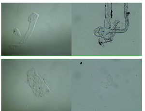
composition distribution and chemical nature of the observed
micro and nano particulates under observation.

Are There Parasites in the Pfizer
"Vaccines"?
A 50 micron elongated body, as seen in Figure 13 is a sharp
mysterious presence in the Pfizer vaccine. It appears and is
identified anatomically as a Trypanosoma cruzi parasite of
which several variants are lethal and is one of many causes of
acquired immune deficiency syndrome or AIDS. [Atlas of
Human Parasitology, 4th Edition, Lawrence Ash and Thomas
Orithel, pages 174 to 178][9]

[Figure 13 shows a Trypanosoma Parasite approximately 20 microns in length found in the so-
called Pfizer “vaccine”. It is composed of carbon, oxygen chromium, sulphur, aluminum,
chloride and nitrogen.]
[A Live Blood pHase Contrast Microscopy Micrograph of Trypanosoma cruzi Parasite[9]]

Figure 14 identifies a composition of nano particulates including
carbon, oxygen chromium, sulpuhr, aluminum, chloride and
nitrogen also found in the CoV-19 "vaccines."

[Figure 13 Identifies a Composite of Nano particulates]

Figures 15 and 16 below show a micrograph of different micro
and nano particulates which have been identified and analyzed
under an Environmental Scanning Electron Microscope (SEM)
coupled with an x-ray microprobe of an Energy Dispersive
System (EDS) that reveals the chemical nature of the observed
micro and nano particulates and their morphology.
The white 2-micron-long particulate is composed of bismuth,
carbon, oxygen, aluminum, sodium, copper and nitrogen.

[Figure 15 shows nano and micron particulates identified in the Pfizer “vaccine”. The white 2
micron long particulate is composed of bismuth, carbon, oxygen, aluminum, sodium, copper and
nitrogen.]

[Figure 16 shows that the white 2 micron particulate found in the so-called Pfizer 'vaccine' is
composed of bismuth, carbon, oxygen, aluminum, sodium, copper and nitrogen.]

Figures 17 and 18 show the identification of organic carbon,
oxygen and nitrogen particulates with an aggregate of embedded
nanoparticles including bismuth, titanium, vanadium, iron,
copper, silicon and aluminum which were all found in the so-
called Pfizer “vaccine.”

[Figure 17 - shows an organic (Carbon-Oxygen-Nitrogen) aggregate with embedded
nanoparticles of bismuth, titanium. vanadium. iron, copper, silicon, aluminum embedded in
Pfizer “vaccine!”]

[Figure 18 - shows an organic (Carbon-Oxygen-Nitrogen) aggregate with embedded
nanoparticles of bismuth, titanium. vanadium. iron, copper, silicon, aluminum embedded in
Pfizer “vaccine!”]
The Astrazeneca "Vaccine" Non-disclosed
Ingredients
Figures 19 and 20 show an engineered aggregate of iron,
chromium and nickel also known as stainless steel of micro and
nano particles embedded and identified in the Astrazeneca
“vaccine” viewed under Transmission Electron Microscopy
(TEM) and quantified with an x-ray microprobe of an Energy
Dispersive System that reveals the chemical nature of the
observed micro and nano particulates and their morphology.

[Figure 19 - Engineered aggregate of iron, chromium and nickel also know as stainless steel.]

[Figure 20 shows the quantified namo particulates in the Astrazeneca "vaccine" with an x-ray
microprobe of an Energy Dispersive System that reveals the chemical nature of the observed
micro and nano particulates.]
Using the XRF (X-ray fluorescence) instrument was used to
evaluate the adjuvants in the Astrazeneca "vaccine", which
identified the following molecules of histidine, sucrose, Poly-
ethylene glycol (PEG) and ethylene alcohol, also contained in
the Pfizer and Moderna "vaccines". The results of this test can
be seen in Figure 20.[10]
The injection of PEG and Ethylene alcohol are both known as
carcinogenic and genotoxic.[10] PEG was the only adjuvant
declared on the data sheet listing the ingredients of the
Astrazeneca “vaccine” but contained in the Pfizer and Moderna
"vaccines".

[Figure 21 Identifies the Spectrum of AstraZeneca Vaccine Adjuvants. Different colors are used
for the four molecules identified by means of reference spectra. Relative concentration is
calculated on integrals of reference signals for molecules in a quantitative spectrum acquired
with a duty cycle of 5 seconds with the longest calculated T1 was 5sec.]
The Janssen "Vaccine" Non-Disclosed
Ingredients
Figures 22 and 23 shows an organic-inorganic aggregate
identified in the Janssen “vaccine”. The particles are composed
of stainless steel and are glued together with a “Carbon-based
glue” of reduced graphene oxide.[11] This aggregate is highly
magnetic and can trigger pathological blood coagulation and
"The Corona Effect" or "The Spike Protein Effect" creation from
the degeneration of the cell membrane due to interactions with
other dipoles.[10] You can view these biological reactions or
cellular transformations in the live blood under pHase Contrast
and Dark Field Microscopy in Figures 24, 25 and 26.[1][12]

[Figure 22 A Stainless Steel Aggregation of Carbon , Oxygen, Iron and Nickel Held Together
With Graphene Oxide.]
[Figure 23 shows the stainless steel aggregation of carbon, oxygen, iron and nickel held together
with graphene oxide.]

The ‘Corona Effect’ and ‘Spike Protein
Effect’
The Endogenously Created "Corona Effect" and "Spike Protein"
ARE Caused by Chemical and Radiation Poisoning from
Reduced Graphene Oxide and Microwave Radiation![12]

[Figure 24 ‘The Corona Effect; and the Endogenous Creation of Exosomes Due to Chemical and
Radiation Poisoning of the Vascular and the Interstitial fluids of the Interstitium.]
[Figure 25 Shows ‘The Corona Effect’ and the the Endogenous Birth of S1 Protein Spikes
Caused by Radiation and Chemical Poisoning or What I Call The ‘Protein Spiking Effect’.

[Figure 26 This Micrograph Shows the Endogenous Creation of the ‘Spike Protein’ as an
Outfection and NOT and Infection.]
Figures 24 and 25 above show 'The CORONA EFFECT' on the
red blood cells with Figure 26 showing 'The SPIKED PROTEIN
EFFECT' both caused by decompensated acidosis of the
interstitial and then vascular fluids from an acidic lifestyle and
specifically, exposure to toxic pulsating electro-magnetic fields
at 2.4gHz or higher, chemical poisoning from the food and water
ingested, toxic acidic air pollution, chem-trails and to top-it-all-
off a nana particulate chemical laden CoV - 19 inoculation!
Please check your feelings and false beliefs at the door before
YOU prematurely cause YOURSELF harm![12]

The Moderna "Vaccine" Non-Disclosed
Ingredients
Figure 26 and 27 identified a mixed entity of organic and
inorganic matter contained in the Moderna “vaccine."
Transmission Electron Microscopy (TMS) and quantified with
an x-ray microprobe of an Energy Dispersive System (EDS)
revealed the chemical nature of the observed micro and nano
particulates.
The so-called Moderna "vaccine' is a carbon-based Reduced
Graphene Oxide substrate where some nanoparticles are
embedded. The nanoparticles are composed of carbon, nitrogen,
oxygen, aluminum, copper, iron and chlorine.[13]
[Figure 26 Transmission Electron Microscopy Reveals a Graphene Oxide Composite of
Embedded Organic and Non-Organic Matter.]

[Figure 27 Reveals Embedded Cytotoxic Nano Particulates.]

Figures 27 and 28 shows an analysis which was also performed
under Transmission Electron Microscopy (TEM) and quantified
with an x-ray microprobe of an Energy Dispersive System
(EDS) and revealed the chemical nature of the observed micro
and nano particulates. Many foreign bodies were identified with
a spherical morphology with some bubble-shaped cavities.
Figure 29 shows they are composed of carbon, nitrogen, oxygen,
silicon, lead, cadmium, and selenium. This highly toxic nano
particulate composition are quantum dots of cadmium selenide
which are cytotoxic and genotoxic.[14][15]

[Figure 27 Reveals the Nano Dots in the Graphene Oxide Found in the Moderna "Vaccine".]
[Figure 28 Reveals the Nano Dots in the Graphene Oxide Found in the Moderna "Vaccine".]

[Figure 29 Reveals the Cytotoxic and Genotoxic Composite of Nano Particulates in Graphene
Oxide Found in the Moderna "Vaccine".]

Figures 30 and 31 further analysis of the so-called Moderna
“vaccine” showed a 100-micron symplast of reduced graphene
oxide nano particulate composite. The rGO is composed of
carbon and oxygen with contamination of nano particulates of
nitrogen, silicon, phosphorus and chlorine Chlorine.[16]
[Figure 30 Transmission Electron Microscopy Reveals a Large 100 micron Symplast Composite
of Reduces Graphene Oxide.]

[Figure 31 Reveals the Nano Particulate Complex Contained in the Moderna "Vaccine".]

Figures 32 and 33 show carbon-based reduced graphene oxdie
entities in the Moderna “vaccine” mixed with aggregates filled
with Aluminium silicate nanoparticulates.[17]
[Figure 32 Reveals a Complex of Graphene Oxide and Aluminium Silicate Using Transmission
Electron Microscopy.]

[Figure 33 Reveals the Nano Elements of Graphene Oxide and Aluminum Silicate Contained in
the Moderna "Vaccine".]

Discussion
The SARS-CVid-2-19 pandemic induced the pharmaceutical
industries to develop new drugs that they called vaccines.
The mechanism of action of these new drugs as declared by the
pharmaceutical industry coupled with what is reported in the
vaccine products’ data sheet is NOT clear for current medical
savants to understand that those new drugs produced by Pfizer--
BioNTech mRNA Vaccine, the Moderna-Lonza mRNA-1273
Vaccine, the Serum Institute Oxford Astrazeneca Vaccine and
the Janssen COVID -19 Vaccine, manufactured by Janssen
Biotech Inc., a Janssen Pharmaceutical Company of Johnson &
Johnson are NOT vaccines but nanotechnological drugs working
as a genetic therapy.
The name “vaccine” is likely to be an escamotage (trickery)
used for bureaucratic and technocratic reasons in order to
receive an urgent approval, ignoring all the normal rules
necessary for new drugs, especially for those involving novel
nanotechnological mechanisms which have never been
developed nor experienced by humans any where, at any time in
the history of World.
All these so-called vaccines” are patented and therefore their
actual content is kept secret even to the buyers, who, of course,
are using taxpayers' money. So, consumers (taxpayers) have no
information about what they are receiving in their bodies by
inoculation. Humanity is kept in the dark as far as the nano
particulate technological processes involved are concerning, on
the negative effects on the cells of the body, but mostly on the
possible magneticotoxic, cytotoxic and genotoxic nano-bio-
interaction effect on the blood and body cells.
This current research study via direct analysis on the
aforementioned so-called “vaccines” by means of nano
particulate technological instrumentation reveals disturbing and
life-altering information concerning the truth about the actual
toxic acidic contents of the so-called vaccines.
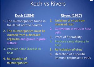
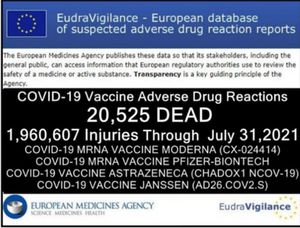
The Pfizer, Moderna, Astrazeneca and Janssen drugs are
NOT "vaccines" but complexed Graphene Oxide nano
particulate aggregates of varying nano elements attached to
genetically modified nucleic acids of mRNA from animal or
vero cells and aborted human fetal cells as viewed and described
above. Once again the ingredients in these so-called vaccines are
highly magneticotoxic, cytotoxic and genotoxic to plant, insect,
bird, animal and human cell membranes and their genetics
which already has lead to serious injuries (estimated at over 500
million) and/or eventual death (estimated at over 35
million).[18][19] through [55]
The so-called “experts” or "medical savants" are telling YOU
that CoV - 2 - 19 vaccines are the ONLY way to stop the spread
of CoV-19... even when there is NO EVIDENCE of its existence
and NO EVIDENCE of it spreading as determined by the
scientific method of Koch or Rivers postulates![54]
That they’re safe — despite the documented evidence is to the
contrary...[54]
That they’re effective — even though millions of “double-
jabbed” people are getting sick, theoretically exposing
themselves to a NON-EXISTENT VIRUS called CoV - 19, and
dying…[55] NOT from some phantom viral infection but from
the FEAR or false evidence appearing real and the toxic acid
contents of reduced graphene oxide delivered via the genetically
modified mRNA to specific targets of the human body leading
to pathological blood coagulation, oxygen deprivation,
hypercapnia, hypoxia and then death by suffocation.[56][57][58]
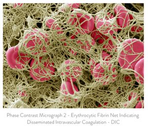
Disseminated Intravascular Coagulation (DIC) or Pathological Blood Coagulation. [56][57]

That YOU MUST get at LEAST two shots PLUS “boosters” to
live “normal lives” ...
And soon, they’ll be telling YOU that YOU have no choice but
to comply with ALL their MANdates even when the CDC and
other Governments, Universities and Medical Institutes have
admitted in writing that they have NO "GOLD STANDARD"
isolation of the CoV - 2 now called CoV - 19 virus![55]
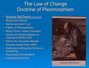
There is NO CORONA VIRUS and NEVER
HAS BEEN! [56]
Remember ...
DON’T LETANYONE TAKE AWAY YOUR HEALTH
FREEDOM!
It is YOUR body, YOUR Life and YOUR Choice!
Knowledge is power. And it’s the key to understanding why the
experimental CoV - 2 -19 vaccines are so dangerous — despite
the corporate media’s official narrative that suppresses and
censors anyone who dares to speak out.
You are in control of your own health. Don’t fall victim to
global governments and bureaucrats that are pushing everyone
to get vaccinated. Billionaire “philanthropist” Bill Gates and
billionaire Big Tech activists think they know what’s best for
you and your family.
You must be free todecide what’s right for you. Do NOT let
governments and employers force you into getting "VAXXD"
“for your own good”.
And never let the cancel culture make you too afraid to stand up
for your rights!
In the words of the great French doctor and scientist, Antione
BeChamp, "there is nothing so false that does NOT contain a
element of truth and so it is with the germ theory." In this case
the viral, vaccine and immunity theory![59]
To learn more about viruses, vaccines and the viral theory please
read and study, A Second Though About Viruses, Vaccines and
the HIV AIDS Hypothesis. You can order this book at:
https://www.phmiracleproducts.com/collections/books-
audio-video/products/second-thoughts-about-viruses-
vaccines-and-the-hiv-aids-hypothesis-booklet
Read and Learn More About Viruses, Vaccines and the
  Viral Theory in the Following Scientific Articles!
Robert O Young MSc, DSc, PhD, Naturopathic Practitioner

1. Scanning & Transmission Electron Microscopy Results
Revealing Graphene Oxide in CoV-19 Vaccines - An
Interview with Dr. Robert O. Young
Absolutely bombshell and major reveals on what is in the
vaccines, with use of electron and other kinds of microscopy
from original research by Dr. Robert Young and his team,
confirming what the La Quinta Columna researchers found-
--toxic nanometallic content with cytotoxic and genotoxic
effects as well as an identified parasite. This is major
revelation: please stay tuned for a major article reporting
this at ECC, meanwhile please share this video widely!
https://www.bitchute.com/video/Z2sAH0Woz38r/
https://rumble.com/vlonug-electron-microscopy-reveals-
graphene-oxide-in-cov-19-vaccines.html

https://odysee.com/@DrRobertYoung:7/MicroscopyEvidenc
eofGOinVaccineszoom_0:c?r=DwWt6qy5ACY9nSS1q6nAq
SpAYbuZDjWf
2. The Epidemic Curves of the Most Vaccinated Countries
https://www.drrobertyoung.com/post/world-health-
organization-who-publishes-epidemic-curves-of-the-most-
vaccinated-countries
3. Sweden Is Following the Biological Science NOT the
Political Science
https://www.drrobertyoung.com/post/sweden-is-following-
the-biological-science-not-the-political-science
4. What is Happening in India
https://www.drrobertyoung.com/post/what-happened-in-
india
5. Surviving the Plague of Corruption
https://www.drrobertyoung.com/post/surviving-the-plague-
of-corruption
6. Dismantling the Viral Theory
https://www.drrobertyoung.com/post/dismantling-the-viral-
theory
7. Corna on Trial -
https://www.drrobertyoung.com/post/corona-on-trial-
explosive-and-exclusive
8. The Havana Cuba Sndrome Caused by Directed Pulsating
EMF Microwaves https://www.drrobertyoung.com/post/the-
havana-syndrome-pulsating-microwaves
9. CDC NOW Admits NO 'Gold Standard' for the Isolation
for ANY Virus! - https://www.drrobertyoung.com/post/cdc-
now-admits-no-gold-standard-for-the-isolation-for-any-
virus?postId=60c4dbc74f7fc9002ba2507e
10. Freedom of Information Responses on CoV - 2 Now
Called CoV - 19 -
https://www.drrobertyoung.com/post/freedom-of-
information-responses-on-cov-2
11. Why Viruses Do Not Exist! -
https://www.drrobertyoung.com/post/why-viruses-do-not-
exist

12. Report 255 | Dr. Robert Young: All Disease is Outfection
Not Infection--Vaccine Nano is Bioweapon!
https://www.bitchute.com/video/rdQhuY455VmK
13. Report 255 | Dr. Robert Young: All Disease is Outfection,
not Infection | The Vaccine's Lipid Nanoparticles with
Graphene Plus Radiation is the Bioweapon--No COVID
Virus Exists!
https://www.brighteon.com/250210af-c8a3-47a1-8922-
c960342fe2fb
14. Report 255 | Dr. Robert Young: All Disease is Outfection
Not Infection--Vaccine Nano is Bioweapon!
https://odysee.com/@RamolaDReports:8/Report-255---Dr.-
Robert-Young-Explains-Disease-by-Outfection,-Lipid-
Nanoparticles-with-Graphene-the:4
15. Forget Everything Else! Look at THE VAER's
NUMBERS on Injuries and Deaths!
https://www.drrobertyoung.com/post/forget-everything-else-
you-ve-heard-just-look-at-the-numbers

16. Dr. Robert O. Young ITNJ Testimony
https://youtu.be/gKjnEz5s37o

Sign up for Dr. Young's FREE Newsletter
NOW at: wwwdrrobertyoung.com
References
[1] Ou, L.,Song, B., Liang, H. et al. Toxicity of graphene-family
nanoparticles: a general review of the origins and mechanisms.
Part Fibre Toxicol13, 57 (2016).
https://doi.org/10.1186/s12989-016-0168-y
[2] Young RO (2016) Pathological Blood Coagulation and the
Mycotoxic Oxidative Stress Test (MOST). Int J Vaccines
Vaccin 2(6): 00048. DOI: 10.15406/ijvv.2016.02.00048
[3] Xu et al, (2019) Identification of graphene oxide and its
structural features in solvents by optical microscopy, RSC Adv.,
9, 18559-18564
1-Extracction RNA Kit
https://www.fishersci.es/shop/products/ambion-purelink-rna-
mini-kit7/10307963
2- NanoDrop™
https://www.thermofisher.com/order/catalog/product/ND-
2000#/ND-2000
3- QUBIT2.0:
https://www.thermofisher.com/es/es/home/references/newsletter
s-andjournals/bioprobes-journal-of-cell-biology-
applications/bioprobes-issues-2011/bioprobes-64-april2011/the-
qubit-2-0-fluorometer-april-2011.html
[4] Muestra RD1, La Quinta Columna Report, June 28, 2021;
Graphene Oxide Detection in Aqueous Suspension; Delgado
Martin, Campra Madrid
[5] Kim et al, Seeing graphene-based sheets, Materials Today,
Volume 13, Issue 3,2010,Pages 28- 38,ISSN 1369-
7021,https://doi.org/10.1016/S1369-7021(10)70031-6
[6] Bano, I. et al , 2019. Exploring the fluorescence properties of
reduced graphene oxide with tunable device performance,
Diamond and Related Materials,Volume 94,59-64,ISSN 0925-
9635,https://doi.org/10.1016/j.diamond.2019.02.021.
[7] Biroju, Ravi & Narayanan, Tharangattu & Vineesh, Thazhe
Veettil. (2018). New advances in 2D electrochemistry—
Catalysis and Sensing. 10.1201/9781315152042-7.
[8] Choucair, M., Thordarson, P. & Stride, J. Gram-scale
production of graphene based on solvothermal synthesis and
sonication. Nature Nanotech 4, 30–33 (2009).
https://doi.org/10.1038/nnano.2008.365
[9] Atlas of Human Parasitology, 4th Edition, Lawrence Ash
and Thomas Orithel, pages 174 to 178
[10] Mano, S.S.; Kanehira, K.; Sonezaki, S.; Taniguchi, A.
Effect of Polyethylene Glycol Modification of TiO2
Nanoparticles on Cytotoxicity and Gene Expressions in Human
Cell Lines. Int. J. Mol. Sci.2012, 13, 3703-3717.
https://doi.org/10.3390/ijms13033703
[11] Srivastava AK, Dwivedi N, Dhand C, et al. Potential of
graphene-based materials to combat COVID-19: properties,
perspectives, and prospects. Mater Today Chem.
2020;18:100385. doi:10.1016/j.mtchem.2020.100385
[12] Young, RO, "The Effects of ElectroMagnetic Frequencies
(EMF) on the Blood and Biological Terrain."
https://www.drrobertyoung.com/post/the-effects-electromagnet-
frequencies-on-the-blood-and-biological-terrain
[13] Gatti AM, Manti A, Valentini L, Montanari S, Gobbi P, et
al. (2016) Nano biointeraction of particulate matter in the blood
circulation. Frontiers 30: 3.
[14] Nikazar, S., Sivasankarapillai, V.S., Rahdar, A. et al.
Revisiting the cytotoxicity of quantum dots: an in-depth
overview. Biophys Rev 12, 703–718 (2020).
https://doi.org/10.1007/s12551-020-00653-0
[15] Ritesh Banerjee, Priya Goswami, Manoswini Chakrabarti,
Debolina Chakraborty, Amitava Mukherjee, Anita Mukherjee,
Cadmium selenide (CdSe) quantum dots cause genotoxicity and
oxidative stress in Allium cepa plants, Mutation
Research/Genetic Toxicology and Environmental Mutagenesis,
Volume 865, 2021, 503338,ISSN 1383-5718,
https://doi.org/10.1016/j.mrgentox.2021.503338.
[16] Wanjun Cao, Lin He, Weidong Cao, Xiaobing Huang, Kun
Jia, Jingying Dai, Recent progress of graphene oxide as a
potential vaccine carrier and adjuvant, Acta Biomaterialia,
Volume 112, 2020, Pages 14-28, ISSN 1742-7061,
https://doi.org/10.1016/j.actbio.2020.06.009.
(https://www.sciencedirect.com/science/article/pii/S1742706120
303305)
[17] Concise Encyclopedia of Composite Materials, ed. Anthony
Kelly, MIT Press, 1989, ISBN0-262-11145-4
[18] L. Harivardhan Reddy, José L. Arias, Julien Nicolas, and
Patrick Couvreur, "Magnetic Nanoparticles: Design and
Characterization, Toxicity and Biocompatibility, Pharmaceutical
and Biomedical Applications." Chemical Reviews 2012 112
(11), 5818-5878 DOI: 10.1021/cr300068p
[19] US Dpt of health and human services (1996) Report
Update: Vaccine Side Effects, Adverse Reactions,
Contraindications, and Precautions. CDC 45(RR-12): 1-35.
[20] Ottaviani G, Lavezzi AM, Matturri L (2006) Sudden infant
death syndrome (SIDS) shortly after hexavalent vaccination:
pathology in suspected SIDS? Virchows Arch 448(1): 100-104.
[21] Taylor B, Miller E, Farrington CP, Petropoulos MC, Favot-
Mayaud I, et al. (1999) Autism and measles, mumps, and rubella
vaccine: no epidemiological evidence for a causal association.
Lancet 353(9169): 2026-2029.
[22] Demicheli V, Rivetti A, Debalini MG, Di Pietrantonj C
(2012) Vaccines for measles, mumps and rubella in children.
Cochrane Database Syst Rev 15(2): CD004407. New Quality-
Control Investigations on Vaccines: Micro- and
Nanocontamination 13/13 Copyright: ©2016 Gatti et al.
Citation: Gatti AM, Montanari S (2016) New Quality-Control
Investigations on Vaccines: Micro- and Nanocontamination. Int
J Vaccines Vaccin 4(1): 00072. DOI:
10.15406/ijvv.2017.04.00072
[23] Carola Bardage, Ingemar Persson, Åke Örtqvist, Ulf
Bergman, Jonas F Ludvigsson, et al. (2011) Neurological and
autoimmune disorders after vaccination against pandemic
influenza A (H1N1) with a monovalent adjuvanted vaccine:
population based cohort study in Stockholm, Sweden. BMJ 343:
d5956.
[24] Johann Liang R (2012) Updating the Vaccine Injury Table
following the 2011 IOM Report on Adverse Effects of vaccines.
HRSA, pp. 1-27.
[25] L Tomljenovic, CA Shaw (2011) Aluminum Vaccine
Adjuvants: Are they Safe? Current Medicinal Chemistry 18(17):
2630-2637.
[26] Shaw CA, Petrik MS (2009) Aluminum hydroxide
injections lead to motor deficits and motor neuron degeneration.
J Inorg Biochem 103(11): 1555-1562.
[27] Authier FJ, Sauvat S, Christov C, Chariot P, Raisbeck G, et
al. (2006) AlOH3-adjuvanted vaccine-induced macrophagic
myofasciitis in rats is influenced by the genetic background.
Neuromuscul Disord 16(5): 347-352.
[28] Exley C, Esiri MM (2006) Severe cerebral congophilic
angiopathy coincident with increased brain aluminium in a
resident of Camelford, Cornwall, UK. J Neurol Neurosurg
Psychiatry 77(7): 877- 879.
[29] Wills MR, Savory J (1985) Water content of aluminium,
dialysis dementia, and osteomalacia. Environ Health Perspect
63: 141-147.
[30] Brinth L, Pors K, Theibel AC, Mehlsen J (2015) Suspected
side effects to the quadrivalent human papilloma vaccine.
Danish Medical J 62(4): 1-12.
[31] Palmieri B, Poddighe D, Vadalà M, Laurino C, Carnovale
C, et al. (2016) Severe somatoform and dysautonomic
syndromes after HPV vaccination: case series and review of
literature. Immunol Res.
[32] Visani G, Manti A, Valentini L, Canonico B, Loscocco F,
et al. (2016) Environmental nanoparticles are significantly over-
expressed in acute myeloid leukemia. Leuk Res 50: 50-56.
[33] Artoni E, Sighinolfi GL, Gatti AM, Sebastiani M, Colaci
M, et al. (2016) Micro and nanoparticles as possible
pathogenetic co-factors in mixed cryoglobulinemia.
Occupational Medicine.
[34] T Hansen, L Klimek, F Bittinger, I Hansen, A Gatti, et al.
(2008) Mast cell reiches Aluminium granuloma Pathologe
29(4): 311-313.
[35] Gatti AM, Manti A, Valentini L, Montanari S, Gobbi P, et
al. (2016) Nano biointeraction of particulate matter in the blood
circulation. Frontiers 30: 3.
[36] Tenzer S, Docter D, Rosfa S, Wlodarski A, Kuharev J, et al.
(2011) Nanoparticle size is a critical physicochemical
determinant of the human blood plasma corona: a
comprehensive quantitative proteomic analysis. ACS Nano 5(9):
7155-167.
[37] Radauer Preiml , Andosch A, Hawranek T, Luetz-Meindl
U, Wiederstein M, et al. (2015) Nanoparticle-allergen
interactions mediate human allergic responses: protein corona
characterization and cellular responses. Fibre toxicology 13: 3.
[38] Cedervall T, Lynch I, Lindman S, Berggård T, Thulin E, et
al. (2016) Understanding the nanoparticle-protein corona using
methods to quantify exchange rates and affinities of proteins for
nanoparticles. PNAS 104 (7): 2050-2055.
[39] Lynch I, Cedervall T, Lundqvist M, Cabaleiro-Lago C,
Linse S, et al. (2007) The nanoparticle-protein complex as a
biological entity; a complex fluids and surface science challenge
for the 21st century. Advances in Colloid and Interface Science
134-135: 167-174.
[40] Gatti AM, Quaglino D, Sighinolfi GL (2009) A
Morphological Approach to Monitor the Nanoparticle-Cell
Interaction. International Journal of Imaging and Robotics 2: 2-
21.
[41] Urban RM, Jacobs JJ, Gilbert JL, Galante JO (1994)
Migration of corrosion products from modular hip prostheses.
Particle microanalysis and histopathological findings. The
Journal of Bone and Joint Surgery 76(9): 1345-1359.
[42] Kirkpatrick CJ, Barth S, Gerdes T, Krump-Konvalinkova
V, Peters (K 2002) Pathomechanisms of impaired wound
healing by metallic corrosion products. Mund Kiefer
Gesichtschir 6(3): 183-190.
[43] Lee SH, Brennan FR, Jacobs JJ, Urban RM, Ragasa DR, et
al. (1997) Human monocyte/macrophage response to cobalt-
chromium corrosion products and titanium particles in patients
with total joint replacements. J Orthop Res 15(1): 40-49.
[44] Shaw CA, Seneff S, Kette SD, Tomljenovic L, Oller Jr JW,
et al. (2014) Aluminum-Induced Entropy in Biological Systems:
Implications for Neurological Disease. Journal of Toxicology
2014: 491316.
[45] Shaw CA, Kette SD, Davidson RM, Seneff S (2013)
Aluminum™s Role in CNS-immune System Interactions
leading to Neurological Disorders. Immunome Research 9: 069.
[46] Seneff S, Swanson N, Chen Li (2015) Aluminum and
Glyphosate Can Synergistically Induce Pineal Gland Pathology:
Connection to Gut Dysbiosis and Neurological Disease.
Agricultural Sciences 6(1): 42- 70.
[47] Pegaz B, Debefve E, Ballini JP, Konan-Kouakou YN, van
den Bergh HJ (2006) Effect of nanoparticle size on the
extravasations and the photothrombic activity of meso(p-
tetracarboxyphenyl)porphyrin. J Photochem Photobiol B 85(3):
216-222.
[48] Brinth LS, Pors K, Hoppe AG, Badreldin I, Mehlsen J
(2015) Is Chronic Fatigue Syndrome/Myalgic
Encephalomyelitis a Relevant Diagnosis in Patients with
Suspected Side Effects to Human Papilloma Virus Vaccine?
International Journal of Vaccines and Vaccination 1(1):1-5.
[49] Moos WH, Faller DV, Harpp DN, Kanara I, Pernokas J, et
al. (2016) Microbiota and Neurological Disorders: A Gut
Feeling. Biores Open Access 5(1): 137-145.
[50] Sekirov I, Russell SL, Caetano L, Antunes M, Brett (2010)
Gut Microbiota in Health and Disease. Physiological Rev 90(3):
859-904.
[51] Umbrello G, Esposito S (2016) Microbiota and neurologic
diseases: potential effects of probiotics. J Transl Med 14(1):
298.
[52] Kinoshita T, Abe RT, Hineno A, Tsunekawa K, Nakane S,
et al. (2014) Peripheral sympathetic nerve dysfunction in
adolescent Japanese girls following immunization with the
human papillomavirus vaccine. Intern Med 53(19): 2185-2200.
[53] Zhang L, Richards A, Khalil A, Wogram E, Ma H, Young
RA, Jaenisch R. SARS-CoV-2 RNA reverse-transcribed and
integrated into the human genome. bioRxiv [Preprint]. 2020 Dec
13:2020.12.12.422516. doi: 10.1101/2020.12.12.422516. PMID:
33330870; PMCID: PMC7743078.
[54] Young, RO, "Forget Everything Else! Look at THE
VAER's NUMBERS on Injuries and Deaths!."
https://www.drrobertyoung.com/post/forget-everything-else-
you-ve-heard-just-look-at-the-numbers5
[55] Young, RO, "CDC NOW Admits NO 'Gold Standard' for
the Isolation for ANY Virus!"
https://www.drrobertyoung.com/post/cdc-now-admits-no-gold-
standard-for-the-isolation-for-any-virus
[56] Young, RO, "The Genesis of Severe Acute Respiratory
(Syndrome) or SARS & Corona Virus or COVID - 19."
https://www.drrobertyoung.com/post/the-genesis-of-severe-
acute-respiratory-syndrome-or-sars-corona-virus-or-covid-19
[57] Young, RO, "What Causes Oxygen Deprivation of the
Blood(DIC) and Then Lungs(SARS - CoV 2 & 19)?"
https://www.drrobertyoung.com/post/what-causes-oxygen-
deprivation
[58] Young RO, Migalko G (2020) What Causes Oxygen
Deprivation of the Blood(DIC) and Then Lungs(SARS - CoV 2
& 12)?. Integ Mol Bio Biotechnol 1: 001-007
http://sciaeon.org/articles/What-Causes-Oxygen-. Deprivation-
of-the-Blood-DIC-and-Then-Lungs-SARS-CoV2and12.pdf
[59] Young RO (2016) Who Had Their Finger on the Magic of
Life - Antoine Bechamp or Louis Pasteur?. Int J Vaccines
Vaccin 2(5): 00047. DOI: 10.15406/ijvv.2016.02.00047
You can also read