A Human Skin Model Recapitulates Systemic Sclerosis Dermal Fibrosis and Identifies COL22A1 as a TGFβ Early Response Gene that Mediates Fibroblast ...
←
→
Page content transcription
If your browser does not render page correctly, please read the page content below
G C A T
T A C G
G C A T
           genes
Article
A Human Skin Model Recapitulates Systemic
Sclerosis Dermal Fibrosis and Identifies COL22A1 as
a TGFβ Early Response Gene that Mediates
Fibroblast to Myofibroblast Transition
Tomoya Watanabe 1 , DeAnna Baker Frost 1 , Logan Mlakar 1 , Jonathan Heywood 1 ,
Willian A. da Silveira 2 , Gary Hardiman 2,3,4 and Carol Feghali-Bostwick 1, *
 1   Division of Rheumatology and Immunology, Department of Medicine, Medical University of South Carolina,
     96 Jonathan Lucas St, MSC 637, Charleston, SC 29425, USA; nabetomo0968@yahoo.co.jp (T.W.);
     bakerde@musc.edu (D.B.F.); logan.mlakar@gmail.com (L.M.); heywood.jonathan@gmail.com (J.H.)
 2   Center for Genomic Medicine, Bioinformatics, Medical University of South Carolina, Charleston, SC 29425,
     USA; willian.abraham@gmail.com (W.A.d.S.); G.Hardiman@qub.ac.uk (G.H.)
 3   Departments of Medicine and Public Health Sciences, Medical University of South Carolina, Charleston,
     SC 29425, USA
 4   School of Biological Sciences & Institute for Global Food Security, Queens University Belfast,
     Belfast BT9 5AG, UK
 *   Correspondence: feghalib@musc.edu; Tel.: +1-843-876-2315; Fax: +1-843-792-7121

 Received: 10 December 2018; Accepted: 14 January 2019; Published: 22 January 2019

 Abstract: Systemic sclerosis (SSc) is a complex multi-system autoimmune disease characterized
 by immune dysregulation, vasculopathy, and organ fibrosis. Skin fibrosis causes high morbidity
 and impaired quality of life in affected individuals. Animal models do not fully recapitulate the
 human disease. Thus, there is a critical need to identify ex vivo models for the dermal fibrosis
 characteristic of SSc. We identified genes regulated by the pro-fibrotic factor TGFβ in human
 skin maintained in organ culture. The molecular signature of human skin overlapped with that
 which was identified in SSc patient biopsies, suggesting that this model recapitulates the dermal
 fibrosis characteristic of the human disease. We further characterized the regulation and functional
 impact of a previously unreported gene in the setting of dermal fibrosis, COL22A1, and show that
 silencing COL22A1 significantly reduced TGFβ-induced ACTA2 expression. COL22A1 expression
 was significantly increased in dermal fibroblasts from patients with SSc. In summary, we identified
 the molecular fingerprint of TGFβ in human skin and demonstrated that COL22A1 is associated with
 the pathogenesis of fibrosis in SSc as an early response gene that may have important implications for
 fibroblast activation. Further, this model will provide a critical tool with direct relevance to human
 disease to facilitate the assessment of potential therapies for fibrosis.

 Keywords: fibrosis; systemic sclerosis; COL22A1; fibroblasts; TGF

1. Introduction
     Systemic sclerosis (SSc) is a multisystem connective tissue disease characterized by immune
dysregulation, vasculopathy, and excessive fibrosis of the skin and internal organs due to fibroblast
proliferation and production of extracellular matrix [1,2]. It has the highest disease-related
mortality and morbidity among the rheumatologic illnesses with impaired quality of life. In fact,
fibroproliferative illnesses are responsible for approximately 45% of deaths in developed countries [3].
The progression of organ fibrosis leads to end-stage organ failure as a result of the loss of normal
structure and function. Skin fibrosis causes hardening and tightening of the skin. These features lead to

Genes 2019, 10, 75; doi:10.3390/genes10020075                                    www.mdpi.com/journal/genes
Genes 2019, 10, 75 2 of 16
limited joint movement or cause joint contractures, diminished mouth opening, and impaired quality
of life. However, the mechanisms underlying skin fibrosis remain to be completely elucidated.
Collagen type XXII alpha 1 chain (COL22A1), which is present on human chromosome 8q24.2,
exhibits a unique localization at the myotendinous junctions, tendons, heart, articular cartilage,
and skin [4]. In the skin, COL22A1 is expressed surrounding the lower third of the anagen hair
follicles [4]. Furthermore, COL22A1 is known to act as a cell adhesion ligand for skin epithelial
cells and fibroblasts [4]. It belongs to the FACIT (fibril-associated collagens with interrupted triple
helix) subset of the collagen superfamily that includes type IX, XII, XIV, XIX, and XXI collagens [5].
These are quantitatively minor collagens that mediate ligand interactions between fibrils and their
milieu. They associate with collagen fibers through their C-terminal collagenous domains, and they
mediate protein-protein interactions through their N-terminal non-collagenous domains [5]. One of
the functions of COL22A1 described in zebrafish is the stabilization of myotendinous junctions and
the strengthening of skeletal muscle attachments during contractile activity [6]. Transcript levels of
COL22A1 are elevated in the head and neck squamous cell carcinoma (HNSCC) and are proposed as
prognostic predictors for HNSCC [7]. In the setting of fibrosis, COL22A1 was identified as a potential
causal variant in patients with diffuse cutaneous SSc (dcSSc) through whole-exome sequencing
(WES) [8]. COL22A1 was significantly enriched in the extracellular matrix–related pathway. However,
the regulation and potential role of COL22A1 in fibrosis and specifically in the pathogenesis of SSc
remain unexplored. Since most studies involving the development of fibrosis have examined the effect
of pro-fibrotic triggers in vitro in cells such as fibroblasts and in vivo in animal models, in this study,
we sought to identify genes regulated by TGFβ in human skin and found COL22A1 as a top regulated
gene. We also investigated the role of COL22A1 in the activation of fibroblasts and the development
of fibrosis.
2. Materials and Methods
Additional methods can be found in the Supplementary section.
2.1. Primary Human Skin and Lung Fibroblasts Culture
Primary fibroblasts were cultured from skin or lung tissues of healthy donors as previously
described [9–11]. Lung fibroblasts were obtained under a protocol (#970946) approved by the
Institutional Review Board (IRB) of the University of Pittsburgh, and skin tissues and fibroblasts
obtained without identifiers were approved as non-human subject research by the IRB of the Medical
University of South Carolina. Fibroblasts were maintained in Dulbecco’s Modified Eagle’s Medium
(DMEM) (Mediatech, Herndon, VA, USA) supplemented with 10% fetal bovine serum (Sigma-Aldrich,
St. Louis, MO, USA), penicillin, streptomycin, and antimycotic agent (Invitrogen, Carlsbad, CA,
USA) and used in passages two to seven. Skin fibroblasts were treated with TGFβ (5 ng/mL) (R&D
Systems, Minneapolis, MN, USA) and harvested 2, 4, 8, 16, 24, 48 h (for RNA), and 72 h (for protein)
post-treatment. Lung fibroblasts were treated with TGFβ (10 ng/mL) and harvested 48 h (for RNA)
and 72 h (for protein) post-treatment. A549 cells were treated similarly to lung fibroblasts.
2.2. Small Interfering RNA (siRNA) Transfection
Primary human skin fibroblasts were seeded at a density of 2 × 10⁵ cells per well in six well
plates 24–48 h prior to transfection with siRNA. ON-TARGETplus COL22A1-specific siRNA and
ON-TARGETplus control siRNA were purchased from Dharmacon (Lafayette, CO, USA). Transfection
was done using Lipofectamine 2000 (Invitrogen) and 100 nmol siRNA diluted in Opti-MEM I
Reduced-Serum Medium (Life Technologies, Carlsbad, CA, USA) following the manufacturer’s
recommendation. TGFβ was added to media of cells 24 h after transfection. Fibroblasts were harvested
24 or 48 h (for RNA) and 72 h (for protein) post-treatment.Genes 2019, 10, 75 3 of 16
2.3. Ex Vivo Human Skin Culture
Normal human skin was obtained from residual tissue following plastic surgery. All tissues
were obtained according to the guidelines of the Medical University of South Carolina Institutional
Review Board without any identifiers. Subcutaneous fat tissue was removed. The skin was cut
with a disposable biopsy 3 mm punch, and the pieces of tissue were cultured in medium containing
TGFβ (10 ng/mL) or a vehicle control. The culture medium was DMEM supplemented with penicillin,
streptomycin, and anti-mycotic agent (Invitrogen). Skin samples were cultured in an air-liquid interface
with the epidermal side up. Skin tissues were harvested at 2, 4, 8, 16, 24, and 48 h (for RNA) or 72 h
(for histological analysis) post-treatment.
2.4. Quantitative PCR
Total RNA was extracted from human skin tissues and cultured fibroblasts using the TRIZOL Lysis
Reagent (Life Technologies) and RNeasy® kit (Qiagen Inc., Valencia, CA, USA). Reverse transcription
was performed with SuperScript IV (Invitrogen). Gene messenger RNA (mRNA) expression levels were
evaluated using quantitative PCR by the TaqMan® real-time PCR system (Life Technologies) according
to the manufacturer’s protocol on a TaqMan® Gene Expression Assays Step One Plus real-time PCR
system (Life Technologies). Premixed PCR primers and TaqMan probes for human COL1A1, COL22A1,
CTGF, FN, ACTA2, and GAPDH were obtained from Life Technologies. Gene expression levels were
normalized to GAPDH and compared with the 2−∆∆Ct method.
2.5. Western Blot Analysis
Western blot analysis of fibroblast lysates and supernatants was done as previously described [11].
The following antibodies were used: Anti-COL22A1 (Novus, Littleton, CO, USA), fibronectin, collagen
1A1, CTGF, GAPDH (Santa Cruz, Dallas, TX, USA), ACTA2 (Sigma-Aldrich), and horseradish
peroxidase-labeled secondary antibody (Santa Cruz). Signals were detected by chemiluminescence
(ProteinSimple, San Jose, CA, USA).
2.6. RNA Expression Profiling
Total RNA was extracted from human skin tissues using TRIZOL Lysis Reagent (Life Technologies)
and the RNeasy® kit (Qiagen Inc.) and was processed by the MUSC Genomics core for 1 × 50 cycles,
using single-end RNA sequencing on an Illumina HiSeq2500. The RNA integrity was verified on an
Agilent 2200 TapeStation (AgilentTechnologies, Palo Alto, CA, USA). A total of 100 ng of total RNA
was used to prepare RNA-Seq libraries using the TruSeq RNA Sample Prep kit following the protocol
described by the manufacturer (Illumina, San Diego, CA, USA). Sequencing was performed on an
Illumina HiSeq2500. Samples were demultiplexed using CASAVA (Illumina).
2.7. Bioinformatics and Statistical Analysis
For the differential gene expression analysis, we utilized the OnRamp’s advanced Genomics
Analysis Engine as previously described [12]. We used human genome build GRCh37/hg19 as the
reference and the following programs and versions: Fastqc version 0.11.5, cutadapt version 1.11,
Star aligner version 2.5.2b, HTseq version 0.7.0, DESEq2 Version 1.18.1. A gene was considered
differentially expressed (DE) if it was below the 0.4 adjusted p-value threshold cut-off as determined
by the Benjamini-Hochberg false discovery rate (FDR) correction [13]. A systems-level analysis of the
biological pathways affected by the DE genes was carried out using the iPathway Guide tool from
Advaita® Bioinformatics [14]. The dataset was deposited into the GEO repository under the accession
number GSE109350.Genes 2019, 10, 75 4 of 16
2.8. Statistical Analysis
All continuous variables were expressed as the mean ± standard deviation. All statistical analyses
were done using IBM SPSS statistics 22 (IBM Corporation, NY, USA). Statistical comparisons were
performed using the unpaired Student’s t-test. Comparison of 3 or more groups was done using
ANOVA with a post-hoc Tukey’s test to evaluate statistical significance.
3. Results
3.1. TGFβ Increases COL22A1 Expression Ex Vivo and In Vitro
To identify new genes that may be involved in the development of dermal fibrosis mediated
by TGFβ in human skin, we performed high-throughput RNA sequencing (RNA-seq) using ex vivo
human skin samples treated with TGFβ or a vehicle control for 48 h. Gene expression profiling
identified several novel transcripts in human skin tissues that were significantly upregulated by TGFβ
including COL22A1, Prostate Transmembrane Protein, Androgen Induced 1 (PMEPA1), Dermatopontin
(DPT), and others (Table 1). A complete list of all differentially-expressed genes regulated by TGFβ (530
using an FDR cutoff of 0.1, and 1051 when the FDR stringency was set at 0.4) is shown in Supplementary
Table S1. A heatmap of the top 100 most significantly differentially expressed mRNAs as determined
by DESeq2 (FDR < 0.1) is shown in Supplementary Figure S1. Pathway analysis suggested that the top
impacted pathway was the ECM-receptor interaction, followed by Proteoglycans in cancer, Cell cycle,
and Cytokine-cytokine receptor interaction (Supplementary Table S2).
Table 1. The significant differentially expressed (DE) messenger RNA (mRNAs) as determined by
DESeq2 (false discovery rate, FDR < 0.1) using ex vivo human skin samples treated with TGFβ or a
vehicle control for 48 h. The top 100 most significant DE mRNAs ranked by FDR (padj) are presented.
Fold
Symbol GeneID Description Padj
Change
COL22A1 169044 collagen, type XXII, alpha 1 6.40 × 10−36 9.19
prostate transmembrane protein,
PMEPA1 56937 6.36 × 10⁻³³ 3.51
androgen induced 1
DPT 1805 dermatopontin 2.89 × 10⁻²⁹ 4.69
latent transforming growth factor beta
LTBP2 4053 6.31 × 10⁻²⁶ 3.18
binding protein 2
COL1A1 1277 collagen, type I, alpha 1 2.49 × 10⁻²³ 3.11
FN1 2335 fibronectin 1 2.78 × 10⁻²⁰ 3.39
MMP10 4319 matrix metallopeptidase 10 6.47 × 10⁻¹³ 2.76
COL3A1 1281 collagen, type III, alpha 1 5.68 × 10⁻¹² 2.34
dehydrogenase/reductase (SDR family)
DHRS2 10202 6.16 × 10⁻¹² 4.55
member 2
COL5A2 1290 collagen, type V, alpha 2 8.53 × 10⁻¹² 2.36
ITGB6 3694 integrin, beta 6 1.66 × 10⁻¹¹ 2.94
ADAM metallopeptidase with
ADAMTS15 170689 1.77 × 10⁻¹¹ 3.55
thrombospondin type 1 motif, 15
LRRC15 131578 leucine rich repeat containing 15 1.77 × 10⁻¹¹ 4.17
transforming growth factor, beta-induced,
TGFBI 7045 6.21 × 10⁻¹¹ 3.12
68kDa
MKI67 4288 marker of proliferation Ki-67 7.29 × 10⁻¹¹ −2.56
COL1A2 1278 collagen, type I, alpha 2 8.85 × 10⁻¹¹ 2.11
ADAM12 8038 ADAM metallopeptidase domain 12 3.00 × 10⁻¹⁰ 2.71
GLS 2744 glutaminase 3.08 × 10⁻¹⁰ 2.08
myocardial infarction associated
MIAT 440823 3.36 × 10⁻¹⁰ 3.23
transcript (non-protein coding)Genes 2019, 10, 75 5 of 16
Table 1. Cont.
Fold
Symbol GeneID Description Padj
Change
C4orf26 152816 chromosome 4 open reading frame 26 3.59 × 10⁻¹⁰ 4.07
wingless-type MMTV integration site
WNT5B 81029 1.01 × 10⁻⁹ 3.58
family, member 5B
KRT77 374454 keratin 77, type II 3.56 × 10⁻⁹ 2.19
CXCL14 9547 chemokine (C-X-C motif) ligand 14 7.49 × 10⁻⁹ 2.61
TPST1 8460 tyrosylprotein sulfotransferase 1 8.75 × 10⁻⁹ 2.58
COL15A1 1306 collagen, type XV, alpha 1 1.03 × 10⁻⁸ 2.75
SNAI1 6615 snail family zinc finger 1 1.04 × 10⁻⁸ 2.65
NKD1 85407 naked cuticle homolog 1 (Drosophila) 2.62 × 10⁻⁸ 3.41
low density lipoprotein receptor class A
LDLRAD4 753 1.43 × 10⁻⁷ 3.30
domain containing 4
NPR3 4883 natriuretic peptide receptor 3 1.46 × 10⁻⁷ 2.96
dehydrogenase/reductase (SDR family)
DHRS9 10170 1.64 × 10⁻⁷ −2.44
member 9
COL5A1 1289 collagen, type V, alpha 1 2.11 × 10⁻⁷ 2.11
ESM1 11082 endothelial cell-specific molecule 1 2.11 × 10⁻⁷ 2.83
DIRAS1 148252 DIRAS family, GTP-binding RAS-like 1 2.17 × 10⁻⁷ 2.51
discs, large (Drosophila)
DLGAP5 9787 3.77 × 10⁻⁷ −2.66
homolog-associated protein 5
RRM2 6241 ribonucleotide reductase M2 7.85 × 10⁻⁷ −2.46
TPM1 7168 tropomyosin 1 (alpha) 7.85 × 10⁻⁷ 2.55
VCAN 1462 versican 8.57 × 10⁻⁷ 2.42
CENPF 1063 centromere protein F, 350/400kDa 9.28 × 10⁻⁷ −2.54
CEP55 55165 centrosomal protein 55kDa 1.84 × 10⁻⁶ −2.49
SOX9 6662 SRY (sex determining region Y)-box 9 1.84 × 10⁻⁶ 1.93
HECT, C2 and WW domain containing E3
HECW2 57520 2.00 × 10⁻⁶ 2.61
ubiquitin protein ligase 2
PRSS23 11098 protease, serine, 23 2.00 × 10⁻⁶ 2.44
SPRR2D 6703 small proline-rich protein 2D 2.42 × 10⁻⁶ −1.78
growth arrest and
GADD45B 4616 2.70 × 10⁻⁶ 1.98
DNA-damage-inducible, beta
KIF4A 24137 kinesin family member 4A 2.84 × 10⁻⁶ −2.55
calcium channel, voltage-dependent, L
CACNA1C 775 2.87 × 10⁻⁶ 2.69
type, alpha 1C subunit
La ribonucleoprotein domain family,
LARP6 55323 3.68 × 10⁻⁶ 2.23
member 6
MMP13 4322 matrix metallopeptidase 13 3.83 × 10⁻⁶ 3.02
FGFBP2 83888 fibroblast growth factor binding protein 2 5.18 × 10⁻⁶ 2.93
TOP2A 7153 topoisomerase (DNA) II alpha 170kDa 6.21 × 10⁻⁶ −2.37
DNAH17 8632 dynein, axonemal, heavy chain 17 7.27 × 10⁻⁶ 1.98
IL11 3589 interleukin 11 8.58 × 10⁻⁶ 2.68
EREG 2069 epiregulin 9.06 × 10⁻⁶ −2.16
pleckstrin homology-like domain, family
PHLDB1 23187 9.42 × 10⁻⁶ 1.95
B, member 1
IQ motif containing GTPase activating
IQGAP3 128239 9.76 × 10⁻⁶ −2.42
protein 3
A2ML1 144568 alpha-2-macroglobulin-like 1 9.83 × 10⁻⁶ −1.84
NDC80 10403 NDC80 kinetochore complex component 1.02 × 10⁻⁵ −2.57
TROAP 10024 trophinin associated protein 1.20 × 10⁻⁵ −2.45
CDCA8 55143 cell division cycle associated 8 1.40 × 10⁻⁵ −2.24
TRIP13 9319 thyroid hormone receptor interactor 13 1.89 × 10⁻⁵ −2.33
G protein-coupled receptor, class C,
GPRC5A 9052 2.19 × 10⁻⁵ 2.37
group 5, member A
ARL15 54622 ADP-ribosylation factor-like 15 2.51 × 10⁻⁵ 2.32
CDC20 991 cell division cycle 20 2.68 × 10⁻⁵ −2.48Genes 2019, 10, 75 6 of 16
Table 1. Cont.
Fold
Symbol GeneID Description Padj
Change
BIRC5 332 baculoviral IAP repeat containing 5 2.69 × 10⁻⁵ −2.34
LYPD1 116372 LY6/PLAUR domain containing 1 2.81 × 10⁻⁵ 2.77
DCLK3 85443 doublecortin-like kinase 3 2.91 × 10⁻⁵ 2.78
AURKB 9212 aurora kinase B 2.91 × 10⁻⁵ −2.28
v-myb avian myeloblastosis viral
MYBL2 4605 3.49 × 10⁻⁵ −2.09
oncogene homolog-like 2
MFAP2 4237 microfibrillar-associated protein 2 3.53 × 10⁻⁵ 1.88
wingless-type MMTV integration site
WNT2 7472 3.53 × 10⁻⁵ 2.76
family member 2
S100A7A 338324 S100 calcium binding protein A7A 3.77 × 10⁻⁵ −2.12
NREP 9315 neuronal regeneration related protein 3.86 × 10⁻⁵ 1.92
protein kinase, membrane associated
PKMYT1 9088 4.74 × 10⁻⁵ −2.07
tyrosine/threonine 1
HMCN2 256158 hemicentin 2 5.10 × 10⁻⁵ 2.69
CDC45 8318 cell division cycle 45 5.23 × 10⁻⁵ −2.05
TOPBP1-interacting checkpoint and
TICRR 90381 6.00 × 10⁻⁵ −2.40
replication regulator
SETBP1 26040 SET binding protein 1 6.31 × 10⁻⁵ 1.87
TPX2 22974 TPX2, microtubule-associated 6.31 × 10⁻⁵ −2.25
KIFC1 3833 kinesin family member C1 6.49 × 10⁻⁵ −2.27
CDCA5 113130 cell division cycle associated 5 6.62 × 10⁻⁵ −2.12
PRICKLE1 144165 prickle homolog 1 (Drosophila) 7.37 × 10⁻⁵ 2.23
LBH 81606 limb bud and heart development 8.71 × 10⁻⁵ 2.31
CCL28 56477 chemokine (C-C motif) ligand 28 8.81 × 10⁻⁵ 2.48
family with sequence similarity 150,
FAM150B 285016 8.81 × 10⁻⁵ 2.65
member B
spindle and kinetochore associated
SKA3 221150 9.14 × 10⁻⁵ −2.41
complex subunit 3
PRC1 9055 protein regulator of cytokinesis 1 9.24 × 10⁻⁵ −2.12
transforming, acidic coiled-coil
TACC3 10460 9.34 × 10⁻⁵ −1.96
containing protein 3
KIF18B 146909 kinesin family member 18B 1.05 × 10⁻⁴ −2.25
CD83 9308 CD83 molecule 1.09 × 10⁻⁴ 2.07
TMEM86A 144110 transmembrane protein 86A 1.09 × 10⁻⁴ −1.88
RUNX1 861 runt-related transcription factor 1 1.18 × 10⁻⁴ 1.99
ATPase, H+/K+ transporting, nongastric,
ATP12A 479 1.25 × 10⁻⁴ −2.26
alpha polypeptide
MAP1B 4131 microtubule-associated protein 1B 1.25 × 10⁻⁴ 1.71
BGN 633 biglycan 1.30 × 10⁻⁴ 2.43
PNMAL1 55228 paraneoplastic Ma antigen family-like 1 1.31 × 10⁻⁴ 2.25
TK1 7083 thymidine kinase 1, soluble 1.42 × 10⁻⁴ −2.08
BMP6 654 bone morphogenetic protein 6 1.46 × 10⁻⁴ 2.19
HPSE 10855 heparanase 1.61 × 10⁻⁴ −1.77
semaphorin 7A, GPI membrane anchor
SEMA7A 8482 1.73 × 10⁻⁴ 1.78
(John Milton Hagen blood group)
CCL8 6355 chemokine (C-C motif) ligand 8 1.82 × 10⁻⁴ −2.56
Among the differentially-regulated genes, COL22A1 was the most highly regulated (fold change
(FC) = 9.19, FDR = 6.40 × 10−36 ). Therefore, we focused on characterizing the levels and potential
function of COL22A1 in skin fibrosis. To confirm the results of the RNA-seq, we first examined the
mRNA and protein levels of COL22A1 in ex vivo human skin samples using quantitative reverse
transcription PCR (qRT-PCR) and immunofluorescence, respectively. TGFβ significantly increased
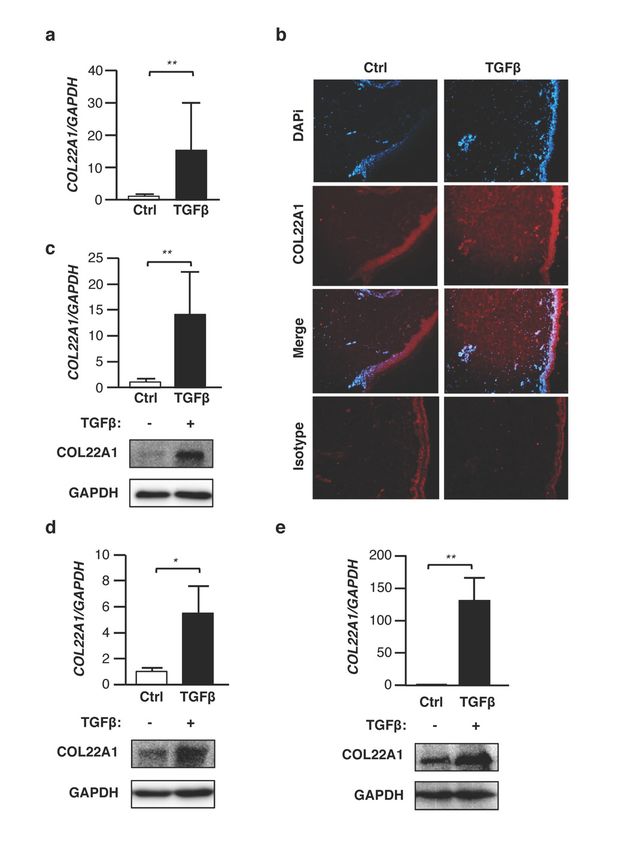
expression levels of COL22A1 in human skin from different donors (Figure 1a). Immunofluorescence
analysis also revealed COL22A1 protein in the dermal layer of human skin treated with TGFβ,Genes 2019, 10, 75 7 of 16
whereas no COL22A1 was detected in human skin treated with the vehicle control (Figure 1b).
Since fibroblasts are effector cells in dermal fibrosis [15], and having observed COL22A1 in the
dermal layer of the skin, we next asked whether COL22A1 expression is increased by TGFβ in skin
fibroblasts. TGFβ significantly increased the expression of COL22A1 in skin fibroblasts from healthy
donors. This was confirmed at the protein level using Western blot analysis (Figure 1c). We also
examined the ability of TGFβ to increase COL22A1 expression in other cell types. Interestingly, mRNA
and protein levels of COL22A1 were significantly increased by TGFβ in normal human lung fibroblasts
(Figure 1d) and A549 cells (Figure 1e), which are adenocarcinomic human alveolar basal epithelial
Genes 2019, 10, 75 7 of 17
cells. Thus, TGFβ can induce COL22A1 expression both in the skin- and lung-derived cells.
Figure 1. TGFβ increases expression levels of COL22A1 ex vivo and in vitro. (a,b) Human skin samples
wereFigure
treated1.with
TGFβ TGFβ (10 ng/mL)
increases for 48
expression andof72COL22A1
levels h. (a) Expression
ex vivo levels COL22A1
and inofvitro. were measured
(a,b) Human skin
in human
samplesskin (N treated
were p < 0.01.
= 5); **with TGFβ(b)(10Localization
ng/mL) for 48 of and 72 h. (a)inExpression
COL22A1 ex vivo normal skin.
levels of COL22A1
COL22A1 werewas
detected usinginimmunofluorescence
measured human skin (N = 5); in ** ap vehicle control
< 0.01. (b) or TGFβ-treated
Localization of COL22A1skinintissue. DAPI
ex vivo normalwasskin.
used to
COL22A1
detect was detected
nuclei (original magnification ×40); scale bars =in100
using immunofluorescence a vehicle
µm. (c)control
Human or normal
TGFβ-treated skin tissue.were
skin fibroblasts
DAPI
treated wasTGFβ
with used (5
to detect
ng/mL) nuclei (original
for 24 or 72 h.magnification
Expression ×40);
levels scale bars = 100 mRNA
of COL22A1 μm. (c) Human normal in
were measured
skin fibroblasts were treated with TGFβ (5 ng/mL) for 24 or 72 h. Expression levels of
human normal skin fibroblasts (N = 9); ** p < 0.01. Protein levels of COL22A1 in the skin fibroblasts COL22A1 mRNA
were measured in human normal skin fibroblasts (N = 9); ** p < 0.01. Protein
of three healthy donors were analyzed by immunoblotting of the lysates; GAPDH is shown levels of COL22A1 in theas a
loading control. (d) Human normal lung fibroblasts were treated with TGFβ (10 ng/mL) for 48 oris72 h.
skin fibroblasts of three healthy donors were analyzed by immunoblotting of the lysates; GAPDH
shown as a loading control. (d) Human normal lung fibroblasts were treated with TGFβ (10 ng/mL)
Expression levels of COL22A1 mRNA were measured in human normal lung fibroblasts treated with
for 48 or 72 h. Expression levels of COL22A1 mRNA were measured in human normal lung fibroblasts
treated with a vehicle control or TGFβ (N = 3); * p < 0.05. The protein levels of COL22A1 in lung
fibroblasts were analyzed by immunoblotting of the lysates. (e) A549 cells were treated with TGFβ (5
ng/mL) for 24 or 72 h. Expression levels of COL22A1 mRNA were measured in A549 cells treated with
a vehicle control or TGFβ (N = 3); ** p < 0.01. Protein levels of COL22A1 in the A549 cells were analyzed
by immunoblotting of the lysates.Genes 2019, 10, 75 8 of 16
a vehicle control or TGFβ (N = 3); * p < 0.05. The protein levels of COL22A1 in lung fibroblasts were
analyzed by immunoblotting of the lysates. (e) A549 cells were treated with TGFβ (5 ng/mL) for 24
or 72 h. Expression levels of COL22A1 mRNA were measured in A549 cells treated with a vehicle
control or TGFβ (N = 3); ** p < 0.01. Protein levels of COL22A1 in the A549 cells were analyzed by
immunoblotting of the lysates.
To 2019,
Genes further investigate the mechanisms involved in COL22A1 expression, we examined
10, 75 8 of 17
whether other fibrosis-promoting growth factors, such as interleukin (IL-6), bleomycin (BLM), and
Derived Growth
Platelet-Derived Factor Factor
Growth (PDGF)-BB, induce COL22A1
(PDGF)-BB, expression
induce COL22A1 in dermalin
expression fibroblasts after 24 h. IL-
dermal fibroblasts after
6 and BLM showed modest increases in COL22A1 expression in skin fibroblasts, while
24 h. IL-6 and BLM showed modest increases in COL22A1 expression in skin fibroblasts, whilePDGF-BB had
no effecthad
PDGF-BB (Supplementary Figure S2A,B).Figure S2A,B).
no effect (Supplementary
3.2.3.2. COL22A1
COL22A1 Is is a TGFβEarly
a TGFβ EarlyResponse
ResponseGene
Gene
To To evaluate
evaluate time-dependentchanges
time-dependent changes induced
induced by by TGFβ,
TGFβ, we we measured
measuredthe themRNA
mRNAlevels of of
levels
COL22A1 in ex vivo human skin samples. Human skin samples treated with
COL22A1 in ex vivo human skin samples. Human skin samples treated with TGFβ were harvested TGFβ were harvested at at
2, 4, 8, 16, and 24 h. TGFβ showed a time-dependent increase in COL22A1 expression.
2, 4, 8, 16, and 24 h. TGFβ showed a time-dependent increase in COL22A1 expression. mRNA levels mRNA levels
of COL22A1
of COL22A1 were
were significantlyupregulated
significantly upregulated starting
starting at
at 88 hh after
after stimulation
stimulationwithwithTGFβ
TGFβ (Figure 2a).2a).
(Figure
We next compared time-dependent changes in normal skin fibroblasts. Similar to ex vivo human skin,
We next compared time-dependent changes in normal skin fibroblasts. Similar to ex vivo human
significantly increased levels of COL22A1 mRNA in skin fibroblasts were noted 4 h after TGFβ
skin, significantly increased levels of COL22A1 mRNA in skin fibroblasts were noted 4 h after TGFβ
treatment (Figure 2b). This upregulation in skin fibroblasts was slightly earlier than that observed in
treatment (Figure 2b). This upregulation in skin fibroblasts was slightly earlier than that observed
ex vivo human skin. We further analyzed the expression of known fibrosis-related genes at the same
in ex vivo human skin. We further analyzed the expression of known fibrosis-related genes at the
time points. qRT-PCR analysis revealed that the expression levels of collagen type I alpha chain
same time points.
(COL1A1), qRT-PCR
fibronectin analysis
(FN), revealed
and alpha that muscle
2 smooth the expression levels of
actin (ACTA2) werecollagen type I alpha
significantly chain
increased
(COL1A1), fibronectin (FN), and alpha 2 smooth muscle actin (ACTA2)
at later time points (after 16 or 24 h of TGFβ treatment) compared with those of COL22A1 (Figure 2c– at
were significantly increased
later
e).time points (after
In contrast, 16 or 24
significant h of TGFβ
increases treatment) compared
in connective tissue growth withfactor of COL22A1
those (CTGF) (Figurewere
expression 2c–e).
In contrast,
observed significant
as early as 2increases in connective
h after TGFβ treatmenttissue growth
(Figure factor
2f). Thus, (CTGF)
TGFβ expression
induces COL22A1 were observed
expression
as early as 2 h after TGFβ treatment (Figure 2f). Thus, TGFβ induces COL22A1 expression
earlier than other extracellular matrix genes and ACTA2, while the early response of the COL22A1 earlier than
other extracellular matrix genes and ACTA2, while the early
expression is comparable to that seen with the early response gene CTGF. response of the COL22A1 expression is
comparable to that seen with the early response gene CTGF.
Figure 2. COL22A1 is an early response gene ex vivo and in vitro. Human skin and fibroblasts treated
with TGFβ
Figure 2. (5 or 10 ng/mL)
COL22A1 were
is an early harvested
response geneatex
the indicated
vivo time Human
and in vitro. points. skin and fibroblasts treated
with TGFβ (5 or 10 ng/mL) were harvested at the indicated time points. (a) The expression levels of
COL22A1 mRNA were measured in normal human skin in organ culture (N = 5); * p < 0.05; ** p < 0.01.
(b) Expression levels of COL22A1 mRNA were measured in primary human skin fibroblasts (N = 3):
* p < 0.05. (c) COL1A1 mRNA levels; * p < 0.05. (d) FN mRNA levels; * p < 0.05; ** p < 0.01; *** p < 0.001.
(e) ACTA2 mRNA levels: ** p < 0.01; *** p < 0.001. (f) CTGF mRNA levels: * p < 0.05; ** p < 0.01; *** p <Genes 2019, 10, 75 9 of 16
(a) The expression levels of COL22A1 mRNA were measured in normal human skin in organ culture
(N = 5); * p < 0.05; ** p < 0.01. (b) Expression levels of COL22A1 mRNA were measured in primary
human skin fibroblasts (N = 3): * p < 0.05. (c) COL1A1 mRNA levels; * p < 0.05. (d) FN mRNA levels;
* p < 0.05; ** p < 0.01; *** p < 0.001. (e) ACTA2 mRNA levels: ** p < 0.01; *** p < 0.001. (f) CTGF mRNA
Genes 2019, 10, 75 9 of 17
levels: * p < 0.05; ** p < 0.01; *** p < 0.001.
3.3. 3.3. TGFβ-Induced
TGFβ-Induced COL22A1
COL22A1 Expression
Expression DependsononALK5
Depends ALK5and
andMEK
MEKActivation
Activation
To investigate
To investigate which
which TGFβ
TGFβ signalingcascades
signaling cascadesare
are involved
involved in
in the
the induction
induction of of COL22A1
COL22A1
expression, normal skin fibroblasts were cultured with TGFβ in combination with specific inhibitors
expression, normal skin fibroblasts were cultured with TGFβ in combination with specific inhibitors of
of ALK5 (SB431542), MEK (U0126), PI3K (LY294002), and JNK signaling. TGFβ induction of COL22A1
ALK5 (SB431542), MEK (U0126), PI3K (LY294002), and JNK signaling. TGFβ induction of COL22A1
expression was abrogated by ALK5 and MEK inhibition (Figure 3a). JNK inhibition partly blocked
expression was abrogated by ALK5 and MEK inhibition (Figure 3a). JNK inhibition partly blocked
the TGFβ-induced expression of COL22A1. On the other hand, TGFβ-induced COL22A1 expression
the TGFβ-induced expression of COL22A1. On the other hand, TGFβ-induced COL22A1 expression
was not affected by PI3K inhibition (Figure 3a). Similar effects were observed at the protein level
was(Figure
not affected by PI3K
3b). These inhibition
results (Figure
suggest that 3a). Similar
the induction effects were
of COL22A1 observed
expression at the
by TGFβ protein level
is mediated by
(Figure 3b). These results
ALK5 and MEK signaling. suggest that the induction of COL22A1 expression by TGFβ is mediated by
ALK5 and MEK signaling.
Figure 3. TGFβ-induced COL22A1 expression is inhibited by MEK and ALK5 inhibitors.
Figure 3. TGFβ-induced COL22A1 expression is inhibited by MEK and ALK5 inhibitors. Skin
Skin fibroblasts were incubated with 10 µmol/L SB431542 (SB), U0126 (U), LY294002 (LY), JNK inhibitor
fibroblasts were incubated with 10 μmol/L SB431542 (SB), U0126 (U), LY294002 (LY), JNK inhibitor II
II (J), or DMSO as a vehicle control for 1 h before the addition of TGFβ (5 ng/mL). Skin fibroblasts
(J), or DMSO as a vehicle control for 1 h before the addition of TGFβ (5 ng/mL). Skin fibroblasts were
werestimulated
stimulated with
with TGFβ
TGFβ for for 24 and
24 and 72 h.72(a)
h.Expression
(a) Expression
levelslevels of COL22A1
of COL22A1 were measured
were measured using
using qPCR
qPCR (N = 4); ** p < 0.01. (b) Representative data from immunoblotting of lysates are shown.
(N = 4); ** p < 0.01. (b) Representative data from immunoblotting of lysates are shown. GAPDH was GAPDH
was used
usedas asaaloading
loadingcontrol.
control.
3.4. Silencing COL22A1 Reduces TGFβ-Induced ACTA2 Expression
3.4. Silencing COL22A1 Reduces TGFβ-Induced ACTA2 Expression
Since COL22A1 expression was induced by TGFβ earlier than other genes, we examined the
Since COL22A1 expression was induced by TGFβ earlier than other genes, we examined the
potential
potential of COL22A1
role role knockdown
of COL22A1 knockdownon TGFβ regulation
on TGFβ of other
regulation fibrosis-related
of other genes.
fibrosis-related Normal
genes. Normalskin
fibroblasts were transfected with COL22A1 siRNA and then stimulated with TGFβ. An average
skin fibroblasts were transfected with COL22A1 siRNA and then stimulated with TGFβ. An average of 60%
silencing of COL22A1 was achieved in unstimulated fibroblasts compared to control siRNA
of 60% silencing of COL22A1 was achieved in unstimulated fibroblasts compared to control siRNA (Figure 4a).
COL22A1
(FiguremRNA levels also
4a). COL22A1 decreased
mRNA levelssignificantly in TGFβ-stimulated
also decreased fibroblasts. Similarly,
significantly in TGFβ-stimulated protein
fibroblasts.
Similarly, protein levels of COL22A1 were decreased by silencing COL22A1 (Figure 4b). We further
investigated whether other fibrosis related genes were affected by COL22A1 knockdown.
Interestingly, TGFβ-induced ACTA2 expression was significantly decreased following COL22A1
silencing (Figure 4c). This effect was also observed at the protein level (Figure 4d). COL22A1 silencing
reduced TGFβ induction of COL1A1 and to a lesser degree that of FN, but the differences did notGenes 2019, 10, 75 10 of 16
levels of COL22A1 were decreased by silencing COL22A1 (Figure 4b). We further investigated whether
other fibrosis related genes were affected by COL22A1 knockdown. Interestingly, TGFβ-induced
ACTA2 expression was significantly decreased following COL22A1 silencing (Figure 4c). This effect
wasGenes
also2019,
observed
10, 75
at the protein level (Figure 4d). COL22A1 silencing reduced TGFβ induction 10 of 17
of
COL1A1 and to a lesser degree that of FN, but the differences did not reach statistical significance. The
protein
reachlevels of COL1A1
statistical wereThe
significance. alsoprotein
reduced. Onofthe
levels other hand,
COL1A1 mRNA
were also and protein
reduced. levelshand,
On the other of CTGF
mRNA
were and protein
not affected levels of CTGF
by COL22A1 were not These
knockdown. affected by COL22A1
results knockdown.
indicate Thesemay
that COL22A1 results indicate
mediate TGFβ
that COL22A1 may mediate TGFβ
induction of ACTA2, and possibly COL1A1. induction of ACTA2, and possibly COL1A1.
Figure 4. Silencing COL22A1 reduces expression levels of ACTA2. Normal skin fibroblasts were
transfected
Figure 4.with control
Silencing or COL22A1
COL22A1 siRNA
reduces and treated
expression levelswith TGFβ (5
of ACTA2. ng/mL)
Normal forfibroblasts
skin 48 and 72 were
h (N = 4).
transfected with control or COL22A1 siRNA and treated with TGFβ (5 ng/mL) for
(a) Expression levels of COL22A1 mRNA were measured; * p < 0.05. (b) Protein levels of COL22A1 48 and 72 h (N = 4).
(a) Expression levels of COL22A1 mRNA were measured; * p < 0.05. (b) Protein
were analyzed by immunoblotting of lysates. GAPDH is shown as a loading control. (c) Expressionlevels of COL22A1
were
levels of analyzed by immunoblotting
fibrosis-related of lysates. GAPDH
genes were measured; is shown
* p < 0.05; as a loading
** p < 0.01. control. (c) Expression
(d) Representative protein levels
levels of fibrosis-related genes were measured; * p < 0.05; **
in lysates from fibroblasts transfected with control or COL22A1 siRNA.p < 0.01. (d) Representative protein levels
in lysates from fibroblasts transfected with control or COL22A1 siRNA.Genes 2019, 10,
Genes 2019, 10,75
75 11 of
11 of 16
17
To determine whether COL22A1 silencing alters the contractile capacity of dermal fibroblasts,
To determine whether COL22A1 silencing alters the contractile capacity of dermal fibroblasts,
we examined the effect of silencing COL22A1 expression on dermal fibroblast-mediated collagen gel
we examined the effect of silencing COL22A1 expression on dermal fibroblast-mediated collagen gel
contraction. Our data show that COL22A1 silencing in the absence of TGFβ modestly decreased the
contraction. Our data show that COL22A1 silencing in the absence of TGFβ modestly decreased the
baseline gel contraction capacity of fibroblasts. However, COL22A1 silencing did not significantly
baseline gel contraction capacity of fibroblasts. However, COL22A1 silencing did not significantly
alter TGFβ-mediated collagen gel contraction of fibroblasts, even though it resulted in a pronounced
alter TGFβ-mediated collagen gel contraction of fibroblasts, even though it resulted in a pronounced
reduction of TGFβ-induced ACTA2 expression (Supplementary Figure S3).
reduction of TGFβ-induced ACTA2 expression (Supplementary Figure S3).
3.5. COL22A1
3.5. COL22A1 Expression
Expression Isis Elevated
Elevated in
in SSc
SSc Skin
Skin Fibroblasts
Fibroblasts
Since SSc
Since SSc is
is aa disease
disease characterized
characterized by by dermal
dermal fibrosis,
fibrosis, we
we compared
compared mRNA
mRNA and and protein
protein levels
levels
of COL22A1
of COL22A1 in in skin
skin fibroblasts
fibroblasts from
from control
control donors
donors and
and patients
patients with
with the
the diffuse
diffuse cutaneous
cutaneous form
form of
of
SSc. COL22A1 expression was significantly increased in fibroblasts from lesional SSc
SSc. COL22A1 expression was significantly increased in fibroblasts from lesional SSc skin (SSc-affected skin (SSc-
affected
skin skin fibroblasts;
fibroblasts; SSc-Aff) andSSc-Aff) and non-lesional
non-lesional SSc skin (SSc-unaffected
SSc skin (SSc-unaffected skin fibroblasts;
skin fibroblasts; SSc-Un)
SSc-Un) compared
compared
to to normal
normal skin skin fibroblasts
fibroblasts (Figure 5a).(Figure 5a). Inno
In contrast, contrast, no significant
significant differencesdifferences
were foundwere found
between
between affected and unaffected SSc fibroblasts. Protein levels of COL22A1 in SSc-affected
affected and unaffected SSc fibroblasts. Protein levels of COL22A1 in SSc-affected fibroblasts were also fibroblasts
were also
higher thanhigher
those in than those
normal in fibroblasts
skin normal skin fibroblasts
(Figure (Figure
5b). These 5b). suggest
findings These findings suggestlevels
that COL22A1 that
are elevated in fibroblasts from patients with SSc-associated dermal fibrosis, and due to its roledue
COL22A1 levels are elevated in fibroblasts from patients with SSc-associated dermal fibrosis, and in
to its role in mediating TGFβ regulation of ACTA2 and myofibroblast differentiation,
mediating TGFβ regulation of ACTA2 and myofibroblast differentiation, COL22A1 may be involved
COL22A1 may
be involved in the pathogenesis of SSc dermal fibrosis.
in the pathogenesis of SSc dermal fibrosis.
Figure 5. Increased expression levels of COL22A1 in SSc skin fibroblasts. Skin fibroblasts from healthy
donors
Figure 5. and patientsexpression
Increased with SSc levels
were cultured
of COL22A1 in serum-free
in SSc skin DMEM for Skin
fibroblasts. 48 and 72 h. (a)from
fibroblasts Expression
healthy
levels
donorsofandCOL22A1
patientsmRNA were
with SSc measured
were culturedin inthe indicated
serum-free groupfor
DMEM (HC; N = 72
48 and 8, SSc-Un; N= 6, SSc-Aff;
h. (a) Expression levels
N 7); * p < 0.05.
of =COL22A1 mRNA (b)were
Protein levels ofinCOL22A1
measured weregroup
the indicated detected in N
(HC; normal skin fibroblasts
= 8, SSc-Un; and SSc
N= 6, SSc-Aff; N skin
= 7);
fibroblasts using immunoblotting. GAPDH is shown as a loading control. NS = normal
* p < 0.05. (b) Protein levels of COL22A1 were detected in normal skin fibroblasts and SSc skin skin fibroblasts;
SSc-Un = SSc-unaffected
fibroblasts skin fibroblasts;
using immunoblotting. SSc-Aff
GAPDH is =shown
SSc-affected
as a skin fibroblasts.
loading control. NS = normal skin
fibroblasts; SSc-Un = SSc-unaffected skin fibroblasts; SSc-Aff = SSc-affected skin fibroblasts.
3.6. Genes Identified as Differentially Expressed in TGFβ-Treated Human Skin Overlap with SSc Patient
Skin Signatures
3.6. Genes Identified as Differentially Expressed in TGFβ-Treated Human Skin Overlap with SSc Patient
Skin To assess the similarity in the response of human skin in organ culture to a well-accepted signature
Signatures
of human SSc skin, we compared the gene signature identified in our model to that identified in
To assess the similarity in the response of human skin in organ culture to a well-accepted
primary fibroblasts stimulated with TGFβ as reported by Sargent et al. [16], and to the SSc skin
signature of human SSc skin, we compared the gene signature identified in our model to that
signature identified by Milano et al. [17]. Sargent et al. carried out in vitro exposure of adult dermal
identified in primary fibroblasts stimulated with TGFβ as reported by Sargent et al. [16], and to the
fibroblasts from healthy and SSc patients and derived a gene signature for TGFβ responsiveness
SSc skin signature identified by Milano et al. [17]. Sargent et al. carried out in vitro exposure of adult
dermal fibroblasts from healthy and SSc patients and derived a gene signature for TGFβGenes 2019, 10, 75 12 of 16
comprised of 674 unique genes. In their study, they reanalyzed a prior data set from Milano et al.,
derived from 75 arrays from patients with different disease severity. Although their 674 unique gene
signatures distinguished the dSSc and healthy patients (53 arrays), this signature was noisy when
applied to the extended data set with different disease progression represented. This signature was
therefore refined to a smaller subset that better differentiated the dSSc and normal groups. We noted
that these array 474 probes described by Sargent et al. corresponded to 371 genes. We looked for
overlaps with all 1051 transcripts differentially expressed (FDR ≤ 0.4) in our study. To minimize
technical noise associated with microarray experimentation and experimental differences between
laboratories, we required that the direction of the fold change was the same between both studies, i.e.,
if a gene was reported as upregulated by Sargent et al., it also needed to be in this study for further
consideration as a biomarker. This stringent filtering resulted in a 48 gene overlaps between our study
and that of Sargent at al., and a heatmap of these transcripts which clearly distinguishes the control
and TGFβ treated groups is presented in Figure 6. These findings suggest that our human skin ex vivo
model is a suitable model for the dermal fibrosis characteristic of SSc.
Figure 6. The heatmap of the 48-gene overlap between our study (significant DE mRNAs as determined
by DESeq2 (FDR < 0.1)) and the 371 gene signature that differentiates the dSSc and the normal state
described by Sargent et al. The red and blue boxes indicate relative over- and under-expression with
respect to a reference which is calculated as the mid-point between the control and TGFβ treated groups.Genes 2019, 10, 75 13 of 16
4. Discussion
Research on fibrosis has relied on the use of in vitro assays and in vivo mouse and rat models.
However, many of the agents tested in animals have not been efficacious in humans in clinical
trials. Using human skin as an ex vivo organ model of fibrosis has great potential to examine
mechanisms underlying fibrosis because this model more closely mimics the skin fibrosis characteristic
of human SSc than the current in vitro and in vivo models. In fact, we have previously shown that
factors such as IGFBP-5, IGFBP-3, and TGFβ can induce fibrosis in human skin maintained in organ
culture [18–20]. We now show that our ex vivo human skin model is a suitable model for the dermal
fibrosis characteristic of SSc. The signature obtained from the human skin stimulated with TGFβ and
maintained in organ culture overlaps with the signature identified in two key studies using human
dcSSc skin biopsies and human fibroblasts stimulated with TGFβ [16,17]. The 48-gene signature in the
overlap between our model and these studies demonstrates the utility and relevance of our model for
research focusing on dermal fibrosis in SSc.
RNA-seq using ex vivo human skin samples is a powerful tool for identifying new genes that
may contribute to disease pathogenesis and their functional analysis. Using RNAseq, we identified
the transcripts regulated by TGFβ in human skin, with COL22A1 showing the greatest differential
expression. Our findings reveal that in normal skin-derived fibroblasts, TGFβ significantly increased
COL22A1 expression. Furthermore, fibroblasts from the skin of dcSSc patients expressed high levels of
COL22A1 compared with normal skin fibroblasts. These findings suggest that upregulated COL22A1
expression may be associated with the pathogenesis of fibrosis in patients with SSc and/or may be a
biomarker for human dermal fibrosis. To the best of our knowledge, this study is the first to investigate
the levels (both mRNA and protein) and the role of COL22A1 in the setting of fibrosis.
Our analysis revealed that COL22A1 is a TGFβ-inducible immediate early gene in skin fibroblasts
while other fibrosis-promoting growth factors had a very modest effect on COL22A1 expression.
TGFβ has been shown to induce several downstream genes such as CTGF, EGR-1, and Endothelin-1
(ET-1) at an early stage compared with other fibrosis-associated genes [21–23]. These factors play
an important role in the deposition of extracellular matrix components including fibronectin and
collagen by fibroblasts in the development and maintenance of fibrosis. In fact, CTGF is an inducible
matricellular protein involved in tissue remodeling, and its expression is increased during wound
repair. CTGF mediates some of the effects of TGFβ, and CTGF transgenic mice were shown to have
multi-organ fibrosis [24]. The fact that COL22A1 expression is induced early while that of other ECM
genes and ACTA2 are increased in a delayed manner raises the possibility that COL22A1 may mediate
the TGFβ induction of pro-fibrotic factors. Indeed, siRNA experiments showed that silencing COL22A1
reduced ACTA2, COL1A1, and FN expression and late-response genes downstream of TGFβ, but not
CTGF expression, which is induced earlier than COL22A1. These results lead us to speculate that
COL22A1 acts upstream of ACTA2, COL1A1, and FN and regulates their expression through the TGFβ
signaling pathway.
In addition, COL22A1 showed a higher magnitude of response to TGFβ than other TGFβ regulated
genes we examined. Other fibrosis-associated genes such as COL1A1, FN, and ACTA2, which are
late response targets of TGFβ, showed increased expression of 2–5-fold following TGFβ stimulation,
whereas the early response genes such as COL22A1, CTGF (Figure 3f), and EGR-1 [25] showed more
than a 30-fold induction in expression in skin fibroblasts. This greater response may reflect the
acute phase of the response to TGFβ, while the late response genes are more characteristic of the
chronic response to TGFβ and other triggers of fibrosis. Although COL22A1 silencing decreased
TGFβ-mediated ACTA2 expression and αSMA levels, it did not affect the contraction of fibroblasts
in the collagen gel. αSMA is a component of stress fibers in fibroblasts; however, its inhibition is
not sufficient to prevent contraction. In fact, αSMA knockout mice show a delay in skin wound
healing, suggesting that αSMA contributes to wound healing but is not necessary [26]. Additionally,
αSMA null mice retained the ability to form myofibroblasts and contractile forces in response to
TGFβ [27]. This may be in part due to the fact that additional proteins can compensate for the absenceGenes 2019, 10, 75 14 of 16
of αSMA. For instance, in fibroblasts and myofibroblasts isolated from αSMA knockout mice, other
actin forms were found at higher levels post-TGFβ simulation [27]. Additionally, non-muscle myosins
colocalize with actin and participate in contraction during wound healing [28]. Thus, fibroblasts have
compensatory proteins that may reduce the functional impact of the αSMA decrease on cell contraction.
Regulation of COL22A1 expression by fibrosis-promoting growth factors also appears to provide
some insights into mechanisms regulating COL22A1 expression by TGFβ. PDGF-BB did not increase
COL22A1 expression, while IL-6 and BLM modestly increased its levels. Our experiments using
TGFβ signaling cascade inhibitors identified pathways regulating COL22A1 expression. PDGF is
known to result in the activation of JAK/STAT, PI3K, phospholipase C-γ (PLC-γ) or MAPK signaling
pathways [29]. The binding of IL-6 to its receptor leads to the activation of JAK/STAT and Ras-mediated
signaling [30]. In addition, IL-6 also activates PI3K [30,31]. Similar to these factors, BLM does not
activate ALK5 or MEK signaling pathways. However, we showed that the activation of PI3K was
not required to induce COL22A1 expression at the mRNA and protein levels. In this regard, ALK5
and MEK signaling pathways seem to play a key role in mediating the TGFβ regulation of COL22A1
expression, explaining, at least in part, why TGFβ was more potent for inducing COL22A1 expression.
COL22A1 is expressed in tissue junctions, tendons, heart, articular cartilage, and skin [4]. However,
no studies to date have assessed COL22A1 expression in lung tissue-derived cells. We demonstrate that
the expression levels of COL22A1 were increased by TGFβ in adenocarcinomic human alveolar basal
epithelial cells and lung fibroblasts. These results suggest that that COL22A1 may be associated with
the development of lung fibrosis as well as skin fibrosis via increased expression in resident fibroblasts
contributing to their activation and transition to myofibroblasts. Furthermore, TGFβ-induced COL22A1
expression may induce epithelial mesenchymal transition (EMT) in human alveolar epithelial cells.
EMT is a biologic process that involves an epithelial cell acquiring a mesenchymal cell phenotype,
which includes enhanced migratory capacity, resistance to apoptosis, and increased production of
ECM components [32]. In fact, TGFβ stimulation promotes alveolar epithelial cell transition to a
myofibroblast-like phenotype in lung tissues [33]. EMT is also found to be associated with fibrosis
occurring in different organs including the kidney, liver, lung, and intestine. Therefore, the high
induction of COL22A1 by TGFβ in A549 cells and lung fibroblasts, in conjunction with the reduction of
ACTA2 and COL1A1 expression by COL22A1 silencing, leads us to speculate that COL22A1 may be
associated with the transition of fibroblasts and epithelial cells to myofibroblasts, effector cells that
play an important role in extracellular matrix production and fibrosis. In fact, in the skin, COL22A1 is
expressed surrounding the lower third of anagen hair follicles and acts as a cell adhesion ligand for skin
epithelial cells and fibroblasts [4]. It has been reported that ACTA2 is expressed in human hair follicles
and that hair follicle-derived smooth muscle cells express ACTA2 [34]. It is thus conceivable that hair
follicle cells can be another source of the myofibroblasts characteristic of fibrotic skin. Furthermore,
since hair follicles are involved in perifollicular fibrosis, and our findings suggest skin localization of
COL22A1, it is likely that COL22A1 may also play an important role in perifollicular fibrosis and may
promote cell adhesion and migration of hair follicle-derived smooth muscle cells.
In conclusion, our findings suggest that the increased expression of COL22A1 is associated with
the pathogenesis of fibrosis in SSc and that the regulation of COL22A1 expression may have important
implications for skin fibrosis as well as lung fibrosis. These results add to our understanding of the
pathogenic mechanism of fibrosis by generating novel insights into genes regulated under pro-fibrotic
conditions in human skin, providing direct relevance to human dermal fibrosis.
Supplementary Materials: The following are available online at http://www.mdpi.com/2073-4425/10/2/75/s1,
Figure S1: Heatmap of significantly differentially expressed mRNAs as determined by DESeq2 (FDR < 0.1) using
ex vivo human skin samples treated with TGFβ (10 ng/mL) or a vehicle control for 48 h, Figure S2: Human
normal skin fibroblasts were treated with IL-6 (20 ng/mL), PDGF-BB (40 ng/mL), or BLM (10 mU/mL) for
24 or 72 h, Figure S3: Decreased expression of ACTA2 in TGFβ-treated fibroblasts deficient in COL22A1 does
not alter TGFβ-induced collagen gel contraction, Table S1: Significantly DE mRNAs as determined by DESeq2
using ex vivo human skin samples treated with TGFβ (10 ng/mL) or a vehicle control for 48 h, Table S2: The
most significantly impacted biological pathways due to TGFβ treatment, ranked by adjusted p value from mostGenes 2019, 10, 75 15 of 16
significant to least significant, Supplemental method 1: Immunofluorescence, Supplemental method 2: Fibroblast
treatment, Supplemental method 3: Collagen Gel Contraction Assay.
Author Contributions: T.W., C.F.B., G.H. contributed to experimental design; T.W., C.F.B., G.H., W.A.d.S., D.B.F.,
L.M., and J.H. contributed to data acquisition; T.W., C.F.B., G.H., W.A.d.S. and D.B.F. contributed to data analysis
and interpretation; all authors contributed to manuscript drafting and editing.
Funding: This work was supported in part by the SmartState and Kitty Trask Holt Endowment and grant
K24AR060297 from NIH/NIAMS (C.F.B.), the E. Carwile LeRoy, MD, Endowed Research Fellowship (T.W.),
KL2TR001452 (D.B.F.), UL1TR001450, and the Genomics Shared Resource, Hollings Cancer Center and the Center
for Genomic Medicine at the Medical University of South Carolina (G.H.). GH acknowledges funding from
SC EPSCoR and G.H. and C.F.B. acknowledge start-up funding from the College of Medicine at the Medical
University of South Carolina.
Conflicts of Interest: The authors declare no conflict of interest.
References
1. Almeida, I.; Faria, R.; Vita, P.; Vasconcelos, C. Systemic sclerosis refractory disease: From the skin to the
heart. Autoimmun Rev. 2011, 10, 693–701. [CrossRef] [PubMed]
2. Steen, V.D.; Medsger, T.A., Jr. Epidemiology and natural history of systemic sclerosis. Rheum. Dis. Clin.
North Am. 1990, 16, 1–10. [PubMed]
3. Wynn, T.A. Common and unique mechanisms regulate fibrosis in various fibroproliferative diseases.
J. Clin. Investig. 2007, 117, 524–529. [CrossRef] [PubMed]
4. Koch, M.; Schulze, J.; Hansen, U.; Ashwodt, T.; Keene, D.R.; Brunken, W.J.; Burgeson, R.E.; Bruckner, P.;
Bruckner-Tuderman, L. A novel marker of tissue junctions, collagen XXII. J. Biol. Chem. 2004, 279,
22514–22521. [CrossRef]
5. Ricard-Blum, S.; Ruggiero, F. The collagen superfamily: From the extracellular matrix to the cell membrane.
Pathol. Biol. 2005, 53, 430–442. [CrossRef] [PubMed]
6. Charvet, B.; Guiraud, A.; Malbouyres, M.; Zwolanek, D.; Guillon, E.; Bretaud, S.; Monnot, C.; Schulze, J.;
Bader, H.L.; Allard, B.; et al. Knockdown of COL22A1 gene in zebrafish induces a muscular dystrophy by
disruption of the myotendinous junction. Development 2013, 140, 4602–4613. [CrossRef] [PubMed]
7. Misawa, K.; Kanazawa, T.; Imai, A.; Endo, S.; Mochizuki, D.; Fukushima, H.; Misawa, Y.; Mineta, H.
Prognostic value of type XXII and XXIV collagen mRNA expression in head and neck cancer patients. Mol.
Clin. Oncol. 2014, 2, 285–291. [CrossRef] [PubMed]
8. Mak, A.C.; Tang, P.L.; Cleveland, C.; Smith, M.H.; Kari Connolly, M.; Katsumoto, T.R.; Wolters, P.J.; Kwok, P.Y.;
Criswell, L.A. Brief Report: Whole-Exome Sequencing for Identification of Potential Causal Variants for
Diffuse Cutaneous Systemic Sclerosis. Arthritis Rheumatol. 2016, 68, 2257–2262. [CrossRef]
9. Feghali, C.A.; Wright, T.M. Identification of multiple, differentially expressed messenger RNAs in dermal
fibroblasts from patients with systemic sclerosis. Arthritis Rheum. 1999, 42, 1451–1457. [CrossRef]
10. Hsu, E.; Shi, H.; Jordan, R.M.; Lyons-Weiler, J.; Pilewski, J.M.; Feghali-Bostwick, C.A. Lung tissues in
patients with systemic sclerosis have gene expression patterns unique to pulmonary fibrosis and pulmonary
hypertension. Arthritis Rheum. 2011, 63, 783–794. [CrossRef]
11. Pilewski, J.M.; Liu, L.; Henry, A.C.; Knauer, A.V.; Feghali-Bostwick, C.A. Insulin-like growth factor binding
proteins 3 and 5 are overexpressed in idiopathic pulmonary fibrosis and contribute to extracellular matrix
deposition. Am. J. Pathol. 2005, 166, 399–407. [CrossRef]
12. Davis-Turak, J.; Courtney, S.M.; Hazard, E.S.; Glen, W.B.; da Silveira, W.A.; Wesselman, T.; Harbin, L.P.;
Wolf, B.J.; Chung, D.; Hardiman, G. Genomics pipelines and data integration: Challenges and opportunities
in the research setting. Expert Rev. Mol. Diagn. 2017, 17, 225–237. [CrossRef] [PubMed]
13. Benjamini, Y.; Hochberg, Y. Controlling the False Discovery Rate: A Practical and Powerful Approach to
Multiple Testing. J. R. Stat. Soc. Ser. B (Methodol.) 1995, 57, 289–300. [CrossRef]
14. Draghici, S.; Khatri, P.; Tarca, A.L.; Amin, K.; Done, A.; Voichita, C.; Georgescu, C.; Romero, R. A systems
biology approach for pathway level analysis. Genome Res. 2007, 17, 1537–1545. [CrossRef]
15. Garrett, S.M.; Baker Frost, D.; Feghali-Bostwick, C. The mighty fibroblast and its utility in scleroderma
research. J. Scleroderma Relat. Disord. 2017, 2, 100–107. [CrossRef] [PubMed]You can also read