Hydrangea serrata (Thunb.) Ser. Extract Attenuate UVB-Induced Photoaging through MAPK/AP-1 Inactivation in Human Skin Fibroblasts and Hairless ...
←
→
Page content transcription
If your browser does not render page correctly, please read the page content below
nutrients
Article
Hydrangea serrata (Thunb.) Ser. Extract Attenuate
UVB-Induced Photoaging through MAPK/AP-1
Inactivation in Human Skin Fibroblasts and
Hairless Mice
Hee-Soo Han 1,2 , Ji-Sun Shin 1 , Da-Bin Myung 1,2 , Hye Shin Ahn 3 , Sun Hee Lee 3 ,
Hyoung Ja Kim 4 and Kyung-Tae Lee 1,2, *
 1   Department of Pharmaceutical Biochemistry, College of Pharmacy, Kyung Hee University, Seoul 02447,
     Korea; heesu3620@hanmail.net (H.-S.H.); jsunvet@naver.com (J.-S.S.); dabin_happy@naver.com (D.-B.M.)
 2   Department of Life and Nanopharmaceutical Science, College of Pharmacy, Kyung Hee University,
     Seoul 02447, Korea
 3   Department of New Material Development, COSMAXBIO, Seongnam 13486, Korea;
     ztztzt08@naver.com (H.S.A.); bt-shlee@cosmax.com (S.H.L.)
 4   Molecular Recognition Research Center, Materials and Life Science Research Division,
     Korea Institute of Science and Technology, Seoul 02792, Korea; khj@kist.re.kr
 *   Correspondence: ktlee@khu.ac.kr; Tel.: +82-2-961-0860; Fax: +82-2-961-0356

 Received: 2 January 2019; Accepted: 4 February 2019; Published: 1 March 2019

 Abstract: Skin photoaging is mainly caused by exposure to ultraviolet (UV) light, which increases
 expressions of matrix metalloproteinases (MMPs) and destroys collagen fibers, consequently inducing
 wrinkle formation. Nutritional factors have received scientific attention for use as agents for normal
 skin functions. The aim of this study was to investigate the effect of hot water extracts from the
 leaves of Hydrangea serrata (Thunb.) Ser. (WHS) against ultraviolet B (UVB)-induced skin photoaging
 and to elucidate the underlying molecular mechanisms in human foreskin fibroblasts (Hs68) and
 HR-1 hairless mice. WHS recovered UVB-reduced cell viability and ameliorated oxidative stress
 by inhibiting intracellular reactive oxygen species (ROS) generation in Hs68 cells. WHS rescued
 UVB-induced collagen degradation by suppressing MMP expression, and reduced the mRNA levels
 of inflammatory cytokines. These anti-photoaging activities of WHS were associated with inhibition
 of the activator protein 1 (AP-1), signal transduction and activation of transcription 1 (STAT1),
 and mitogen-activated protein kinase (MAPK) signaling pathways. Oral administration of WHS
 effectively alleviated dorsal skin from wrinkle formation, epidermal thickening, collagen degradation,
 and skin dehydration in HR-1 hairless mice exposed to UVB. Notably, WHS suppressed UVB
 activation of the AP-1 and MAPK signaling pathways in dorsal mouse skin tissues. Taken together,
 our data indicate that WHS prevents UVB-induced skin damage due to collagen degradation and
 MMP activation via inactivation of MAPK/AP-1 signaling pathway.

 Keywords: Hydrangea serrata (Thunb.) Ser.; ultraviolet B; photoaging; MMPs; collagen; MAPK; AP-1

1. Introduction
     The skin, as the most vulnerable organ of the human body, functions as the first line of defense
protecting the body against toxic chemicals, infections, and ultraviolet (UV) radiation [1]. The process
of skin aging is classified either as intrinsic (chronological) or extrinsic (photoaging). Intrinsic skin
aging is a natural process and is typically influenced by genetic factors. In contrast, extrinsic aging is
mainly caused by repeated exposure to solar ultraviolet light, especially UVB, and is characterized by

Nutrients 2019, 11, 533; doi:10.3390/nu11030533                                 www.mdpi.com/journal/nutrients
Nutrients 2019, 11, 533                                                                            2 of 15

a thickened epidermis, mottled discoloration, deep wrinkles, loss of elasticity, and slowing of skin cell
growth associated with slower wound healing [2]. Excessive UVB irradiation penetrates the skin and
generates intracellular reactive oxygen species (ROS), which consequently results in cellular oxidative
stress and prolonged skin inflammation [3] through activation of mitogen-activated protein kinase
(MAPK), activator protein 1 (AP-1), nuclear factor kappa B (NF-κB)/p65, and signal transduction and
activation of transcription (STAT) pathways [4]. Activation of these pathways results in overexpression
of inflammatory cytokines like tumor necrosis factor (TNF)-α, interleukin (IL)-1β, and IL-6 [5].
Moreover, UVB-induced ROS upregulates the expression of matrix metalloproteinases (MMPs),
contributing to extracellular matrix (ECM) degradation, including the destruction of collagen fibers,
and consequently resulting in wrinkle formation [6,7]. Increased levels of MMPs and degradation of
ECM components such as collagen, hyaluronic acid (HA), and elastin, are thought to be prominent
characteristics of photo-damaged skin [8].
     Several studies have indicated that dietary ingredient supplementation may improve or delay
skin aging [9,10]. Various chemicals or medicines proposed to alleviate skin aging have disadvantages,
including high prices, chemical instability, and side effects [11]. Therefore, there has been growing
interest in the development of safe and effective materials from botanical sources to prevent or
treat damaged skin. Hydrangea (Hydrangeaceae) leaves have been consumed as a tea and as
medicine in far-east Asian countries like Korea, China, and Japan [12]. Several studies have
demonstrated that extracts or isolated compounds of hydrangea leaves possess anti-inflammatory [13],
anti-diabetic [14], renal protective [15], and hepatoprotective activities [16] through an ameliorating
effect on oxidative stress. Hydrangea serrata (Thunb.) Ser. is native to the Korean mountains known
as “San-soogook, Mountain Hydrangea, or Tea of Heaven”. Its leaves are used to make herbal
teas. Our recent phytochemical study showed that a variety of compounds isolated from H. serrata
possess anti-photoaging activity in UVB-exposed human fibroblasts. Among these compounds,
hydrangenol has potential protective effects on cell viability, production of procollagen type I, MMP-1,
and pro-inflammatory cytokines [17]. Based on our previous report, we concluded that H. serrata
may have the potential for application as a beneficial natural anti-photoaging agent. However, due to
the lack of evidence on the anti-photoaging activity of H. serrata extract per se, we investigated the
anti-photoaging effect of hot water extracts from the leaves of H. serrata (WHS) in UVB-irradiated Hs68
human fibroblasts and HR-1 hairless mice.

2. Materials and Methods

2.1. Preparation and Standardization of WHS
     Hydrangea serrata (Thunb.) Ser. were cultivated in Yeongju, Korea and were identified by Dr.
Hyung-Jun Kim from Forest Medicinal Resources Research Center in National Institute of Forest
Science (NIFoS, Seoul, Korea). A specimen voucher has been deposited in COSMAXBIO (COSMAXBIO,
Seongnam, Korea). The dried leaves of H. serrata (1000 g) were extracted with distilled water at 98 ◦ C
for 5 h followed by filtration and then spray-dried to give a dried extract residue WHS (261 g, 26.1%).
The HPLC system consisted of a Waters model 2695 HPLC pump with photodiode array detector
(Waters model 2998) set to 228 nm (Waters, Milford, MA, USA). Separations were achieved at 30 ◦ C
using a Luna C18 (4.6 mm, 250 mm, I.D., 5 µm, Phenomenex) column. The gradient mobile phases
consisted of solvent A (100% acetonitrile) and solvent B (water) with a gradient elution as follows: 0 to
15 min, 20% to 25%; 15 to 30 min, 25% to 50%; 30 to 40 min, 50% to 100%; 40 to 50 min, 100% to 20% as
percent of solvent A at a flow rate of 1.0 mL/min.

2.2. Cell Culture
      Human fibroblasts (Hs68) were obtained from the American Type Culture Collection (ATCC,
Manassas, VA, USA) and cultured in Dulbecco’s modified eagle’s medium (DMEM) containing 10%
fetal bovine serum (FBS), penicillin-streptomycin sulfate (100 U/mL and 100 µg/mL) at 37 ◦ C with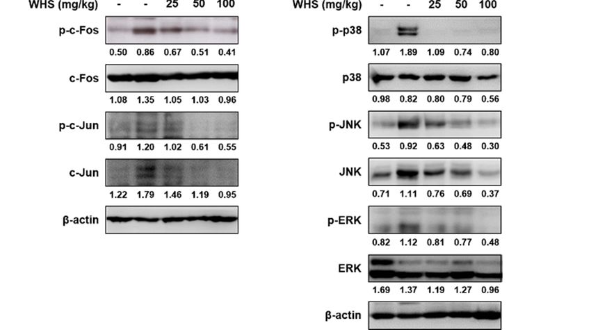
Nutrients 2019, 11, 533                                                                         3 of 15

5% CO2 . Hs68 cells were plated at 1 × 10⁵ /mL in culture area. After 24 h incubation, the cells were
washed once with PBS and then exposed to UVB irradiation (15 mJ/cm2 ) using UVP Crosslinker
(Analytik Jena AG, Jena, Germany). After UVB irradiation, cells were incubated in fresh culture media
in the presence or absence of WHS (6.25, 12.5, or 25 µg/mL).

2.3. Determination of Cell Viability
     After UVB irradiation, Hs68 cells were treated with WHS for 24 h. Cell viability was estimated
using 3-(4,5-Dimethylthiazol-2-yl)-2,5-diphenyl tetrazolium bromide (MTT) assay. 20 µL MTT solution
(5 mg/mL) was added to each well, and the cells were further incubated for an additional 4 h.
The supernatant was removed and the formazan was resolved with 1 mL/well of DMSO. The optical
density was measured by microplate reader (Molecular Devices Inc., San Jose, CA, USA) at 540 nm.

2.4. Fluorescence Assay of Intracellular ROS
     After UVB irradiation, Hs68 cells were treated with WHS (6.25, 12.5, or 25 µg/mL) or
N-acetyl-L-cysteine (NAC, 10 mM) for 24 h. For measuring intracellular ROS generations, the cells
were stained with 20 µM H2 DCFDA for 30 min. The stained cells were resuspended in PBS and
fluorescence intensities were detected by flow cytometry (Beckman Coulter Inc., Brea, CA, USA).

2.5. Elastase Activity Assay
     Elastase activity was measured using N-succinyl-tri-alanyl-p-nitroanilide (STANA, elastase
substrate) as previously reported, with some modifications [18]. Cells were collected and dissolved in
0.1% Triton X-110 in 0.2 M Tris-HCl buffer solution (pH 8.0). The cell resuspension was frozen and
thawed for three times. Cell debris was removed by microcentrifugation (3000 rpm, 20 min, 4 ◦ C).
100 µg of each cellular protein was loaded in 96-well plates and the volume of each well was adjusted
with 0.2 M Tris-HCl buffer by 98 µL. 2 µL of 50 mM N-succinyl-tri-alanyl-p-nitroanilide (elastase
substrate) solution was dispensed into each well and then incubated for 90 min at 37 ◦ C. The elastase
activity was determined by measuring the absorbance of each well at 405 nm using a microplate reader
(Molecular Devices Inc., San Jose, CA, USA).

2.6. Measurement of Pro-Collagen Type I, MMPs, and HA Production
     The production of pro-collagen type I (cat#. MK101) and MMP-1/-3 (cat#. ab100603/ab100607)
in cell culture media and the levels of hyaluronic acid (HA) (cat#. DY3614-05) in skin tissues were
quantified by ELISA kits according to the manufacturer’s instructions (TaKaRa Bio Inc., Shiga, Japan;
Abcam, Cambridge, UK; and R&D Systems, Minneapolis, MN, USA, respectively).

2.7. RNA Extraction and Quantitative Real-Time RT-PCR (qRT-PCR)
     Total cellular RNA was extracted by using Easy Blue® kits (Intron Biotechnology, Seoul, Korea).
RNA (1 µg) was reverse-transcribed (RT) using 0.5 mg/mL random oligonucleotide primers (Promega,
Madison, WI, USA) and TOPscriptTM RT DryMIX (Enzynomics, Daejeon, Korea). PCR amplification
was performed using the incorporation of SYBR green using SYBR Primix Ex Taq (TaKaRa Bio Inc.,
Shiga, Japan). The PCR primers used in this study are described in Table S1. Steady-state mRNA
levels were determined by real time qPCR using the TaKaRa thermal cycler device. Mean Ct values of
genes were calculated from triplicate measurements and normalized versus the mean Ct of GAPDH.
The PCR primers used in this study are described in Table S1.

2.8. Western Blot Analysis
     Total cellular or nuclear protein extracts from UVB-treated or UVB plus WHS-treated cells were
prepared as described previously [19]. Proteins (25 to 50 µg) were resolved by SDS-PAGE on 8%
to 12% polyacrylamide gel and electrotransferred to polyvinylidene fluoride (PVDF) membrane.
Nutrients 2019, 11, 533 4 of 15
The immunoblot was incubated in blocking solution (5% skim milk) for 1 h at room temperature and
then incubated overnight with a 1:1000 dilution of primary antibody at 4 ◦ C. The blots were washed
three times with tris-buffered saline with 0.1% tween 20 (TBST), incubated with a 1:2000 dilution of
horseradish peroxidase-conjugated secondary antibody for 2 h at room temperature, washed again
three times with TBST, and finally developed using an enhanced chemiluminescence (ECL) substrate
(Santa Cruz Biotechnology, Santa cruz, CA, USA). The protein bands were visualized by LAS-4000
luminescent image analyzer (FUJIFILM, Tokyo, Japan). The antibodies used in this study are listed in
Table S2. The optical density of each representative blot was presented on the basis of internal control
proteins (Histone H3 or β-actin).
2.9. Animals
Five-week-old male HR-1 hairless mice were purchased from SLC Inc. (SLC Inc., Shizuoka, Japan).
The animals were housed under consistent conditions (temperature: 22 ± 1 ◦ C, humidity: 40% to
60%, light/dark cycle: 12 h). All experiments were conducted under university guidelines of ethical
committee for Animal Care and Use of the Kyung Hee University according to an animal protocol
(KHUASP (SE) -18-107). After the acclimation period (1 week), mice were randomly divided into five
groups (n = 8): Vehicle-treated control (without UVB irradiation), UVB only-treated, UVB + WHS
(25 mg/kg/day, p.o.), UVB + WHS (50 mg/kg/day, p.o.), and UVB + WHS (100 mg/kg/day, p.o.).
2.10. UVB-Irradiated Skin Aging Model
UVB irradiation was applied to the backs of mice, three times a week during 10 weeks using UVP
Croslinker (Analytik Jena AG, Jena, Germany). The energy of UVB was progressively increased from
60 mJ/cm2 at first 4 weeks, to 240 mJ/cm2 at final irradiation with regular intervals of 60 mJ/cm2 for
every 2 weeks.
2.11. Evaluation of Skin Wrinkle Formation
The skin wrinkle formation on the dorsal skin of mice was observed at 10 weeks shortly after
a sacrifice. The skin replicas were cast on the dorsal skin surface of mice using SILFLO (Flexico
developments LTD., Tokyo, Japan) and then analyzed to measure skin wrinkles (Visioline® VL 650).
The parameters for the assessment of skin wrinkles were total wrinkle area, mean length, mean depth,
and max wrinkle depth.
2.12. Histological Analysis
The dorsal skin specimens were obtained, fixed in 4% paraformaldehyde overnight, dehydrated
in ethanol and then embedded in paraffin. The sliced sections were stained with hematoxylin and
eosin (H&E) for skin layers and Masson’s trichrome for collagen fiber analysis at the Seoul Medical
Science Institute (SCL. Co. Ltd., Seoul, Korea).
2.13. Physiological Analysis of the Skin Surface
On the day of sacrifice, the dorsal skin thickness of the mice was measured using a caliper.
Skin hydration and skin transepidermal water loss (TEWL) on the back of the mice were measured
using a GPSkin Barrier® (GPOWER Inc., Seoul, Korea) [20]. The instrument’s probe tip is designed as
a closed chamber with a water loss sensor on the top and two skin hydration sensors on the edge of
the chamber. The values of skin hydration and TEWL were automatically calculated and expressed in
arbitrary units (AU) and g/m2 /h, respectively.
2.14. Statistical Analysis
Values are presented as the mean ± SD of triplicate experiments (in vitro). In the animal study,
data were expressed as the mean ± SD (n = 8 for skin aging indicators, n = 5 for qRT-PCR, and n = 3 forNutrients 2019, 11, 533 5 of 15
Western blotting). Comparison between groups was made by using analysis of variance and Dunnett’s
test. p-values
post hocNutrients of 0.05
2019, 11, x FOR or less were considered statistically significant.
PEER REVIEW 5 of 15
3. Results
for Western blotting). Comparison between groups was made by using analysis of variance and
Dunnett’s post hoc test. p-values of 0.05 or less were considered statistically significant.
3.1. Identification of Active Compounds from WHS
3. Results
Previously, using Hs68 fibroblasts, we showed that hydrangenol is a valuable skin protective
compound3.1. Identification of Active Compounds
against UVB-induced from WHS [17]. To understand the anti-photoaging activity of
skin damage
WHS, we first standardized
Previously, WHSfibroblasts,
using Hs68 with hydrangenol
we showedandthat then performed
hydrangenol a chromatographic
is a valuable skin protectiveanalysis
using ancompound
HPLC/UV system
against with an skin
UVB-induced analytical
damageC[17].
18 column. As shown
To understand in Figure S1,activity
the anti-photoaging a hydrangenol
of
peak wasWHS, we first standardized
identified in WHS at 33.07WHS with
minhydrangenol
compared and then
to the performed a chromatographic
hydrangenol standard. Theanalysis
proportion of
using an HPLC/UV system with an analytical C18 column. As shown in Figure S1, a hydrangenol peak
hydrangenol in WHS was recorded as 0.63 ± 0.05%.
was identified in WHS at 33.07 min compared to the hydrangenol standard. The proportion of
hydrangenol in WHS was recorded as 0.63 ± 0.05%.
3.2. WHS Attenuates UVB-Reduced Cell Proliferation, UVB-Induced ROS Generation, Elastase Activity,
and Degradation of Collagen in Hs68 Fibroblasts
3.2. WHS Attenuates UVB-Reduced Cell Proliferation, UVB-Induced ROS Generation, Elastase Activity,
We and Degradation
initially of Collagenan
conducted in Hs68
MTT Fibroblasts
assay to investigate the cytoprotective effect of WHS in
UVB-induced Hs68 fibroblasts. As shown in Figure
We initially conducted an MTT assay 1A, UVBtheexposure
to investigate cytoprotective effect 2of) reduced
(15 mJ/cm cell viability
WHS in UVB-
induced Hs68 fibroblasts. As shown in Figure 1A, UVB exposure (15 mJ/cm 2) reduced cell viability
by 80.18 ± 2.57% compared to control cells; however, WHS diminished the decrease of cell proliferation
by 80.18 ± 2.57% compared to control cells; however, WHS diminished the decrease of cell
induced by UVB (86.75 ± 0.46% at 6.25 µg/mL, 98.65 ± 2.66% at 12.5 µg/mL, and 99.69 ± 1.51% at
proliferation induced by UVB (86.75 ± 0.46% at 6.25 μg/mL, 98.65 ± 2.66% at 12.5 μg/mL, and 99.69 ±
25 µg/mL). Asatshown
1.51% in Figure
25 μg/mL). 1B, in
As shown UVB induced
Figure 1B, UVB aninduced
increase anin ROS generation,
increase whichwhich
in ROS generation, was significantly
was
suppressed by WHS treatment. We also evaluated the effect of WHS treatment
significantly suppressed by WHS treatment. We also evaluated the effect of WHS treatment on elastase
on activity
and procollagen type Iand
elastase activity production.
procollagen As Ⅰ production.
typeshown in FigureAs1C, WHS
shown in treatment
Figure 1C, significantly
WHS treatmentreduced
significantlyelastase
the UVB-induced reduced the UVB-induced
activity, elastasethat
suggesting activity,
the WHSsuggesting that theagainst
inhibited WHS inhibited
elastinagainst
degradation.
elastin degradation. Cells exposed to UVB exhibited markedly reduced procollagen type Ⅰ
Cells exposed to UVB exhibited markedly reduced procollagen type I production to 78.27 ± 7.59%.
production to 78.27 ± 7.59%. Production of procollagen type Ⅰ was significantly improved with
Production of procollagen type I was significantly improved with WHS treatment (99.07 ± 3.65% at
WHS treatment (99.07 ± 3.65% at 6.25 μg/mL, 119.8 ± 15.83% at 12.5 μg/mL, and 139.26 ± 3.95% at 25
6.25 µg/mL,
μg/mL;119.8 ± 1D).
Figure 15.83% at 12.5 µg/mL, and 139.26 ± 3.95% at 25 µg/mL; Figure 1D).
Figure 1.Figure 1. Effects
Effects of Hydrangea
of Hydrangea serrata
serrata (Thunb.)Ser.
(Thunb.) Ser. (WHS)
(WHS)onon cell proliferation,
cell reactive
proliferation, oxygenoxygen
reactive species species
(ROS) generation, elastase activity, and procollagen type Ⅰ production
(ROS) generation, elastase activity, and procollagen type I production in UVB-induced Hs68 in UVB-induced Hs68
fibroblasts.
fibroblasts. Cells were exposed to UVB irradiation 2 (15 mJ/cm2) and then treated with WHS (6.25, 12.5,
Cells were exposed to UVB irradiation (15 mJ/cm ) and then treated with WHS (6.25, 12.5, or 25 µg/mL)
or 25 μg/mL) for 24 h. (A) Cell viability was evaluated with an MTT assay. (B) After incubation, cells
for 24 h.were
(A) Cell viability was evaluated with an MTT assay. (B) After incubation, cells were stained
stained with H2DCFDA (20 μM) for 30 min. ROS levels were evaluated by flow cytometry. N-
with H2 DCFDA (20
acetyl-L-cysteinµM)(NAC)for(10
30 mM),
min.aROS levels
common ROSwere evaluated
inhibitor, was usedbyasflow control.N-acetyl-
cytometry.
a positive L-cystein
(C) Cellular
(NAC) (10 mM),
protein wasa prepared
common to ROS inhibitor,
examine was used
elastase activity, as a positive
determined using thecontrol. (C) Cellular
elastase substrate STANA protein
in was
prepared to examine elastase activity, determined using the elastase substrate STANA in WHS-treated
cells. (D) The cell culture media were collected to determine the levels of procollagen type I. Values are
expressed as means ± SD of three independent experiments. # p < 0.05 vs. the control group; ** p < 0.01,
and *** p < 0.001 as compared to the UVB-irradiated group.Nutrients 2019, 11, x FOR PEER REVIEW 6 of 15
WHS-treated cells. (D) The cell culture media were collected to determine the levels of procollagen
Nutrientstype 11, Values
2019,Ⅰ. 533 are expressed as means ± SD of three independent experiments. # p < 0.05 vs. the 6 of 15
control group; p < 0.01, and p < 0.001 as compared to the UVB-irradiated group.
** ***
3.3.3.3.
WHSWHS Attenuates
AttenuatesUVB-Induced
UVB-InducedMMP-1/-3
MMP-1/-3 Production
Production andandmRNA
mRNAExpression,
Expression,asasWell
Wellasas Skin
Skin
Inflammation, in Hs68 Fibroblasts
Inflammation, in Hs68 Fibroblasts
Since
SinceMMPs
MMPsare arethe
themajor
majorendopeptidases
endopeptidases inducing collagendegradation,
inducing collagen degradation,overexpression
overexpression of of
MMPs is a major characteristic of photo-damaged skin [21,22]. We found that MMP-1/-3
MMPs is a major characteristic of photo-damaged skin [21,22]. We found that MMP-1/-3 production production
andandmRNAmRNA expression
expression were
wereboth increased
both by by
increased UVB UVBirradiation, butbut
irradiation, WHS WHSsignificantly inhibited
significantly these
inhibited
upregulations (Figure 2A,B).
these upregulations (FigureIrradiation with UVB
2A,B). Irradiation alsoUVB
with has the
alsopotential
has the topotential
induce pro-inflammatory
to induce pro-
cytokines that contribute
inflammatory cytokines thatto skin inflammation
contribute [23]. We then[23].
to skin inflammation investigated the effect ofthe
We then investigated WHS
effectonofthe
mRNA
WHS levels
on theof pro-inflammatory
mRNA cytokines by qRT-PCR.
levels of pro-inflammatory cytokines As by shown
qRT-PCR.in Figure 2C–F,
As shown in WHS
Figure(25
2Cµg/mL)
to F,
significantly
WHS (25 μg/mL)downregulated UVB-induced
significantly mRNA
downregulated expression mRNA
UVB-induced levels ofexpression
TNF-α, IL-1β,
levelsIL-6, and IL-8
of TNF-α, IL- by
1β, ±
23.67 IL-6, and IL-8
15.12%, 62.29by±23.67 ± 15.12%,
12.14%, ± 0.48%,
49.26 62.29 ± 12.14%, 49.26±
and 65.45 ± 0.48%,
8.95%, and 65.45 ± 8.95%,
respectively, respectively,
compared to that in
compared
cells exposedtoonlythat to
in UVB.
cells exposed only to UVB.
Figure
Figure 2. Effects
2. Effects of WHS
of WHS on MMP-1/-3
on MMP-1/-3 protein
protein and mRNAand mRNA expression
expression levels,
levels, and and on expression
on mRNA mRNA
of expression of pro-inflammatory
pro-inflammatory cytokines in UVB-exposed
cytokines in UVB-exposed Hs68
Hs68 fibroblasts. fibroblasts.
Cells Cells were
were exposed to UVB exposed to
irradiation
(15UVB
mJ/cm 2 ) prior to
irradiation (15WHS
mJ/cm 2) prior to WHS treatment (6.25, 12.5, or 25 μg/mL). (A) Following a 24 h
treatment (6.25, 12.5, or 25 µg/mL). (A) Following a 24 h incubation, the cell
incubation,
culture mediathe cell culture
were media
collected were collected
to determine MMP to determine MMP
levels, using levels,kits.
ELISA using ELISA
(B–F) kits. (B toaF)3 h
Following
Following a 3 h incubation, total cellular RNA was extracted from WHS-treated
incubation, total cellular RNA was extracted from WHS-treated cells. The mRNA levels cells. The ofmRNA
MMPs,
TNF-α, IL-1β, IL-6, and IL-8 were quantified by qRT-PCR and adjusted to GAPDH expression. Values
are expressed as means ± SD of three independent experiments. # p < 0.05 vs. the control group;
** p < 0.01, and *** p < 0.001 as compared to the UVB-irradiated group.Nutrients 2019, 11, x FOR PEER REVIEW 7 of 15
levels of MMPs, TNF-α, IL-1β, IL-6, and IL-8 were quantified by qRT-PCR and adjusted to GAPDH
expression.
Nutrients Values are expressed as means ± SD of three independent experiments. # p < 0.05 vs. the7 of 15
2019, 11, 533
control group; ** p < 0.01, and *** p < 0.001 as compared to the UVB-irradiated group.
3.4. WHS Inhibits the Activation of the AP-1, STAT1,
STAT1, and
and MAPK
MAPK Signaling
Signaling Pathways
Pathways in
in UVB-Exposed
UVB-Exposed
Hs68
Hs68 Fibroblasts
Fibroblasts
MAPKs,
Irradiation with UVB has been reported to activate AP-1 and its upstream regulators, MAPKs,
thus accelerating MMP transcription and skin inflammation
inflammation [24]. Our results
[24]. Our results showed that UVB
induced the phosphorylation
phosphorylation andand expression
expression of the AP-1 subunits c-Fos and c-Jun, as well as the
phosphorylation of signal transduction and activation of transcription 1 (STAT1) at Ser727, but WHS
effectively inhibited these effects (Figure 3A,B). Moreover, UVB stimulated overall MAPK signaling,
signaling,
and WHS suppressed the phosphorylation of p38 and c-Jun N-terminal kinases (JNK), but not
extracellular signal-regulated kinase (ERK) (Figure
(Figure 3C).
3C).
Figure 3. Effects
EffectsofofWHS
WHSon onthe
theactivator
activatorprotein
protein1 1(AP-1),
(AP-1),signal
signal transduction
transduction and
andactivation of
activation
transcription 1 (STAT1),
of transcription andand
1 (STAT1), mitogen-activated
mitogen-activatedprotein kinase
protein (MAPK)
kinase (MAPK)signaling pathways
signaling in UVB-
pathways in
exposed Hs68 Hs68
fibroblasts. Cells were irradiated with UVB (15 mJ/cm 2) and 2then treated with WHS
UVB-exposed fibroblasts. Cells were irradiated with UVB (15 mJ/cm ) and then treated with
(6.25, (6.25,
WHS 12.5, or 25 μg/mL)
12.5, for 4 hfor
or 25 µg/mL) (AP-1) or 30 min
4 h (AP-1) or 30(STAT1 and MAPK).
min (STAT1 and MAPK). (A) Nuclear or (B,C)
(A) Nuclear total
or (B,C)
cellular protein
total cellular was prepared
protein and resolved
was prepared by SDS-PAGE,
and resolved by SDS-PAGE,transferred to PVDF
transferred membranes,
to PVDF and
membranes,
detected withwith
and detected specific p-c-Fos
specific (Ser32),
p-c-Fos p-c-Jun
(Ser32), (Ser63),
p-c-Jun p-STAT1
(Ser63), p-STAT1(S727),
(S727),p-p38,
p-p38,p-JNK,
p-JNK, and
and p-ERK
Histone H3 and β-actin
antibodies. Histone were used
β-actin were used as
as internal
internal controls
controls for
for nuclear
nuclear andand whole-cell
whole-cell lysates,
lysates,
respectively. Presented
respectively. Presented data
data are
are the
the representative
representative blots
blotsof
ofthree
threeindependent
independentexperiments.
experiments.
3.5. WHS
3.5. WHS Ameliorates
Ameliorates UVB-Induced
UVB-Induced Wrinkle
Wrinkle Formation
Formation on
on the
the Dorsal
Dorsal Skin
Skin of
of HR-1
HR-1 Hairless
Hairless Mice
Mice
To verify
To verify the anti-photoaging effect
the anti-photoaging effect of WHS in
of WHS in vivo,
vivo, we
we used
used UVB-irradiated
UVB-irradiated HR-1
HR-1 hairless
hairless mice.
mice.
We
We examined
examined the the alleviative
alleviative effect
effect ofof WHS
WHS on on UVB-induced
UVB-induced wrinkle
wrinkle formation
formation byby observing
observing the
the
photographs and skin replicas of the mice (Figure 4). The repetitive and gradual
photographs and skin replicas of the mice (Figure 4). The repetitive and gradual increase of increase of UVB
UVB
exposure induced
exposure induced significant wrinkle formation
significant wrinkle formation in in the
the dorsal
dorsal skin. However, treatment
skin. However, treatment with
with WHS
WHS
(25, 50, or 100 mg/kg; p.o.) potently reduced the wrinkle formation induced by UVB
(25, 50, or 100 mg/kg; p.o.) potently reduced the wrinkle formation induced by UVB irradiation. Skin irradiation.
Skin wrinkle
wrinkle severity,
severity, including
including totaltotal wrinkle
wrinkle area,
area, mean
mean length
length anddepth
and depthofofwrinkles,
wrinkles, and
and maximum
maximum
wrinkle depth, were quantified by analysis of the skin replicas. These wrinkle
wrinkle depth, were quantified by analysis of the skin replicas. These wrinkle grade biomarkersgrade biomarkers
significantly increased
significantly increased in in the
the UVB
UVB only-treated
only-treated group,
group, whereas
whereas they
they dose-dependently
dose-dependently decreased
decreased inin
the UVB + WHS-treated groups (Figure
the UVB + WHS-treated groups (Figure 4B to E). 4B–E).Nutrients 2019, 11, 533 8 of 15
Nutrients 2019, 11, x FOR PEER REVIEW 8 of 15
Figure
Figure 4.4. Effects
Effects of of WHS
WHS on on skin
skin wrinkle
wrinkle formation
formation inin UVB-irradiated
UVB-irradiated hairless
hairless mice.
mice. HR-1
HR-1 hairless
hairless
mice were orally administrated WHS (25, 50, or 100 mg/kg) daily and exposed
mice were orally administrated WHS (25, 50, or 100 mg/kg) daily and exposed to UVB irradiation to UVB irradiation
three
three times
times aa week
week for for 10
10 weeks.
weeks. The
The skin
skin replica
replica samples
samples ofof the
the dorsal
dorsal areas
areas werewere taken
taken shortly
shortly after
after
sacrifice.
sacrifice. (A) Representative examples of external appearance of dorsal skin surface. The parameters
(A) Representative examples of external appearance of dorsal skin surface. The parameters
for
for wrinkle
wrinkle formation,
formation, including
including (B)
(B) total
total wrinkle
wrinkle area,
area, (C)
(C) mean
mean length,
length, (D) (D) mean
mean depth,
depth, and
and (E)
(E)
maximum wrinkle depth, were obtained from the skin replica analysis. Values are
maximum wrinkle depth, were obtained from the skin replica analysis. Values are expressed as means expressed as means
± SD (n
± SD 8). ## ppNutrients 2019, 11, 533 9 of 15
Nutrients 2019, 11, x FOR PEER REVIEW 9 of 15
The UVB-induced reduction of pro-COL1A1 expression and HA production in dorsal skin tissues
was significantly
significantly inhibited
inhibited by WHS
by WHS administration
administration (Figure
(Figure 5D,E).Moreover,
5D,E). Moreover,administration
administrationof
of WHS
WHS
significantly restored
significantly restored the
the epidermal
epidermal water
water content
content and
and TEWL
TEWL (Figure
(Figure 5F,G).
5F,G).
Figure
Figure 5.5. Effects of WHS
Effects of WHS on on skin
skin thickness
thickness and
and skin
skin moisturizing
moisturizing factors
factors in
in UVB-irradiated
UVB-irradiated hairless
hairless
mice.
mice. HR-1
HR-1 hairless
hairlessmice
micewere
wereorally
orallyadministrated
administratedWHS WHS(25, (25,50,
50,oror100
100mg/kg)
mg/kg) daily and
daily andexposed
exposedto
UVB irradiation three times a week for 10 weeks. (A) Skin tissues were stained
to UVB irradiation three times a week for 10 weeks. (A) Skin tissues were stained with hematoxylin with hematoxylin and
eosin (H&E)
and eosin to evaluate
(H&E) epidermal
to evaluate thickness
epidermal and (B)
thickness andthe
(B)dorsal thickness
the dorsal of theofskin
thickness theof hairless
skin mice
of hairless
was
mice measured
was measured withwith
a caliper before
a caliper sacrifice.
before (C)(C)
sacrifice. Collagen
Collagen fibers
fiberswere
wereidentified
identifiedwith
with Masson’s
trichrome staining. Skin tissues were homogenized for protein extraction. (D) Representative blots of
pro-COL1A1 protein expression (n = 3). (E) (E) Hyaluronic
Hyaluronic acidacid (HA)
(HA) production
production was measured with the
lysates using
usingELISA
ELISAkits.
kits. (F) Skin hydration and (G) transepidermal
(F) Skin hydration and (G) transepidermal water loss water loss were
(TEWL) (TEWL) were
measured
measured
in the backinofthe
theback of Values
mice. the mice.areValues are expressed
expressed as means as ± means
SD (n =±8). #
SD (np< 0.05pvs
= 8). # < 0.05 vs the vehicle-
the vehicle-treated
treated
control control
group; *group; * p < 0.05, ** p < 0.01, and *** p < 0.001 as compared to the UVB only-treated group.
p < 0.05, ** p < 0.01, and *** p < 0.001 as compared to the UVB only-treated group.
3.7. WHS
3.7. WHS Attenuates
Attenuates the
the Increased
Increased MMP-1/-3
MMP-1/-3 Protein
ProteinLevels
Levelsand
andIL-1β
IL-1βand
andIL-6
IL-6mRNA
mRNAExpression
ExpressionLevels
Levelsby
Inhibiting the AP-1 and MAPK Signaling Pathways in UVB-Exposed HR-1 Hairless Mice
by Inhibiting the AP-1 and MAPK Signaling Pathways in UVB-Exposed HR-1 Hairless Mice
In accordance with the in vitro data, we examined the levels of MMP-1/-3 and pro-inflammatory
In accordance with the in vitro data, we examined the levels of MMP-1/-3 and pro-inflammatory
cytokines. The protein levels of MMP-1/-3 and the mRNA expression levels of IL-1β and IL-6 were
cytokines. The protein levels of MMP-1/-3 and the mRNA expression levels of IL-1β and IL-6 were
elevated in the UVB only-treated group compared to the vehicle-treated control group, but treatment
elevated in the UVB only-treated group compared to the vehicle-treated control group, but treatment
with WHS effectively attenuated these increases (Figure 6A,B). As shown in Figure 6C, exposure
with WHS effectively attenuated these increases (Figure 6A,B). As shown in Figure 6C, exposure to
to UVB enhanced c-Fos and c-Jun phosphorylation, which was significantly suppressed by WHS
UVB enhanced c-Fos and c-Jun phosphorylation, which was significantly suppressed by WHS
administration. Interestingly, WHS inhibited c-Fos phosphorylation with unchanged total c-Fos
administration. Interestingly, WHS inhibited c-Fos phosphorylation with unchanged total c-Fos
expression, but inhibition of c-Jun phosphorylation by WHS resulted from the downregulation of total
expression, but inhibition of c-Jun phosphorylation by WHS resulted from the downregulation of
c-Jun expression. We then examined whether WHS affects UVB-induced activation of MAPKs (p38,
total c-Jun expression. We then examined whether WHS affects UVB-induced activation of MAPKs
JNK, and ERK), the upstream regulators of AP-1, and found that the UVB-induced phosphorylation of
(p38, JNK, and ERK), the upstream regulators of AP-1, and found that the UVB-induced
phosphorylation of p38, ERK, and JNK was markedly suppressed by WHS. Interestingly, decreased
JNK phosphorylation resulted from the down-regulation of JNK protein expression (Figure 6D).Nutrients 2019, 11, 533 10 of 15
p38, ERK, and JNK was markedly suppressed by WHS. Interestingly, decreased JNK phosphorylation
resulted from11,the
Nutrients 2019, down-regulation
x FOR PEER REVIEW of JNK protein expression (Figure 6D). 10 of 15
Figure 6.
Figure 6. Effects
Effects ofof WHS
WHS on on protein
protein expression,
expression, pro-inflammatory
pro-inflammatory cytokine
cytokine mRNA
mRNA levels,
levels, and
and the
the
AP-1 and
AP-1 and MAPK
MAPK signaling
signaling pathways
pathways in in UVB-irradiated
UVB-irradiated hairless
hairless mice.
mice. HR-1
HR-1 hairless
hairless mice
mice were
were orally
orally
administrated WHS
administrated WHS (25,
(25, 50,
50, or
or 100
100 mg/kg)
mg/kg) daily and exposed to UVB irradiation three times aa week week
for 10
for 10 weeks.
weeks. Skin tissues were homogenized
homogenized for for protein
protein or
or RNA
RNA extraction.
extraction. (A) Representative
Representative blots
blots
of MMP-1/-3 protein expression (n = 3). (B) The mRNA levels of IL-1β and IL-6
of MMP-1/-3 protein expression (n = 3). (B) The mRNA levels of IL-1β and IL-6 were quantified were quantified by
qRT-PCR and adjusted to β-actin expression (n = 5). Representative blots of (C)
by qRT-PCR and adjusted to β-actin expression (n = 5). Representative blots of (C) AP-1 and (D) AP-1 and (D) MAPK
signaling
MAPK pathway
signaling (n = 3).(n
pathway The protein
= 3). lysates lysates
The protein were resolved by SDS-PAGE,
were resolved by SDS-PAGE,transferred to PVDF
transferred to
membranes,
PVDF and detected
membranes, withwith
and detected specific MMP-1/-3,
specific MMP-1/-3, p-c-Fos, p-c-Jun,
p-c-Fos, p-c-Jun, p-p38,
p-p38,p-JNK,
p-JNK, and p-ERK
and p-ERK
antibodies. β-actin was used as the internal control for total protein lysates.
antibodies. lysates. Values areare expressed
expressed as
as
means ±
means #
± SD. p < 0.05 vs. the vehicle-treated
# vehicle-treated control
controlgroup;
group;* ppNutrients 2019, 11, 533 11 of 15
Irradiation with UVB induces cytotoxicity by increasing ROS production, which triggers the skin
damage process [30]. In this study, we found that WHS not only rescued the UVB-induced cytotoxicity,
but also substantially inhibited intracellular ROS generation in human fibroblasts. Together with elastin
and hyaluronic acid, procollagen type I, a major protein component of the ECM, is responsible for skin
elasticity and moisture. Normal collagen turnover is regulated by MMPs [8]. Among the different
MMPs, MMP-1, a collagenase, and MMP-3, an activator of proMMP-1, are two main contributors to
the collagen degradation process [31]. In our study, UVB exposure altered the expression of MMP-1/-3
and procollagen type I in human fibroblasts, while WHS notably reversed these patterns.
The molecular mechanisms of skin aging are complex signaling cascades mostly initiated by
UVB-induced ROS, which subsequently activate various intracellular transcription factors, including
AP-1, NF-κB, and STATs [7]. AP-1 is a major regulatory protein consisting of two subunits,
c-Fos and c-Jun, and is strongly implicated in mediating the photoaging response [32]. It was
also reported that there is an AP-1 binding sites in the regulatory sites of MMP genes. Several
growth factors and cytokines stimulate the expression of AP-1 transcription factors (c-fos and c-jun),
bind to the AP-1 binding site of MMP genes and activate their gene expression [8,33]; STAT1 is
an additional transcription factor controlling UVB-induced stress signals and skin inflammation [34].
These transcription factors not only regulate UVB-induced MMP expression, but also mediate skin
inflammation responses by increasing the production of pro-inflammatory cytokines [31,33]. It has also
been described that inhibition of MAPKs using specific inhibitors (SB202190 or SB203580, p38 inhibitor;
U0126 or PD98059, ERK inhibitor; SP600125, JNK inhibitor) down-regulated the UVB-induced AP-1
activation and MMP-1 expression [35–38]. MAPKs, which include three subgroups (p38, JNK,
and ERK), play a central role in mediating the transduction of a series of distinct intracellular events
that control various downstream transcription factors, including AP-1 and STATs [39]. Previous reports
have described that activation of the AP-1 signaling pathway is regulated by MAPK [40], moreover,
p38 has been shown to be required for the stress-induced phosphorylation of STAT1 at Ser727 [41].
Here, we revealed that WHS significantly decreased the UVB-stimulated phosphorylation and nuclear
expression of c-Fos and c-Jun, as well as the phosphorylation of STAT1 (Ser727). In addition, treatment
with WHS suppressed the UVB-induced phosphorylation of p38 and JNK, but not ERK. It is well known
that p38 and JNK respond strongly to inflammation or stress signals [39]. In contrast, ERK signaling
is involved in transmitting signals activated by growth factors and regulates cell growth, survival,
and differentiation [42]. Thus, our results suggest that WHS mainly contributes to the inhibition of
inflammatory signals in cells exposed to UVB-triggered cellular stress.
To investigate the effect of WHS on photo-damaged skin in vivo, we repeatedly irradiated HR-1
hairless mice with UVB. We initially investigated the acute toxicity of WHS in mice prior to evaluating
its anti-photoaging properties. In a toxicity test, at doses between 800 mg/kg and 2500 mg/kg (p.o.),
WHS did not elicit mortality, gross behavioral changes, or toxic symptoms in organs during a 14 day
observational period. This suggests that WHS is not toxic in vivo at the administered doses, and that
the approximate lethal dose is higher than 2500 mg/kg. In the animal studies, we used up to 1/25th
of this dose (25 to 100 mg/kg) based on the toxicity reported in previous in vivo experiments [14,43].
Ultraviolet rays penetrate the skin in a wavelength-dependent manner: UVA (long wavelength; 320 to
400 nm) deeply penetrates the dermis, while UVB (short wavelength; 290 to 320 nm) is mostly absorbed
by the epidermis, damaging the superficial epidermal layers [44]. Consistent with this, our results
showed that UVB exposure caused thickening of the epidermis in mice, while the dermis remained
unchanged. Irradiation with UVB accelerated wrinkle formation and skin dehydration, whereas oral
administration of WHS potently diminished epidermal thickness, wrinkling, and TEWL in the dorsal
skin of mice. Treatment with WHS also rescued UVB-induced collagen degradation and reduction of
skin moisturizing factors, including HA production and epidermal water content of the dorsal skin
of mice. According to our previous research, WHS contains various types of chemical ingredients,
with hydrangenol having been identified as a main active component of WHS, possessing the most
effective anti-photoaging properties [17]. Hydrangenol and its derivatives have also been shown toNutrients 2019, 11, 533 12 of 15
effectively inhibit skin conditions, such as allergic [45] or passive cutaneous anaphylaxis reaction [46].
Based on these previous results, we speculate that hydrangenol is the most active component of WHS
for improving damaged skin.
UVB can induce DNA damage by producing DNA photoproducts such as cyclobutane pyrimidine
dimers (CPD) and pyrimidine (6-4) pyrimidone photoproducts [47]. These mutagenic and cytotoxic
DNA lesions can cause severe structural distortions in DNA molecules and limit vital cellular
processes including DNA replication and transcription, which subsequently lead to mutagenesis,
carcinogenesis, and cell death [48]. Some antioxidant compounds have been reported to protect against
UVB-induced DNA damage by inhibiting the formation of these photoproducts [49–51]. To elucidate
the photoprotective effect of WHS against UVB damage, it needs to be further studied whether WHS
influences enhanced DNA repair or inhibits DNA damage.
5. Conclusions
In summary, our in vitro and in vivo experiments showed that WHS effectively prevents skin
photoaging by enhancing collagen deposition and inhibiting MMPs and inflammatory cytokines
via the MAPK/AP-1 signaling pathway. Moreover, the restoration of various skin aging markers
such as a reduction in skin wrinkle formation, skin hydration, TEWL, and HA and procollagen
type I production in the dorsal skin tissues of mice also support the effect of WHS on ameliorating
photo-damaged skin. These results support the argument that WHS is a potential therapeutic candidate
for improving photoaged-skin.
Supplementary Materials: The following are available online at http://www.mdpi.com/2072-6643/11/3/533/s1,
Figure S1: HPLC chromatogram of WHS. Table S1: The primer sequence for qRT-PCR. Table S2: The catalog
numbers and sources of antibodies for Western blot analysis.
Author Contributions: H.-S.H. performed the experiments and wrote the paper; J.-S.S. participated in data
analysis and manuscript writing; D.-B.M. performed the experiments; H.S.A. and S.H.L. provided the plant
materials; H.J.K. analyzed the active compound; K.-T.L. critically revised the manuscript.
Funding: This work was supported by Korea Institute of Planning and Evaluation for Technology in Food,
Agriculture, Forestry and Fisheries (IPET) through High Value-added Food Technology Development Program,
funded by Ministry of Agriculture, Food and Rural Affairs (MAFRA) (117070-03).
Conflicts of Interest: The authors declare no conflict of interest.
References
1. Hwa, C.; Bauer, E.A.; Cohen, D.E. Skin biology. Dermatol. Ther. 2011, 24, 464–470. [CrossRef] [PubMed]
2. Ganceviciene, R.; Liakou, A.I.; Theodoridis, A.; Makrantonaki, E.; Zouboulis, C.C. Skin anti-aging strategies.
Dermatoendocrinol 2012, 4, 308–319. [CrossRef] [PubMed]
3. Subedi, L.; Lee, T.H.; Wahedi, H.M.; Baek, S.H.; Kim, S.Y. Resveratrol-Enriched Rice Attenuates
UVB-ROS-Induced Skin Aging via Downregulation of Inflammatory Cascades. Oxid Med. Cell. Longev. 2017,
2017, 8379539. [CrossRef] [PubMed]
4. Bosch, R.; Philips, N.; Suarez-Perez, J.A.; Juarranz, A.; Devmurari, A.; Chalensouk-Khaosaat, J.; Gonzalez, S.
Mechanisms of Photoaging and Cutaneous Photocarcinogenesis, and Photoprotective Strategies with
Phytochemicals. Antioxidants (Basel) 2015, 4, 248–268. [CrossRef] [PubMed]
5. Kim, H.K. Protective Effect of Garlic on Cellular Senescence in UVB-Exposed HaCaT Human Keratinocytes.
Nutrients 2016, 8, 464. [CrossRef] [PubMed]
6. Wen, K.C.; Fan, P.C.; Tsai, S.Y.; Shih, I.C.; Chiang, H.M. Ixora parviflora Protects against UVB-Induced
Photoaging by Inhibiting the Expression of MMPs, MAP Kinases, and COX-2 and by Promoting Type I
Procollagen Synthesis. Evid. Based Complement. Alternat. Med. 2012, 2012, 417346. [CrossRef] [PubMed]
7. Chiang, H.M.; Chen, H.C.; Chiu, H.H.; Chen, C.W.; Wang, S.M.; Wen, K.C. Neonauclea reticulata (Havil.)
Merr Stimulates Skin Regeneration after UVB Exposure via ROS Scavenging and Modulation of the
MAPK/MMPs/Collagen Pathway. Evid. Based Complement. Alternat. Med. 2013, 2013, 324864. [CrossRef]
[PubMed]Nutrients 2019, 11, 533 13 of 15
8. Lu, J.; Guo, J.H.; Tu, X.L.; Zhang, C.; Zhao, M.; Zhang, Q.W.; Gao, F.H. Tiron Inhibits UVB-Induced AP-1
Binding Sites Transcriptional Activation on MMP-1 and MMP-3 Promoters by MAPK Signaling Pathway in
Human Dermal Fibroblasts. PLoS ONE 2016, 11, e0159998. [CrossRef] [PubMed]
9. Proksch, E.; Schunck, M.; Zague, V.; Segger, D.; Degwert, J.; Oesser, S. Oral intake of specific bioactive
collagen peptides reduces skin wrinkles and increases dermal matrix synthesis. Skin Pharmacol. Physiol. 2014,
27, 113–119. [CrossRef] [PubMed]
10. Boelsma, E.; Hendriks, H.F.; Roza, L. Nutritional skin care: Health effects of micronutrients and fatty acids.
Am. J. Clin. Nutr. 2001, 73, 853–864. [CrossRef] [PubMed]
11. Im, A.R.; Song, J.H.; Lee, M.Y.; Yeon, S.H.; Um, K.A.; Chae, S. Anti-wrinkle effects of fermented and
non-fermented Cyclopia intermedia in hairless mice. BMC Complement. Altern. Med. 2014, 14, 424. [CrossRef]
[PubMed]
12. Jung, C.H.; Kim, Y.; Kim, M.S.; Lee, S.; Yoo, S.H. The establishment of efficient bioconversion, extraction,
and isolation processes for the production of phyllodulcin, a potential high intensity sweetener, from sweet
hydrangea leaves (Hydrangea macrophylla Thunbergii). Phytochem. Anal. 2016, 27, 140–147. [CrossRef]
[PubMed]
13. Kim, H.J.; Kang, C.H.; Jayasooriya, R.; Dilshara, M.G.; Lee, S.; Choi, Y.H.; Seo, Y.T.; Kim, G.Y. Hydrangenol
inhibits lipopolysaccharide-induced nitric oxide production in BV2 microglial cells by suppressing the
NF-kappaB pathway and activating the Nrf2-mediated HO-1 pathway. Int. Immunopharmacol. 2016, 35,
61–69. [CrossRef] [PubMed]
14. Zhang, H.; Matsuda, H.; Yamashita, C.; Nakamura, S.; Yoshikawa, M. Hydrangeic acid from the processed
leaves of Hydrangea macrophylla var. thunbergii as a new type of anti-diabetic compound. Eur. J. Pharmacol.
2009, 606, 255–261. [CrossRef] [PubMed]
15. Zhang, S.; Ma, J.; Sheng, L.; Zhang, D.; Chen, X.; Yang, J.; Wang, D. Total Coumarins from Hydrangea
paniculata Show Renal Protective Effects in Lipopolysaccharide-Induced Acute Kidney Injury via
Anti-inflammatory and Antioxidant Activities. Front. Pharmacol. 2017, 8, 872. [CrossRef] [PubMed]
16. Akanda, M.R.; Tae, H.J.; Kim, I.S.; Ahn, D.; Tian, W.; Islam, A.; Nam, H.H.; Choo, B.K.;
Park, B.Y. Hepatoprotective Role of Hydrangea macrophylla against Sodium Arsenite-Induced
Mitochondrial-Dependent Oxidative Stress via the Inhibition of MAPK/Caspase-3 Pathways. Int J. Mol. Sci.
2017, 18, 1482. [CrossRef] [PubMed]
17. Shin, J.S.; Han, H.S.; Lee, S.B.; Myung, D.B.; Lee, K.; Lee, S.H.; Kim, H.J.; Lee, K.T. Chemical constituents
from leaves of Hydrangea serrata and their anti-photoaging effects on UVB-irradiated human fibroblasts.
Biol. Pharm. Bull. 2019, 42. [CrossRef]
18. Kim, S.Y.; Go, K.C.; Song, Y.S.; Jeong, Y.S.; Kim, E.J.; Kim, B.J. Extract of the mycelium of T. matsutake
inhibits elastase activity and TPA-induced MMP-1 expression in human fibroblasts. Int. J. Mol. Med. 2014,
34, 1613–1621. [CrossRef] [PubMed]
19. Shin, J.S.; Park, Y.M.; Choi, J.H.; Park, H.J.; Shin, M.C.; Lee, Y.S.; Lee, K.T. Sulfuretin isolated from
heartwood of Rhus verniciflua inhibits LPS-induced inducible nitric oxide synthase, cyclooxygenase-2,
and pro-inflammatory cytokines expression via the down-regulation of NF-kappaB in RAW 264.7 murine
macrophage cells. Int. Immunopharmacol. 2010, 10, 943–950. [CrossRef] [PubMed]
20. Ye, L.; Wang, Z.; Li, Z.; Lv, C.; Man, M.Q. Validation of GPSkin Barrier((R)) for assessing epidermal
permeability barrier function and stratum corneum hydration in humans. Skin Res. Technol. 2018. [CrossRef]
[PubMed]
21. Pittayapruek, P.; Meephansan, J.; Prapapan, O.; Komine, M.; Ohtsuki, M. Role of Matrix Metalloproteinases
in Photoaging and Photocarcinogenesis. Int J. Mol. Sci. 2016, 17, 868. [CrossRef] [PubMed]
22. Pyun, H.B.; Kim, M.; Park, J.; Sakai, Y.; Numata, N.; Shin, J.Y.; Shin, H.J.; Kim, D.U.; Hwang, J.K. Effects of
Collagen Tripeptide Supplement on Photoaging and Epidermal Skin Barrier in UVB-exposed Hairless Mice.
Prev. Nutr. Food Sci. 2012, 17, 245–253. [CrossRef] [PubMed]
23. Halliday, G.M. Inflammation, gene mutation and photoimmunosuppression in response to UVR-induced
oxidative damage contributes to photocarcinogenesis. Mutat. Res. 2005, 571, 107–120. [CrossRef] [PubMed]
24. Kim, J.M.; Kim, S.Y.; Noh, E.M.; Song, H.K.; Lee, G.S.; Kwon, K.B.; Lee, Y.R. Reversine inhibits MMP-1 and
MMP-3 expressions by suppressing of ROS/MAPK/AP-1 activation in UV-stimulated human keratinocytes
and dermal fibroblasts. Exp. Dermatol. 2018, 27, 298–301. [CrossRef] [PubMed]Nutrients 2019, 11, 533 14 of 15
25. Cosgrove, M.C.; Franco, O.H.; Granger, S.P.; Murray, P.G.; Mayes, A.E. Dietary nutrient intakes and
skin-aging appearance among middle-aged American women. Am. J. Clin. Nutr. 2007, 86, 1225–1231.
[CrossRef] [PubMed]
26. Choi, S.H.; Choi, S.I.; Jung, T.D.; Cho, B.Y.; Lee, J.H.; Kim, S.H.; Yoon, S.A.; Ham, Y.M.; Yoon, W.J.; Cho, J.H.;
et al. Anti-Photoaging Effect of Jeju Putgyul (Unripe Citrus) Extracts on Human Dermal Fibroblasts and
Ultraviolet B-induced Hairless Mouse Skin. Int J. Mol. Sci. 2017, 18, 2052. [CrossRef] [PubMed]
27. Park, H.C.; Jung, T.K.; Kim, M.J.; Yoon, K.S. Protective effect of Cornus walteri Wangerin leaf against
UVB irradiation induced photoaging in human reconstituted skin. J. Ethnopharmacol. 2016, 193, 445–449.
[CrossRef] [PubMed]
28. Park, B.; Hwang, E.; Seo, S.A.; Cho, J.G.; Yang, J.E.; Yi, T.H. Eucalyptus globulus extract protects against
UVB-induced photoaging by enhancing collagen synthesis via regulation of TGF-beta/Smad signals and
attenuation of AP-1. Arch. Biochem. Biophys. 2018, 637, 31–39. [CrossRef] [PubMed]
29. Park, E.K.; Lee, H.J.; Lee, H.; Kim, J.H.; Hwang, J.; Koo, J.I.; Kim, S.H. The Anti-Wrinkle Mechanism
of Melatonin in UVB Treated HaCaT Keratinocytes and Hairless Mice via Inhibition of ROS and Sonic
Hedgehog Mediated Inflammatory Proteins. Int J. Mol. Sci. 2018, 19, 1995. [CrossRef] [PubMed]
30. Xu, H.; Yan, Y.; Li, L.; Peng, S.; Qu, T.; Wang, B. Ultraviolet B-induced apoptosis of human skin fibroblasts
involves activation of caspase-8 and -3 with increased expression of vimentin. Photodermatol. Photoimmunol.
Photomed. 2010, 26, 198–204. [CrossRef] [PubMed]
31. Rittie, L.; Fisher, G.J. UV-light-induced signal cascades and skin aging. Ageing Res. Rev. 2002, 1, 705–720.
[CrossRef]
32. Angel, P.; Szabowski, A.; Schorpp-Kistner, M. Function and regulation of AP-1 subunits in skin physiology
and pathology. Oncogene 2001, 20, 2413–2423. [CrossRef] [PubMed]
33. Pillai, S.; Oresajo, C.; Hayward, J. Ultraviolet radiation and skin aging: Roles of reactive oxygen species,
inflammation and protease activation, and strategies for prevention of inflammation-induced matrix
degradation - a review. Int J. Cosmet. Sci. 2005, 27, 17–34. [CrossRef] [PubMed]
34. Zykova, T.A.; Zhang, Y.; Zhu, F.; Bode, A.M.; Dong, Z. The signal transduction networks required for
phosphorylation of STAT1 at Ser727 in mouse epidermal JB6 cells in the UVB response and inhibitory
mechanisms of tea polyphenols. Carcinogenesis 2005, 26, 331–342. [CrossRef] [PubMed]
35. Chen, W.; Bowden, G.T. Activation of p38 MAP kinase and ERK are required for ultraviolet-B induced c-fos
gene expression in human keratinocytes. Oncogene 1999, 18, 7469–7476. [CrossRef] [PubMed]
36. Kim, H.H.; Shin, C.M.; Park, C.H.; Kim, K.H.; Cho, K.H.; Eun, H.C.; Chung, J.H. Eicosapentaenoic acid
inhibits UV-induced MMP-1 expression in human dermal fibroblasts. J. Lipid Res. 2005, 46, 1712–1720.
[CrossRef] [PubMed]
37. Yang, B.; Ji, C.; Kang, J.; Chen, W.; Bi, Z.; Wan, Y. Trans-Zeatin inhibits UVB-induced matrix
metalloproteinase-1 expression via MAP kinase signaling in human skin fibroblasts. Int J. Mol. Med.
2009, 23, 555–560. [PubMed]
38. Piao, M.J.; Zhang, R.; Lee, N.H.; Hyun, J.W. Phloroglucinol attenuates ultraviolet B radiation-induced matrix
metalloproteinase-1 production in human keratinocytes via inhibitory actions against mitogen-activated
protein kinases and activator protein-1. Photochem. Photobiol. 2012, 88, 381–388. [CrossRef] [PubMed]
39. Bode, A.M.; Dong, Z. Mitogen-activated protein kinase activation in UV-induced signal transduction. Sci
STKE 2003, 2003, RE2. [CrossRef] [PubMed]
40. Khan, M.F.; Kannan, S.; Wang, J. Activation of transcription factor AP-1 and mitogen-activated protein
kinases in aniline-induced splenic toxicity. Toxicol. Appl. Pharmacol. 2006, 210, 86–93. [CrossRef] [PubMed]
41. Ramsauer, K.; Sadzak, I.; Porras, A.; Pilz, A.; Nebreda, A.R.; Decker, T.; Kovarik, P. p38 MAPK enhances
STAT1-dependent transcription independently of Ser-727 phosphorylation. Proc. Natl. Acad. Sci. USA 2002,
99, 12859–12864. [CrossRef] [PubMed]
42. Zhu, X.; Jiang, M.; Song, E.; Jiang, X.; Song, Y. Selenium deficiency sensitizes the skin for UVB-induced
oxidative damage and inflammation which involved the activation of p38 MAPK signaling. Food Chem.
Toxicol. 2015, 75, 139–145. [CrossRef] [PubMed]
43. Zhang, H.; Matsuda, H.; Kumahara, A.; Ito, Y.; Nakamura, S.; Yoshikawa, M. New type of anti-diabetic
compounds from the processed leaves of Hydrangea macrophylla var. thunbergii (Hydrangeae Dulcis
Folium). Bioorg. Med. Chem. Lett. 2007, 17, 4972–4976. [CrossRef] [PubMed]Nutrients 2019, 11, 533 15 of 15
44. D’Orazio, J.; Jarrett, S.; Amaro-Ortiz, A.; Scott, T. UV radiation and the skin. Int J. Mol. Sci. 2013, 14,
12222–12248. [CrossRef] [PubMed]
45. Kakegawa, H.; Matsumoto, H.; Satoh, T. Inhibitory effects of hydrangenol derivatives on the activation of
hyaluronidase and their antiallergic activities. Planta. Med. 1988, 54, 385–389. [CrossRef] [PubMed]
46. Matsuda, H.; Shimoda, H.; Yamahara, J.; Yoshikawa, M. Effects of phyllodulcin, hydrangenol, and their
8-O-glucosides, and thunberginols A and F from Hydrangea macrophylla SERINGE var. thunbergii
MAKINO on passive cutaneous anaphylaxis reaction in rats. Biol. Pharm. Bull. 1999, 22, 870–872. [CrossRef]
[PubMed]
47. Ravanat, J.L.; Douki, T.; Cadet, J. Direct and indirect effects of UV radiation on DNA and its components. J.
Photochem. Photobiol. B. 2001, 63, 88–102. [CrossRef]
48. Rastogi, R.P.; Singh, S.P.; Incharoensakdi, A.; Häder, D.-P.; Sinha, R.P. Ultraviolet radiation-induced
generation of reactive oxygen species, DNA damage and induction of UV-absorbing compounds in the
cyanobacterium Rivularia sp. HKAR-4. South African J. Botany. 2014, 90, 163–169. [CrossRef]
49. Stewart, M.S.; Cameron, G.S.; Pence, B.C. Antioxidant nutrients protect against UVB-induced oxidative
damage to DNA of mouse keratinocytes in culture. J. Invest. Dermatol. 1996, 106, 1086–1089. [CrossRef]
[PubMed]
50. De Haes, P.; Garmyn, M.; Verstuyf, A.; De Clercq, P.; Vandewalle, M.; Degreef, H.; Vantieghem, K.;
Bouillon, R.; Segaert, S. 1,25-Dihydroxyvitamin D3 and analogues protect primary human keratinocytes
against UVB-induced DNA damage. J. Photochem. Photobiol. B. 2005, 78, 141–148. [CrossRef] [PubMed]
51. Rybchyn, M.S.; De Silva, W.G.M.; Sequeira, V.B.; McCarthy, B.Y.; Dilley, A.V.; Dixon, K.M.; Halliday, G.M.;
Mason, R.S. Enhanced Repair of UV-Induced DNA Damage by 1,25-Dihydroxyvitamin D3 in Skin Is Linked
to Pathways that Control Cellular Energy. J. Invest. Dermatol. 2018, 138, 1146–1156. [CrossRef] [PubMed]
© 2019 by the authors. Licensee MDPI, Basel, Switzerland. This article is an open access
article distributed under the terms and conditions of the Creative Commons Attribution
(CC BY) license (http://creativecommons.org/licenses/by/4.0/).You can also read