HUMAN ADENOVIRUS SEROTYPE 5 IS SENSITIVE TO IGM-INDEPENDENT NEUTRALIZATION IN VITRO AND IN VIVO - MDPI
←
→
Page content transcription
If your browser does not render page correctly, please read the page content below
viruses
Article
Human Adenovirus Serotype 5 Is Sensitive to
IgM-Independent Neutralization In Vitro and In Vivo
Andor Doszpoly 1,†,‡ , Fernando de la Cuesta 1,† , Estrella Lopez-Gordo 2,§ , Cécile Bénézech 1 ,
Stuart A. Nicklin 2 and Andrew H. Baker 1, *
 1    Centre for Cardiovascular Science, Queen’s Medical Research Institute, University of Edinburgh,
      Edinburgh EH16 4TJ, UK
 2    Institute of Cardiovascular and Medical Sciences, BHF Glasgow Cardiovascular Research Centre,
      University of Glasgow, Glasgow G12 8TA, UK
 *    Correspondence: Andy.Baker@ed.ac.uk; Tel.: +447-495-498-085
 †    These authors have contributed equally to this work.
 ‡    Current affiliation: Institute for Veterinary Medical Research, Centre for Agricultural Research, Hungarian
      Academy of Sciences, Budapest, Hungary.
 §    Current affiliation: Cardiovascular Institute, Icahn School of Medicine at Mount Sinai, New York City,
      NY, USA.

 Received: 25 April 2019; Accepted: 3 July 2019; Published: 5 July 2019

 Abstract: Human adenovirus 5 (HAdV-5) is used as a vector in gene therapy clinical trials, hence
 its interactions with the host immune system have been widely studied. Previous studies have
 demonstrated that HAdV-5 binds specifically to murine coagulation factor X (mFX), inhibiting IgM
 and complement-mediated neutralization. Here, we examined the physical binding of immune
 components to HAdV-5 by nanoparticle tracking analysis, neutralization assays, mass spectrometry
 analysis and in vivo experiments. We observed that purified mouse Immunoglobulin M (IgM)
 antibodies bound to HAdV-5 only in the presence of complement components. Active serum
 components were demonstrated to bind to HAdV-5 in the presence or absence of mFX, indicating
 that immune molecules and mFX might bind to different sites. Since binding of mFX to HAdV-5
 blocks the neutralization cascade, these findings suggested that not all complement-binding sites
 may be involved in virion neutralization. Furthermore, the data obtained from serum neutralization
 experiments suggested that immune molecules other than IgM and IgG may trigger activation of the
 complement cascade in vitro. In vivo experiments were conducted in immunocompetent C57BL/6
 or immuno-deficient Rag2-/- mice. HAdV-5T* (a mutant HAdV-5 unable to bind to human or mFX)
 was neutralized to some extent in both mouse models, suggesting that murine immunoglobulins
 were not required for neutralization of HAdV-5 in vivo. Liquid Chromatography-Mass Spectrometry
 (LC-MS/MS) analysis of HAdV-5 and HAdV-5T* after exposure to murine sera showed stable
 binding of C3 and C4b in the absence of mFX. In summary, these results suggest that HAdV-5
 neutralization can be mediated by both the classical and alternative pathways and that, in the absence
 of immunoglobulins, the complement cascade can be activated by direct binding of C3 to the virion.

 Keywords: human adenovirus 5; virus neutralization; immune response; mouse IgM;
 complement components

1. Introduction
      Human adenoviruses have been used as gene therapy vectors for the past four decades. Adenoviral
vectors have large DNA packaging capacity (7.5–36 kbp), can transduce both quiescent and dividing
cells, and present a minimal risk of integration of vector DNA into the host [1]. Amongst human
adenoviruses, the most widely studied and used in gene therapy preclinical studies is human adenovirus

Viruses 2019, 11, 616; doi:10.3390/v11070616                                        www.mdpi.com/journal/viruses
Viruses 2019, 11, 616                                                                          2 of 15

5 (HAdV-5). Nonetheless, use is hampered by several factors such as the high level of pre-existing
neutralizing antibodies against HAdV-5 virions in the clinical population [2,3] and hepatic tropism
following intravenous administration, which can lead to acute liver toxicity in humans, non-human
primates and rodent models [4–7].
     The high hepatic tropism of HAdV-5 is putatively mediated by the binding of the capsid hexon
hypervariable regions (HVRs) of HAdV-5 hexon with blood coagulation factor X (FX) [8], which in turn
interacts with heparan sulphate proteoglycans (HSPGs) present on the surface of hepatocytes [9,10],
and thus results in virion accumulation in the liver. Moreover, when FX is bound to the virions it
also serves as a shield to protect the virus against immune neutralization both in vitro and in vivo.
FX binding to HAdV-5 prevents complement activation and binding of C3 to the viral capsid [11].
However, FX shielding is not functionally relevant for some serotypes, since the serotypes HAdV-35
and HAdV-50, which also bind FX, are not neutralized in vitro by mouse serum, even when FX binding
is abrogated [12]. Both liver tropism and immune shielding appear to be mediated primarily by
the HVRs, specifically HVR loops 5 and 7. In fact, hexon HVRs are highly variable among AdV
serotypes and represent the primary determinant of neutralization specificity. Modification of the
capsid HVRs of HAdV-5 by genetically exchanging HVR regions or nucleotides encoding specific
amino acids of the HAdV-5 HVR5 and HVR7 for those equivalent regions from a non-FX-binding HAdV
has proven an effective strategy to ablate the virion:FX interaction in order to study neutralization
and liver tropism effects [8,10,13]. Interestingly, FX shielding is not necessary for protection of
virions against neutralization in mice lacking either antibodies, C1q or C4 complement molecules,
although liver transduction was decreased when administering a mutant HAdV-5 unable to bind
FX (termed AdHVR7) [11]. Mouse Immunoglobulin M (IgM) has a pivotal role in triggering the
classical complement pathway in vitro, which can lead to neutralization of adenovirus virions [11].
Furthermore, we recently reported that binding of human FX to the HAdV-5 capsid prevents binding
of human IgMs but not binding of human IgGs [14]. When HAdV-5 virions were co-incubated with
human FX and IgM, there was a reduction in the size shift of the capsid complex compared to when
human IgM was added alone. However, the size shift observed when human IgG was incubated with
the capsid was equivalent in both the absence and presence of human FX, demonstrating that the
binding was FX-independent [14].
     Previously, NanoSight particle size tracking was shown to be a useful method to study the direct
binding of human IgM and other serum components to HAdV-5 virions [14]. In the studies presented
here we conducted similar experiments with murine IgM and serum and demonstrate that IgM isolated
from mouse serum only binds HAdV-5 when C3 and C4b were co-isolated as impurities. This guided
us to further assess the interactions of HAdV-5 with the different components of the mouse immune
system for a better understanding of these complex mechanisms. These results led us to hypothesize
that the complement may play a significant role in adenovirus neutralization and that the alternative
pathway may possibly lead to IgM-independent neutralization of HAdV-5 in vitro and/or in vivo.

2. Material and Methods

2.1. Ethics Statement
      All animal experiments were approved by the University of Glasgow Animal Procedures and
Ethics Committee and Performed under UK Home Office license (PPL 60/4429) in strict accordance
with UK Home Office guidelines. Animals were housed under standard 24 h light/dark cycle, with
ad libitum access to food and drinking water. All efforts were made to minimize suffering.

2.2. Cells and Viral Vectors
     SKOV3 (human ovarian carcinoma: ATCC® HTB-77 TM) cells were cultured in RPMI-1640
medium (Invitrogen, USA) supplemented with 2 mM L-glutamine (Invitrogen, Paisley, UK), 10%
FBS (PAA Laboratories, Leonding, Austria) and 1% penicillin-streptomycin (Invitrogen, Paisley, UK)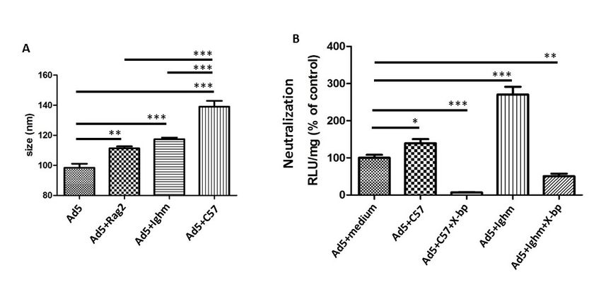
Viruses 2019, 11, 616                                                                               3 of 15

at 37 ◦ C and 5% CO2 . HEK-293 cells (human embryonic kidney: ATCC® CRL-1573 TM) were
grown in MEM (Invitrogen, USA) with 2 mM L-glutamine, 10% FBS, 1 mM sodium pyruvate and
1% penicillin-streptomycin (Invitrogen, USA) at 37 ◦ C and 5% CO2 . Vectors (HAdV-5 luc and LacZ
[β-galactosidase]; HAdV-5T* luc and LacZ containing the following mutations I421G, T423N, E424S,
L426Y, E451Q in HVR7 and T270P and E271G in HVR5 showing reduced FX-binding capacity [8])
were propagated in HEK-293 cells and purified by CsCl gradient centrifugation [15]. Viral particles
were determined by micro BCA assay (Life Technologies, Camarillo, CA, USA) using the formula 1 mg
protein = 4 × 10⁹ virus particles (vp). Laser-based nanoparticle tracking analysis (NTA) was used to
characterize the size of adenoviral particles from pure preparations with Nanosight NTA v2.3 software
in a NanoSight LM14 (Malvern Panalytical, Malvern, UK). Plaque forming units (pfu)/mL were
calculated by end-point dilution assay [15]. The luc vectors were used for all the in vitro experiments,
whereas LacZ vectors were used for the in vivo analyses.

2.3. Fluorescent Labelling of HAdV Particles
      HAdV-5 and HAdV-5T* were fluorescently labelled using an the Alexa Fluor 532 protein labelling
kit (Invitrogen, Paisley, UK) described in detail in [14].

2.4. Nanoparticle Tracking Analysis (NTA)
      Alexa Fluor 532 labelled HAdV-5 or HAdV-5T* vectors (1 × 10⁹ vp) were incubated at room
temperature for 90 min in PBS (10 µL final volume) with physiologically relevant concentrations of
purified mouse IgM (1000 µg/mL) (Rockland Immunochemicals, Limerick, PA, USA) and mouse IgG
(1000 µg/mL) (Sigma-Aldrich, Gillingham, UK) in the presence or absence of 5 µg/mL human blood
coagulation factor X (FX) (Cambridge Biosciences, Cambridge, UK) or with different naïve murine sera
(C57BL/6, Rag2-/- , Ighmtm1Che ; native or inactivated) in the presence or absence of 40 µg/mL X-bp (25 µL
final volume). Human FX has been shown to be able to be substituted by mouse FX [16]. The mutant
Ighmtm1Che C57BL/6 mice have B cells that do not secrete IgM, but that express membrane-bound
IgM. Fresh serum from C57BL/6, Rag2-/- , or Ighmtm1Che C57BL/6 mice was separated from whole blood
after clotting by centrifugation (10 min, 4 ◦ C, 2000× g). Serum inactivation was carried out by 30 min
56 ◦ C incubation. Samples were then diluted in 1.5 mL PBS and injected into the Nanosight LM14
(Malvern Panalytical, Malvern, UK), where the fluorescent virus particle sizes were tracked along
time. Results are shown as data from a minimum of three separate experiments with approximately
700 completed tracks.

2.5. Serum Neutralization Assay
     Human cells were seeded in 96-well plates (1 × 10⁴ cells/well) and incubated overnight at 37 ◦ C
and 5% CO2 . Fresh serum from naïve C57BL/6 mice, Rag2-/- , or Ighmtm1Che C57BL/6 mice was incubated
with 1 × 10⁹ vp of vector (HAdV-5 luc) in a final volume of 50 µL (30 min at 37 ◦ C). The potential
interactions with mouse FX were inhibited by the addition of 40 µg/mL X-bp. Samples were then
diluted 200-fold in serum-free medium. A total of 1000 vp/cell were added onto cells for 2 h at 37 ◦ C,
then media was replaced with fresh media containing 2% FBS, and after further ~16 h at 37 ◦ C cells
were rinsed with PBS and harvested by freezing and thawing in Reporter Lysis Buffer (Promega, UK)
for both luciferase assay (Promega, Southampton, UK) and protein content measurement (BCA assay)
using a VICTORTM X3 Multilabel Plate Reader (PerkinElmer, Waltham, MA, USA).

2.6. Isolation of Proteins Bound to the Virions
     50 µg (corresponding to 2 × 10¹¹ vp) of each of the adenovirus vectors HAdV-5 and HAdV-5T*
was incubated with 250 µL of C57BL/6 mouse serum during 30 min at 37 ◦ C. After that, virion particles
and proteins bound to the virus were purified by CsCl gradient ultracentrifugation, as previously
described [15]. Briefly, 2 layers of CsCl were placed in an appropriate tube: 4 mL of 1.25 density
CsCl and 4 mL of 1.40 carefully added under the other, in order to create the gradient. The mixture
of virus and sera was placed on top of the CsCl cushion drop-wise. Tubes were then centrifuged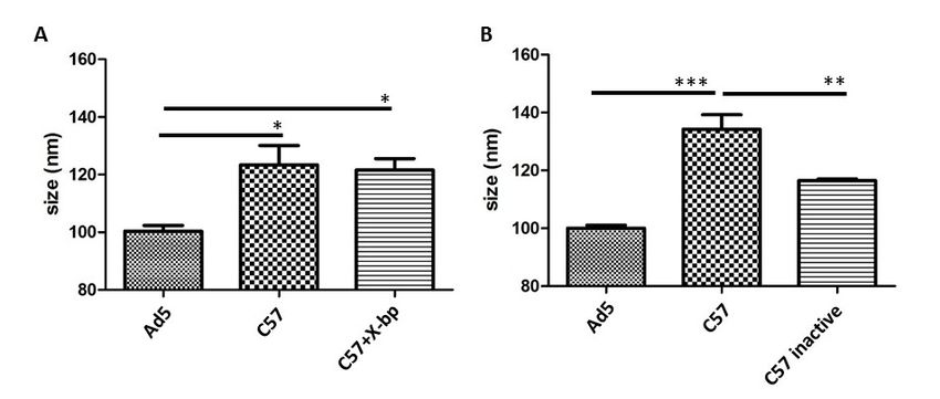
Viruses 2019, 11, 616                                                                            4 of 15

in a SW41 rotor on a Sorvall WX + Ultracentrifuge (Thermofisher, Waltham, MA, USA) for 1.5 h at
room temperature. The white band corresponding to the virions and the proteins bound was evident
after this ultracentrifugation step was carefully collected by piercing the plastic tube with a needle
from the side. As an internal control, a similar tube with only C57BL/6 mouse serum was run in
parallel and a wide amount of liquid at a similar level to that of the virus sample was collected to
rule out contamination of the sample with serum protein complexes co-migrating with the virus.
To remove CsCl, sample buffer was exchanged to 10 mM Tris, 1 mM EDTA pH = 8 buffer (TE 1x) using
Amicon 10 kDa filters (Merck, Darmstadt, Germany). Proteins were additionally concentrated by
precipitation using cold acetone and resuspended in Laemmli sample buffer. Analysis by SDS-PAGE
gel electrophoresis showed no detectable protein in controls, while virus exposed to sera sample
showed a similar pattern to that of the virus alone (Supplementary Figure S1).

2.7. Liquid Chromatography-Mass Spectrometry (LC-MS/MS)
     Five µg of total protein from each sample was loaded into an SDS-PAGE gel and stopped
immediately after the samples entered the gel. The gel was stained with InstantBlue Protein Stain
(Expedeon, Heidelberg, Germany) for 1 h and destained in 10% acetic acid overnight to visualize
the sample bands. Subsequently, each sample was isolated as a unique band and in-gel digestion
with trypsin performed as described elsewhere [17]. Peptide samples were then subjected to liquid
chromatography (LC) in an Ultimate 3000 RSLCnano Systems (Dionex; Thermofisher, Waltham, MA,
USA), being separated on a 50 cm EASY-Spray column (Thermofisher, Waltham, MA, USA) assembled
in an EASY-Spray source (Thermofisher, Waltham, MA, USA) and operated at 50 ◦ C. The LC was
directly coupled to an Orbitrap Fusion Lumos Tribrid mass spectrometer (Thermofisher, Waltham,
MA, USA) for protein identification. Label-free quantification analysis was performed by employing
the MaxLFQ algorithm as described elsewhere [18]. Peptide and protein identifications were filtered
using a 1% FDR. All Label-free quantification (LFQ) MS analyses were performed using two complete
biological replicates of each of the conditions being compared. Amounts of C3, C4b and IgM were
calculated as LFQ of the protein of interest normalised to adenovirus 5 hexon protein’s LFQ.

2.8. In Vivo Experiments
      For in vivo experiments, male C57BL/6 inbred mice and Rag2-/- (B6(Cg)-Rag2tm1.1Cgn /J) knockout
mice aged between 10 to 12 weeks were used. A total of 4 × 10¹² vp/kg of HAdV-5 LacZ or HAdV-5T*
LacZ in 150 µL Dulbecco’s phosphate-buffered saline (DPBS) were administered via the tail vein.
Animals were humanely killed 48 hr post-injection, perfused with DPBS, and internal organs (liver
and spleen) were either collected and fixed in 2% PFA overnight at 4 ◦ C, frozen in liquid nitrogen or
embedded in Optimal Cutting Temperature compound (OCT) in cryomolds (Tissue-Tek, Torrance, CA,
USA) and frozen at −80 ◦ C. DNA extraction was carried out from frozen liver and spleen samples using
the QIAamp DNA mini kit (QIAGEN, Hilden, Germany) according to the manufacturer’s instructions.
Viral genome content was quantified by SYBR green-based qPCR [8]. Protein was extracted from frozen
liver (~25 mg) and spleen (~10 mg). Samples were placed in a 2 mL microcentrifuge tube containing
1 mL of lysis buffer provided in the anti-β-galactosidase enzyme-linked immunosorbent assay (ELISA)
kit (Roche, Welwyn Garden City, UK) and two stainless steel beads (3 mm). Tissues were homogenized
using a TissueLyser II (QIAGEN, Manchester, UK) for 1 min at 25 mHz twice, tissue lysates were
subjected to centrifugation at 24,000× g for 1 min at 4 ◦ C and supernatant was transferred to a clean
1.5 mL microcentrifuge tube for storage at −80 ◦ C. β-galactosidase content was quantified by the
anti-β-galactosidase ELISA kit (Roche, Basel, Switzerland) according to the manufacturer’s instructions
and values were normalized by the total protein concentration in the samples measured by the BCA
Protein assay (Thermofisher, Waltham, MA, USA ). Immunohistochemistry analyses of β-galactosidase
were carried out on 6 µm frozen liver and spleen sections [13]. Rabbit anti-β-galactosidase primary
antibody and Alexa Fluor 488-conjugated goat anti-rabbit IgG secondary antibody were used. Nuclei
were counterstained with DAPI. X-Gal staining was performed in liver and spleen samples previously
fixed in 2% PFA.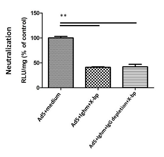
Viruses 2019, 11, x FOR PEER REVIEW 5 of 15
Viruses 2019, 11, 616 5 of 15
2.9. Detection of IgM and IgG
Commercially available ELISA kits (Abcam, Cambridge, UK) were used according to the
2.9.
instructionsofofIgM
Detection theand IgG
manufacturer for the detection of IgM and IgG in murine serum. IgG depletion
wasCommercially available ELISA kitsG(Abcam,
carried out by Dynabeads Protein (Thermofisher, Waltham,
Cambridge, UK)MA, USA).
were used according to the
instructions of the manufacturer for the detection of IgM and IgG in murine serum. IgG depletion was
2.10. Statistical
carried Analysis Protein G (Thermofisher, Waltham, MA, USA).
out by Dynabeads
Statistical significance was calculated using one-way ANOVA followed by Tukey post hoc test
2.10. Statistical Analysis
with GraphPad Prism 4.00 (GraphPad Software, San Diego, CA, USA). p-values of 0.05 or below were
considered
Statisticalstatistically
significancesignificant. Each using
was calculated in vitro result presented
one-way is data from
ANOVA followed a minimum
by Tukey post hocof test
three
separate
with experiments
GraphPad with
Prism 4.00 at least four
(GraphPad experimental
Software, San Diego,replicates per group.
CA, USA). LC-MS
p-values of 0.05experiments
or below werewere
performedstatistically
considered twice to assure reliability
significant. Eachofinthe results
vitro resultobtained.
presentedInisvivo
dataexperiments
from a minimumwere performed
of three
with a minimum
separate experiments of three animals
with at per group
least four (n = 5–6replicates
experimental for HAdVper treated groups,
group. LC-MSn =experiments
3 for DPBS groups).
were
Values are
performed shown
twice as thereliability
to assure mean ± SEM and
of the unpaired
results Student’s
obtained. t-test
In vivo was applied.
experiments were performed with
a minimum of three animals per group (n = 5–6 for HAdV treated groups, n = 3 for DPBS groups).
3. Results
Values are shown as the mean ± SEM and unpaired Student’s t-test was applied.
3. 3.1.
Results
Differential Binding of Different Sources of IgM
Using NTA,
3.1. Differential we of
Binding aimed to first
Different analyse
Sources the potential binding of murine IgM and IgG to HAdV-5.
of IgM
There was no size shift of the HAdV-5 particles observed following incubation of virions with
Using NTA,available
commercially we aimedpurified
to first analyse
murinethe IgMpotential
and IgGbinding
(Figureof murine IgM andincubation
1A). However, IgG to HAdV-5.
with a
There
purified murine IgM (kindly provided by Dr. A.P. Byrnes, Maryland, USA; herein virions
was no size shift of the HAdV-5 particles observed following incubation of with
called “home-
commercially available
purified murine IgM”) purified
did showmurine IgM
a size and
shift, IgG (Figure
suggesting 1A).
that However,
binding incubation
of serum with a purified
components to HAdV-
murine IgM (kindly provided by Dr. A.P. Byrnes, Maryland, USA; herein called “home-purified
5 occurred in a FX-dependent manner (Figure 1B), in similarity to that previously observed for murine
human
IgM”) did show a size shift, suggesting that binding of serum components to HAdV-5
IgM [14]. These surprising results pointed to a potentially different nature of home-purified occurred inand
a
FX-dependent manner (Figure 1B), in similarity to that previously observed for human
commercial murine IgM and suggested the possibility that IgM might not be able to bind HAdV-5 IgM [14]. These
surprising results
under certain pointed to a potentially different nature of home-purified and commercial murine
conditions.
IgM and suggested the possibility that IgM might not be able to bind HAdV-5 under certain conditions.
Figure 1. Mouse IgM does not bind to HAdV-5 in the absence of complement molecules.
(A) Commercially available purified IgM and IgG are not able to bind to HAdV-5. Alexa Fluor
Figure 1. HAdV-5
532-labelled Mouse IgMvectordoes
was not bind to
incubated HAdV-5
at room in the absence
temperature for 90 minof in
complement molecules. (A)
PBS with physiologically
relevant concentrations of purified mouse IgM and mouse IgG. Samples were diluted in PBS andFluor
Commercially available purified IgM and IgG are not able to bind to HAdV-5. Alexa injected532-
labelled HAdV-5 vector was incubated at room temperature for 90 min in PBS
into the Nanosight. Data from a minimum of three separate experiments with ~700 completed tracks are with physiologically
relevant
shown as theconcentrations
mean ± SEM. of purifiedANOVA
One-way mouse andIgMTukey’s
and mousepost IgG. Samples
hoc test applied.were
(B) diluted in PBS and
Non-commercial
injected into the Nanosight. Data from a minimum of three separate
mouse IgM (provided by Dr. A.P. Byrnes; “home-purified murine IgM”) binds to HAdV-5 experiments with ~700 completed
in a
FX-dependent manner, similarly to human IgM [14]. Further investigations showed that these(B)
tracks are shown as the mean ± SEM. One-way ANOVA and Tukey’s post hoc test applied. Non-
IgMs
commercial mouse IgM (provided by Dr. A.P. Byrnes; “home-purified murine
were contaminated by complement components. Alexa Fluor 532-labelled HAdV-5 vector was incubated IgM”) binds to HAdV-
at 5room
in a FX-dependent
temperature for manner,
90 minsimilarly to human
in PBS with IgM [14]. relevant
physiologically Further investigations
concentrationsshowed
of mouse thatIgM
these
in IgMs were contaminated
the presence or absence ofbyhuman
complement
FX, then components.
diluted in PBSAlexa Fluor
and 532-labelled
injected into the HAdV-5
Nanosight vector
LM14. was
Data from a minimum of three separate experiments with ~700 completed tracks are shown as the of
incubated at room temperature for 90 min in PBS with physiologically relevant concentrations
mouse
mean IgMOne-way
± SEM. in the presence
ANOVA or andabsence
Tukey’s ofposthuman FX,
hoc test applied. *** p < 0.001
then diluted in PBS vs.and injected
matched intoorthe
control
IgMNanosight LM14. Data from a minimum of three separate experiments with ~700 completed tracks
conditions.Viruses 2019, 11, x FOR PEER REVIEW 6 of 15
are shown as the mean ± SEM. One-way ANOVA and Tukey’s post hoc test applied. *** p < 0.001 vs.
Virusesmatched control or IgM conditions.
2019, 11, 616 6 of 15
3.2. Differential Binding of Serum Components to HAdV-5 Virions
3.2. Differential Binding of Serum Components to HAdV-5 Virions
We next investigated the binding of proteins present in mouse serum to HAdV-5 virions.
We
Incubationnextof
investigated
HAdV-5 the withbinding of proteins present
immunocompetent in mousemurine
(C57BL/6) serum toserum
HAdV-5 virions.that
showed Incubation
serum
of
components are able to bind to the virion at similar levels in both the presence or absence of theare
HAdV-5 with immunocompetent (C57BL/6) murine serum showed that serum components FX
able to bind
inhibitor to (Figure
X-bp the virion2A,atcompare
similar levels in both
the “C57” the presence
condition to “C57or+ absence of theisFX
X-bp”). X-bp inhibitor
a protein X-bp
isolated
(Figure
from the 2A, compare
venom the “C57” condition
of Trimeresurus to that
flavoviridis + X-bp”).
“C57belongs X-bp
to the is a protein
C-type isolated fromand
lectin superfamily thebinds
venom to
of Trimeresurus flavoviridis that belongs to the C-type lectin superfamily and binds
the GLA domain of FX, inhibiting its interaction with the hexon of HAdV-5 [19]. The fact that X-bp to the GLA domain
of FX, inhibiting
could its interaction
indeed inhibit FX bindingwith thein hexon of HAdV-5 [19].
our experimental The fact
setting wasthatconfirmed
X-bp couldby indeed inhibit
performing
FX binding in our experimental setting was confirmed by performing neutralization
neutralization assays in parallel (Figure 3B, 3rd column). Since Figure 1B showed that binding of assays in parallel
(Figure
murine 3B,IgM3rdto column).
virions was Since Figure
greatly 1B showed
reduced in the that binding
presence of murine
of FX, IgM the
we suggest to virions was
size shift greatly
observed
reduced
in Figure in the
2A presence of FX, we suggestof
in the presence/absence theFX
sizebinding
shift observed in Figure
(+/- X-bp) may 2A be in the presence/absence
predominantly due to
of FX binding (+/– X-bp) may be predominantly due to complement
complement components binding to the virion. Moreover, the size shift observed components binding to the virion.
was complement-
Moreover,
dependent,the size ashift
since observed
significant was complement-dependent,
reduction in particle size was since a significant
observed reduction
for HAdV-5 in particle
incubated with
size was observed for HAdV-5 incubated with heat-inactivated immunocompetent
heat-inactivated immunocompetent murine serum (heat is reported to inactivate the complement murine serum (heat
is reported
[11]), to inactivate
compared the untreated
to that with complement [11]),
serum compared
(Figure to that
2B). These with suggested
results untreated aserum (Figure
potential 2B).
binding
These results
of serum suggested
proteins a potential
to the virions binding
both inoftheserum proteins
presence andto the virions
absence ofboth
FX, in the presence
with and
a substantial
absence of FX, with a substantial
contribution of complement factors. contribution of complement factors.
Figure 2. HAdV-5 binds to murine serum components in a FX-independent manner, but in a
Figure 2. HAdV-5 binds to murine serum components in a FX-independent manner, but in a
complement-dependent way. (A) HAdV-5 binds to immunocompetent murine serum components
complement-dependent way. (A) HAdV-5 binds to immunocompetent murine serum components
regardless of the presence of FX. Alexa Fluor 532-labelled HAdV-5 was incubated at room temperature
regardless of the presence of FX. Alexa Fluor 532-labelled HAdV-5 was incubated at room
for 90 min in murine sera -/+ X-bp. Samples were diluted in PBS and injected into the Nanosight LM14.
temperature for 90 min in murine sera -/+ X-bp. Samples were diluted in PBS and injected into the
Data from a minimum of three separate experiments with ~700 completed tracks are shown as the
Nanosight LM14. Data from a minimum of three separate experiments with ~700 completed tracks
mean ± SEM. One-way ANOVA and Tukey’s post hoc test applied. * p < 0.05 vs. matched control. (B)
are shown as the mean ± SEM. One-way ANOVA and Tukey’s post hoc test applied. * p < 0.05 vs.
Significant particle size drop was observed incubating HAdV-5 in heat-inactivated immunocompetent
matched control. (B) Significant particle size drop was observed incubating HAdV-5 in heat-
murine serum compared to active serum. Alexa Fluor 532-labelled HAdV-5 was incubated at room
inactivated immunocompetent murine serum compared to active serum. Alexa Fluor 532-labelled
temperature for 90 min in murine sera. Serum inactivation was carried out by a 30 min 56 ◦ C incubation.
HAdV-5 was incubated at room temperature for 90 min in murine sera. Serum inactivation was
Samples were diluted in PBS and injected into the Nanosight. Data from a minimum of three separate
carried out by a 30 min 56 °C incubation. Samples were diluted in PBS and injected into the Nanosight.
experiments with ~700 completed tracks are shown as the mean ± SEM. One-way ANOVA and Tukey’s
Data from a minimum of three separate experiments with ~700 completed tracks are shown as the
post hoc test applied. ** p < 0.01, *** p < 0.001 vs. matched control or inactive serum.
mean ± SEM. One-way ANOVA and Tukey’s post hoc test applied. ** p < 0.01, *** p < 0.001 vs. matched
control
To or inactive
further serum. which serum immunoglobulins might bind to HAdV-5, sera from
characterize
immunocompetent and two types of immunoglobulin-deficient mice was utilised. Mouse strains
were To
the further
B and Tcharacterize which
cell deficient Rag2-/-serum
mice in immunoglobulins might bindisotypes
which all immunoglobulin to HAdV-5, sera from
are absent and
immunocompetent
Ighm tm1Che mice, in which B cells cannot secrete IgM [20] but are competent to secrete allstrains
and two types of immunoglobulin-deficient mice was utilised. Mouse other
were the B and Tisotypes.
immunoglobulin cell deficient Rag2-/- mice
Comparison in size
of the which allbetween
shift immunoglobulin isotypes with
virions incubated are absent and
sera from
Ighm tm1Che mice, in which mice
immunoglobulin-deficient B cells cannot
(Rag2 secrete
-/- , Ighm IgM
tm1Che [20] buttoare
C57BL/6) thatcompetent to secrete allmouse
of immunocompetent other
immunoglobulin isotypes. Comparison of the size shift between virions incubated
sera (C57BL/6) showed that serum components from all types of sera were able to bind to HAdV-5 with sera from
immunoglobulin-deficient mice (Rag2 -/-, Ighmtm1Che C57BL/6) to that of immunocompetent mouse sera
virions (Figure 3A, comparing the last three bars with HAdV-5). The binding observed when HAdV-5
(C57BL/6)
was showed
incubated withthat
the serum components from allsera
immunoglobulin-deficient types
mayof sera were
be due to able to bind tobiding
complement HAdV-5 virions
directly to
the viral capsid. This was consistent with the aforementioned results obtained with heat-inactivated
immunocompetent murine serum (Figure 2B, compare “C57 inactive” to “C57”). However, there was aViruses 2019, 11, x FOR PEER REVIEW 7 of 15
(Figure 3A, comparing the last three bars with HAdV-5). The binding observed when HAdV-5 was
incubated with the immunoglobulin-deficient sera may be due to complement biding directly to the
Viruses 2019, 11, 616
viral capsid. 7 of 15
This was consistent with the aforementioned results obtained with heat-inactivated
immunocompetent murine serum (Figure 2B, compare “C57 inactive” to “C57”). However, there was
a significant increase in particle size when HAdV-5 was incubated with the immunocompetent
significant increase in particle size when HAdV-5 was incubated with the immunocompetent C57BL/6
C57BL/6 serum (Figure 3A, bar 4), in comparison to Rag2-/- or Ighmtm1Che C57BL/6 sera (Figure 3A,
serum (Figure 3A, bar 4), in comparison to Rag2-/- or Ighmtm1Che C57BL/6 sera (Figure 3A, bars 2 and 3,
bars 2 and 3, respectively). This size shift may be due to the binding of immunoglobulins, presumably
respectively). This size shift may be due to the binding of immunoglobulins, presumably IgM. Together
IgM. Together the results obtained suggest that complement could bind HAdV-5 in a FX-independent
the results obtained suggest that complement could bind HAdV-5 in a FX-independent manner while
manner while pure IgM alone cannot bind. While binding was FX-independent, virion neutralization
pure IgM alone cannot bind. While binding was FX-independent, virion neutralization was shielded
was shielded by FX. This implied that the mechanism by which FX protected HAdV-5 might not be
by FX. This implied that the mechanism by which FX protected HAdV-5 might not be only relying on
only relying on impeding the binding of immune response proteins to the capsid.
impeding the binding of immune response proteins to the capsid.
Figure 3. Murine IgM is not crucial for neutralization in vitro. (A) HAdV-5 binds to Rag2-/- , Ighmtm1Che
Figure
and wild3. Murine
type (WT) IgM(immunocompetent
is not crucial for neutralization
C57BL/6) serum in vitro. (A) HAdV-5
components. binds
Rag2 to Rag2
-/- serum -/-, Ighmtm1Che
lacks mature T
and
and Bwild type (WT) while
lymphocytes, (immunocompetent
Ighm tm1Che C57BL/6)
lacks serumIgM.
only soluble components.
Alexa Fluor Rag2 -/- serum lacks
532-labelled HAdV-5maturewas T
and B lymphocytes,
incubated while Ighmfor
at room temperature tm1Che lacks only soluble IgM. Alexa Fluor 532-labelled HAdV-5 was
90 min in murine sera. Samples were diluted in PBS and injected
incubated at room temperature
into the Nanosight LM14. Data fromfor 90amin in murine
minimum sera.separate
of three Samplesexperiments
were dilutedwith in PBS
~700 and injected
completed
into theare
tracks Nanosight
shown asLM14.meanData± SEM.from a minimum
One-way ANOVA of three
andseparate
Tukey’sexperiments
post hoc testwith ** p < 0.01,
~700 completed
applied.
*** p < are
tracks 0.001shown as meancontrol
vs. matched ± SEM.orOne-way
WT serum. ANOVA(B) Ighmandtm1Che
Tukey’s postishoc
serum testtoapplied.
able neutralize** pHAdV-5
< 0.01, ***
in
pthe
< 0.001
absencevs.of
matched
FX. Human control orwere
cells WT serum.
seeded (B) Ighmtm1Che
in 96-well serum
plates. Thenis different
able to neutralize
murine sera (Ighmtm1Che
HAdV-5 in the
absence of FX.were
or wild type) Human cells were
incubated withseeded
HAdV-5 in in
96-well plates. or
the presence Then different
absence murine
of X-bp. sera of
A total (Ighm
1000tm1Che or
vp/cell
wild type) were incubated with HAdV-5 in the ◦
presence or absence of X-bp.
were added onto cells, and after further ~16 h at 37 C, cells were harvested in Reporter Lysis Buffer A total of 1000 vp/cell
were added
(Promega, onto
UK) forcells, and after
luciferase assayfurther ~16 h content
and protein at 37 °C,measurement
cells were harvested in Reporter
using a VICTOR TM X3 Lysis Buffer
Multilabel
(Promega,
Plate Reader. UK)Dataforfrom
luciferase
a minimum assay and separate
of four protein experiments
content measurement
are shown asusing meana± VICTOR
SEM. One-way TM X3
Multilabel
ANOVA and Plate Reader.
Tukey’s postData
hoc from a minimum
test applied. * p < 0.05,
of four < 0.01, ***
** pseparate p < 0.001 vs.
experiments arematched
shown as mean ±
controls.
SEM. One-way ANOVA and Tukey’s post hoc test applied. * p < 0.05, ** p < 0.01, *** p < 0.001 vs.
3.3. Murine IgM and IgG are not Necessary for Neutralization In Vitro
matched controls.
Both Ighmtm1Che C57BL/6 and immunocompetent sera enhanced virus transduction (Figure 3B,
3.3.
bars 4 and IgM
Murine and IgG are not
2, respectively) asNecessary
a result for
of Neutralization
mFX shielding In the
Vitrovirions and thus preventing virus
neutralization tm1Che C57BL/6 serum,
Both Ighmand also
tm1Che facilitating
C57BL/6 cell attachment [11].sera
and immunocompetent Incubation
enhanced with Ighm
virus transduction (Figure 3B,
which4 does
bars and not contain IgM, as
2, respectively) notaonly enhanced
result of mFXtransduction
shielding the butvirions
also showed a greater
and thus transduction
preventing virus
than that with immunocompetent sera (Figure 3B, compare bar 4
neutralization and also facilitating cell attachment [11]. Incubation with Ighmto bar 2). Interestingly,
tm1Che when HAdV-5
C57BL/6 serum,
was incubated
which does not with
containX-bp to inhibit
IgM, not onlymFX binding,
enhanced Ighmtm1Chebut
transduction C57BL/6 serumaneutralized
also showed HAdV-5,
greater transduction
similarly
than thattowith
that immunocompetent
observed with immunocompetent
sera (Figure 3B, mouse serumbar
compare (Figure
4 to3B,barbars
2). 5Interestingly,
and 3, respectively)
when
suggesting that murine IgM was not necessary for
HAdV-5 was incubated with X-bp to inhibit mFX binding, Ighm neutralization in vitro.
tm1Che C57BL/6 serum neutralized
Next,similarly
IgG was depleted from Ighm tm1Che C57BL/6 mouse serum, and a serum neutralization assay
HAdV-5, to that observed with immunocompetent mouse serum (Figure 3B, bars 5 and 3,
was performed
respectively) to determine
suggesting whether
that murine IgMIgGwaswas responsible
not necessary forfor neutralization
triggering neutralization
in vitro. (Figure 4).
Depletion of IgG did not inhibit
Next, IgG was depleted from Ighm serum neutralization,
tm1Che C57BL/6 mouse serum, and a serum neutralizationfor
thus showing that IgG was not necessary this
assay
process. The absence of solublewhether
IgM in Ighm tm1Che C57BL/6 serum or IgG inneutralization
IgG-depleted Ighm tm1Che
was performed to determine IgG was responsible for triggering (Figure 4).
C57BL/6 serum was confirmed using ELISA, where the immunoglobulin
Depletion of IgG did not inhibit serum neutralization, thus showing that IgG was not necessary concentration was below
for
the detection limit. Therefore, both IgM
this process. The absence of soluble IgM in Ighm and IgG were found
tm1Che to be unnecessary for the
C57BL/6 serum or IgG in IgG-depletedFX-dependent
neutralisation
Ighm of HAdV-5.
tm1Che C57BL/6 serum was confirmed using ELISA, where the immunoglobulin concentrationViruses 2019, 11, x FOR PEER REVIEW 8 of 15
was below the detection limit. Therefore, both IgM and IgG were found to be unnecessary for the FX-
Viruses 2019, 11, 616 8 of 15
dependent neutralisation of HAdV-5.
Figure 4. IgG depletion does not inhibit serum neutralization. This assay was performed to determine
whether tm1Che serum. Human cells were
Figure 4.IgG
IgGwas responsible
depletion for inhibit
does not triggering neutralization
serum in Ighm
neutralization. This assay was performed to determine
seeded in IgG
96-well tm1Che
whether was plates. Thenfor
responsible Ighm
triggering murine serum in
neutralization (with
Ighm ortm1Che
without depletion
serum. Humanofcells
IgGwere
by
Dynabeads Protein G) was incubated with HAdV-5 for 30 min at 37 ◦ C. A total of 1000 vp/cell were
seeded in 96-well plates. Then Ighmtm1Che murine serum (with or without depletion of IgG by
added onto cells, andG)after further ~16 hwith ◦ C cells were harvested for luciferase assay and protein
at 37HAdV-5
Dynabeads Protein was incubated for 30 min at 37 °C. A total of 1000 vp/cell were
content measurement TM
added onto cells, andusing a VICTOR
after further ~16 h atX337Multilabel Plate
°C cells were Reader. for
harvested Data from a minimum
luciferase assay andof four
protein
separate experiments are shown as mean ± SEM. One-way ANOVA and Tukey’s
content measurement using a VICTOR X3 Multilabel Plate Reader. Data from a minimum of four
TM post hoc test applied.
p < 0.01 experiments
**separate vs. matched arecontrols.
shown as mean ± SEM. One-way ANOVA and Tukey’s post hoc test applied.
** p C3
3.4. IgM, < 0.01
andvs.C4b
matched
Bind tocontrols.
HAdV-5 in the Presence of FX
3.4. The
IgM,NTA results
C3 and showed
C4b Bind that commercially
to HAdV-5 available
in the Presence of FX human IgM and non-commercial mouse
IgM produced a shift in the HAdV-5 size, while commercially available murine IgM failed to do so
The NTA results showed that commercially available human IgM and non-commercial mouse
(Figure 1). Next the purity of the different immunoglobulins were therefore analysed by LC-MS.
IgM produced a shift in the HAdV-5 size, while commercially available murine IgM failed to do so
These data demonstrated that human (Sigma, USA) and non-commercial mouse IgM were, among
(Figure 1). Next the purity of the different immunoglobulins were therefore analysed by LC-MS.
other proteins, contaminated with C3, C4b and C4b-binding protein, while the commercially available
These data demonstrated that human (Sigma, USA) and non-commercial mouse IgM were, among
murine IgM (Rockland Immunochemicals, USA) was free of these or contained substantially less
other proteins, contaminated with C3, C4b and C4b-binding protein, while the commercially
(Supplementary Table S1). These observations suggest that IgM was only able to bind HAdV-5 when
available murine IgM (Rockland Immunochemicals, USA) was free of these or contained
complement proteins were present. Next, LC-MS was used to assess which serum components bound
substantially less (Supplementary Table S1). These observations suggest that IgM was only able to
to adenovirus vectors (Supplementary Table S2). It was found that complement components (C3, C4b)
bind HAdV-5 when complement proteins were present. Next, LC-MS was used to assess which
and IgM bound to HAdV-5 (Figure 5), confirming the NTA data (Figure 2). When HAdV-5T* (a HAdV-5
serum components bound to adenovirus vectors (Supplementary Table S2). It was found that
vector which is unable to bind to either murine or human FX [8]) was tested, the amounts of bound C3
complement components (C3, C4b) and IgM bound to HAdV-5 (Figure 5), confirming the NTA data
and C4b detected were higher compared to those bound to HAdV-5 (fold changes: C3 = 40.2 ± 20.3;
(Figure 2). When HAdV-5T* (a HAdV-5 vector which is unable to bind to either murine or human FX
C4 = 87.8 ± 10.6), whereas IgM binding to HAdV-5T* was not different to that observed for HAdV-5
[8]) was tested, the amounts of bound C3 and C4b detected were higher compared to those bound to
(Figure 5). These results provide evidence for the binding of IgM, C3 and C4b to HAdV-5 even in the
HAdV-5 (fold changes: C3 = 40.2 ± 20.3; C4 = 87.8 ± 10.6), whereas IgM binding to HAdV-5T* was not
presence of FX. Moreover, the binding of C3 and C4b was higher for HAdV-5T*, a mutant vector that
different to that observed for HAdV-5 (Figure 5). These results provide evidence for the binding of
cannot bind FX.
IgM, C3 and C4b to HAdV-5 even in the presence of FX. Moreover, the binding of C3 and C4b was
higher for HAdV-5T*, a mutant vector that cannot bind FX.Viruses 2019, 11, x FOR PEER REVIEW 9 of 15
Viruses 2019, 11, 616 9 of 15
Figure 5. Binding of complement proteins C3 and C4b is substantially higher on HAdV-5T* virions. A
total of5.50Binding
Figure µg (corresponding
of complement × 1011 vp)
to 2 proteins C3ofand
HAdV-5
C4b isorsubstantially
HAdV-5T* were incubated
higher with 250
on HAdV-5T* µL of
virions.
◦
A total of 50 µg (corresponding to 2 × 10¹¹ vp) of HAdV-5 or HAdV-5T* were incubated with 250were
C57BL/6 mouse sera at 37 C for 30 min. Next, virion particles and proteins bound to the virus µL
purified
of C57BL/6 bymouse
CsCl gradient. Thefor
sera at 37 °C mixture
30 min.ofNext,
viralvirion
and serum proteins
particles was loaded
and proteins boundinto an virus
to the SDS-PAGE
were
gel and by
purified subjected to electrophoresis
CsCl gradient. The mixture until immediately
of viral and serumafter the samples
proteins entered
was loaded intothe gel. Following
an SDS-PAGE gel
in-gel tryptic digestion, peptide samples were analysed by LC-MS/MS in an Orbitrap Fusion Lumos
and subjected to electrophoresis until immediately after the samples entered the gel. Following in-gel
Tribrid mass spectrometer (Thermo Fisher Scientific) for protein identification. Label-free quantification
tryptic digestion, peptide samples were analysed by LC-MS/MS in an Orbitrap Fusion Lumos Tribrid
analysis were performed by employing the MaxLFQ algorithm. Peptide and protein identifications were
mass spectrometer (Thermo Fisher Scientific) for protein identification. Label-free quantification
filtered using a 1% FDR. All LFQ MS analyses were performed using two complete biological replicates
analysis were performed by employing the MaxLFQ algorithm. Peptide and protein identifications
of each of the conditions being compared. Amounts of C3, C4b and IgM were calculated as LFQ of the
were filtered using a 1% FDR. All LFQ MS analyses were performed using two complete biological
protein of interest normalised to adenovirus 5 hexon protein’s LFQ. LFQ: label-free quantification.
replicates of each of the conditions being compared. Amounts of C3, C4b and IgM were calculated as
LFQ of the Isprotein
3.5. HAdV-5T* of Neutralized
Partially interest normalised to adenovirus 5 hexon
in Immunoglobulin-Deficient protein’s LFQ. LFQ: label-free
Mice
quantification.
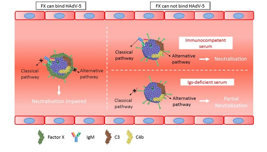
Since IgM and IgG were not crucial for neutralization in vitro, next in vivo experiments were
conducted
3.5. HAdV-5T* to further analyse
is Partially these findings.
Neutralized C57BL/6 or Rag2-/- mice
in Immunoglobulin-Deficient Micewere administered HAdV-5 LacZ
or HAdV-5T* LacZ vectors. In C57BL/6 mice, both transduction levels and the number of genomes
SinceinIgM
detected the and
liverIgG
andwere notfollowing
spleen crucial for neutralization
HAdV-5T* in vitro, next
administration in significantly
were vivo experiments
lowerwere
than
conducted to further analyse these findings. C57BL/6 or Rag2 -/- mice were administered HAdV-5 LacZ
those detected for HAdV-5 (Figure 6A,C). Immunohistochemical analysis of β-galactosidase on liver
or
andHAdV-5T* LacZ vectors.
spleen sections confirmedIn these
C57BL/6 mice,(Figure
findings both transduction
7A,C). Theselevels
data and the number
suggested of genomes
that either the lack
detected in the liver and spleen following HAdV-5T* administration were
of HAdV-5T* binding to FX, and/or shielding of the capsid from complement by FX, leads significantly lower than
to virus
those detected for HAdV-5 (Figure 6A,C). Immunohistochemical analysis of β-galactosidase
neutralization. Interestingly, a similar pattern (reduced transduction levels and number of genomes
on liver
and spleen sections confirmed these findings (Figure 7A,C). These data suggested that either the lack
mediated by HAdV-5T* compared to HAdV-5) was observed in Rag2-/- mouse livers (Figure 6B,D and
of HAdV-5T* binding to FX, and/or shielding of the capsid from complement by FX, leads to virus
Figure 7B,D), indicating that partial neutralization of HAdV-5T* still occurred, even in the absence
neutralization. Interestingly, a similar pattern (reduced transduction levels and number of genomes
of mature B and T cells. There was no significant difference in the transduction levels or number of
mediated by HAdV-5T* compared to HAdV-5) was observed in Rag2-/- mouse livers (Figure 6B,D and
genomes detected from each vector in the spleen of Rag2-/- mice (Figure 6B,D and Figure 7B,D). The
Figure 7B,D), indicating that partial neutralization of HAdV-5T* still occurred, even in the absence of
differential splenic tropism for FX-binding-ablated HAdV-5 that has been formerly reported [13] might
mature B and T cells. There was no significant difference in-/-the transduction levels or number of
have attenuated the effect of the partial neutralization in Rag2 mice spleens. These results altogether
genomes detected from each vector in the spleen of Rag2-/- mice (Figure 6B,D and Figure 7B,D). The
suggest that immunoglobulins are not required for partial neutralization of HAdV-5 in vivo.
differential splenic tropism for FX-binding-ablated HAdV-5 that has been formerly reported [13]
might have attenuated the effect of the partial neutralization in Rag2-/- mice spleens. These results
altogether suggest that immunoglobulins are not required for partial neutralization of HAdV-5 in
vivo.Viruses 2019, 11, x FOR PEER REVIEW 10 of 15
Viruses 2019, 11, 616 10 of 15
Viruses 2019, 11, x FOR PEER REVIEW 10 of 15
Figure 6. Accumulation of adenoviral genomes and transduction levels in liver and spleen. In
C57BL/6 mice, the transduction levels and number of genomes detected in the liver and spleen
Figure
Figure 6.6.Accumulation
following Accumulation of of
adenoviral
HAdV-5T* administration
adenoviral genomes
were and transduction
significantly
genomes levels
lower than
and transduction in liver
those
levels inand
of HAdV-5. spleen.
liver In C57BL/6
Interestingly,
and spleen. Ina
mice, the
similar transduction
pattern was levels
observed and
in Rag2number of genomes detected in the liver and spleen
-/- mouse livers, indicating that neutralization of HAdV-5T* still following
C57BL/6 mice, the transduction levels and number of genomes detected in the liver and spleen
HAdV-5T* administration
occurred, HAdV-5T*
even wereofsignificantly
in theadministration
absence mature lower than those of(AHAdV-5. Interestingly, anda D)
similar
following wereBsignificantly
and T cells. C57BL/6
lower than and C)
those of or Rag2-/- (B
HAdV-5. were
Interestingly, a
pattern was observed in Rag2-/- mouse-/-livers, indicating that neutralization of HAdV-5T* still occurred,
administered
similar pattern 1was × 10 11 vp ofinHAdV-5
observed Rag2 mouse LacZlivers,
or HAdV-5T*
indicatingLacZ by intravascular
that neutralization delivery still
of HAdV-5T* and
even in the absence of mature B and T cells. C57BL/6 (A and C) or Rag2-/- (B and D) were administered
euthanized
occurred, andinperfused
even the absencewithofDPBS
mature 48Bhand post-injection. Viral (A
T cells. C57BL/6 genome
and C)content
or Rag2was -/- (B quantified
and D) wereby
1 × 10¹¹ vp of HAdV-511LacZ or HAdV-5T* LacZ by intravascular delivery and euthanized and perfused
SYBR green-based QPCR analysis in liver (grey bars) and spleen
administered 1 × 10 vp of HAdV-5 LacZ or HAdV-5T* LacZ by intravascular delivery and(black bars) (A–B). β-galactosidase
with DPBS 48 h post-injection. Viral genome content was quantified by SYBR green-based QPCR
expression was
euthanized and quantified
perfused withby ELISA
DPBSin48 liver and spleen and
h post-injection. normalised
Viral genometocontenttotal mg wasof protein (C–D).
quantified by
analysis in liver (grey bars) and spleen (black bars) (A–B). β-galactosidase expression was quantified by
n = 5–6.
SYBR Values areQPCR
green-based expressed as the
analysis mean
in liver ± SEM.
(grey bars)Unpaired
and spleenStudent’s t-test(A–B).
(black bars) applied. * p < 0.05 vs.
β-galactosidase
ELISA in liver and spleen and normalised to total mg of protein (C–D). n = 5–6. Values are expressed
HAdV-5, $was
expression p < 0.01 vs. HAdV-5.
quantified by ELISA in liver and spleen and normalised to total mg of protein (C–D).
as the mean ± SEM. Unpaired Student’s t-test applied. * p < 0.05 vs. HAdV-5, $ p < 0.01 vs. HAdV-5.
n = 5–6. Values are expressed as the mean ± SEM. Unpaired Student’s t-test applied. * p < 0.05 vs.
HAdV-5, $ p < 0.01 vs. HAdV-5.
Figure 7. β-galactosidase expression in mouse liver and spleen. C57BL/6 (A and C) or Rag2-/--/- (B and
Figure 7. β-galactosidase expression in mouse liver and spleen. C57BL/6 (A and C) or Rag2 (B and
D) mice were administered 1 × 101111vp of HAdV-5 LacZ, HAdV-5T* LacZ or DPBS by intravascular
D) mice were administered 1 × 10 vp of HAdV-5 LacZ, HAdV-5T* LacZ or DPBS by intravascular
delivery and euthanized and perfused with DPBS 48 h post-injection. (A and B) Representative-/- images
delivery
Figure and euthanizedexpression
7. β-galactosidase and perfused with liver
in mouse DPBSand 48 spleen.
h post-injection.
C57BL/6 (A (Aand
andC)B)orRepresentative
Rag2 (B and
of X-Gal staining of liver and spleen11fixed in 2% paraformaldehyde are shown. Scale bars 1 cm. (upper
images of X-Gal staining of liver and spleen fixed in 2% paraformaldehyde are
D) mice were administered 1 × 10 vp of HAdV-5 LacZ, HAdV-5T* LacZ or DPBS by intravascular shown. Scale bars 1
panels) or 0.25 cm (lower panels). (C) and (D) Immunohistochemistry analysis of β-galactosidase on
cm. (upper
delivery andpanels) or 0.25
euthanized andcmperfused
(lower panels).
with DPBS(C) and
48 h (D) Immunohistochemistry
post-injection. analysis of β-
(A and B) Representative
6 µm frozen liver and spleen sections. Rabbit anti-β-galactosidase primary antibody and Alexa Fluor
galactosidase
images on staining
of X-Gal 6 µm frozen liverand
of liver andspleen
spleenfixed
sections.
in 2% Rabbit anti-β-galactosidase
paraformaldehyde primary
are shown. antibody
Scale bars 1
488-conjugated goat anti-rabbit IgG secondary antibody (green) were used. Nuclei were counterstained
and (upper
cm. Alexa Fluor 488-conjugated
panels) goat anti-rabbit
or 0.25 cm (lower panels). (C)IgGandsecondary antibody (green) wereanalysis
(D) Immunohistochemistry used. Nuclei
of β-
with DAPI (blue). n = 3 mice analysed per group with 1 technical replicate. Representative merged
were counterstained with DAPI (blue). n = 3 mice analysed per group with 1 technical
galactosidase on 6 µm frozen liver and spleen sections. Rabbit anti-β-galactosidase primary antibody replicate.
images are shown. Magnification 40×. Scale bars 50 µm.
Representative
and Alexa Fluormerged images are
488-conjugated goatshown. Magnification
anti-rabbit 40×. Scale
IgG secondary bars 50(green)
antibody µm. were used. Nuclei
were counterstained with DAPI (blue). n = 3 mice analysed per group with 1 technical replicate.
Representative merged images are shown. Magnification 40×. Scale bars 50 µm.Viruses 2019, 11, 616 11 of 15
Viruses 2019, 11, x FOR PEER REVIEW 11 of 15
In summary, the data generated suggest that (1) immune response proteins can bind HAdV-5
In summary, the data generated suggest that (1) immune response proteins can bind HAdV-5
virions in naïve mice but neutralization only occurs if FX is absent or the virion is unable to bind FX,
virions in naïve mice but neutralization only occurs if FX is absent or the virion is unable to bind FX,
(2) purified mouse IgM antibodies bind to HAdV-5 only in the presence of complement components
(2) purified mouse IgM antibodies bind to HAdV-5 only in the presence of complement components
and (3) in serum from immunocompetent mice both classical and alternative complement pathways are
and (3) in serum from immunocompetent mice both classical and alternative complement pathways
activated, while in serum from immuno-deficient mice with absent immunoglobulins, the alternative
are activated, while in serum from immuno-deficient mice with absent immunoglobulins, the
complement pathway mediates partial neutralization (Figure 8).
alternative complement pathway mediates partial neutralization (Figure 8).
Figure 8. Schematic model of the interactions of HAdV-5 with the host immune system. Neutralization
of HAdV-5
Figure in naïve mice
8. Schematic occursofif FX
model thebinding to the virions
interactions is ablated.
of HAdV-5 In immunocompetent
with the host immunemice both
system.
classical and alternative complement pathways are activated, while in immunoglobulin-deficient
Neutralization of HAdV-5 in naïve mice occurs if FX binding to the virions is ablated. In mice
partial neutralization
immunocompetent is caused
mice by the alternative
both classical pathway.
and alternative complement pathways are activated, while in
immunoglobulin-deficient mice partial neutralization is caused by the alternative pathway.
4. Discussion
In this study, we demonstrated that murine IgM is not pivotal for either in vitro or in vivo HAdV-5
4. Discussion
neutralization. It was observed that IgM alone does not bind to HAdV-5, however IgM does bind in
In this study, we demonstrated that murine IgM is not pivotal for either in vitro or in vivo
the presence of specific complement proteins. Interestingly, these binding events were observed even
HAdV-5 neutralization. It was observed that IgM alone does not bind to HAdV-5, however IgM does
in the presence of FX. The in vitro and in vivo data suggest that serum proteins other than IgM or
bind in the presence of specific complement proteins. Interestingly, these binding events were
IgG might also trigger the immune response, implying the importance of the alternative complement
observed even in the presence of FX. The in vitro and in vivo data suggest that serum proteins other
pathway in HAdV-5 neutralisation.
than IgM or IgG might also trigger the immune response, implying the importance of the alternative
It has been previously reported that HAdV-5 specifically binds to murine and human FX,
complement pathway in HAdV-5 neutralisation.
and that the capsid:FX interaction inhibits the neutralization mediated by IgM and complement
It has been previously reported that HAdV-5 specifically binds to murine and human FX, and
components [11,21]. A previous study based on particle size tracking demonstrated that purified
that the capsid:FX interaction inhibits the neutralization mediated by IgM and complement
human IgM and IgG were able to bind to HAdV-5 virions, with IgM only binding in a FX-dependent
components [11,21]. A previous study based on particle size tracking demonstrated that purified
manner [14]. Given the pivotal role for murine IgM in virus neutralization both in vivo and in vitro [11],
human IgM and IgG were able to bind to HAdV-5 virions, with IgM only binding in a FX-dependent
it was expected that murine IgM would behave in a similar fashion to human IgM. However, the results
manner [14]. Given the pivotal role for murine IgM in virus neutralization both in vivo and in vitro
here revealed that while non-commercial IgM was able to bind to HAdV-5 virions, commercially purified
[11], it was expected that murine IgM would behave in a similar fashion to human IgM. However,
murine IgM showed no binding (Figure 1). Interestingly, LC-MS analysis showed that non-commercial
the results here revealed that while non-commercial IgM was able to bind to HAdV-5 virions,
IgM preparations contained C3, C4b and C4b-binding protein (Supplementary Table S1). Similarly,
commercially purified murine IgM showed no binding (Figure 1). Interestingly, LC-MS analysis
commercially available human IgM, which we have previously proved to be able to bind HAdV-5 [14],
showed that non-commercial IgM preparations contained C3, C4b and C4b-binding protein
was also analysed by LC-MS and found to be contaminated with C3, C4b and C4b-binding protein.
(Supplementary Table S1). Similarly, commercially available human IgM, which we have previously
These findings suggest that either murine IgM is not able to bind to HAdV-5 virions in the absence of
proved to be able to bind HAdV-5 [14], was also analysed by LC-MS and found to be contaminated
complement molecules or that it is actually the complement proteins that bind to the virions. Since
with C3, C4b and C4b-binding protein. These findings suggest that either murine IgM is not able to
C3 and C4b can covalently bind to pathogens to trigger an immune response [22], it might also be
bind to HAdV-5 virions in the absence of complement molecules or that it is actually the complement
possible that C4b binds to C3, forming a complex that protects C3 from inactivation. Further studies
proteins that bind to the virions. Since C3 and C4b can covalently bind to pathogens to trigger an
immune response [22], it might also be possible that C4b binds to C3, forming a complex that protects
C3 from inactivation. Further studies with completely purified human IgM would clarify whetherYou can also read