A RAGE-Targeted Antibody-Drug Conjugate: Surface Plasmon Resonance as a Platform for Accelerating Effective ADC Design and Development - MDPI
←
→
Page content transcription
If your browser does not render page correctly, please read the page content below
antibodies
Article
A RAGE-Targeted Antibody-Drug Conjugate: Surface
Plasmon Resonance as a Platform for Accelerating
Effective ADC Design and Development
Gareth D. Healey 1, * , Asa Frostell 2 , Tim Fagge 3 , Deyarina Gonzalez 1 and R. Steven Conlan 1
 1    Institute of Life Science, Swansea University Medical School, Swansea University, Swansea, SA2 8PP, UK;
      d.gonzalez@swansea.ac.uk (D.G.); r.s.conlan@swansea.ac.uk (R.S.C.)
 2    GE Healthcare Bio-Sciences, SE-751 84 Uppsala, Sweden; asafrostell@gmail.com
 3    GE Healthcare, Little Chalfont, Buckinghamshire, HP7 9NA, UK; tim.fagge@ge.com
 *    Correspondence: g.d.healey@swansea.ac.uk; Tel.: +44-1792-606474

 Received: 25 November 2018; Accepted: 21 December 2018; Published: 7 January 2019

 Abstract: Antibodies, antibody-like molecules, and therapeutics incorporating antibodies as a
 targeting moiety, such as antibody-drug conjugates, offer significant potential for the development of
 highly efficacious drugs against a wide range of disorders. Despite some success, truly harnessing
 the superior targeting properties of these molecules requires a platform from which to effectively
 identify the best candidates for drug development. To streamline the development of antibody-drug
 conjugates targeting gynecological cancers within our laboratory, we incorporated surface plasmon
 resonance analysis (Biacore™ T200) into our development toolkit. Antibodies, selected based on
 positive ELISA screens as suitable for development as antibody-drug conjugates, were evaluated
 using surface plasmon resonance to determine a wide range of characteristics including specificity,
 kinetics/affinity, the effect of linker binding, the impact of the drug to antibody ratio, and the effect
 of endosomal pH on antibody-antigen binding. Analysis revealed important kinetics data and
 information regarding the effect of conjugation and endosomal pH on our antibody candidates that
 correlated with cell toxicity and antibody internalization data. As well as explaining observations
 from cell-based assays regarding antibody-drug conjugate efficacies, these data also provide important
 information regarding intelligent antibody selection and antibody-drug conjugate design. This study
 demonstrates the application of surface plasmon resonance technology as a platform, where detailed
 information can be obtained, supporting the requirements for rapid and high-throughput screening
 that will enable enhanced antibody-drug conjugate development.

 Keywords: surface plasmon resonance; antibody-drug conjugates; antibodies; gynecological cancers;
 binding kinetics

1. Introduction
     Antibodies and antibody-like molecules offer the potential to develop highly efficacious drugs
against a wide range of disorders from cancers to autoimmune diseases to rheumatic and cardiovascular
disease. Although the beginnings of this potential have been glimpsed, truly harnessing the superior
targeting properties of these molecules requires a platform from which to effectively identify the best
candidates for drug development.
     The idea of an immunotherapeutic treatment strategy for cancer emerged in the 1920s focused
around the treatment of Hodgkin’s Lymphoma with lymph node extract. However, it is only
more recently that immunotherapies have become an established treatment modality, leading to
the development of several novel therapeutics for hematological cancers and solid tumors [1]. Over the
past 20 years, antibody-based therapies have seen particular success with nearly 20 antibodies gaining

Antibodies 2019, 8, 7; doi:10.3390/antib8010007                               www.mdpi.com/journal/antibodies
Antibodies 2019, 8, 7                                                                              2 of 15

US Food and Drug Administration (FDA) approval for use in oncologic care since 1997 [1]. The more
recent emergence of chimeric, humanized, and human monoclonal antibodies, has led to a rapid
increase in antibody-based therapeutics, which, with 75 Billion USD global sales in 2013, are now the
dominant class of molecules within the global biopharmaceutical market [2].
      However, antibodies by themselves can, depending on their mechanism of action, display
low therapeutic efficacy, meaning alternative approaches are required to increase the potency of
antibody-based therapeutics. To address such limitations, antibody-drug conjugates (ADCs) have
emerged as a promising therapeutic approach, which combine the selectivity of a targeted treatment
with the cytotoxic potency of chemotherapy agents.
      The first ADC gemtuzumab ocogamicin (Mylotarg® ) gained clinical approval in 2000 [3], paving
the way for three further ADCs, brentuximab vedotin (Adectris® ), ado-trastuzumab emtansine
(Kadcyla® ), and Inotuzumab ozogamicin (Besponsa® ), which were licensed for the treatment of
Hodgkin’s and anaplastic large-cell lymphomas, HER-2 positive breast cancer, and relapsed or
refractory B-cell precursor acute lymphoblastic leukemia, respectively [4–6]. The need to develop
efficacious, novel antibody-based therapies means that over 50 different ADCs are currently in
preclinical or clinical development [7,8].
      In such a competitive marketplace, there is an increasing focus on the potential developability
of early-stage molecules to prevent costly late-stage failures. This responsibility falls on analytical
techniques, which are used to study structural and functional properties including affinity, kinetics,
potency, aggregation, solubility, stability, immunogenicity, and pharmacokinetics, as well as cell-based
assays to study toxicity and off-target effects.
      One such technology, rapidly adopted to study antibody-antigen interactions following its
introduction in 1990, is surface plasmon resonance (SPR) [9]. First applied to the study of antibody-
antigen interactions and epitope mapping [10], SPR has several advantages over traditional immunoassays
such as enzyme-linked immunosorbent assay (ELISA) or radioimmune assay (RIA). It is a label-free
technique that monitors the formation and dissociation of biomolecular complexes in real-time,
allowing binding kinetics and affinities to be measured. It is also sensitive, requires small sample
volumes, works well with crude samples, and has the automation and throughput capability required
to support high throughput screening and characterization studies [11–13].
      Current uses for SPR technology include early-stage screening of hybridoma/phage libraries
to monitor expression and triage antibodies based on binding affinity, profiling binding specificity,
and providing a detailed understanding of binding kinetics and affinity to characterize antibody-
antigen interactions. During therapeutic antibody development, SPR is part of a suite of analytical
methods used to study stability, drug-target binding interactions, and binding to Fc receptors,
complement and the neonatal receptor (FcRn) to assess the critical quality attributes that determine the
efficacy and clinical safety of the final product. As a core technology in analytical and Quality Control
(QC) labs, SPR is also used to monitor batch-to-batch variation, support Chemistry, Manufacturing,
and Controls (CMC), and as a potency assay to support clinical batch and final product release.
      Given the prevalent use of SPR in the selection and development of standard antibody
biotherapeutics, together with its increasing use in many aspects of ADC development, including
target selection, antibody kinetics characterization, epitope mapping, and optimization [14,15],
we explored how the technology could be used within our lab for the selection and characterization of
next-generation ADCs. The importance of the antibody component of an ADC to therapeutic efficacy
means that careful consideration must be given to the selection of antibodies for this purpose. Previous
work within our laboratory demonstrated variability in the efficacy of antibodies characterized using
standard immunoassay techniques such as ELISA, which led us to investigate ADC characteristics
using SPR (unpublished).
      Aiming to streamline the design and development of ADCs, we study multiple aspects of effective
ADC design, each assessed by SPR. Biacore™ technology is employed to characterize four ADCs
that target the Receptor for Advanced Glycation End Products (RAGE), a multi-ligand signaling
system that drives innate immune inflammatory responses via nuclear factor-kappa beta (NF-kB)
Antibodies 2019, 8, 7                                                                                3 of 15

mediated gene activation and is associated with gynecological disease [16]. We demonstrate that a
wide range of antibody characteristics can be evaluated including specificity, kinetics/affinity, the effect
of linker binding, the impact of drug to antibody ratio (DAR), and the effect of endosomal pH on
antibody-antigen binding. In doing so, this study demonstrates the application of SPR Biacore™
technology as a platform, where detailed information can be obtained, supporting the requirements
for rapid and high-throughput screening that will enable enhanced ADC development.

2. Materials and Methods

2.1. Antibody Production
      Monoclonal antibody production was performed by Bio-Rad Antibodies (formerly AbD Serotec,
Bio-Rad Laboratories, Oxford, UK). All procedures were performed in accordance with the Animals
(Scientific Procedures) Act 1986, and the guidance issued in ‘Responsibility in the case of Animals
in Bioscience research: expectations of the major research council and charitable funding bodies.’
Monoclonal antibodies against RAGE were produced using standard protocols for monoclonal
antibody production [17,18]. Briefly, BALB/c mice, obtained from Charles River, Oxford, UK, were
immunized with keyhole limpet hemocyanin (KLH)-conjugated RAGE, or KLH-conjugated peptides
corresponding to amino acids (aa) 198–217 or 327–344 of the RAGE protein. Clones were selected
based on a positive ELISA screen using bovine serum albumin (BSA)-conjugated peptides. Post-fusion,
individual clones were then selected by limiting dilution and clonal expansion to identify genetically
stable, antibody-producing cells for subsequent antibody production. One clone with an affinity for the
full-length rRAGE protein (RBGO1), two clones with an affinity for aa198–217 (RBGO2 and RBGO3),
and one with an affinity for aa327–344 (RBGO4) were selected for antibody production. Antibodies
were purified from the tissue culture medium using protein G affinity purification.

2.2. Antibody-Drug Conjugation
      Murine antibodies against RAGE were reconstituted in 10 mM Tris/HCl (Sigma, Dorset, UK)
and 2 mM EDTA (Sigma) pH 8.0. Antibodies were reduced with 3.5 equivalents TCEP:Ab (10 mM
in water, Sigma) for 2 h at 37 ◦ C. Without purification the reduced antibody was split in two equal-
volume aliquots and each aliquot alkylated with 6.5 equivalents of drug linker: Ab (10 mM MC-ValCit-
PAB-MMAE or MC-MMAF, see Figure S1, in DMA with additional DMA added to achieve 5%
v/v final DMA, ADC Biotechnology, St Asaf, UK) for 2 h at 22 ◦ C. Following alkylation, N-acetyl
cysteine (Sigma) was used to quench any unreacted toxin linker. The conjugates were purified using
a HiTrap™ G25 column (GE Healthcare, Uppsala, Sweden) equilibrated in 5 mM histidine/HCl,
50 mM trehalose (Sigma), 0.01% w/v polysorbate 20 (Sigma), pH 6.0. Conjugates were analysed by
size exclusion chromatography (SEC) for monomeric content and concentration using a calibration
curve of naked antibody. Running conditions: Agilent 1100 High Pressure Liquid Chromatography
(HPLC), Tosoh TSKgel®G3000SWXL 7.8 mm × 30 cm, 5 µm column (Tosoh Bioscience, Reading, UK),
0.5 mL/min in, 0.2 M Potassium Phosphate, 0.25 M Potassium Chloride, 10% isopropyl alcohol (IPA),
pH 6.95. Drug loading of the conjugates was confirmed using a combination of hydrophobic interaction
chromatography (HIC) and reverse phase chromatography. HIC was carried out using a TOSOH
Butyl-NPR 4.6 mm × 3.5 cm, 2.5 µm column (Tosoh Bioscience) run at 0.8 mL/min with a 12 min linear
gradient between A—1.5 M (NH4)2SO4, 25 mM NaPi, pH 6.95±0.05 and B—75% 25 mM NaPi, pH
6.95 ± 0.05, 25% IPA. Reverse phase analysis was performed on a Polymer Lab’s polymeric reversed
phase (PLRP) 2.1 mm × 5 cm, 5 µm column (Tosoh Bioscience) run at 1 mL/min at 80 ◦ C with a 25 min
linear gradient between 0.05% trifluoracetic acid (TFA)/H2 O and 0.04% TFA/CH3 CN. Samples were
first reduced by incubation with 1, 4-Dithiothritol (DTT, Sigma) at pH 8.0 at 37 ◦ C for 15 min.

2.3. Surface Plasmon Resonance
   SPR reagents used were Series S Sensor Chip CM5, HBS-EP+ buffer (10 mM Hepes, 150 mM
NaCl, 3 mM ethylenediaminetetraacetic acid, and 0.05% Surfactant P20, pH 7.4), Amine Coupling Kit,
Antibodies 2019, 8, 7                                                                              4 of 15

Mouse Antibody Capture Kit, including 10 mM glycine-HCl pH 1.7 regeneration solution (all from GE
Healthcare).
      SPR analysis was performed using a Biacore™ T200 system (GE Healthcare) and HBS-EP+ buffer
was used as sample and running buffer. The analysis temperature and sample compartment were
set to 25 ◦ C. Immobilization of α-mouse antibody was performed using the Amine Coupling Kit in
accordance with the manufacturer’s instructions. The anti-mouse antibody was immobilized in all
flow cells, but flow cells 1 and 3 were used as reference cells for antibodies captured in flow cells 2
and 4, respectively. Antibody capture levels were typically in the range 500–1000 RU for the kinetic
experiments. Protein or peptide was injected for 60 s in order of increasing concentration over reference
and active flow cells, applying a single cycle kinetics procedure using five concentrations. Following
each binding cycle, the surface was regenerated with a 180 s injection of regeneration solution from the
capture kit, removing the bound antibody. Blank cycles (antibody + buffer injections + regeneration)
were performed between each antibody. Data were double referenced by first subtracting responses
from the reference flow cell and then subtracting the blank cycles. Data were fitted to a one-to-one
binding model using Biacore™ T200 Evaluation Software 2.0.

2.4. Enzyme Linked Immunosorbent Assay (ELISA)
      The 96-well micro plates were coated with peptide-BSA conjugates (10 µg/mL) in 20 mM
carbonate-bicarbonate buffer (Sigma) at room temperature (RT) for 2 h. After coating, plates were
washed (×3) with washing solution, which comprised phosphate buffered saline (PBS; Sigma)
containing 0.1% Tween-20 (Sigma). Plates were then blocked with PBS containing 0.1% Tween-20 and
0.2% (w/v) Gelatin (Sigma) for 30 min at RT. After blocking, plates were washed (×3) with washing
solution and doubling-dilutions of primary antibody (10 to 0.0006 µg/mL) prepared in PBS containing
0.1% Tween-20. Primary antibody dilutions were added to the appropriate wells of the plate in 100 µL
volumes and plates incubated at RT for 2 h. At the end of the period, plates were washed (×4) with
washing solution and horseradish peroxidase (HRP) conjugated α-mouse IgG1 added at a dilution
of 1:2000. Plates were incubated at RT for 30 min before washing (×4) and the addition of HRP
substrate in accordance with the manufacturer’s instructions (3,30 ,5,50 -Tetramethylbenzidine, TMB,
liquid substrate system, BD Biosciences, Oxford, UK). After sufficient color development, the TMB
substrate reaction was stopped by the addition of 2 M sulphuric acid (BD Biosciences) and plates read
at 405 nm using a FLUOstar Omega (BMG Labtech, Aylesbury, UK) spectrophotometer. Data were
fitted to a 4-parameter logistic model using MARS data analysis software v3.01R2 (BMG Labtech).

2.5. Protein Analysis
      Protein or peptides (100 µg/mL) were immobilized onto activated polyvinylidene difluoride
(PVDF) membranes (Bio-Rad, Watford, UK) by spotting the desired volume onto the membrane and
allowing air-drying at RT. Non-specific sites were blocked with 5% bovine serum albumin (BSA) in
tris-buffered saline containing 0.05% Tween-20 (TBS-T) at RT for 1 h. After blocking, membranes
were incubated with primary antibody (1 µg/mL) in BSA/TBS-T at RT for 2 h. Membranes were
then washed (3 × 5 min) in TBS-T before incubation with HRP-conjugated α-mouse IgG at RT for 1 h.
Membranes were then washed with TBS-T (1 × 15 min, 2 × 5 min), then once with TBS (5 min) before
visualization using luminol reagent in accordance with the manufacturer’s instructions (Bio-Rad).
Images were acquired using a ChemiDoc™ MP imaging system (Bio-Rad) and analyzed with Image
Lab™ (Version 3.0) software (Bio-Rad).

2.6. Cell Culture and α-RAGE Antibody Cell Surface Binding and Internalization
     The HEC1A (endometrial cancer) cell line was obtained from the European Collection of
Authenticated Cell Cultures (ECACC, Public Health England, UK). Cells were grown to 80%
confluence before passage in complete medium, which comprised a 1:1 mixture of Dulbecco’s Modified
Eagle Medium and Ham’s F-12 nutrient medium (DMEM/F12, Thermo Fisher, Gloucester, UK)
Antibodies 2019, 8, 7                                                                             5 of 15

supplemented with 10% heat-inactivated fetal bovine serum (FBS, Thermo Fisher), 100 units/mL
penicillin, and 100 µg/mL streptomycin (Thermo Fisher). Cells were maintained in a humidified,
5% CO2 in air atmosphere incubator at 37 ◦ C, and the culture medium was changed every 48 h.
      HEC1A cells were seeded (1 × 10⁵ cells/mL) in 8-well chamber slides (BD Biosciences, Oxford,
UK) in 200 µL of stripped medium (complete medium using charcoal stripped FBS) and cultured for
24 h in a humidified, 5% CO2 in air atmosphere incubator at 37 ◦ C. After culture, cells were washed in
pre-warmed (37 ◦ C) Dulbecco’s phosphate buffered saline (DPBS) and slides were placed on ice. Cells
were treated with control medium or medium containing one of the α-RAGE antibodies at 10 µg/mL,
and the 8-well chamber slides were incubated on ice for 30 min. Slides were then transferred to the
incubator at 37 ◦ C for 240 min, before washing in DPBS and then fixing in 4% paraformaldehyde at
4 ◦ C for 20 min. Where appropriate, cells were permeabilized following fixation, by incubation in 0.01%
Triton X-100 in DPBS at 4 ◦ C for 10 min. Cells were then washed and stained with goat anti-mouse
IgG-Alexafluor488 diluted 1:1000 in DPBS before nucleus staining with 40 ,6-Diamidine-20 -phenylindole
dihydrochloride (DAPI).
      Images were acquired on a Zeiss LSM 710 confocal microscope (Carl Zeiss Microscopy, Jena,
Germany), and analyzed using the Zen 2012 (blue edition) image analysis software v10 (Carl Zeiss).

2.7. RAGE-ADC in vitro Efficacy Screening
     HEC1A endometrial cancer cells were seeded (5 × 10² cells/mL) in 96-well tissue culture plates
(TPP) in 100 µL of stripped medium and cultured for 24 h in a humidified, 5% CO2 in air atmosphere
incubator at 37 ◦ C. After culturing was carried out, cells were treated with control medium or medium
containing ADCs (0.01–100 µg/mL) for 96 h. Positive controls were cells treated with 0.01% Triton
X-100 in stripped medium for the last 4 h of the experiment. Cell growth was monitored over a 96-h
period using the RealTime-Glo™ MT Cell Viability Assay (Promega, Southampton, UK) in accordance
with the manufacturer’s instructions. Fluorescence was measured using a FLUOstar Omega microplate
reader (BMG Labtech, Aylesbury, UK).

2.8. Statistical Analyses
      Statistical analyses were performed using IBM SPSS Statistics 22 (IBM Corp. Armonk, NY, USA).
Initially, the data were tested for homogeneity. Data were analysed by a Student’s t-test and are
represented mean (SD). A p-value < 0.05 was considered statistically significant.

3. Results

3.1. SPR Provides an Enhanced Platform for Antibody Clone Selection.
     To aid the design of novel ADCs for gynecological cancers, we undertook an immunogenicity
and sequence alignment analysis of the RAGE protein using the online software tools UniProt and
NHLBI-AbDesigner. [19,20] In addition to developing antibodies using the whole RAGE protein,
we explored the possibility of designing antibodies against specific regions of the RAGE protein.
Specifically, we were interested in targeting a conserved, highly immunogenic region of the RAGE
protein, so that subsequent ADCs would be effective against as many RAGE isoforms as possible and
the extracellular region adjacent to the transmembrane domain, to enable RAGE targeting following
ectodomain shedding. Immunogenicity analysis revealed several highly immunogenic regions within
the RAGE protein. We considered all peptides with an NHLBI-AbDesigner immunogenicity score
greater than 4.0 and conducted a basic local alignment search tool (BLAST) analysis and sequence
alignment of these peptides to identify highly conserved peptides amongst the immunogenic set.
This analysis identified the peptide GGDPRPTFSCSFSPGLPRH, corresponding to aa198–217 of the
RAGE protein, that was highly conserved amongst human and murine RAGE isoforms and had an
immunogenicity score of 10.03. Next, we considered the extracellular region of the RAGE protein that
remained following ectodomain shedding (aa317–344) with the aim of identifying a peptide that could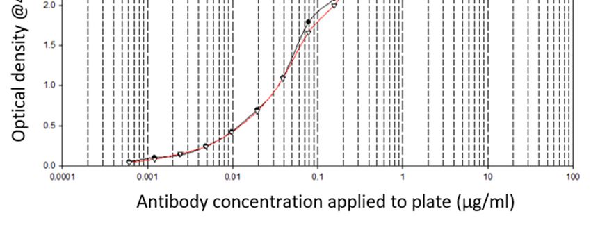
Antibodies 2019, 8, 7 6 of 15
be used for immunization. Immunogenicity analysis identified several peptides within this region of
theAntibodies
RAGE 2018, 7, x FOR PEER REVIEW
protein that met our immunogenicity criteria; however, these peptides were not as6 highly of 15
conserved as aa198–217. Based on a balance between immunogenicity and conservation, we selected
immunogenicity and conservation, we selected the peptide GPTAGSVGGSGLGTLALA,
thecorresponding
peptide GPTAGSVGGSGLGTLALA, corresponding to aa327–344 of the RAGE protein, which had
to aa327–344 of the RAGE protein, which had an immunogenicity score of 7.31 and
an was
immunogenicity score of 7.31 and
conserved in eight human RAGE isoforms. was conserved in eight human RAGE isoforms.
Subsequently,
Subsequently,we we generated
generated aa small
smallpanel
panelof of mouse
mouse antibodies
antibodies targeted
targeted against
against RAGE,RAGE,
see
seeFigure
Figure S2, selecting antibodies raised against the full-length rRAGE protein
S2, selecting antibodies raised against the full-length rRAGE protein (RBGO1); the C1 domain(RBGO1); the C1
domain
peptide peptide corresponding
corresponding to aa198–217
to aa198–217 of theof RAGE
the RAGE protein
protein (RBGO2
(RBGO2 andand RBGO3)
RBGO3) andandthethe
transmembrane
transmembrane proximal
proximalregion
regionpeptide
peptidecorresponding
corresponding to aa327–344
aa327–344of ofthe
theRAGE
RAGEprotein
protein(RBGO4).
(RBGO4).
Typically,
Typically, antibody
antibody selection
selectionrelies
relieson
ondata
data from
from ELISA
ELISA asas an
an indicator
indicatorofofimmunogenicity
immunogenicity andand
a criterion
a criterionforfor
clone
clone selection.
selection.Indeed,
Indeed,thethe antibodies RBGO2,see
antibodies RBGO2, seeFigure
Figure1A,
1A,RBGO3,
RBGO3, seesee Figure
Figure 1A,1A,
andand RBGO4,
RBGO4, seesee Figure
Figure 1B,1B,
werewere selected
selected based
based onon ELISA
ELISA against
against the
the immunizationpeptides
immunization peptidesaa198–217
aa198–
217 (0.0006
(0.0006 to 10 μg/mL:
to 10 µg/mL: RBGO2RBGO2 andand RBGO3)
RBGO3) or or aa327–344(0.0006
aa327–344 (0.0006toto10
10 µg/mL:
μg/mL: RBGO4)
RBGO4)conjugated
conjugated
to bovine serum albumin (BSA), which showed good immunogenicity
to bovine serum albumin (BSA), which showed good immunogenicity and, therefore, grounds and, therefore, grounds forfor
antibody
antibody clone
clone selection.
selection.
Figure 1. Antibody selection based upon enzyme linked immunosorbent assay (ELISA) against the
Figure 1. Antibody selection based upon enzyme linked immunosorbent assay (ELISA) against the
immunizing
immunizing peptide. Clones
peptide. ClonesRBGO2
RBGO2&&33(A) (A) and
and RBGO4
RBGO4 (B) (B)were
wereselected
selectedbased
basedonona positive
a positive ELISA
ELISA
screen using bovine serum albumin (BSA)-conjugated peptides. For ELISA, BSA-conjugated
screen using bovine serum albumin (BSA)-conjugated peptides. For ELISA, BSA-conjugated peptides peptides
(aa198-217
(aa198-217forfor
RBGO2
RBGO2andandRBGO3;
RBGO3;aa327–344
aa327–344 for
for RBGO4)
RBGO4) were were immobilized
immobilizedonto
ontomicro
micro plates
plates and
and
incubated
incubatedwith two-fold
with serial
two-fold dilutions
serial ofofantibodies.
dilutions antibodies.ThisThismethod
methodrepresents
representsthe
thestandard
standard method
method of
clone selection
of clone and good
selection responses
and good to the
responses RBGO2,
to the RBGO2, 3 or3 4orantibodies were
4 antibodies apparent.
were apparent.
However, we noted that these antibodies had lower lethal dose (LD)50 values and reduced
internalization rates in HEC1A endometrial cancer cells, compared to an RBGO1-based ADC, when
used to make anti-RAGE ADCs, see Table 1. Antibodies raised against peptides can lack affinity to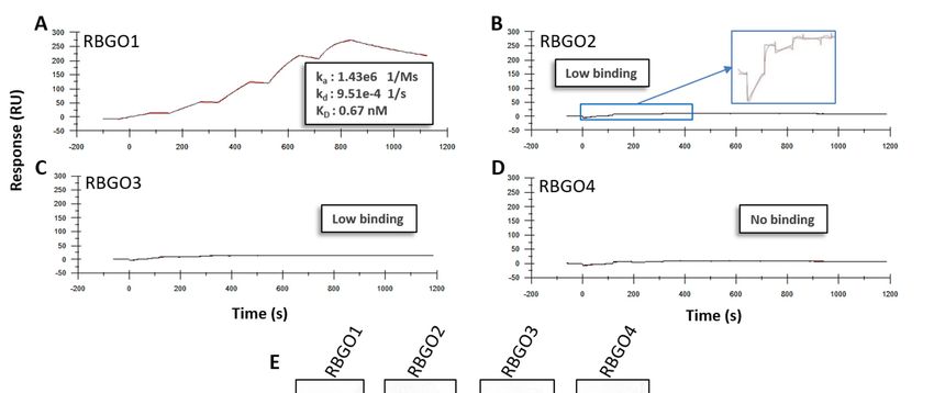
Antibodies 2019, 8, 7                                                                                                     7 of 15

      However, we noted that these antibodies had lower lethal dose (LD)50 values and reduced
internalization rates in HEC1A endometrial cancer cells, compared to an RBGO1-based ADC, when
used to make anti-RAGE ADCs, see Table 1. Antibodies raised against peptides can lack affinity to the
full-length protein since protein folding can result in the binding epitope being obscured. To explore the
reduced efficacy observation, we evaluated antibody binding kinetics to recombinant (r) RAGE protein
with a combination of SPR, dot blot analysis, and confocal microscopy. We selected an approach where
antibodies were captured onto a CM5 sensor chip via amine coupled anti-mouse antibody, to minimize
the time needed for assay development. Kinetic experiments were carried out using rRAGE (2.5 to
200 nM) and the resulting kinetics profiles for RBGO1, see Figure 2A, RBGO2, see Figure 2B, RBGO3,
see Figure 2C, and RBGO4, see Figure 2D, showed that binding affinity between the RBGO1 antibody
and rRAGE was high, in the picomolar range, whilst binding to the other three antibodies was poor
or undetectable. Dot blot analysis using rRAGE, supported the SPR kinetics data, demonstrating
the high-binding affinity of RBGO1, weak binding-affinity of RBGO2 and RBGO3, and an absence of
binding for the RBGO4 antibody, see Figure 2E.
      Additionally, confocal analysis of antibody binding to HEC1A endometrial cancer cells,
see Figure 3, corroborated the SPR and dot blot analysis demonstrating strong binding for RBGO1,
see Figure 3A, weaker binding for RBGO2, see Figure 3B, and RBGO3, see Figure 3C, and very poor
binding for RBGO4, see Figure 3D. Continuing our analysis, we explored the binding kinetics between
the RBGO2, 3 or 4 antibodies and their respective peptides, see Figure 4. The RBGO2, see Figure 4A,
and RBGO3, see Figure 4B, antibodies bound with high-affinity to the aa198-217 peptide used for
clone generation (0.52 ± 0.02 nM and 0.46 ± 0.03 nM, respectively). However, the RBGO4 antibody,
did not bind to the aa327–344 peptide used to generate the RBGO4 clone, see Figure 4C. Additionally,
we performed a dot blot analysis against the immunization peptides (inset images), which confirmed
binding of the RBGO2 and RBGO3 antibodies to the aa198–217 peptide, and the absence of RBGO4
antibody binding to the aa327–344 peptide.
      These data highlighted the benefit of validating antibodies raised using specific peptide regions
against the full-length target protein in native conditions, a role that SPR is readily amenable to, prior
to further development along a therapeutic development pipeline. Additionally, they demonstrate the
benefit of adopting a multi-faceted approach to ADC development where multiple technologies are
used to give a thorough characterization of ADC candidate antibodies.

      Table 1. Internalization and cell toxicity of Receptor for Advanced Glycation End Products (RAGE)
      targeting antibody drug conjugates.

                                                 Internalization
                          Antibody                                             ADC        LD50 (µM)
                                            (Fluorescence/Cell Area)
                                                                                vcE        0.3 ± 0.02
                           RBGO1                    0.31 ± 0.04
                                                                                mcF       0.09 ± 0.01
                                                                                vcE         2 ± 0.05
                           RBGO2                    0.11 ± 0.02
                                                                                mcF          >100 *
                                                                                vcE        1.5 ± 0.07
                           RBGO3                    0.12 ± 0.03
                                                                                mcF          >100 *
                                                                                vcE        2.4 ± 0.06
                           RBGO4                    0.03 ± 0.02
                                                                                mcF        2.2 ± 0.03
      LD50 values and internalization, as a function of fluorescence/cell area, of antibody-drug conjugates (ADCs) within
      HEC1A endometrial cancer cells. * less than 50% cell killing was observed for the range of ADC concentrations tested.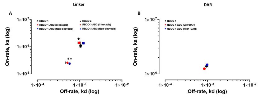
mcF >100 *
vcE 2.4 ± 0.06
RBGO4 0.03 ± 0.02
mcF 2.2 ± 0.03
LD50 values and internalization, as a function of fluorescence/cell area, of antibody-drug conjugates
(ADCs) within HEC1A endometrial cancer cells. * less than 50% cell killing was observed for the range
Antibodies 2019, 8, 7 8 of 15
of ADC concentrations tested.
Antibodies 2018, 7, x FOR PEER REVIEW 8 of 15
Displayed sensorgrams and overlapping fittings are exemplars from three independent experiments
and the data shown are the mean. (E) Full-length, recombinant RAGE (at the volumes displayed) at
100 μg/mL was immobilized onto activated polyvinylidene difluoride (PVDF) membrane and probed
with each of the four α-RAGE antibodies (1 μg/mL). Images were acquired using a Gel-Doc Image
acquisition system.
Additionally, confocal analysis of antibody binding to HEC1A endometrial cancer cells, see
Figure 3, corroborated the SPR and dot blot analysis demonstrating strong binding for RBGO1, see
Figure 2. RBGO1
RBGO1 has
has a higher
a higher binding
binding affinity
affinity (K(K ) to
D )Dto full-length
full-length Receptor
Receptor for Advanced Glycation
Figure
Figure 3A, 2.
weaker binding for RBGO2, see Figure 3B, and RBGO3, see for Advanced
Figure 3C, andGlycation
very poor
End
End Products
Products (RAGE)
(RAGE) protein
protein than
than RBGO2,
RBGO2, RBGO3,
RBGO3, andand RBGO4.
RBGO4. (A–D)
(A–D) Antibodies
Antibodies were
were captured
captured toto
binding for RBGO4, see Figure 3D. Continuing our analysis, we explored the binding kinetics
a aSensor
SensorChip
ChipCM5CM5viaviaanan amine
amine coupled
coupled anti-mouseantibody
anti-mouse antibodyfollowed
followedbybysingle-cycle
single-cyclekinetics
kinetics
between the RBGO2, 3 or 4 antibodies and their respective peptides, see Figure 4. The RBGO2, see
experiments.
experiments. RBGO1
RBGO1 (A),(A), RBGO2
RBGO2 (B),(B), RBGO3
RBGO3(C) (C)ororRBGO4
RBGO4(D) (D)antibodies
antibodieswere
wereexposed
exposedtoto
Figure 4A, and RBGO3, see Figure 4B, antibodies bound with high-affinity to the aa198-217 peptide
recombinant
recombinant RAGE
RAGE (2.5 toto
(2.5 200
200nM)
nM)and
anddata
datawere
werefitted
fittedusing
usinga aone-to-one
one-to-oneLangmuir
Langmuirbinding
bindingmodel.
model.
used for clone generation (0.52 ± 0.02 nM and 0.46 ± 0.03 nM, respectively). However, the RBGO4
Displayed sensorgrams and overlapping fittings are exemplars from three independent experiments
antibody, did not bind to the aa327–344 peptide used to generate the RBGO4 clone, see Figure 4C.
and the data shown are the mean. (E) Full-length, recombinant RAGE (at the volumes displayed) at
Additionally,
100 µg/mLwe was performed
immobilized a onto
dot blot analysis
activated against the
polyvinylidene immunization
difluoride peptides and
(PVDF) membrane (inset images),
probed
which confirmed binding of the RBGO2 and RBGO3 antibodies to the aa198–217
with each of the four α-RAGE antibodies (1 µg/mL). Images were acquired using a Gel-Doc Image peptide, and the
absence of RBGO4
acquisition antibody binding to the aa327–344 peptide.
system.
Figure 3. RBGO1
RBGO1 cellcell surface binding is greater than RBGO2, RBGO3 and RBGO4. (A–D) (A–D) HEC1A
endometrial
endometrial cancer
cancer cells
cells were incubated
incubated in
in medium
medium containing
containing RBGO1
RBGO1 (A),
(A), RBGO2
RBGO2 (B),
(B), RBGO3
RBGO3 (C),
(C),
or RBGO4 (D) antibodies at 10 μg/mL for 240 min. After incubation, the cells were washed and fixed.
µg/mL for 240 min. After incubation, the cells were washed and fixed.
Cell surface bound antibody was imaged via fluorescently labeled secondary antibodiesantibodies and nuclei
stained with 0 ,6-Diamidine-20 -phenylindole dihydrochloride
with44',6-Diamidine-2'-phenylindole dihydrochloride (DAPI).
(DAPI). Images
Images werewere acquired
acquired on a
on a Zeiss
Zeiss LSM
LSM 710 710 confocal
confocal microscope
microscope and analyzed
and analyzed using
using the Zenthe Zenimage
2012 2012 image
analysisanalysis software.
software. Scale
Scale bars =
bars = 50 μm.
50 µm.Antibodies 2019, 8, 7 9 of 15
Antibodies 2018, 7, x FOR PEER REVIEW 9 of 15
Figure 4.4.Surface
Figure Surfaceplasmon
plasmon resonance (SPR)
resonance provides
(SPR) an enhanced
provides platform
an enhanced for antibody
platform clone selection.
for antibody clone
Antibody clones RBGO2 (A), RBGO3 (B) and RBGO4 (C) were assessed for binding
selection. Antibody clones RBGO2 (A), RBGO3 (B) and RBGO4 (C) were assessed for binding kineticskinetics to the
immunization peptides used to generate the clones by ELISA. For SPR, antibodies were
to the immunization peptides used to generate the clones by ELISA. For SPR, antibodies were captured onto
acaptured
Sensor Chip
ontoCM5 via an
a Sensor amine
Chip CM5coupled
via ananti-mouse antibody.
amine coupled Single-cycle
anti-mouse kinetics
antibody. experiments
Single-cycle were
kinetics
then
experiments were then performed using unconjugated peptides; aa198–217 peptide (RBGO2 nM)
performed using unconjugated peptides; aa198–217 peptide (RBGO2 and RBGO3; 2.5 to 200 and
or the aa327–344 peptide (RBGO4; 2.5 to 200 nM). Kinetics were determined using a one-to-one
RBGO3; 2.5 to 200 nM) or the aa327–344 peptide (RBGO4; 2.5 to 200 nM). Kinetics were determined binding
model. Curves displayed are exemplar curves from three independent experiments and the data are
using a one-to-one binding model. Curves displayed are exemplar curves from three independent
the mean. Inset images = aa198–217 (A,B) or aa327–344 (C) were immobilized onto activated PVDF
experiments and the data are the mean. Inset images = aa198–217 (A,B) or aa327–344 (C) were
membrane (the volumes displayed at 100 µg/mL) and probed with each appropriate α-RAGE antibody
immobilized onto activated PVDF membrane (the volumes displayed at 100 μg/mL) and probed with
(1 µg/mL). Images were acquired using a ChemiDoc™ MP Imaging system and analyzed with Image
each appropriate α-RAGE antibody (1 μg/mL). Images were acquired using a ChemiDoc™ MP
Lab™ software.
Imaging system and analyzed with Image Lab™ software.
3.2. The Effect of Conjugation on Antigen Binding Kinetics is Antibody Dependent.
These data highlighted the benefit of validating antibodies raised using specific peptide regions
The production
against the of target
full-length an ADC requires
protein the conjugation
in native conditions,ofacytotoxic
role that molecules withamenable
SPR is readily a molecular
to, mass
prior
in the region of two orders of magnitude smaller than the antibody, via synthetic
to further development along a therapeutic development pipeline. Additionally, they demonstratelinkers (again of
significantly smaller mass than the antibody), to enable cell killing following cell-surface
the benefit of adopting a multi-faceted approach to ADC development where multiple technologies target
recognition and abinding
are used to give thorough bycharacterization
the antibody component of the ADC.
of ADC candidate Characterization of the effect
antibodies.
of antibody conjugation on thermal stability and antigen binding using SPR has previously been
described [21].ofWhilst
3.2. The Effect different
Conjugation conjugation
on Antigen chemistries
Binding Kinetics isvary in their
Antibody effect on thermal stability, thiol
Dependent.
conjugation of IgG1 antibodies is reported to reduce the antibody melting temperature but have no
The production of an ADC requires the conjugation of cytotoxic molecules with a molecular
effect on antigen binding in vitro [21]. To investigate the effect of drug-linker conjugation, two of our
mass in the region of two orders of magnitude smaller than the antibody, via synthetic linkers (again
antibodies, RBGO1 and RBGO3, were conjugated to the antimitotic agents monomethyl auristatin
of significantly smaller mass than the antibody), to enable cell killing following cell-surface target
E (MMAE), via a lysosomally cleavable dipeptide valine-citrulline (vc) linker (vcE; see Figure S1A);
recognition and binding by the antibody component of the ADC. Characterization of the effect of
antibody conjugation on thermal stability and antigen binding using SPR has previously beenAntibodies 2018, 7, x FOR PEER REVIEW 10 of 15
described [21]. Whilst different conjugation chemistries vary in their effect on thermal stability, thiol
conjugation of IgG1 antibodies is reported to reduce the antibody melting temperature but have no
effect on antigen binding in vitro [21]. To investigate the effect of drug-linker conjugation, two of our
Antibodies 2019, 8, 7 10 of 15
antibodies, RBGO1 and RBGO3, were conjugated to the antimitotic agents monomethyl auristatin E
(MMAE), via a lysosomally cleavable dipeptide valine-citrulline (vc) linker (vcE; see Figure S1A); or
monomethyl
or monomethyl auristatin
auristatin F (MMAF),
F (MMAF), via via
a non-cleavable
a non-cleavable maleimido
maleimido caproyl
caproyl (mc)(mc) linker (mcF;
linker see
(mcF;
Figure
see FigureS1B)S1B)
andand captured
captured onto ontoa a CM5CM5sensor
sensorchipchipviavia amine
amine coupled
coupled anti-mouse
anti-mouse antibodies.
antibodies.
Characterization of binding kinetics before and after conjugation to vcE
Characterization of binding kinetics before and after conjugation to vcE was performed using rRAGE was performed using rRAGE
(2.5 toto200nM;
(2.5 200nM; RBGO1)
RBGO1) or aa198-217
or aa198-217 (2.5 to(2.5 to 200
200 nM; nM; and
RBGO3) RBGO3) and binding/dissociation
binding/dissociation rates determinedrates
determined
using using abinding
a one-to-one one-to-one model,binding model,
see Figure 5. see Figure 5. Conjugation
Conjugation of the RBGO3 ofantibody,
the RBGO3 seeantibody,
Figure 5A,B,see
Figure 5A,B, resulted in a four-fold reduction in antigen binding affinity
resulted in a four-fold reduction in antigen binding affinity (KD : conjugated = 1.95 ± 0.03 nM vs (K D : conjugated = 1.95 ± 0.03
nM vs unconjugated
unconjugated = 0.47 ±=0.04 0.47nM,± 0.04 nM, pwhilst
p < 0.05), < 0.05), whilst conjugation
conjugation of the RBGO1 of the RBGO1had
antibody antibody had no
no discernible
discernible effect on K
effect on KD (conjugated = 0.63 ± 0.02 nM vs unconjugated = 0.67 ± 0.03 nM, see Figure Figure
D (conjugated = 0.63 ± 0.02 nM vs unconjugated = 0.67 ± 0.03 nM, see 5C,D).
5C,D). Although
Although the dissociation
the dissociation rate was rate wasby
affected affected by conjugation
conjugation of the RBGO3 of the RBGO3
antibody (kdantibody
: conjugated (kd:
conjugated
= 5.12 × 10 = 5.12
− 4 ± 1.3 ××10 10± 1.3s x 10
-4 − 4 − 1 vs. sunconjugated
-4 -1 vs. unconjugated= 9.39=×9.39
10 x ±
− 4 10 0.9
-4 ±×0.910 x 10s s ),
− 4 -4− -1
1 ), the
the predominant
predominant
factor driving the reduced K
factor driving the reduced KD was a ten-fold reduction in the association rate (ka : conjugated ==2.62
D was a ten-fold reduction in the association rate (k a: conjugated 2.62×x
10 5 ± 1.1 x 10² Ms
5 2 -1 vs
− 1 unconjugated = 2.0 x 10 6 ± 2.8
6 x 10 2 Ms-1 2p < 0.01).
10 ± 1.1 × 10 Ms vs unconjugated = 2.0 × 10 ± 2.8 × 10 Ms p < 0.01). These data suggest that− 1 These data suggest that whilst
conjugation
whilst can impact
conjugation binding
can impact kinetics,
binding as has
kinetics, been
as has beenpreviously
previously demonstrated
demonstrated[21], [21],the
the effect is
effect is
variable for different antibodies.
variable for different antibodies.
Figure
Figure 5. The effect
5. The effect of
of conjugation
conjugation on on antibody–antigen
antibody-antigen binding
binding kinetics
kinetics isis antibody-dependent.
antibody-dependent.
Antibodies were captured onto a Sensor Chip CM5 via an amine coupled
Antibodies were captured onto a Sensor Chip CM5 via an amine coupled anti-mouse anti-mouse antibody.
antibody.
Single-cycle
Single-cycle kinetics experiments were performed. RBGO3 antibody (A) and RBGO3-ADC (B)
kinetics experiments were performed. RBGO3 antibody (A) and RBGO3-ADC (B) were
were
exposed to the aa198–217 peptide (2.5 to 200 nM), and the RBGO1 antibody (C) and
exposed to the aa198–217 peptide (2.5 to 200 nM), and the RBGO1 antibody (C) and RBGO1-ADC (D)RBGO1-ADC (D)
were
were exposed
exposed toto rRAGE
rRAGE (2.5
(2.5 to
to 200
200 nM). Kinetics were
nM). Kinetics were determined
determined using
using aa one-to-one
one-to-one binding
binding model.
model.
Curves displayed are exemplar curves from three independent experiments and data
Curves displayed are exemplar curves from three independent experiments and data are the mean. are the mean.*
* p < 0.05, ** p < 0.01 compared to the unconjugated antibody.
p < 0.05, ** p < 0.01 compared to the unconjugated antibody.
3.3. The Use of Cleavable or Non-Cleavable Linkers does not Affect Binding Kinetics.
3.3. The Use of Cleavable or Non-Cleavable Linkers does not Affect Binding Kinetics.
To further explore the effect of the conjugation described above on binding kinetics, we compared
To further
the influence explore the
of cleavable andeffect of the conjugation
non-cleavable described
linkers on the antigen above
bindingonaffinities
bindingofkinetics,
the RBGO1 we
compared
and RBGO3the influence see
antibodies, of cleavable
Figure 6. and
Bothnon-cleavable
antibodies werelinkers on the antigen
conjugated binding
to vcE or mcF andaffinities of the
the binding
RBGO1 and RBGO3 antibodies, see Figure 6. Both antibodies were conjugated to vcE
kinetics compared between conjugated and unconjugated forms. Antibodies were captured to a CM5 or mcF and the
binding kinetics compared between conjugated and unconjugated forms. Antibodies
sensor chip via amine coupled anti-mouse antibodies. Binding kinetics experiments were performedwere captured
to a CM5
using rRAGEsensor
(2.5chip vianM;
to 200 amine coupled
RBGO1) anti-mouse
or aa198–217 (2.5antibodies.
to 200 nM;Binding
RBGO3)kinetics experiments were
and binding/dissociation
rates determined, see Figure 6A. As previously shown in Figure 5A,B, conjugation ofRBGO3)
performed using rRAGE (2.5 to 200 nM; RBGO1) or aa198–217 (2.5 to 200 nM; the RBGO3and
binding/dissociation rates determined, see Figure 6A. As previously shown in Figure
antibody to vcE or mcF resulted in a reduced kd and significantly reduced ka (p < 0.01), whilst no 5A,B,
conjugation of the RBGO3 antibody to vcE or mcF resulted in a reduced kd and significantly reduced
difference in either kd or ka was observed following conjugation of the RBGO1 antibody. These data
suggest that whilst conjugation had an impact, the type of linker used did not affect antibody-antigen
binding affinity for this set of antibodies.prepared using varying antibody to TCEP (tris(2-carboxyethyl) phosphine) molar ratios to enable
ADCs with low or high drug to antibody ratios (DAR) to be generated. Drug loading of the conjugates
was analyzed using a combination of hydrophobic interaction chromatography (HIC) and reverse
phase chromatography—Polymer Laboratories Reverse Phase (PLRP). Analysis of the traces (Area
Under Curve) indicated average DAR of 1 (low DAR) and 4 (high DAR) were achieved for the
Antibodies 2019, 8, 7
test
11 of 15
conjugates, see Figure S3.
Figure 6. Antibody–antigen kinetics are not affected by the type of linker used or the drug to antibody
Figure 6. Antibody–antigen kinetics are not affected by the type of linker used or the drug to antibody
ratio. Single-cycle kinetics experiments were performed. (A) RBGO1 antibody ( ), RBGO1-ADC
ratio. Single-cycle kinetics experiments were performed. (A) RBGO1 antibody (●), RBGO1-ADC
(Cleavable; and Non-cleavable; ) were exposed to rRAGE (2.5 to 200 nM). RBGO3 antibody (u),
(Cleavable; ● and Non-cleavable; ●) were exposed to rRAGE (2.5 to 200 nM). RBGO3 antibody ( ),
RBGO3-ADC (Cleavable; u and Non-cleavable; u) were exposed to the aa198-217 peptide (2.5 to 200
RBGO3-ADC (Cleavable; and Non-cleavable; ) were exposed to the aa198-217 peptide (2.5 to
nM). (B) RBGO1 antibody ( ), RBGO1-ADC (Low DAR; and High DAR; ) were exposed to rRAGE
200 nM). (B) RBGO1 antibody (●), RBGO1-ADC (Low DAR; ● and High DAR; ●) were exposed to
(2.5 to 200 nM). On-rates and off-rates, ka and kd were determined using a one-to-one binding model.
rRAGE (2.5 to 200 nM). On-rates and off-rates, ka and kd were determined using a one-to-one binding
Data displayed are mean ± SD (n = 3). ** p < 0.01 for ka compared to the unconjugated antibody.
model. Data displayed are mean ± SD (n = 3). ** pThe continuous internalization of cell surface receptors into the endosomal compartment of cells
is essential to the efficacy of the ADC therapeutic approach, facilitating effective delivery of ADC
payload to the internal cell environment where the cytotoxic drug mechanism of action is typically
focused. Accordingly, a comprehensive understanding of the molecular mechanisms governing ADC
Antibodies 2019, 8, 7 12 of 15
intracellular trafficking is critical to ADC design and selection. Therefore, we wished to investigate
the impact of an endosomal-like environment on antigen–antibody dissociation, together with the
possible compounding/detrimental effects
possible compounding/detrimental effects of
of conjugation
conjugation to to the
the binding
binding kinetics
kinetics of
of our
our lead
lead antibody
antibody
when exposed
when exposed to to endosomal
endosomal pH.pH. Using
Using thethe dual-inject
dual-inject functionality
functionality inin Biacore™
Biacore™ T200,
T200, which
which enables
enables
two freely selected solutions to be injected in immediate sequence whilst keeping
two freely selected solutions to be injected in immediate sequence whilst keeping the same running the same running
buffer.
buffer. Once
Once again,
again, unconjugated
unconjugated RBGO1
RBGO1 antibody
antibody or or RBGO1-ADC
RBGO1-ADC (vcE; (vcE; High
High DAR)
DAR) werewere captured
captured
to
to the
the same
same CM5
CM5 sensor
sensor chip
chip with
with amine
amine coupled
coupled anti-mouse
anti-mouse antibodies
antibodies that
that had
had also
also been
been used
used for
for
the previous analyses. We characterized antibody/ADC association at pH
the previous analyses. We characterized antibody/ADC association at pH 7.4, whilst determining 7.4, whilst determining
dissociation
dissociation kinetics
kinetics at
ateither
eitherpH
pH7.47.4ororpH
pH 6.06.0
with
withrRAGE
rRAGE (10(10
nM;nM;Figure 7). The
Figure dissociation
7). The rate
dissociation
between RBGO1 and rRAGE was increased in a pH 6.0 solution compared
rate between RBGO1 and rRAGE was increased in a pH 6.0 solution compared to a pH 7.4 solution to a pH 7.4 solution (%
dissociation over 180 s = 45% and 25%, respectively, Figure 7A.), which is in keeping
(% dissociation over 180 s = 45% and 25%, respectively, Figure 7A.), which is in keeping with good with good ADC
design.
ADC Additionally,
design. conjugation
Additionally, of the
conjugation of RBGO1
the RBGO1 to vcE hadhad
to vcE nonodiscernible
discernibleeffect
effectononthe
the rate
rate of
of
dissociation at pH 6.0, see Figure
dissociation at pH 6.0, see Figure 7B. 7B.
Figure 7.
Figure The dissociation
7. The dissociation rate
rate of
of the
the RBGO1
RBGO1 antibody/ADC
antibody/ADC is is increased
increased in
in acidic
acidic pH.
pH. Antibody/ADC
Antibody/ADC
was captured to a Sensor Chip CM5 via amine coupled anti-mouse
was captured to a Sensor Chip CM5 via amine coupled anti-mouse antibodies andantibodies and binding/dissociation
experiments were performed
binding/dissociation using rRAGE
experiments (10 nM). Using
were performed usingthe dual-inject
rRAGE functionality
(10 nM). Using thetransition from
dual-inject
extracellular to endosomal conditions was mimicked. (A) rRAGE was injected
functionality transition from extracellular to endosomal conditions was mimicked. (A) rRAGE was in pH 7.4 buffer
(extracellular)
injected in pHand allowed
7.4 buffer to bind to unconjugated
(extracellular) and allowedRBGO-1,
to bind immediately
to unconjugatedfollowed by dissociation
RBGO-1, immediately in
either pH 6.0 (endosomal) or pH 7.4 buffer. (B) Overlapping sensorgrams showing
followed by dissociation in either pH 6.0 (endosomal) or pH 7.4 buffer. (B) Overlapping sensorgramssimilar binding
profiles for
showing unconjugated
similar and conjugated
binding profiles antibodyand
for unconjugated at the two different
conjugated pHs. at
antibody The
thesensorgrams
two different shown
pHs.
are exemplars from three independent experiments.
The sensorgrams shown are exemplars from three independent experiments.
4. Discussion
Antibody–drug conjugates are a proven example of the type of novel, precision medicines required
to combat the increasing incidence of diseases such as the gynecological cancers [24,25]. Aiming to
streamline the design and development of ADCs within our laboratory, we evaluated antibodies beingAntibodies 2019, 8, 7 13 of 15
developed as ADCs, using SPR technology. This technology enabled us to study ADC characteristics
such as specificity, antigen kinetics/affinity, any effects that the pH or our choice of linker type and
DAR may have on antigen binding kinetics and demonstrate the applicability of this technology to
ADC design and development. SPR analysis within our lab had indicated that antibodies raised against
our therapeutic target, RAGE, bound poorly to the full-length protein, data that we verified using
dot blot analysis and confocal microscopy. Clone selection based on ELISA against the immunization
peptide conjugated to BSA is typical, so we compared ELISA data to kinetics data from SPR analysis
using the same peptides. Whilst two of the antibodies in question, RBGO2 and RBGO3, bound with
high-affinity to the immunization peptide, the RBGO4 antibody did not, suggesting that the use of
ELISA alone might lack the specificity required for the selection of effective, high-affinity binding
antibodies for therapeutic development. Indeed, dot blot analysis verified the SPR data, confirming
that the RBGO4 antibody did not bind to the aa327-344 peptide. It is unclear why a positive ELISA
screen was obtained for the RBGO4 antibody, although interestingly, repeat ELISAs using a non-BSA
conjugated aa327-344 or BSA alone were negative, see Figure S4, suggesting that the RBGO4 epitope
may span the BSA-peptide junction.
A variable effect of conjugation on antibody characteristics is known, but this can range from a
large effect to none depending on the antibody and conjugation chemistry employed. Our cell-based
data demonstrated variability in the cell killing efficacy of our ADCs. An effect that was variable for
each antibody and so could not be attributed to the drug or linker being used. Non-specific conjugation
often alters the electrostatic properties and hydrophobicity of an antibody with implications for ADC
stability and pharmacokinetics [26]. Thiol conjugation, in particular, has a dramatic, DAR-related
effect on antibody thermostability compared to alternative techniques such as amine or carbohydrate
conjugation; however, the effect is not consistent [21]. Our data showed antibody to antibody variability
regarding the effect of conjugation. Although antibody-antigen affinity was comparable for different
types of linker and DARs, conjugation caused a four-fold reduction of the affinity (primarily due
to a 10-fold reduction in the association rate) for the RBGO3 antibody, whilst the RBGO1 antibody
was unaffected despite both antibodies having similar DARs. Utilizing inter-chain, disulfide bridge
cysteines is an effective, inexpensive strategy for ADC production. However, variability in the effect of
conjugation on antibody binding kinetics means it is important to quantify the impact of conjugation.
An important consideration when developing ADCs is also that affinity is not the whole story. ADCs
with similar affinity may, in fact, have very different kinetics and, depending on the receptor being
targeted, it may be acceptable to have a reduced on-rate if the off-rate is slow enough. The strength of
using SPR as a development tool is that affinity can be dissolved into kinetics to obtain the desired
kinetic profile for the ADC being developed.
The efficacy of an ADC is dependent on internalization and the release of the cytotoxic payload.
Consequently, recent developments in antibody therapeutics have included the design of antibodies
that, in addition to binding with high-affinity at the extracellular pH 7.4, also dissociate at a higher
rate under endosomal pH 6.0 conditions [27]. Although this concept is yet to be demonstrated for
ADCs, such an approach is plausible and could be beneficial for ADCs targeting antibodies that are
rapidly recycled back to the cell surface such as HER2 and RAGE [27–29]. To explore this aspect of
our ADCs, we used an SPR-based method to qualitatively assess the effect of pH on antibody-antigen
dissociation. Whilst association was performed at extracellular pH 7.4, it was possible to monitor and
compare dissociation at pH 7.4 or endosomal pH 6.0. Interestingly, our lead ADC candidate, based
upon the RBGO1 antibody, exhibits high-binding affinity at pH 7.4 and increased dissociation rate at
pH 6.0 compared to the dissociation rate at pH 7.4.
5. Conclusions
SPR analysis provides an effective platform for the development of ADCs and can be used to
assess multiple aspects of these complex advanced biological therapeutic molecules. Of notable value,
is the ability to deselect candidate antibodies early in the development process preserving resourcesAntibodies 2019, 8, 7 14 of 15
that can then be focused on candidates with a greater likelihood of successful development. Using SPR,
we were able to determine the specific properties that could explain the superior efficacy of our RBGO1
ADCs compared to others being developed by our lab. The combination of high affinity to the target
protein, favorable binding kinetics (fast on-rate, slow off-rate), resistance to loss of binding affinity
following conjugation and effective dissociation within an endosomal-like environment, are all key
aspects underpinning the efficacy of RAGE targeting RBGO1-ADC and provide a basis for intelligent
ADC design. SPR technology has multiple benefits, which when used in combination with alternative
approaches, demonstrate its suitability as a key enabler in rational ADC development.
Supplementary Materials: The following are available online at http://www.mdpi.com/2073-4468/8/1/7/s1,
Figure S1: Linkers used during ADC manufacture, title, Figure S2: RAGE antibody binding locations, Figure S3:
PLRP traces of high and low DAR ADCs, Figure S4: ELISA of BSA conjugated peptide.
Author Contributions: G.D.H., A.F., and T.F. prepared the manuscript and contributed to the experimental
procedures and design; D.G contributed to the experimental design; R.S.C. conceived the study and contributed
to experimental design and the preparation of the manuscript.
Funding: This research received no external funding.
Acknowledgments: The authors gratefully acknowledge the contribution of loan of a Biacore™ T200 system from
GE Healthcare for the period of this research. The work described in this study is protected under International
Patent Application PCT/GB2015/053156.
Conflicts of Interest: Asa Frostell and Tim Fagge were employed by GE Healthcare during the preparation of this
article. GE Healthcare is the provider of Biacore™ systems, sensor chips, and reagents.
References
1. Chester, C.; Dorigo, O.; Berek, J.S.; Kohrt, H. Immunotherapeutic approaches to ovarian cancer treatment.
J. Immunother. Cancer 2015, 3, 7. [CrossRef]
2. Ecker, D.M.; Jones, S.D.; Levine, H.L. The therapeutic monoclonal antibody market. MAbs 2015, 7, 9–14.
[CrossRef] [PubMed]
3. Sievers, E.L. Efficacy and safety of gemtuzumab ozogamicin in patients with CD33-positive acute myeloid
leukemia in first relapse. J. Clin. Oncol. 2001, 19, 3244–3254. [CrossRef] [PubMed]
4. Senter, P.D.; Sievers, E.L. The discovery and development of brentuximab vedotin for use in relapsed
Hodgkin lymphoma and systemic anaplastic large cell lymphoma. Nat. Biotechnol. 2012, 30, 631–637.
[CrossRef] [PubMed]
5. Verma, S.; Miles, D.; Gianni, L.; Krop, I.E.; Welslau, M.; Baselga, J.; Pegram, M.; Oh, D.Y.; Diéras, V.;
Guardino, E.; et al. Trastuzumab emtansine for HER2-positive advanced breast cancer. N. Engl. J. Med. 2012,
367, 1783–1791. [CrossRef] [PubMed]
6. Lamb, Y.N. Inotuzumab Ozogamicin: First Global Approval. Drugs 2017, 77, 1603–1610. [CrossRef]
7. Perez, H.L.; Cardarelli, P.M.; Deshpande, S.; Gangwar, S.; Schroeder, G.M.; Vite, G.D.; Borzilleri, R.M.
Antibody-drug conjugates: Current status and future directions. Drug Discov. Today 2014, 19, 869–881.
[CrossRef]
8. Beck, A.; Goetsch, L.; Dumontet, C.; Corvaïa, N. Strategies and challenges for the next generation of
antibody–drug conjugates. Nat. Rev. Drug Discov. 2017, 16, 315. [CrossRef]
9. Löfås, S.; Malmqvist, M.; Rönnberg, I.; Stenberg, E.; Liedberg, B.; Lundström, I. Bioanalysis with surface
plasmon resonance. Sens. Actuators B Chem. 1991, 5, 79–84. [CrossRef]
10. Karlsson, R.; Michaelsson, A.; Mattsson, L. Kinetic analysis of monoclonal antibody-antigen interactions
with a new biosensor based analytical system. J. Immunol. Methods 1991, 145, 229–240. [CrossRef]
11. Kim, M.; Park, K.; Jeong, E.J.; Shin, Y.B.; Chung, B.H. Surface plasmon resonance imaging analysis of
protein-protein interactions using on-chip-expressed capture protein. Anal. Biochem. 2006, 351, 298–304.
[CrossRef] [PubMed]
12. Madeira, A.; Vikeved, E.; Nilsson, A.; Sjogren, B.; Andren, P.E.; Svenningsson, P. Identification of protein-
protein interactions by surface plasmon resonance followed by mass spectrometry. Curr. Protoc. Protein Sci.
2011, 19, 19–21.You can also read