Characterization of the High-Affinity Drug Ligand Binding Site of Mouse Recombinant TSPO - MDPI
←
→
Page content transcription
If your browser does not render page correctly, please read the page content below
International Journal of
           Molecular Sciences

Article
Characterization of the High-Affinity Drug Ligand
Binding Site of Mouse Recombinant TSPO
Soria Iatmanen-Harbi 1 , lucile Senicourt 1 , Vassilios Papadopoulos 2 , Olivier Lequin 1         and
Jean-Jacques Lacapere 1, *
 1    Sorbonne Université, Ecole Normale Supérieure, PSL University, CNRS, Laboratoire des
      Biomolécules (LBM), 4 place Jussieu, F-75005 Paris, France; soriatmanen@hotmail.com (S.I.-H.);
      lucile.senicourt@gmail.com (I.S.); olivier.lequin@upmc.fr (O.L.)
 2    Department of Pharmacology and Pharmaceutical Sciences, School of Pharmacy, University of Southern
      California, Los Angeles, CA 90089, USA; vpapadop@usc.edu
 *    Correspondence: jean-jacques.lacapere@upmc.fr; Tel.: +33-1-44-27-61-83

 Received: 4 February 2019; Accepted: 19 March 2019; Published: 21 March 2019

 Abstract: The optimization of translocator protein (TSPO) ligands for Positron Emission Tomography
 as well as for the modulation of neurosteroids is a critical necessity for the development of TSPO-based
 diagnostics and therapeutics of neuropsychiatrics and neurodegenerative disorders. Structural hints
 on the interaction site and ligand binding mechanism are essential for the development of efficient
 TSPO ligands. Recently published atomic structures of recombinant mammalian and bacterial TSPO1,
 bound with either the high-affinity drug ligand PK 11195 or protoporphyrin IX, have revealed the
 membrane protein topology and the ligand binding pocket. The ligand is surrounded by amino acids
 from the five transmembrane helices as well as the cytosolic loops. However, the precise mechanism of
 ligand binding remains unknown. Previous biochemical studies had suggested that ligand selectivity
 and binding was governed by these loops. We performed site-directed mutagenesis to further test
 this hypothesis and measured the binding affinities. We show that aromatic residues (Y34 and
 F100) from the cytosolic loops contribute to PK 11195 access to its binding site. Limited proteolytic
 digestion, circular dichroism and solution two-dimensional (2-D) NMR using selective amino acid
 labelling provide information on the intramolecular flexibility and conformational changes in the
 TSPO structure upon PK 11195 binding. We also discuss the differences in the PK 11195 binding
 affinities and the primary structure between TSPO (TSPO1) and its paralogous gene product TSPO2.

 Keywords: translocator protein (TSPO); ligand binding site; nuclear magnetic resonance (NMR);
 trypsin digestion; circular dichroism (CD); intrinsic fluorescence

1. Introduction
     The 18 kDa translocator protein (TSPO), previously named PBR for peripheral-type benzodiazepine
receptor [1], is an evolutionarily conserved membrane protein [2] located in eukaryotic cell mitochondria.
TSPO is highly expressed in steroidogenic and bile salt-synthesizing cells [1,3], but it has been also
observed in many other tissues [1]. Despite its implication in many cellular functions and the high number
of pharmacological studies, little is known about its structure–function relationships that may limit its
pharmacological efficiency. However, TSPO overexpression in neuroinflammation and neurodegenerative
disorders has made TSPO and its possible ligands extremely attractive subcellular targets for diagnostics
and therapeutics [4–6].
     Many TSPO ligands belonging to different chemical classes have been identified over the last
decades [4], but overly complex binding profiles likely due to the number of genetic variants [7] as
well as the lack of atomic structures have not permitted the optimization of drug design [8]. Recently,

Int. J. Mol. Sci. 2019, 20, 1444; doi:10.3390/ijms20061444                       www.mdpi.com/journal/ijms
Int. J. Mol. Sci. 2019, 20, 1444                                                                     2 of 18

the atomic structure of recombinant mouse TSPO (rec-mTSPO) was determined by NMR [9] (PDB
ID-2MGY), after stabilization by its high-affinity drug ligand, PK 11195 [10]. Previous studies sought
to understand the origin of the selectivity of TSPO toward PK 11195. Various mutations and deletions
have been reported in mammalian and bacterial species [11–14], suggesting the involvement of the five
transmembrane helices and the cytosolic loops, which were then confirmed by the atomic structures
determined for mammalian and bacterial TSPO [9,15,16]. The ligand binding pocket that has been
characterized in these atomic structures reveals the involvement of several conserved residues [9,15,16].
Some are rigorously identical among the different species of TSPO isoform 1, but others are just
homologous. The most significant amino acid differences are observed when comparing TSPO (TSPO1)
to its paralogous gene product TSPO2 that correlates with the observed differences in the PK 11195
binding affinities for these proteins [17].
      The precise mechanism of ligand access to its pocket from the cytosol remains unknown.
The crystal structures of bacterial TSPO (BcTSPO and RsTSPO for Bacillus cereus and Rhodobacter
sphaeroides, respectively) with and without ligands give highly superimposable atomic structures with
cytosolic loops closing the entrance to the binding site [15,16], giving no indication of the dynamic
changes needed to understand ligand binding. The NMR structure of the A147T polymorph of
mammalian TSPO, described as decreasing the binding affinity for PK 11195 [7], exhibits a very similar
structure to the wild type (WT) with ligand bound [18], as measured by a root-mean-square deviation
(CA-RMSD) of 1.3 Å for all carbon alpha atoms. A147T substitution modulates the structure around
the site mutation but also induces local rearrangements of the cytosolic domains and, in particular,
the cytosolic end of the first transmembrane helix [18]. A larger CA-RMSD of 3.0 Å can be measured
for the TM1-TM2 connecting loops (E29-R46) for A147T compared to WT protein, suggesting that this
part might be important for the access of PK 11195 to the binding pocket [18]. Interestingly, in the
A147T mutant, residue Y34 is oriented toward the binding site and is facing the aliphatic moiety of
PK 11195, whereas it is oriented outward in the WT protein (corresponding to a change in the chi1
rotamer). The dynamics of this TSPO region and of Y34, in particular, suggest a contribution of this
residue to the accessibility of the binding site.
      Interestingly, a large fluctuation of the structure is also observed between mTSPO and BcTSPO
with bound PK 11195 for the segment connecting TM3 and TM4 and comprising a highly conserved
F100. The side chain of this residue is solvent-exposed in the WT-mTSPO but is oriented toward the
PK 11195 binding site at 4 Å in BcTSPO [16], suggesting a possible contribution of this residue in
the accessibility of the ligand to the pocket and/or the positioning of the ligand within its binding site.
      Deletion mutants previously described [13,14] are interesting to overview (Figure S1) because
they concern large protein fraction deletions and they could strongly affect protein folding, specifically
within the TM helices and, thus, ligand binding. However, among the various deletion mutants
described, only the ∆15–35 mutant of human TSPO, i.e., involving a large part of TM1, loses its binding
capacity when overexpressed in yeast. Interestingly, two deletion mutants in the cytosolic part of
the TSPO (∆41–51 and ∆153–169) show reduced binding properties (55% and 75% of the control,
respectively). The first one (∆41–51) involves several residues from the TM2 involved in the binding of
PK 11195 within the atomic structure. It also lacks several highly conserved residues, W42, P44 and
P45, that are not part of the TM2 but might contribute to the access of the binding site. The second
(∆153–169) is the C-terminus domain where the first residues (153–159) belong to TM5 and makes
several contacts with the TM1-TM2 and TM3-TM4 loops [18]. Such a deletion might destabilize the
folding and stability and, thus, alter the affinity and stoichiometry of binding.
      In this report, we have repeated the studies illustrating mTSPO1 structural changes upon
ligand binding using circular dichroism and quantify the binding of PK 11195 to the protein in
detergent. We have specifically labelled the two unique lysine residues located on opposite sides of
the protein and recorded their NMR spectra in the presence and absence of the ligand, revealing the
large conformational change. We have designed limited proteolysis studies to show changes in the
accessibility to trypsin upon ligand binding. We have focused our mutagenesis study on conserved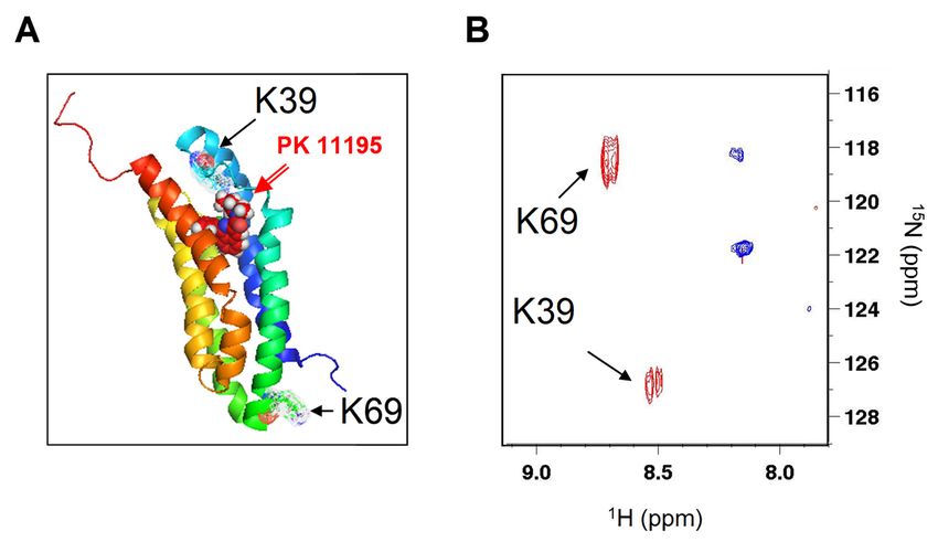
Int. J. Mol. Sci. 2019, 20, 1444 3 of 18
aromatic amino
Int. J. Mol. Sci. 2019,acids residues (Y34 and F100) located in the cytoplasmic loops that could be involved
20, 1444 3 of 18
in the access of the ligand to its binding pocket. We have also included two deletion mutants of
cytosolic
cytosolicfacing
facingdomains
domains(∆41–51
(Δ41–51andand∆153–169)
Δ153–169)toto compare
compare their
their contribution
contribution to
to the
the point mutations.
mutations.
We
Weexpressed
expressedthese thesepoint
pointmutants,
mutants,as aswell
wellas
asdeletion
deletionmutants,
mutants,investigated
investigatedtheir
theirsecondary
secondarystructure
structure
and
and characterized the effects of the mutations on PK 11195 binding. We also discussed theglobal
characterized the effects of the mutations on PK 11195 binding. We also discussed the global
conformation
conformationchange changeof ofTSPO
TSPOupon
uponligand
ligandbinding,
binding,the
thespecific
specificinvolvement
involvementof ofthe
thecytoplasmic
cytoplasmicloops
loops
and the differences in amino acid sequences between TSPO1 and TSPO2 proteins
and the differences in amino acid sequences between TSPO1 and TSPO2 proteins that may account that may account for
the
for ability of TSPO1
the ability of TSPO1to bind drugdrug
to bind ligands.
ligands.
2.2.Results
Results
The
Theoverexpression
overexpressionof ofrec-mTSPO
rec-mTSPOin inheterologous
heterologouscellscellspermits
permitsthetheproduction
productionof oflarge
largeamounts
amounts
of
of protein by extraction from bacterial inclusion bodies using sodium dodecyl sulfate (SDS) as
protein by extraction from bacterial inclusion bodies using sodium dodecyl sulfate (SDS) as
detergent
detergent [19].
[19]. SDS-solubilized
SDS-solubilized rec-mTSPO
rec-mTSPO showed
showed partial
partial helical
helical folding
folding asaspreviously
previouslydescribed
described
and
and shown
shown in in the
thecircular
circulardichroism
dichroism (CD)
(CD) spectrum
spectrum [19][19] (Figure
(Figure S2). However,
S2). However, no PK no PKbinding
11195 11195
binding was observed
was observed in theinpresence
the presence of this
of this detergent
detergent [19].Exchanging
[19]. Exchangingthe theSDS
SDS detergent
detergent forfor
dodecylphosphocholine
dodecylphosphocholine (DPC) increased the helix folding percentage of rec-mTSPO as previously
(DPC) increased the helix folding percentage of rec-mTSPO as previously
described
described and
and inferred from the
inferred from the change
changeininCD CDspectrum
spectrum[19] [19](Figure
(FigureS2).S2).
PKPK 11195
11195 binding
binding to
to rec-
rec-mTSPO was observed in the presence of DPC [10]. The addition of increasing
mTSPO was observed in the presence of DPC [10]. The addition of increasing amounts of PK 11195 amounts of PK 11195
induced
inducedincreasing
increasingchanges
changesin inthe
theCD
CDspectrum,
spectrum,as asshown
shownin inFigure
Figure1,1,but
butrequired
requiredhigh
highligand
ligandover
over
protein molar ratios (inset of Figure 1), suggesting a low affinity compared to the native
protein molar ratios (inset of Figure 1), suggesting a low affinity compared to the native membrane membrane [12].
This
[12].ratio does does
This ratio not reflect just just
not reflect binding, since
binding, partpart
since of the PKPK
of the 11195 is is
11195 bound
boundinto
intothe
theDPC
DPCmicelles,
micelles,
reducing the free PK 11195 concentration available for the protein
reducing the free PK 11195 concentration available for the protein [10]. [10].
Figure 1. The circular dichroism spectra of rec-mTSPO: The spectra recorded in the presence of
dodecylphosphocholine
Figure 1. The circular (DPC) detergent
dichroism andofincreasing
spectra rec-mTSPO: amounts of PK 11195
The spectra (its structure
recorded is given in
in the presence of
the bottom right of the panel).
dodecylphosphocholine (DPC)The insert shows
detergent the increase
and increasing in the of
amounts total
PKhelix
11195content of rec-mTSPO
(its structure is given
upon
in thePK 11195right
bottom addition,
of theexpressed asinsert
panel). The a molar ratiothe
shows over protein.
increase in the total helix content of rec-mTSPO
upon PK 11195 addition, expressed as a molar ratio over protein.
NMR studies previously showed that ligand binding stabilizes TSPO tertiary folding, [9,10,19] as
revealed
NMR in the 1-D 1previously
studies H NMR spectrum
showedwith that the presence
ligand of several
binding upfield
stabilizes TSPOshifted methyl
tertiary resonances
folding, [9,10,19]
as 1 15
as revealed in the 1-D H NMR spectrum with the presence of several upfield shifted amethyl
described [10,19]. In the1 2-D H– N HSQC spectra, the addition of excess PK 11195 induces large
spectral dispersion of the amide signals, indicating conformational changes from
resonances as described [10,19]. In the 2-D H– N HSQC spectra, the addition of excess PK 11195
1 15 the partially folded
to foldedastructure
induces in agreement
large spectral dispersion with previously
of the described
amide signals, CD experiments
indicating [10,18].changes
conformational The NHfromindole
the
resonances of tryptophan (boxes in Figure S3A,B) were particularly sensitive to PK 11195
partially folded to folded structure in agreement with previously described CD experiments [10,18]. binding, since
aThe
broad
NHmassif
indolewas observedofintryptophan
resonances the absence(boxes
of ligand (Figure
in Figure S3A),were
S3A,B) whereas several well-dispersed
particularly sensitive to PK
11195 binding, since a broad massif was observed in the absence of ligand (Figure S3A), whereas
several well-dispersed peaks were detected in its presence (Figure S3B) as previously described
[10,19]. The line widths and low chemical shift dispersion of tryptophan NH indole resonances in the
absence of ligand suggests the presence of conformational exchange and the absence of a stable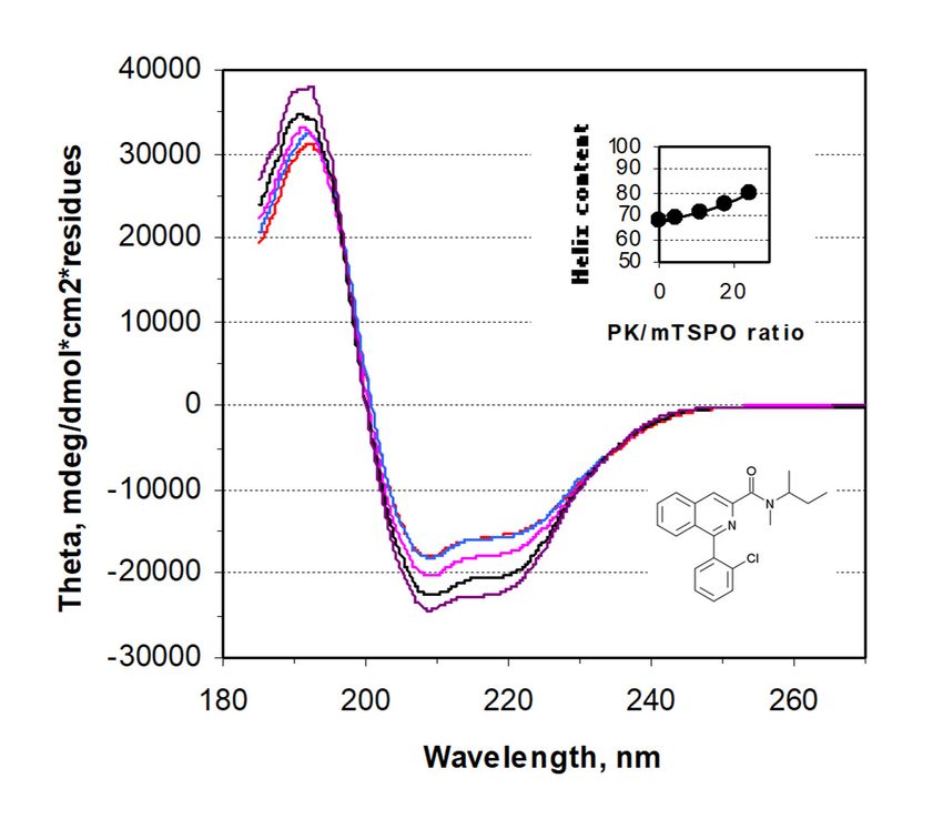
Int. J. Mol. Sci. 2019, 20, 1444 4 of 18
peaks were detected in its presence (Figure S3B) as previously described [10,19]. The line widths and
low
Int. J.chemical shift
Mol. Sci. 2019, 20,dispersion
1444 of tryptophan NH indole resonances in the absence of ligand suggests 4 ofthe
18
presence of conformational exchange and the absence of a stable tertiary structure [10,19]. The atomic
tertiary structure
structure obtained [10,19].
by NMR The [9]
atomic structure
reveals obtained
that the by NMR
two unique [9] reveals
lysine residues that the twothe
(among unique lysine
189 amino
residues
acids (among
of the the 189are
rec-mTSPO) amino acids
located at of
twothe rec-mTSPO)
opposite sides are located
of the protein at (Figure
two opposite sides of the
2A). Therefore, to
protein study
further (Figure the2A). Therefore,
rec-mTSPO to further
structural study the
changes, we rec-mTSPO structuralamino
performed selective changes,acidwe performed
labelling and
selective amino
recorded the 2-Dacid 1 H– 15 N NMR
labelling and recorded
HSQC the 2-D
spectra. 1H–15N 15
Selective [ NMR
N]-LysHSQC spectra.
labelling an[151 H–
Selective
provided 15 N
N]-Lys
labelling
HSQC provided
spectrum with antwo1H–15N HSQC spectrum with two peaks, as expected from the amino acid
peaks, as expected from the amino acid sequence (Figure 2B, blue). The two
sequence (Figure
well-separated peaks 2B,were
blue). The two
strongly well-separated
shifted upon PK 11195 peaks were(Figure
binding strongly shifted
2B, red), upon PKthat
suggesting 11195
the
bindingare
lysines (Figure
either2B, red), involved
directly suggesting in that the lysines
PK 11195 binding areoreither
lie indirectly
regions involved
affected by in PK
PK11195-induced
11195 binding
or lie in regions changes.
conformational affected by PKfirst
The 11195-induced
lysine (K39) conformational
is in a flexiblechanges.
loop and The firstthe
caps lysine (K39) isininitsa
PK 11195
flexible loop
binding and caps
site (Figure theThe
2A). PKsecond
11195 in its binding
lysine (K69) issite (Figure
located 2A).opposite
at the The secondside lysine (K69) is located
of the membrane and,
at therevealed
thus, oppositeaside of the membrane
long-range and, thus,
conformational changerevealed
in thisaregion
long-range
upon conformational
PK 11195 binding. change
The in this
peaks
region upon PK
corresponding to 11195
K39 and binding. Thedetected
K69 were peaks corresponding to K39 and
both in the presence andabsence
K69 were of detected bothligand,
the PK 11195 in the
presence and
suggesting absenceof of
the absence the PK 11195
conformational ligand, suggesting
heterogeneous states in thethevicinity
absence of conformational
of these residues.
heterogeneous states in the vicinity of these residues.
Figure 2.2.The
Figure Theligand-induced
ligand-induced stabilization
stabilization of rec-mTSPO
of the the rec-mTSPO structure
structure in dodecylphosphocholine
in dodecylphosphocholine (DPC)
detergent: (A) the(A)
(DPC) detergent: atomic NMR structure
the atomic of rec-mTSPO
NMR structure with PK
of rec-mTSPO 11195
with PKbound
11195 [9] (PDB
bound [9]ID-2MGY)
(PDB ID-
emphasizing the positions of the two 1 H–15 N HSQC
2MGY) emphasizing the positions oflysines
the two(K39 and K69)
lysines (K39 and
and (B) theand
K69) 2-D(B) the 2-D 1H–15spectra
N HSQC of
selectively 15
[ N]-Lys labelled rec-mTSPO in the absence (blue) and (blue)
in the presence
spectra of selectively [ N]-Lys
15 labelled rec-mTSPO in the absence and in the(red) of PK (red)
presence 11195.
of
PK 11195.
Figure 3 shows that not only lysines but also arginines are located on both sides of the mTSPO.
Both Figure
types of3 residues are not
shows that selectively digested
only lysines but by trypsin;
also thus,
arginines arewelocated
took advantage of this
on both sides of to
theperform
mTSPO. a
limited proteolysis digestion that has been previously used to probe the conformational changes
Both types of residues are selectively digested by trypsin; thus, we took advantage of this to perform of
proteins
a limited[20,21].
proteolysis digestion that has been previously used to probe the conformational changes of
proteins [20,21].Int. J. Mol. Sci. 2019, 20, 1444 5 of 18
Int. J. Mol. Sci. 2019, 20, 1444 5 of 18
Figure 3. (A) The mTSPO sequence in a 2-D diagram with transmembrane helices shown as boxes
Figure 3. (A) The mTSPO sequence in a 2-D diagram with transmembrane helices shown as boxes
crossing the membrane (Mb): The point mutations and deletion mutants used in the present work are
crossing
showntheasmembrane
red-coloured (Mb): Thecircles
double point ormutations and deletionThe
ellipses, respectively. mutants
amino used
acids in the present
involved in thework
are shown
bindingaspocket
red-coloured double
of the atomic circles
structure areor ellipses,
shown respectively.
in orange-filled boldThe amino
circles. Lysineacids involved
(K), arginine (R)in the
binding
andpocket of the
tryptophan (W)atomic structure
are shown are grey-
in blue-, shown andingreen-filled
orange-filled boldrespectively.
circles, circles. Lysine (K),position
(B) The arginine (R)
and tryptophan
of Y34 in the (W)
NMR are shownofinA147T
structure blue-,polymorph
grey- andof green-filled
mammaliancircles,
TSPO [18]respectively.
(PDB ID-2NO2) (B) The
andposition
(C)
of Y34theinposition
the NMR of Y30 and F93 of
structure in A147T
the X-ray structure ofof
polymorph bacterial TSPO [16]
mammalian TSPO(PDB ID-4RYI):
[18] These two and
(PDB ID-2NO2)
residues
(C) the areof
position homologous
Y30 and F93 to positions Y32 and
in the X-ray F100 ofofmammalian
structure TSPO.[16]
bacterial TSPO (D) (PDB
The top view of atomic
ID-4RYI): These two
NMR structure with bound PK 11195 [9] (PDB ID-2MGY) emphasizes
residues are homologous to positions Y32 and F100 of mammalian TSPO. (D) The top the positions of theview
mutations
of atomic
NMRand deletions.
structure with bound PK 11195 [9] (PDB ID-2MGY) emphasizes the positions of the mutations
and deletions.
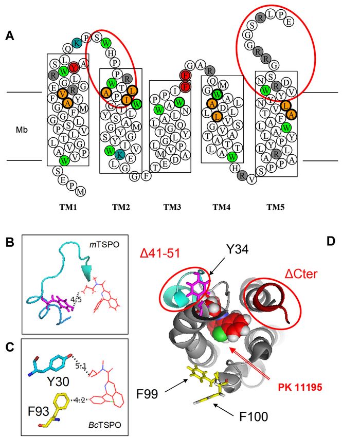
The treatment of detergent-solubilized rec-mTSPO with trypsin produced protein fragments
which the composition of changed with time (Figure 4A). The presence of PK 11195 protected against
The treatment of detergent-solubilized rec-mTSPO with trypsin produced protein fragments
which the composition of changed with time (Figure 4A). The presence of PK 11195 protected against
cleavage by slowing down the kinetics and led to a different pattern of protein fragments (Figure 4B,
left panel). A higher PK 11195 concentration showed increased protection against cleavage for theInt. J. Mol. Sci. 2019, 20, 1444 6 of 18
Int. J. Mol. Sci. 2019, 20, 1444 6 of 18
cleavage by slowing down the kinetics and led to a different pattern of protein fragments (Figure 4B,
leftincubation
same panel). A higher PK 11195
time (Figure concentration
4B, right showed increased
panel). Interestingly, at a highprotection against cleavage
PK 11195 concentration for the
(1 mM),
same
part incubation
of the timenot
protein was (Figure 4B,and
cleaved, rightonly
panel).
largeInterestingly, at a high
protein fragments PKobserved.
were 11195 concentration (1 the
This suggests mM),
part of the protein was not cleaved, and only large protein fragments were observed.
existence of conformational changes induced by ligand binding to its pocket that very likely reduces This suggests
the existence
trypsin access tooflysines
conformational changes
and arginines. induced
It has by ligand
to be noted binding
that most to arginines
of the its pocketare
that very
part likely
of the
reducesloops
cytosolic trypsin
andaccess to lysinesregion
the C-terminal and arginines. It hasconfirming
of the mTSPO, to be notedthethat most of theof
involvement arginines are in
these loops part
of the cytosolic loops
the ligand binding process. and the C-terminal region of the mTSPO, confirming the involvement of these
loops in the ligand binding process.
Figure
Figure4. The limited
4. The limitedtrypsin digestion
trypsin digestion of rec-mTSPO:
of rec-mTSPO: (A) The timetime
(A) The course of theofproteolytic
course cleavage
the proteolytic of
cleavage
rec-mTSPO solubilized
of rec-mTSPO in DPCin
solubilized byDPCtrypsin.
by In the first
trypsin. Inminutes
the firstofminutes
trypsin digestion,
of trypsintwo main fragments
digestion, two main
around 15 andaround
fragments 6.5 kDa15 were andobserved.
6.5 kDa The werelarger one disappeared
observed. The larger over
one time, whereasover
disappeared shorter peptides
time, whereas
below 6.5 kDa
shorter started
peptides below to appear.
6.5 kDa After
started 48toh appear.
of digestion,
After only
48 h very short peptides
of digestion, only veryaround
short2.6–3.7
peptides
kDa, probably
around 2.6–3.7corresponding
kDa, probably to corresponding
the transmembrane to the TM helices, wereTM
transmembrane visible. (B)were
helices, The visible.
effect of(B)PKThe
11195 upon
effect proteolytic
of PK 11195 upon cleavage: the time
proteolytic coursethe
cleavage: in time
the presence
course inofthe
100presence
µM PK 11195
of 100 (left
µM PK panel)
11195and(left
thepanel)
effect and
of increasing
the effect of theincreasing
concentration of PK 11195 of
the concentration with
PK 311195
min with
of trypsin
3 min digestion
of trypsin(right
digestionpanel):
(right
Thepanel):
regionsThehighlighted in red (in panel
regions highlighted in red(A))(incorrespond to the fragment
panel A) correspond at fragment
to the 5 kDa thatatis5notkDagenerated
that is not
in the presence of PK 11195, whereas the fragments around 15 kDa (highlighted
generated in the presence of PK 11195, whereas the fragments around 15 kDa (highlighted in in red in left panel
red in
in (B))
left panel in B) remain. Moreover, only large fragments are observed in the presence of of
remain. Moreover, only large fragments are observed in the presence of high concentrations high
PKconcentrations
11195 (red circle of in
PKright
11195panel
(redin (B)).in right panel in B).
circle
To further study the mechanism of PK 11195 ligand binding, we decided to mutate amino acids
and to measure the binding affinities of the mutants. Figure 3 displays the location of the mTSPOInt. J. Mol. Sci. 2019, 20, 1444 7 of 18
To further study the mechanism of PK 11195 ligand binding, we decided to mutate amino acids
and to measure the binding affinities of the mutants. Figure 3 displays the location of the mTSPO amino
acids involved in the binding pocket (2-D diagram, top panel). In determining the mutation strategy, we
took into consideration (i) our limited proteolysis data, (ii) previous reports of mutagenesis, and (iii) the
possible hydrophobic interactions between PK 11195 and aromatic residues. Not all trypsin cleavage
sites are immediately accessible to cleavage. Some of them are located close to the PK 11195 binding
site, such as R27, R32, K39, and R46, which are in the region connecting TM1 and TM2, as well as
R103, which is in the region connecting TM3 and TM4. Our selective labeling NMR spectra confirmed
the involvement in ligand interactions of the small helical loop containing K39. The large chemical
shift of K39 upon PK 11195 binding supports a strong environment change induced by the ligand.
Moreover, based on the available TSPO structural information, with and without ligand [22], together
with the hindered location of the binding pocket, we can speculate on a potential conformational
change involving hydrophobic interactions between PK 11195 and the aromatic residues. In line with
this, several residues are highly conserved in these regions, among which are the W33 and Y34 parts
of the end of TM1. A comparison of the NMR structures of WT and A147T mutant showed that
Y34 has two distinct orientations (toward and opposite of ligand, Figure 3), whereas W33 only has
one, suggesting a potential contribution of Y34 to the PK 11195 binding process and conformational
change. Thus, we mutated the highly conserved tyrosine Y34 into either serine or phenylalanine to
maintain the hydroxyl or phenyl group of the tyrosine, respectively. This was our first mutant. Our
second mutant was the ∆41–51, a large fragment at the end of the connecting region between TM1 and
TM2, previously described as having reduced binding properties [14] and containing two conserved
tryptophans (W42 and W47) and several other residues as part of the binding pocket (Figure 3A).
The third mutant targeted a highly conserved aromatic residue F100 from the short loop connecting
TM3 and TM4 that exhibited two different orientations toward the ligand when comparing the mTSPO
and BcTSPO structures (Figure 3), which was mutated into alanine, a short chain residue. Our fourth
mutant was the ∆153–169, a large fragment at the C-terminus of the protein (Figure 3A), previously
described as having reduced binding properties [14] that may stabilize the global 3-D structure of
the TSPO by interaction with the TM1-TM2 and TM3-TM4 loops [18]. It contains the cholesterol
recognition/interaction amino acids consensus (CRAC) pattern, i.e. the cholesterol binding site [13],
and atomic NMR structure shows close interactions between Y153 and Q38S (TM1) on one side and
between W155 and M105 (TM4) on the other side that might stabilize folding and stability. Thus, its
deletion might change the affinity and stoichiometry of binding by destabilisation. We also constructed
double aromatic mutants involving the two loops to test if there may be a “sandwich effect” that may
drive PK 11195 to its binding site.
In order to check if the mutations and, in particular, the deletions had an effect on the folding of
rec-mTSPO, we decided to characterize the secondary structure by recording the circular dichroism
spectra (Figure 5A) of the following point mutations and deletion mutants: Y34S, Y34F, F100A,
Y34F/F100A, Y34F/F99A, ∆41–51 and ∆153–169. First, these mutants were expressed in E. coli and
purified in SDS [13,19,23,24]. All mutants seemed folded since they exhibited at least a helical content
of around 35%, except mutants F100A and ∆41–51 which showed a level of helicity above 40%. We
previously showed (Figure S2) that rec-mTSPO had a higher level of helical content when SDS was
exchanged for DPC [19,25]. Therefore, we also recorded the circular dichroism spectra of each mutant
in DPC (Figure 5A). We observed an increase in the helical content close to 10% compared to SDS, but
some mutants such as Y34S and F100A showed smaller changes, indicating that aromatic interactions
might be involved in the folding process. The deletion mutant ∆41–51 that exhibited the highest
helical content in SDS (close to 50%) showed an even higher helical content in DPC (close to 60%).
Interestingly, both deletion mutants showed a high secondary structure content even when ten to
fifteen residues were removed.
The wild type (WT) TSPO has 12 tryptophans, and the intrinsic fluorescence can easily be
recorded. We previously showed that the fluorescence intensity is highly dependent on the detergentInt. J. Mol. Sci. 2019, 20, 1444 8 of 18
environment, with large increases when SDS was replaced by DPC [19]. Such increases reflect a change
in the tryptophan environment and/or conformational change. We measured the fluorescence intensity
Int. J. Mol. Sci. 2019, 20, 1444
of each mutant in the presence of both SDS and DPC and observed a large increase (more than a 8factor
of 18
of 2) for all mutants (Figure 5B). Interestingly, ∆41–51 that exhibits the largest helicity change still
than a factor of 2) for all mutants (Figure 5B). Interestingly, Δ41–51 that exhibits the largest helicity
shows a large fluorescence change, although 2 tryptophans have been deleted. Former studies reported
change still shows a large fluorescence change, although 2 tryptophans have been deleted. Former
the binding affinity measurements of the ligand to bacterial TSPO [26,27] using intrinsic fluorescence
studies reported the binding affinity measurements of the ligand to bacterial TSPO [26,27] using
changes. However, these previous reports showed that the addition of PK 11195 to WT TSPO induced
intrinsic fluorescence changes. However, these previous reports showed that the addition of PK 11195
a 100% decrease of fluorescence, which does not seem to be a measure of binding but rather a full
to WT TSPO induced a 100% decrease of fluorescence, which does not seem to be a measure of
quenching of the intrinsic fluorescence indicative of more nonspecific interactions. The measurement
binding but rather a full quenching of the intrinsic fluorescence indicative of more nonspecific
of “true” binding affinities is not really possible with detergent-solubilized rec-mTSPO, as it usually
interactions. The measurement of “true” binding affinities is not really possible with detergent-
gives affinities in the micro-millimolar range, probably due to an additional interaction between PK
solubilized rec-mTSPO, as it usually gives affinities in the micro-millimolar range, probably due to
11195 and detergent [10].
an additional interaction between PK 11195 and detergent [10].
A B
0,8
80
70 0,7
Fluorescence Arb Units
60 0,6
50 0,5
Helix, %
SDS SDS
40 0,4
DPC DPC
30 0,3
20 0,2
10 0,1
0 0,0
4F 0 A
T
4F
4S
A
A
4F
A
s
4S
W
sd
00
00
00
Y3
Y3
Y3 10
Y3
Y3
F1
F1
F1
T
W
F
4F
Y3
Figure 5. The circular dichroism (CD) and fluorescence studies of rec-mTSPO mutants: the CD-inferred
Figure 5. The circular dichroism (CD) and fluorescence studies of rec-mTSPO mutants: the CD-
alpha-helix percentage (A) and the fluorescence emission intensity (B) are shown for the wild type
inferred alpha-helix percentage (A) and the fluorescence emission intensity (B) are shown for the wild
(WT) as well as the mutants in the presence of sodium dodecyl sulfate (SDS) and DPC (blue and red
type (WT) as well as the mutants in the presence of sodium dodecyl sulfate (SDS) and DPC (blue and
bars, respectively).
red bars, respectively).
In our hands, the reconstitution of rec-mTSPO into proteoliposomes is the most efficient way to
In our
measure thehands, the reconstitution
PK 11195 of rec-mTSPO
high-affinity drug into proteoliposomes
ligand binding constants usingisradioactive
the most efficient way to
assays [23,28].
measure the PK 11195 high-affinity drug ligand binding constants using radioactive assays
The saturation curves of PK 11195 binding to the WT and mutants of TSPO reconstituted in liposomes [23,28].
Thepresented
are saturationincurves
Figureof6.PK 11195
The databinding to thetoWT
were fitted and the
obtain mutants of TSPO
affinity reconstituted
constant in liposomes
(Kd ) and stoichiometry
are presented
(B in Figure 6. The data were fitted to obtain the affinity constant (Kd) and stoichiometry
max ) (see Table 1). Kd varied from 8 nM for the WT to 150 nM for the double mutants (Y34F/F100A
(B max) (see Table 1). Kd varied from 8 nM for the WT to 150 nM for the double mutants (Y34F/F100A
and Y34F/F99A), whereas all the stoichiometries obtained were very close and equal to 25–30 nmol of
and Y34F/F99A), whereas all the stoichiometries obtained were very close and equal to 25–30 nmol
PK 11195 bound per mg of recombinant TSPO. This value of Bmax is consistent with a saturation of
of PK 11195 bound per mg of recombinant TSPO. This value of Bmax is consistent with a saturation of
1 PK 11195 per TSPO, taking into account that (i) about 50% of the protein is facing the inside of the
1 PK 11195 per TSPO, taking into account that (i) about 50% of the protein is facing the inside of the
proteoliposome, a classical observation after membrane protein reconstitution in proteoliposomes [29],
proteoliposome, a classical observation after membrane protein reconstitution in proteoliposomes
and (ii) a calculated maximal stoichiometry of approx. 50 nmol of PK 11195 bound per mg of
[29], and (ii) a calculated maximal stoichiometry of approx. 50 nmol of PK 11195 bound per mg of
recombinant TSPO (i.e., 1 mol of PK 11195 per mol of TSPO). Figure 6B shows that the point mutation
recombinant TSPO (i.e., 1 mol of PK 11195 per mol of TSPO). Figure 6B shows that the point mutation
of Y34 to serine (Y34S) led to a decrease in the apparent affinity compared with WT (Figure 6A),
of Y34 to serine (Y34S) led to a decrease in the apparent affinity compared with WT (Figure 6A),
which is even more significant than when tyrosine is replaced by phenylalanine (Y34F). Both deletion
which is even more significant than when tyrosine is replaced by phenylalanine (Y34F). Both deletion
mutants (∆41–51 and ∆153–169) had the same affinity decrease as for Y34F. Interestingly, compared to
mutants (Δ41–51 and Δ153–169) had the same affinity decrease as for Y34F. Interestingly, compared
the effect expected for the suppression of 10 residues, we observed only a mild decrease in affinity
to the effect expected for the suppression of 10 residues, we observed only a mild decrease in affinity
(Figure 6C [13,25]. The point mutation of the highly conserved F100 to alanine (F100A) alone induced
(Figure 6C) in agreement with previous studies [13,25]. The point mutation of the highly conserved
a small decrease in affinity for PK 11195 binding, but the double mutant (Y34F/F100A) of the two
F100 to alanine (F100A) alone induced a small decrease in affinity for PK 11195 binding, but the
highly conserved Y34 and F100 had a large effect (20-fold decrease) (Figure 6D). It has to be noted
double mutant (Y34F/F100A) of the two highly conserved Y34 and F100 had a large effect (20-fold
that the double mutant (Y34F/F99A), involving the less conserved F99, also had a large effect (20-fold
decrease) (Figure 6D). It has to be noted that the double mutant (Y34F/F99A), involving the less
conserved F99, also had a large effect (20-fold decrease, Table 1), enforcing the hypothesis of
hydrophobic interactions between PK 11195 and the aromatic residues driving the ligand to its
binding pocket.Int. J. Mol. Sci. 2019, 20, 1444 9 of 18
decrease, Table 1), enforcing the hypothesis of hydrophobic interactions between PK 11195 and the
Int. J. Mol. Sci. 2019, 20, 1444 9 of 18
aromatic residues driving the ligand to its binding pocket.
A B
25 25
20 20
15 15
10 10
5 5
0 0
0 10 20 30 40 0 10 20 30 40
P K 1119 5 , nM P K 1119 5 , nM
C D
25 25
20 20
15 15
10 10
5 5
0 0
0 10 20 30 40 0 10 20 30 40
P K 1119 5 , nM P K 1119 5 , nM
Figure 6. The saturation isotherms of [3 H]-PK 11195 binding to reconstituted rec-mTSPO in
proteoliposomes: (A) the wild type (WT) rec-mTSPO, (B) mutants Y34 (F and S opened and closed
Figure 6. The saturation isotherms of [3H]-PK 11195 binding to reconstituted rec-mTSPO in
squares, respectively), (C) deletion mutants (∆41–51 and ∆153–169 open and closed diamonds,
proteoliposomes: (A) the wild type (WT) rec-mTSPO, (B) mutants Y34 (F and S opened and closed
respectively) and (D) mutant F100A, double mutants Y34F/F100A and Y34F/F99A (open and closed
squares, respectively), (C) deletion mutants (Δ41–51 and Δ153–169 open and closed diamonds,
triangles and filled red circles, respectively). The lines represent the best-fit regression curves.
respectively) and (D) mutant F100A, double mutants Y34F/F100A and Y34F/F99A (open and closed
The dashed line in panels (B–D) corresponds to that of WT presented in panel (A).
triangles and filled red circles, respectively). The lines represent the best-fit regression curves. The
dashed
Table 1. line
Theinmutant
panels B, C and D
binding correspondsThe
parameters: to that of WTand
affinities presented in panel A.were determined
stoichiometries
by fitting the saturation curves of bound radioactive [3 H]-PK 11195 to rec-mTSPO reconstituted
inTable 1. The mutant binding parameters: The affinities and stoichiometries were determined by
proteoliposomes.
fitting the saturation curves of bound radioactive [3H]-PK 11195 to rec-mTSPO reconstituted in
proteoliposomes. Mutation Kd (nM) Bmax (nmol/mg)
WT 8±3 25 ± 3
Mutation
Y34S Kd±(nM)
40 7 Bmax (nmol/mg)
25 ± 5
Y34FWT 208±±33 27 ±±33
25
∆41–50Y34S 20 ± 8
40 ± 7 25 ±±58
25
∆C-ter (153–169) 20 ± 5 30 ± 10
Y34F 20 ± 3 27 ± 3
F100A 20 ± 5 25 ± 7
Δ41–50
Y34F/F100A 20 ±
150 ± 20 8 25
25 ±± 810
ΔC-ter (153–169) 15020±±205
Y34F/F99A 30
25 ±±1010
F100A 20 ± 5 25 ± 7
Y34F/F100A 150 ± 20 25 ± 10
Y34F/F99A 150 ± 20 25 ± 10
3. DiscussionInt. J. Mol. Sci. 2019, 20, 1444 10 of 18
3. Discussion
The recent determination of the atomic structure of TSPO from different species produced valuable
information about the PK 11195 ligand binding site [9,15,16,18]. However, the binding mechanism
still remains elusive. The mammalian mTSPO in DPC micelles, studied by NMR, was stabilized upon
ligand binding and displayed larger mobility and lower helix packing in the absence of PK 11195 than
in its presence [9,22]. Conversely, bacterial BcTSPO, studied by X-ray crystallography, displayed a
similar atomic structure [16] with or without PK 11195. Therefore, it is not clear whether or how the
ligand entered the binding pocket. Ligand binding is a dynamic process that involves several amino
acids in helix packing and binding pocket formation. In its stabilized form, 61 NOEs between mTSPO
and PK 11195 have been exploited for the NMR structure determination [9]. They involve 10 amino
acids (A23, V26, L49, A50, I52, W107, A110, L114, A147 and L150, displayed in orange in Figure 3
(top), except for W107 shown in green), but an atomic structure analysis suggested the involvement
of 5 additional amino acids (R46, W53, W95, D111 and W143), which are at a short distance from the
bound PK 11195. Nevertheless, the transition between the free and bound structures involves probably
even more residues and, in particular, those from cytosolic loops that are needed to reach the PK 11195
binding site located in between transmembrane helices.
Surprisingly, the deletion of several segments of TSPO did not induce a complete loss of PK 11195
binding as previously described [12,13] (Figure S1). The first part of TM1 (N-terminus, i.e., residues
2–20 or 5–20) can be removed, leading only to a reduced affinity [12,13]. The first part of TM2 (∆41–51)
can also be removed without a great loss of the PK 11195 binding (herein and in Reference [13]). L49
and A50, which have been described as part of the binding pocket [9], can be deleted (∆41–51) without
large decreases in the affinity (herein and in Reference [13]). These two amino acids are then neither
important to binding nor to the folding, in agreement with the conservation of the secondary structure
that we observed by circular dichroism. Similarly, W107, A110 and L114 can be removed (∆108–119)
while still measuring the PK 11195 binding [13]. Further on, a study of the ∆141–152 mutant showed
that the deletion of A147 and L150, two of the ligand-protein contact points, still kept almost 75% of
the PK 11195 binding [13]. This observation suggests that these two amino acid contributors to the
binding pocket are not essential.
Instead, other amino acids are crucial contacts, since a single point mutation at V26T, as well as a
deletion of 15–35 that includes V26, induces a complete loss of PK 11195 binding [12]. ∆15–35 lacks
Y34 for which we showed the contribution of its aromatic part in PK 11195 binding, suggesting the
necessity of aromatic residues in targeting PK 11195 to its hydrophobic pocket. This observation is
also supported by the effect of mutating F100 in the second loop and is reinforced by the effect of
the double mutation (Y34F/F100A) that had an even larger contribution than the single mutations
of Y34 and F100. However, not all aromatic residues present in the region connecting the two first
transmembrane helices are involved, since the mutation of W42 and W47 had no effect on the PK 11195
binding [12]. In addition, the mutation of charged residues in the first and second loops (R32G, K39G
and R103A) had no effect on the PK 11195 binding [12], suggesting that electrostatic interactions are
not required for the ligand–protein complex. Since the C-terminus (∆153–169 or ∆158–169) decreased
the binding of PK 11195 (herein and in References [12,13,25]), we can therefore propose a contribution
from the three cytosolic-facing regions to the targeting of PK 11195 to its binding pocket.
Human polymorphism rs6971, i.e., a natural mutation of A147T, is responsible for differences
in the affinity of some TSPO ligands [7,30,31]. The presence of alanine instead of threonine induces
a decrease of the affinity of one or two orders of magnitude for the PBR28 ligand [7], whereas it has
no effect on the PK 11195 ligand [30]. The mouse TSPO mutation of A147T reveals that the binding
pocket can adopt different “conformations” leading to slightly different structures [18]. Notably, V26 in
TM1 is significantly more dynamic than the rest of the residues [31] and is placed opposite to A147 in
TM5 in the PK 11195 binding cavity. Moreover, the TM1-TM2 loop conformation is divergent between
the two polymorphs. The bacterial RsTSPO A/T mutant reveals an "open" structure without the
ligand rather than a “closed” structure for native TSPO [15]. Several differences are clearly observedInt. J. Mol. Sci. 2019, 20, 1444 11 of 18
between the two structures; TM2 was more tilted, and TM5 was less kinked for the A/T mutant, but
more interestingly, the greatest change was observed in the loop connecting TM1 to TM2 (res29–40).
Namely, in the “open” structure, the loop was not observed in the 3-D crystal, presumably due to a
high flexibility. It is worth mentioning that the same loop was also poorly defined in the 2-D crystals
of the same RsTSPO by cryo-electron microscopy [26].
Interestingly, the atomic structure of TSPO from Rhodobacter sphaeroides has been determined [15]
with the protoporphyrin IX (PPIX) bound in the same pocket as PK 11195 in both mouse and Bacillus
cereus TSPO. Amino acids interacting with both ligands are either conserved or homologous (Figure 7).
Among them, F92 of RsTSPO, highly conserved among TSPOs, is closer to the PPIX ligand than
the equivalent F93 of BcTSPO to the PK 11195 ligand. We showed that the mTSPO mutation of
F100 to a short and nonaromatic residue such as alanine has a strong effect on the PK 11195 affinity
when coupled to another mutation of an aromatic residue in the first loop. Our data suggesting a
role for the loops in ligand recognition coupled with previous data indicating that the gating of the
high-affinity ligand binding site is controlled by TM1 and TM5 tend to clarify the mechanism of ligand
binding in TSPO. Whereas the C-terminus extending TM5 seems to participate to this process, the
N-terminus located on the opposite side of the ligand binding pocket is not involved. Accordingly,
the deletion of a long sequence from the N-terminus of Arabidopsis thaliana TSPO, AtTSPO, has no
effect on ligand binding [32]. Ligand binding might involve large changes in the structure of TSPO
as observed in different NMR studies [9,10,18,22]. However, one may raise the question of the effect
of detergents upon the structures determined by NMR when compared with structures determined
by x-ray crystallography that show smaller conformational changes. On the one hand, PK 11195
bound did not have the same conformation by itself and is not superposable in the binding pocket of
different atomic structures solved by NMR or x-ray crystallography. On the other hand, even if the
three-dimensional fold of the 5 TM domains is conserved, the one-to-one superposition of individual
TMs is different for the various TSPOs [9,15,16]. A recent paper suggests that high-resolution NMR
studies obtained for some membrane proteins in DPC detergent correspond to nonfunctional states [33].
Several atomic structures of membrane proteins have been resolved by x-ray crystallography in a
unique conformation, impairing our understanding of transport mechanisms, such as the case with
the ATP/ADP Carrier, which the structure of has been determined only in the presence of the strong
inhibitor carboxyatractyloside CATR [34]. Crystal formation may also constrain the conformation of
cytosolic domains, as is the case of the first atomic structure of Ca-ATPase [35], which exhibited a
domain separation that differs from the compact structure described by electron microscopy [36].
TSPO1 exhibits a high affinity for PK 11195, while its paralogous TSPO2 has been described as not
binding to PK 11195 [17]. The question raised is the origin of such a difference. Looking at the binding
of PK 11195 to mTSPO1 and BcTSPO1, which are selective to the same (R) enantiomer of PK 11195, the
bound ligand has different orientations of the rings and carbonyl group within the cavity, supporting a
possible plasticity of the binding site. However, ligand binding requires the presence of various amino
acids to make the pocket, and an analysis of the sequence alignment of the two proteins (Figure 8)
reveals little amino acid conservation between TSPO1 and TSPO2. The crucial residues in the TM1
(A23 and V26), as well as in TM3 (W95) and TM4 (W107), that make up the binding site of PK 11195 in
TSPO1 are not conserved in TSPO2. In particular, Y34 present in TSPO1 that we describe as important
for targeting PK 11195 to the binding pocket is absent in TSPO2. While the two paralogous TSPOs do
not share the same capacity to bind PK 11195, the site of cholesterol binding in the transmembrane
region, one of TSPO’s hallmark functions, is highly conserved [37–39].Int. J. Mol. Sci. 2019, 20, 1444 12 of 18
Int. J. Mol. Sci. 2019, 20, 1444 12 of 18
10 20 30 40
MPESWVPAVG LTLVPSLGGF MGAYFVRGEG LRWYASLQKP Mouse
TM1 --MKKSSIIV FFLTYGLFYV SSVLFPIDR- -TWYDALEKP 36 Bacillus
MNMDWALFLT FLAACGAPAT TGALLKPD-- -EWYDNLNKP 37 Rhodocbacter
. : ** * **
50 60 70
SWHPPRWTLA PIWGTLYSAM GYGSYIVWKE LG
TM2 SWTPPGMTIG MIWAVLFGLI ALSVAIIYNN YG 68
WWNPPRWVFP LAWTSLYFLM SLAAMRVA-- -- 67
* ** : * *: : . . :
80 90 100
GFTEDAMV PLGLYTGQLA LNWAWPPIFF G
TM3 ---FKPKT FWFLFLLNYI FNQAFSYFQF S 94
-QLEGSGQ ALAFYAAQLA FNTLWTPVFF G 93
:: : :* :. *
110 120
ARQMGWALA DLLLVSGVAT ATTLAWHRV
TM4 QKNLFLATV DCLLVAITTL LLIMFSSNL 123
MKRMATALA VVMVMWLFVA ATMWAFFQL 121
: . . ::: :.
130 140 150 160
S PPAARLLYPY LAWLAFATVL NYYVWRDNSG RRGGSRLPE
TM5 S KVSAWLLIPY FLWSAFATYL SWTIYSIN-- --------- 151
D TWAGVLFVPY LIWATATTGL NFEAMRLNWN RPEARA--- 158
: *: ** : * : * * .: *
Figure
Figure The
7. 7. sequence
The sequencealignment
alignment of mouse, Bacillus
of mouse, cereuscereus
Bacillus and Rhodobacter sphaeroides
and Rhodobacter TSPO: The
sphaeroides TSPO:boxes
The
depict the five transmembrane domains (labelled TM1 to TM5) as well as the short
boxes depict the five transmembrane domains (labelled TM1 to TM5) as well as the short alpha helix alpha helix in
continuity with the first TM for BcTSPO and RsTSPO. The black bold amino
in continuity with the first TM for BcTSPO and RsTSPO. The black bold amino acids are thoseacids are those involved
ininvolved
the transmembrane helices in the
in the transmembrane NMR
helices in atomic
the NMR structure
atomic obtained
structure from (mTSPO—PDB-2MGY).
obtained from (mTSPO—PDB-
The aminoThe
2MGY). acids involved
amino acidsininvolved
the PK 11195
in thebinding pocket
PK 11195 of thepocket
binding atomicofstructure obtained
the atomic either
structure from
obtained
NMR (mTSPO—PDB-2MGY) or x-ray (BcTSPO—PDB-4RYI) data are written
either from NMR (mTSPO—PDB-2MGY) or x-ray (BcTSPO—PDB-4RYI) data are written in red bold in red bold characters.
Those involved
characters. in the
Those protoporphyrin
involved IX (PPIX) binding
in the protoporphyrin pocketbinding
IX (PPIX) of the atomic
pocketstructure from the
of the atomic x-ray
structure
data of RsTSPO (PDB-4UC1) are in green.
from the x-ray data of RsTSPO (PDB-4UC1) are in green.Int. J. Mol. Sci. 2019, 20, 1444 13 of 18
Int. J. Mol. Sci. 2019, 20, 1444 13 of 18
10 20 30
MQLQGPVF VG-VPLLGPI LICMLIHQPS SRC-EDERKL Mouse 2
MRLQGAI- FVLLPHLGPI LVWLFTRDHM SGWCEGPRML
TM1 MAPPWVPAMG FTLAPSLGCF VGSRFVHGEG LRWYAGLQKP
Human
Human
2
1
MPESWVPAVG LTLVPSLGGF MGAYFVRGEG LRWYASLQKP Mouse 1
10 20 30 40
40 50 60
PWCPPHKVIL LVWVTIYSVM GYASYLVWKE LG
SWCPFYKVLL LVQTAIYSVV GYASYLVWKD LG
TM2 SWHPPHWVLG PVWGTLYSAM GYGSYLVWKE LG
SWHPPRWTLA PIWGTLYSAM GYGSYIVWKE LG
50 60 70
70 80 90
GGFRWPLAL PLGLYSFQLA LSWTFLVLF
TM3 GGLGWPLAL
G-FTEKAVV
PLGLYAVQLT
PLGLYTGQLA
ISWTVLVLF
LNWAWPPIF
G-FTEDAMV PLGLYTGQLA LNWAWPPIFF
80 90 100
100 110 120
LAADSPGLAL LDLLLLYGLV ASLVFIWQPI
TVHNPGLALL HLLLLYGLVV STALIWHPI
TM4 GARQMGWALV DLLLVSGAA AATTVAWYQV
GARQMGWALA DLLLVSGVAT ATTLAWHRV
110 120
130 140 150 160
NKLAALLLLP YLAWLTVTTA ITYRLWRDSL CPTYQP
NKLAALLLLP YLAWLTVTSA LTYHLWRDSL CPVHQPQPTEKSD
TM5 SPLAARLLYP YLAWLAFTTT LNYCVWRDNH GWRGGRRLPE
SPPAARLLYP YLAWLAFATV LNYYVWRDNS GRRGGSRLPE
130 140 150 160
Human Polymorphism A/T
Figure 8. The sequence alignment and helix assignment of isoforms 1 and 2 of mouse and human
Figure 8. The sequence alignment and helix assignment of isoforms 1 and 2 of mouse and human
TSPO: The boxes depict the five transmembrane helices (labelled TM1 to TM5). The numbers on top
TSPO: The boxes depict the five transmembrane helices (labelled TM1 to TM5). The numbers on top
correspond to the isoform 2 positions, whereas the numbers at the bottom correspond to isoform 1.
correspond to the isoform 2 positions, whereas the numbers at the bottom correspond to isoform 1.
The amino acids involved in the PK 11195 binding pocket of the atomic structure obtained from NMR
The amino acids involved in the PK 11195 binding pocket of the atomic structure obtained from NMR
data are written in red bold characters, and those observed at a short distance (3 angstroms) are in
data are written in red bold characters, and those observed at a short distance (3 angstroms) are in
normal red characters.
normal red characters.
4. Materials and Methods
4. Materials and Methods
4.1. Expression and Purification of Recombinant Mouse TSPO
4.1.Mouse
Expression
TSPOand(mTSPO)
Purification of Recombinant
was expressed inMouse
E. coliTSPO
BL21 DE3, grown up in an LB medium and
purified by His-binding to Ni-NTA chelation resin in
Mouse TSPO (mTSPO) was expressed in E. coli BL21 the presence
DE3,ofgrown
1% SDSupaccording
in an LB to published
medium and
protocols
purified [13,19,22,40].
by His-binding The Protein purity
to Ni-NTA wasresin
chelation analyzed
in the by SDS-PAGE
presence of 1%(12% acrylamide)
SDS according to run on a
published
protocols [13,19,22,40]. The Protein purity was analyzed by SDS-PAGE (12% acrylamide) run on aInt. J. Mol. Sci. 2019, 20, 1444 14 of 18
Protean II system (BioRad, Marnes la coquette, France). The protein levels were quantified by UV
spectra, using an absorption coefficient calculated from the amino acid sequence composition. When
needed, SDS was exchanged with 0.2 % dodecylphosphocholine (DPC-h38 or DPC-d38) before protein
elution from the Ni-NTA column (Qiagen, Les Ulis, France). Stable isotope labeled recombinant
mTSPO was expressed in a M9 minimal medium complemented with 1 g/L [15 N]-NH4 Cl and 4 g/L
glucose for fully labeled mTSPO, whereas selectively labeled mTSPO was obtained by adding a mixture
of amino acids containing the desired [15 N]-Lys to the M9 minimal medium [41,42].
4.2. NMR Experiments
The 2-D 1 H–15 N HSQC spectra were recorded at 30 ◦ C on a Bruker 500 MHz spectrometer
equipped with a cryogenic triple resonance probe with samples containing 0.1 mM mTSPO in
10 mM sodium phosphate (pH6) 90:10 H2 O:D2 O solutions containing 0.2% w/w DPC-d38 without
or with PK 11195 at 10–20 molar ratios of PK 11195 over mTSPO. The HSQC spectra (pulse program
hsqcetfp3gpsi, Bruker, Palaiseau, France) were collected with 2048 and 256 complex points in the direct
and indirect dimension, respectively. The data were processed with Bruker Topspin 3.2.
4.3. Trypsin Digestion
Proteolysis was performed by mixing rec-mTSPO (0.2 mg/mL) solubilized in DPC (0.1%) and
a phosphate buffer (10 mM at pH 7.0) with protease at a TSPO:Trypsin ratio of 30:1 (w/w). After
various incubation times at 30 ◦ C, the reaction was stopped by adding a Tosyl-L-lysyl-chloromethane
hydrochloride (TLCK trypsin inhibitor at a ratio over trypsin of 2.5 (w/w), and the sample was placed
on ice. PK 11195 from the stock solution (25 mM in ethanol) was added at chosen concentrations before
trypsin addition. A control reaction without PK 11195 was performed to confirm that ethanol up to
4% had no effect on proteolysis. Tricine SDS-PAGE (16.5%) silver stained gels were used to follow
proteolysis [43,44]. The apparent molecular weights of peptides generated by trypsin digestion were
calculated using the relative migration distance to that of a set of molecular weight standards.
4.4. Site-Directed Mutagenesis
Mutations were performed using the QuikChange Site-Directed Mutagenesis kit (Stratagene,
France) according to published protocols [25]. In brief, miniprep pET-PBR plasmid double-strand
DNA was used as a template. Synthetic oligonucleotide primer pairs containing a point mutation or
deletion, each complementary to the opposing strand of the vector, were extended during temperature
cycling by pfu DNA polymerase. The nicked vector DNAs containing the desired mutations were then
transfected into E. coli. The mutations and deletions generated were confirmed by DNA sequencing.
4.5. CD Spectroscopy
Far UV circular dichroism spectra were recorded on a Jobin-Yvon CD6 spectropolarimeter
operated at room temperature according to published protocols [19,25,45]. Briefly, a detergent
solubilized mTSPO sample (5 µM) in a 10 mM sodium phosphate buffer solution was placed in
a 0.2 mm path length quartz cuvette (Hellma, Paris, France). The CD spectra were recorded in the
185 to 270 nm wavelength range with a 0.2 nm step resolution, 1 s signal averaging time and 1 nm
bandwidth. The spectra were averaged over five scans, corrected for background and smoothed
over 25 points. A consensus secondary structure content was estimated by spectral deconvolution
using CONTINLL, CDSSTR and Selcon software and datasets of reference proteins SMP50 (37 soluble
proteins and 13 membrane proteins) and SP37 (37 soluble proteins) [46], as well as CDFriend.You can also read