Crystal structure of the BEACH domain reveals an unusual fold and extensive association with a novel PH domain
←
→
Page content transcription
If your browser does not render page correctly, please read the page content below
The EMBO Journal Vol. 21 No. 18 pp. 4785±4795, 2002

Crystal structure of the BEACH domain reveals an
unusual fold and extensive association with a novel
PH domain

Gerwald Jogl, Yang Shen, Damara Gebauer,                               A domain of ~300 amino acid residues near the
Jiang Li, Katja Wiegmann1,                                          C-terminus of CHS is highly conserved in a large family
Hamid Kashkar1, Martin KroÈnke1 and                                 of eukaryotic proteins (Figure 1B). This domain is known as
Liang Tong2                                                         the BEACH (for beige and CHS) domain (beige is the name
                                                                    for the CHS disease in mice) (Figure 1A) (Nagle et al.,
Department of Biological Sciences, Columbia University, New York,   1996). Many of the proteins that contain this domain are
NY 10027, USA and 1Institute of Medical Microbiology, Immunology
and Hygiene, University of Cologne, D-50935 KoÈln, Germany          very large (with >2000 residues) (Figure 1A) and have
2
                                                                    putative functions in vesicular transport or membrane
 Corresponding author                                               dynamics, such as CHS, neurobeachin (Nbea) (Wang
e-mail: tong@como.bio.columbia.edu
                                                                    et al., 2000), LBA (also known as BGL, CDC4L)
                                                                    (Feuchter et al., 1992; Wang et al., 2001) and LvsA
The BEACH domain is highly conserved in a large
                                                                    (Kwak et al., 1999; Cornillon et al., 2002). Nbea has been
family of eukaryotic proteins, and is crucial for their
                                                                    implicated in membrane traf®c in neuronal cells. It also
functions in vesicle traf®cking, membrane dynamics
                                                                    contains a motif that binds to the regulatory subunit of
and receptor signaling. However, it does not share any
                                                                    protein kinase A (PKA; Wang et al., 2000), and therefore
sequence homology with other proteins. Here we
                                    Ê resolution of the             can be classi®ed as an A kinase anchoring protein (AKAP)
report the crystal structure at 2.9 A
                                                                    (Colledge and Scott, 1999) (Figure 1A). LBA may have a
BEACH domain of human neurobeachin. It shows
                                                                    function in polarized vesicle traf®cking, and is localized to
that the BEACH domain has a new and unusual poly-
                                                                    vesicles after stimulation by lipopolysaccharide (LPS)
peptide backbone fold, as the peptide segments in its
                                                                    (Wang et al., 2001). Another function for BEACH-
core do not assume regular secondary structures.
                                                                    containing proteins is illustrated by the protein FAN,
Unexpectedly, the structure also reveals that the
                                                                    which is involved in signaling by the p55 tumor necrosis
BEACH domain is in extensive association with a
                                                                    factor receptor (TNFRI) (Figure 1A) (Adam-Klages et al.,
novel, weakly conserved pleckstrin-homology (PH)
                                                                    1996; Kreder et al., 1999).
domain. Consistent with the structural analysis, bio-
                                                                       The BEACH domain is crucial for the normal function
chemical studies show that the PH and BEACH
                                                                    of these proteins. Studies with FAN showed that its
domains have strong interactions, suggesting they may
                                                                    BEACH domain is required for downstream signaling by
function as a single unit. Functional studies in intact
                                                                    TNFRI (Adam-Klages et al., 1996). Genetic analyses of
cells demonstrate the requirement of both the PH and
                                                                    the naturally occurring mutations associated with CHS also
the BEACH domains for activity. A prominent groove
                                                                    reveal the functional importance of the BEACH domain.
at the interface between the two domains may be used
                                                                    These are all non-sense or frame-shift mutations, leading to
to recruit their binding partners.
                                                                    premature termination of the protein and the loss of the
Keywords: Chediak±Higashi syndrome/protein structure/
                                                                    BEACH domain (Figure 1A) (Introne et al., 1999; Certain
TNF signaling/vesicle traf®cking
                                                                    et al., 2000). In particular, one of these mutations occurs
                                                                    within the BEACH domain itself (Figure 1A) (Karim et al.,
                                                                    1997). Moreover, the severity of the disease is not
                                                                    correlated with the length of the remaining CHS protein,
Introduction
                                                                    suggesting the full-length protein is required for the normal
Chediak±Higashi syndrome (CHS) is a rare, autosomal                 function (Certain et al., 2000).
recessive disorder that can cause severe immunode®-                    The exact molecular function of the BEACH domain is
ciency, albinism and other diseases in humans and other             currently unknown. Moreover, the domain does not share
mammals (Spritz, 1998; Introne et al., 1999; Ward et al.,           any recognizable sequence homology to other proteins in
2000). Unless treated by bone marrow transplantation,               the database. To help obtain a complete understanding of
CHS patients generally die in childhood (Spritz, 1998;              this highly conserved domain, we have determined the
Introne et al., 1999; Certain et al., 2000). At the cellular        crystal structure of the BEACH domain of human Nbea at
level, a hallmark of the CHS disease is the presence of             2.9 AÊ resolution. The crystal structure reveals that the
giant, perinuclear vesicles (lysosomes, melanosomes and             BEACH domain has a new and unusual polypeptide
others) in many cells of the patients (Introne et al., 1999;        backbone fold. In addition, our structural studies reveal the
Dell'Angelica et al., 2000). Therefore, it has been                 presence of a novel, weakly conserved pleckstrin-hom-
proposed that the CHS protein, with 3801 amino acid                 ology (PH) domain just before the BEACH domain in the
residues (Figure 1A) (Barbosa et al., 1996; Nagle et al.,           primary sequence (Figure 1A). The structural analysis
1996; Perou et al., 1996; Ward et al., 2000), may have a            suggests strong interactions between the PH and the
crucial function in the fusion, ®ssion or traf®cking of these       BEACH domains, which we have con®rmed using protein
vesicles (Introne et al., 1999; Ward et al., 2000).                 binding assays. Functional studies with the protein FAN

ã European Molecular Biology Organization                                                                                  4785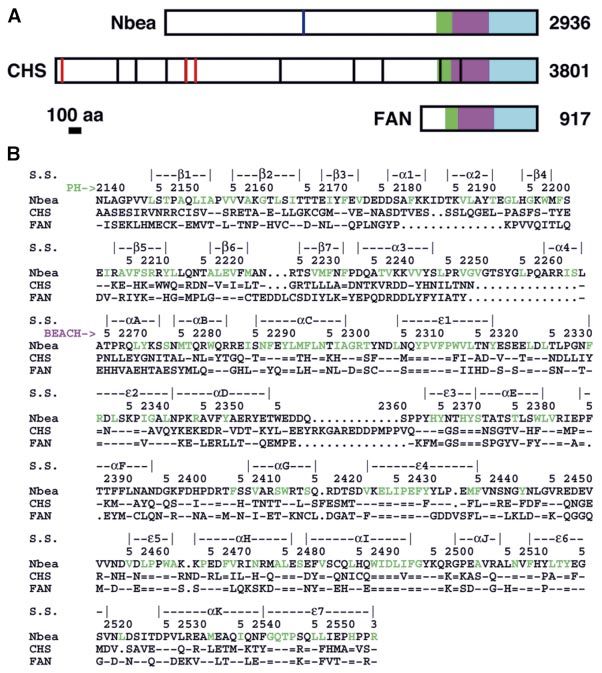
G.Jogl et al.

Fig. 1. Primary structures of the BEACH domain. (A) Schematic drawing of the primary structures of neurobeachin (Nbea), CHS and FAN. The BEACH
and WD40 domains are shown in purple and cyan, respectively. The PH domains, identi®ed from the current study, are shown in green. The bars in CHS
indicate naturally occurring frame-shift (black) or non-sense (red) mutations in CHS patients. The blue bar in Nbea represents the A-kinase anchoring
(AKAP) motif. (B) Alignment of the PH and BEACH domain sequences. The start of the PH and BEACH domains is indicated with the green and purple
arrows, respectively. The secondary structure elements (S.S.) are shown and labeled. The residue numbers shown are for Nbea. Residues in the core of
the structure are colored green in the Nbea sequence. The symbol `=' represents a residue that is strictly conserved among 30 BEACH domain sequences,
and 17 out of 18 identi®ed PH domain sequences. `±' indicates a residue that is identical to that in Nbea, and `´' represents a deletion.

demonstrate that both the PH and the BEACH domains are                        Nbea produced crystals suitable for X-ray structure
required to transduce the signal from TNFRI. Our studies                      determination. It covers residues 2137±2553 of human
therefore suggest that these two domains may function as a                    Nbea, and contains ~130 additional residues N-terminal
single unit. A prominent groove at the interface between                      to the conserved BEACH domain (Figures 1B and 3A,
these two domains may be important for binding the                            see below).
partner of these domains. The structural information                             The crystal structure of this BEACH domain was
provides a foundation for studying and understanding the                      determined at 2.9 A  Ê resolution. Initial phases for the
role of these BEACH-containing proteins in vesicle                            re¯ections were obtained from the selenomethionyl (Se-
traf®cking, membrane dynamics and receptor signaling.                         Met) single- and multi-wavelength anomalous diffraction
                                                                              method (Hendrickson, 1991). After solvent-¯attening,
                                                                              several a-helices could be recognized in the electron
Results                                                                       density map, but the overall quality of the map was still
Structure determination                                                       rather poor. The parameters of a non-crystallographic
To obtain crystals for structural analysis, we screened                       symmetry (NCS) axis were obtained based on the Se
many BEACH domains from several organisms for their                           positions, and re®ned in reciprocal space using the solvent-
bacterial expression, solution properties and crystalliza-                    ¯attened phases (Tong et al., 1992; Tong, 1993). The
tion behavior. An expression construct based on human                         electron density map after 2-fold NCS averaging was of

4786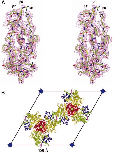
Crystal structure of the BEACH domain

Fig. 2. Electron density and crystal packing of the BEACH domain. (A) The electron density after 2-fold NCS averaging at 2.9 A  Ê resolution for strands
b5, b6 and b7 in the PH domain. The contour level is at 1s. (B) The crystal packing. The crystal unit cell is viewed along the c-axis. The PH domains
are shown in red and blue, and the BEACH domains are shown in yellow and green. The locations of the 61, 31 and 21 screw axes are shown. The
channel along the c-axis of the unit cell extends through the entire crystal. (A) was produced with Setor (Evans, 1993) and (B) was produced with
Molscript and Raster3D (Kraulis, 1991; Merritt and Bacon, 1997).

excellent quality (Figure 2A), and allowed the tracing of                      two molecules in the asymmetric unit are related by an
the entire protein. The current R-factor for the structure is                  improper NCS, with an angle of rotation of 163° and a
23.0% for all observed re¯ections to 2.9 A Ê resolution. The                   translation element of 32 AÊ . Each molecule has ~700 AÊ 2 of
statistics for structure determination and re®nement are                       buried surface area in this improper dimer, half of which is
summarized in Table I.                                                         due to the docking of a loop (residues 2438±2444) from
   There are two copies of the BEACH domain molecule                           one monomer into a groove on the surface of the other
in the asymmetric unit of the crystal, which belongs to                        monomer (see below). The equivalent residues in the other
space group P61. This gives rise to rather high solvent                        monomer (2440¢±2452¢, with `¢' indicating residues in the
content (75%) and Vm value (4.5 A    Ê 3/Da) for the crystal.                  second monomer) are disordered.
The molecules are clustered around the 31-screw axes in
the unit cell, forming long ®bers along the direction of the                   The BEACH domain has a new and unusual fold
c-axis of the unit cell (Figure 2B). The contacts between                      The structural analysis reveals that the BEACH domain
neighboring ®bers in the crystal are mediated by crystal-                      has a new polypeptide backbone fold, consistent with its
lographic 21-screw axes, also along the c-axis. Such a                         unique amino acid sequence. Moreover, the fold of the
packing arrangement produces large channels (~140 A      Ê in                  BEACH domain is quite unusual in that it contains several
diameter) along the 61-screw axes that extend throughout                       segments that are either completely buried in the
the entire crystal (Figure 2B). The high solvent content of                    hydrophobic core or help to enclose it, but these segments
these crystals may be correlated with their poor X-ray                         can not be classi®ed as b-strands as they are not fully
diffraction quality.                                                           extended (Figure 3A and B). In addition, only a few main-
   Light scattering studies showed that the protein exists as                  chain hydrogen bonds are made among these segments.
monomers in solution (data not shown). In the crystal, the                     Many of the main-chain amides and carbonyls of these

                                                                                                                                                 4787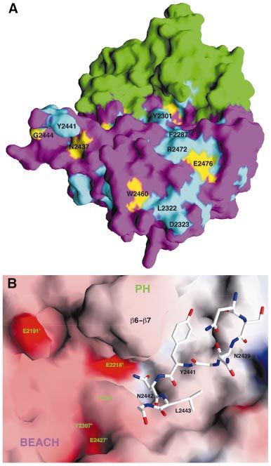
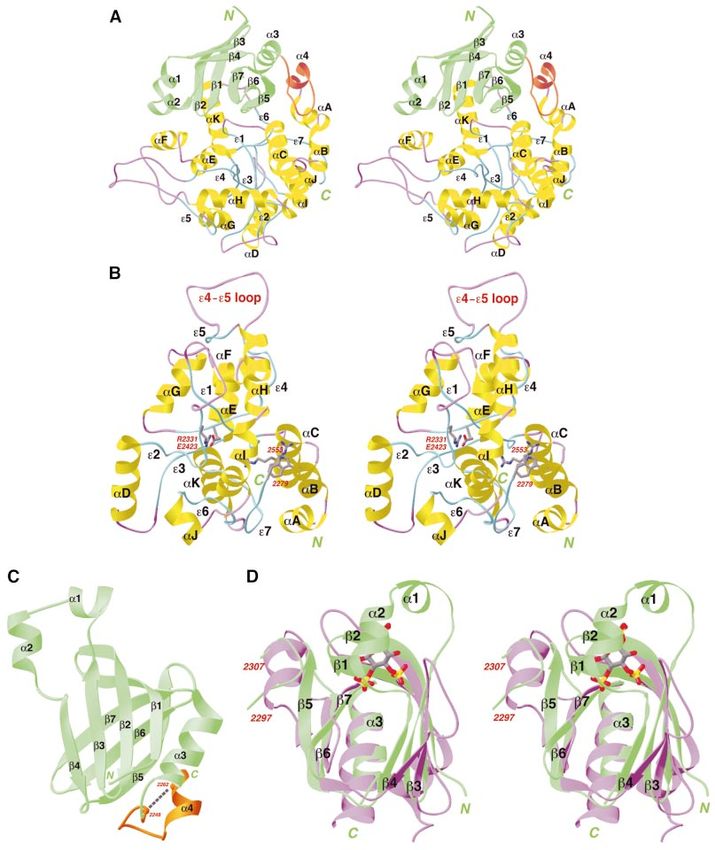
G.Jogl et al.

Fig. 3. Structures of the BEACH and PH domains. (A) Schematic stereoview of the structure of the PH±BEACH domains of human Nbea. The PH
domain is shown in green, and the linker between the two domains in orange. (B) Schematic stereoview of the structure of the BEACH domain of
human Nbea. The extended segments are shown in cyan, the a-helices in yellow, and the loops in purple. The side chains of residues W2279, R2299,
R2331, E2423 and R2553 are shown as stick models. (C) Schematic drawing of the structure of the PH domain of human Nbea. The b-strands are
shown as arrowed ribbons. Also shown is the linker segment that connects to the BEACH domain (in orange). (D) Superposition of the structure of
the PH domains of Nbea (in green) and phospholipase C (PLC, in purple) (Ferguson et al., 1995). The phospholipid ligand in the latter structure is
shown as a stick model. The aC±e3 segment from the BEACH domain (residues 2297±2307) is located in the same position as the b5±b6 insertion in
the PLC PH domain, which is also the place where several other PH domains bind peptide ligands. Produced with Ribbons (Carson, 1987).

4788
Crystal structure of the BEACH domain
segments are hydrogen-bonded to the side-chains of residues (Figure 1B). However, the ®rst conserved residue
conserved amino acids in the domain instead. of the domain is Trp2279, and the sequence conservation
For ease of discussion, these partially extended seg- for the 15 residues N-terminal to this Trp residue
ments that help to form the hydrophobic core of the (2264±2278) is much weaker than the rest of the
domain are named e1 to e7, starting from the N-terminus BEACH domain (Figure 1B). This exempli®es the dif®-
of the domain (Figure 3B). Of the seven segments, e1, e4 culty of domain parsing based solely on sequence com-
and e7 contain regions that are completely buried in the parisons. Remarkably, the side chain of Trp2279 shows
core of the domain (Figure 3B). These residues are highly amino-aromatic interactions with the side chain of
conserved among the BEACH domains (Figure 1B), Arg2553 (Figure 3B), the last highly conserved residue
including a buried ion pair between Arg2331 and in the BEACH domain (Figure 1B).
Glu2423 (Figure 3B). In addition to these segments, the
structure also contains 11 a-helices (aA through aK). The structure reveals a novel PH domain just prior
They are arranged on the periphery of the structure, but to the BEACH domain
help to enclose the core of the domain (Figure 3B). While the BEACH domain is highly conserved, the amino
Based on the structural analysis, the BEACH domain of acid sequences outside this region show much greater
human Nbea covers residues 2264±2553 and contains 290 variation (Figure 1B). From a detailed sequence analysis,
we found that a 130-residue segment just N-terminal to the
BEACH domain in the primary sequence is weakly
Table I. Summary of crystallographic information conserved among many of these proteins, with ~20%
sequence identity (Figure 1B). Therefore, we engineered
Data processing statistics expression constructs that contained this additional 130-
Wavelength l2 l3 l4
residue segment at the N-terminus. The crystal structure
Ê)
Maximum resolution (A 2.9 2.9 2.9 revealed that this N-terminal segment forms a complete
No. of observations 316 221 355 843 391 076 domain in itself (Figure 3A and C). Unexpectedly, the
Rmerge (%)a 8.3 7.9 8.5 backbone fold of this domain is identical to that of PH
No. of re¯ections 80 562 80 025 79 148 domains (Blomberg et al., 1999). Extensive sequence
Completeness (%) 99.4 99.2 98.7
searches failed to classify this region in Nbea as a PH
Structure re®nement statistics domain, identifying this as a PH domain with a novel
sequence.
Resolution range for re®nement Ê
30±2.9 A Like the other PH domains, the PH domain in Nbea
Completeness (%) 99.2
R-factorb (%) 23.0
contains a seven-stranded b-sandwich (b1 to b7), with an
Free R-factor (%) 26.4 a-helix (a3) near the C-terminus that closes off one of the
R.m.s. deviation in bond lengths (AÊ ) 0.008 open ends of the sandwich (Figure 3C). The structure of
R.m.s. deviation in bond angles (°) 1.3 the b-sandwich core of this PH domain is highly
a
XX XX homologous to those of other PH domains, with a root
Rmerge jIhi ÿ hIh ij= Ihi Ê for ~80 equiva-
h i h i
mean squares (r.m.s.) distance of ~2.5 A
X X lent Ca atoms (Figure 3D). The homology to the other PH
b
R jFho ÿ Fhc j= Fho domains at the amino acid sequence level, however, is
h h
much lower, in the 6±12% range for structurally equiva-
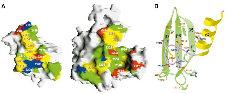
Fig. 4. The interface between the PH and BEACH domains. (A) Molecular surface of the PH and BEACH domains. Residues in the PH±BEACH inter-
face are shown in yellow for hydrophobic residues, green for polar residues, red for acidic residues, and blue for basic residues. (B) Schematic drawing
of part of the interface between the PH and BEACH domains. The exposed residues of the back sheet (b5, b6 and b7) of the PH domain, shown in
green, interact with the aC±e1 linker of the BEACH domain (in purple). (A) was produced with Grasp (Nicholls et al., 1991) and (B) was produced
with Ribbons (Carson, 1987).
4789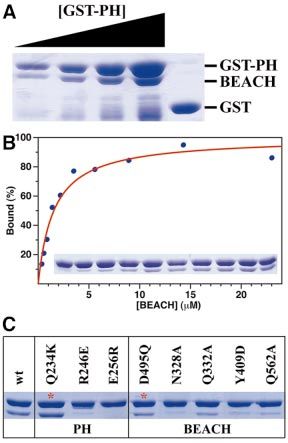
G.Jogl et al.

                                                                           the interface, which is mostly hydrophobic or polar in
                                                                           nature (Figure 4A). A major portion of this interface
                                                                           consists of residues 2295±2306 from the BEACH domain
                                                                           (the aC±e1 linker) packing against the `back sheet'
                                                                           (consisting of strands b5, b6, b7 and b1) of the b-sandwich
                                                                           of the PH domain (Figure 4B). Interestingly, the equiva-
                                                                           lent surface areas in several other PH domains are also
                                                                           used for protein±protein interactions (Blomberg et al.,
                                                                           1999).
                                                                              Many of the residues in the PH±BEACH interface are
                                                                           conserved among these proteins. For example, four of the
                                                                           ®ve highly conserved residues in the PH domains
                                                                           (Figure 1B) are located in this interface, a remarkable
                                                                           distribution considering the low degree of sequence
                                                                           conservation of the PH domains. These conserved residues
                                                                           include Arg2208 (in strand b5) and Glu2218 (b6), which
                                                                           form an ion pair on the surface of the PH domain and are
                                                                           buried in this interface (Figure 4B). In addition, Arg2208
                                                                           is located near the C-terminus of helix aC in the BEACH
                                                                           domain, and may therefore have favorable interactions
                                                                           with the dipole of this helix (Figure 4B). This sequence
                                                                           conservation suggests that similar PH±BEACH interfaces
                                                                           may be present in other BEACH-containing proteins.
                                                                              The linker between the PH and BEACH domains covers
                                                                           residues 2246±2263 in Nbea (Figure 3A). Neither its
Fig. 5. Biochemical evidence for interactions between the PH and           sequence nor its length are conserved among the different
BEACH domains. (A) Protein binding assays with wild-type PH (as
GST fusion) and BEACH domains of human FAN. To show the dose-
                                                                           proteins. Nbea appears to have an insertion of >13 residues
responsive nature of the interaction, the amount of GST±PH was in-         as compared with CHS or FAN in this region (Figure 1B),
creased, while that of BEACH was held constant. The speci®city of the      while the yeast YCR032W protein has an insertion of >40
interaction was indicated with the GST control. (B) Binding isotherm       residues. The structure shows, however, that the Ca atoms
of GST±PH and BEACH. The experimental observations from the GST            of 2248 and 2262 in Nbea are within 5 A     Ê of each other
pull-down experiment are shown in the inset and plotted as the blue
dots. The red line represents a hyperbolic ®t to the experimental data.    (Figure 3C), suggesting that insertions (such as in
(C) Mutations in the PH±BEACH interface disrupts their interactions.       YCR032W) or deletions (such as in CHS and FAN) in
Mutations in the PH and BEACH domains were selected based on the           this linker may be accommodated with minimal disturb-
structural information. The two control mutations are indicated with red   ances to the PH±BEACH interface.
asterisks.
                                                                           Biochemical evidence for interactions between the
                                                                           PH and BEACH domains
lent residues. In addition, the Nbea PH domain contains an                 The extensive interface between the PH and BEACH
insertion of two helices (a1 and a2) between strands b3                    domains suggests there may be strong interactions
and b4 (Figure 3C). The low sequence homology, together                    between the two domains. To obtain biochemical evidence
with the unique pattern of insertions/deletions, may be the                for such interactions, we performed protein binding
reason why sequence analyses failed to identify this region                assays using puri®ed PH domain fused with glutathione
in Nbea as a PH domain.                                                    S-transferase (GST±PH) and the puri®ed His-tagged
   PH domains are known to have a variety of functions,                    BEACH domain of the protein FAN (Figure 1A) (Adam-
including phospholipid binding, phosphotyrosine binding                    Klages et al., 1996). The experiments clearly demonstrate
[for the related phosphotyrosine binding (PTB) domains]                    the strong and dose-responsive interactions between the
and protein±protein interactions (Blomberg et al., 1999).                  PH and the BEACH domains of FAN (Figure 5A). This
The phospholipids generally bind on either side of the loop                con®rms the structural information, and also supports the
connecting b1 and b2 (Figure 3D). In the PH domain of                      conserved nature of the PH±BEACH interface as observed
Nbea, however, one of these binding sites is occupied by                   in our Nbea structure.
the a2 helix, while the other is blocked by residues                          To obtain an estimate for the Kd of the interactions
2302±2307 from the BEACH domain (Figure 3D). The                           between these two domains, we immobilized a constant
structural analyses therefore suggest that the PH domain of                amount of the GST±PH domain on glutathione±agarose
Nbea is probably not involved in phospholipid binding.                     beads and introduced increasing amounts of the BEACH
Instead, this domain may be involved in protein±protein                    domain. The resulting binding data can be readily ®tted to
interactions (see below).                                                  a hyperbolic curve, suggesting a 1:1 ratio in the interaction
                                                                           between these two domains (Figure 5B). The Kd value
An extensive, conserved interface between the PH                           obtained from this binding isotherm is ~1 mM, consistent
and BEACH domains                                                          with the structural information. However, in the native
The structural analysis revealed an extensive interface                    protein, the two domains are linked covalently. This
between the PH and BEACH domains in Nbea (Figure 3A).                      should produce an even stronger interaction between the
About 1100 AÊ 2 of surface area in each domain is buried at                two domains.

4790
Crystal structure of the BEACH domain
interface from the structural information. Similarly, the
effects of mutations in the BEACH domain are generally
in agreement with the structural observation (Figure 4B).
For example, the N328A mutation has a greater effect on
the interaction than the Q332A mutation, and N328 has a
larger surface area burial (120 A Ê 2) than Q332 (41 AÊ 2) in
the interface. Overall, the results from the protein binding
experiments con®rm our observations from the crystal
structure. Therefore, the strong association between the
PH and BEACH domains may be a conserved feature, and
the two domains may function as a single unit.
Both the PH and the BEACH domains are required
for signaling by FAN
Next, we characterized the effects of the PH and BEACH
domains on the biological functions of the protein FAN.
We selected this protein for assessing the structural
information as it is one of the few BEACH-containing
proteins that have been examined in some detail (Adam-
Klages et al., 1996; Kreder et al., 1999). Prior studies
demonstrated that the WD40 domain in this protein,
located at the extreme C-terminus (Figure 1A), is neces-
sary and suf®cient for binding to a peptide segment in the
intracellular domain of TNFRI (Adam-Klages et al.,
1996). Transfection experiments showed that this domain
by itself has dominant-negative effects on FAN signaling,
suggesting that the other parts of the molecule (BEACH
and N-terminal domains; Figure 1A) may have crucial
functions in downstream events in the pathway.
To assess the functional roles of the PH and BEACH
domains in the signal transduction by FAN, we created the
Fig. 6. Functional studies of the PH and BEACH domains of FAN. BEACH-WD40 (missing the N-terminal 275 residues,
(A) The activity of neutral sphingomyelinase after TNF activation was D1±275) and the PH±BEACH-WD40 (D1±178) deletion
determined for mouse FAN±/± ®broblasts transfected with vector con- mutants. The ability of these mutants to rescue signaling in
trol, full-length FAN, BEACH-WD40 (D1±275) and PH±BEACH- FAN±/± mouse ®broblasts (Kreder et al., 1999) were then
WD40 (D1±178) deletion mutants. (B) The effects of FAN mutants
in the activation of neutral sphingomyelinase in mouse FAN±/± determined. While the BEACH-WD40 mutant could not
®broblasts. The error bars represent observations from three rescue the knock-out phenotype, the PH±BEACH-WD40
independent experiments. mutant restored wild-type level FAN activity to these cells
(Figure 6A). Moreover, the BEACH-WD40 mutant pro-
duced a dominant-negative effect (95% inhibition) when
To characterize further the interactions at the transfected into human embryonic kidney (HEK) 293 cells
PH±BEACH interface, we introduced mutations in this that express endogenous wild-type FAN. In contrast,
interface in the protein FAN based on the structural transfection of the wild-type or the PH±BEACH-WD40
information, and determined their effects on the inter- mutant has little effect on the function of the endogenous
actions by the protein binding assay. The mutants created FAN. This shows that the BEACH domain cannot function
include R246E (R246 in FAN is equivalent to R2208 in independently of the PH domain in FAN signaling, and is
Nbea) and E256R (2218) in the PH domain, and N328A therefore consistent with our structural and biochemical
(2302), Q332A (2306), Y409D (2388) and Q562A (2540) observations that the PH and BEACH domains function as
in the BEACH domain. (R2208, E2218, N2302 and Q2306 a single unit.
can be seen in Figure 4B, and residues 2388 and 2540 can Moreover, single-site mutations in the PH±BEACH
be seen in Figure 4A.) Two additional mutants, Q234K interface can also reduce the signaling by the protein FAN
(2196) in the PH domain and D495Q (2473) in the (Figure 6B), supporting the functional importance of this
BEACH domain, were generated as controls, as these interface. The expression levels of the different mutants
residues are poorly conserved and located outside the were checked using anti-FLAG western analyses.
PH±BEACH interface based on the structure (Figure 1B). Although there are some inconsistencies in individual
Our results showed that mutations at the interface in experiments, we obtained functional data on every single-
either the PH or the BEACH domain can signi®cantly site mutant that is expressed at signi®cant levels. A clear
affect the interactions between the two domains, whereas reduction in the signaling activity of FAN was observed
mutations outside this interface had little effect for those mutants in the PH±BEACH interface (R246A,
(Figure 5C). In particular, the R246E and E256R muta- F258A and N328A) that have comparable expression
tions in the PH domain reduced the interactions such that levels. The effects of mutations at the Arg246 and Asn328
they were barely detectable by Coomassie Blue staining, positions have also been assessed by the GST pull-down
consistent with the important roles of these residues in the experiments (Figure 5C). The effects of the mutations in
4791G.Jogl et al.
(Figure 6B). Glu448 (equivalent to Glu2427 in Nbea) is
highly conserved and partly exposed on the surface
(Figure 7B). Our mutation result shows that this residue
is not required for the function of FAN (Figure 6B).
A possible binding site at the interface between
the PH and BEACH domains
To identify possible biological functions for the BEACH
domain based on the crystal structure, we ®rst examined
the locations of the conserved residues in this domain. This
analysis showed that most of these residues are located in
the core of the domain (Figure 1B). Few of the strictly
conserved residues that could have catalytic activities are
exposed on the surface of the BEACH domain (Figure 7A),
and we have mutated some of these residues in the
functional assay (Figure 6). Therefore, based on the
currently available structural and mutagenesis data, it is
unlikely that the BEACH domain is an enzyme.
Our structural and biochemical evidence showed that
the BEACH domain may function together with the PH
domain. There is a prominent groove at the interface
between the two domains in the current structure, and there
is a higher concentration of exposed, mostly conserved
residues in this region (Figure 7A). Moreover, in the
improper dimer in the crystal, the loop between the e4 and
e5 segments of one molecule is docked into part of this
groove of the other molecule (Figure 7B). The strongest
interaction in this area is mediated by residues Tyr2441-
Asn-Leu2443 (Figure 7B). Preliminary binding assays
based on ¯uorescence perturbation measurements suggest
that the af®nity between a 12-residue peptide (2436-
VNSNGYNLGVRE-2447) and Nbea is in the micromolar
range, whereas little binding was observed for the
equivalent peptide carrying the Y2441A mutation (data
not shown). While it is probably unlikely that this mode of
interaction between the two BEACH domain molecules is
biologically relevant, this observation does offer the
Fig. 7. A groove at the interface between PH and BEACH domains. tantalizing suggestion that this groove between the PH
(A) Molecular surface of the PH±BEACH domains, with the PH do- and BEACH domains may be involved in binding a partner
main in green and BEACH domain in purple. Strictly conserved resi-
dues are colored in yellow, and mostly conserved residues in cyan.
(possibly a peptide segment) in the natural function of the
(B) Docking of residues in the e4±e5 loop of one BEACH molecule PH±BEACH domains.
into the groove of the other in the crystal. The molecular surface of the
second PH±BEACH molecule (residue numbers 2137¢±2553¢) is col-
ored according to electrostatic potential. Produced with Grasp (Nicholls Discussion
et al., 1991).
The BEACH domain has so far only been found in
eukaryotes, and the sequences of this domain are highly
conserved among these proteins. For example, the
the signaling assay (Figure 6B) are smaller than those in BEACH domains of human Nbea (Wang et al., 2000)
the biochemical assay (Figure 5C), which may be for two and yeast YCR032W share 46% amino acid sequence
reasons. First, the biochemical assays are based on identity. A total of 31 BEACH domain sequences are
Coomassie Blue staining. Weaker interactions cannot be currently known, which show that many genomes carry
detected with this assay, but such interactions might still more than one BEACH-containing protein. The human
be able to function in FAN signaling. Secondly, and genome may have eight such proteins, including CHS,
perhaps more importantly, the biochemical assays detect Nbea, FAN, LBA and KIAA1607 (Nagase et al., 2000).
the interactions of PH and BEACH domains as separate Nbea and LBA belong to a subfamily of BEACH-
entities, while the two domains are covalently linked in the containing proteins that have orthologs in many other
natural FAN protein. This should increase the af®nity organisms, such as AKAP550 in Drosophila melanogaster
between the two domains and dampen the effect of (Han et al., 1997) and the uncharacterized open reading
mutations in their interface. We have also checked the frame F10F2.1 in the Caenorhabditis elegans genome. In
effects of mutations outside this interface. The Asn459 and addition, many of the proteins in this subfamily can also be
Leu463 residues are in the e4±e5 loop, one of the longest classi®ed as AKAPs, as they contain the motif that binds to
loops in the BEACH domain structure (Figure 3B). Their the regulatory subunit of PKA (Colledge and Scott, 1999).
mutations have little impact on the function of FAN Therefore, one function of these proteins may be to direct
4792Crystal structure of the BEACH domain
PKA to proper locations in the cell. However, the AKAP amino acid sequences of these proteins appear to be
motif covers only ~20 amino acid residues, and therefore unique. As illustrated by the BEACH domain, they are not
accounts for only a tiny fraction of the residues in these homologous to other protein sequences in the database,
proteins (Figure 1A). which makes it nearly impossible to infer the biological
In all the BEACH-containing proteins, the BEACH functions of these proteins based solely on sequence
domain is located just prior to a WD40 domain in the analysis. Therefore, elucidating the three-dimensional
primary sequence, which is located at the extreme structures of these proteins is a crucial component in
C-terminus of most of these proteins (Figure 1A). The understanding their functions. Our structural, biochemical
WD40 domain is found in a large family of proteins, and is and functional analyses of the PH and BEACH domains of
believed to mediate protein±protein interactions (Neer human Nbea represent the ®rst step in this process, and
et al., 1994). This domain in the protein FAN is necessary suggest that there may be many more surprises in the
and suf®cient for the interactions with TNFRI (Adam- studies of this important family of proteins.
Klages et al., 1996), thereby mediating the recruitment of
FAN to the receptor. The function of the WD40 domains in
the other BEACH-containing proteins is currently un- Materials and methods
known, although it is likely that these domains are also Protein expression and puri®cation
involved in recruiting the proteins to the proper locations Residues 229±645 of the KIAA1544 protein (Nagase et al., 2000), a
in the cell. putative human ortholog of murine Nbea (sharing 99% amino acid
The BEACH domain is the only domain that is highly sequence identity) (Gilbert et al., 1999; Wang et al., 2000), was
conserved among this family of proteins, many of which subcloned into the pET28a vector (Novagen) and overexpressed in
Escherichia coli at 20°C. The expression construct contains an
contain >2000 amino acid residues (Figure 1A). This N-terminal His6 tag, and covers the conserved BEACH domain together
strong conservation gives the impression that this domain with an additional 130 residues at the N-terminal end. The soluble protein
might function as an independent module (a cassette) in was bound to nickel±agarose af®nity resin (Qiagen), and eluted with a
these proteins. Surprisingly, our studies showed that the buffer containing 20 mM Tris pH 8.5, 250 mM NaCl and 150 mM
imidazole. The protein was puri®ed further by anion exchange
BEACH domain has intimate contacts with a novel PH chromatography at pH 8.5, using a linear gradient of 10±500 mM NaCl
domain just before it in the primary seqeunce. The concentration. Finally, the protein sample was puri®ed by gel ®ltration
biochemical and biological data con®rm that the two chromatography, in a running buffer containing 20 mM Tris pH 8.5,
domains may function as a unit, even though the PH 200 mM NaCl and 10 mM dithiothreitol (DTT). The protein fractions
from this column were pooled and concentrated to 30 mg/ml. Glycerol
domain is conserved at a much lower level. Therefore was added to 5% (v/v) concentration, and the protein sample was ¯ash-
these two domains do not behave like `beads on a string', frozen in liquid nitrogen and stored at ±80°C. The N-terminal His tag was
as might be implied from sequence comparisons. It will be not removed for crystallization.
interesting to study whether the functions of these proteins For the production of selenomethionyl proteins, the expression
require close associations between the PH±BEACH unit construct was transformed into DL41(DE3) cells. Bacterial growth was
carried out in de®ned LeMaster media (Hendrickson et al., 1990), and the
and other domains within them (e.g. the WD40 domain). protein was puri®ed using the same protocol as for the wild-type protein.
In most protein structures, the main-chain segments that The successful incorporation of selenomethionyl residues was con®rmed
are buried in the core of the structure assume regular by MALDI-TOF mass spectrometry.
secondary structures (a-helix or b-sheet). The structure of
Protein crystallization
the BEACH domain is rather unusual in that none of the Crystals of the BEACH domain were obtained at 4°C by the sitting-drop
segments in the core of the structure assume regular a- or vapor diffusion method. The reservoir solution contained 100 mM Tris
b-conformation (Figure 3B), and the BEACH domain pH 7.6, 4% (w/v) PEG8000 and 2 mM DTT. Crystals in the shape
lacks extensive main-chain hydrogen-bonding interactions of hexagonal prisms generally took 2 weeks to grow to full size
among the segments (e1±e7) in its core. Therefore, (0.3 3 0.3 3 1 mm3). For cryo protection, the crystals were transferred
in a few steps to an arti®cial mother liquor containing 20 mM Tris pH 8.3,
interactions involving side chains are likely to be 5% (w/v) PEG8000 and 25% (v/v) PEG200. The largest crystals generally
extremely important for the stability of this domain. This did not survive this treatment, and X-ray diffraction analyses and data
is consistent with our observation that most of the collection were performed with crystals of size 0.1 3 0.1 3 0.6 mm3.
conserved residues are located in the core of the domain, Most of the crystals were highly mosaic and had very weak X-ray
diffraction.
and the high degree of sequence conservation of this
domain may be required to stabilize its fold. Data collection
In contrast, our identi®cation of a PH domain with novel X-ray diffraction data to 2.9 AÊ resolution were collected on a Mar CCD at
amino acid sequences demonstrates the strong stability of the 32-ID beamline (ComCAT) of the Advanced Photon Source (APS)
the PH fold, which is probably provided by the many (Table I). Four wavelengths were used for collecting the selenomethionyl
MAD data: l1 (12 500 eV, 0.9764 A Ê , low-energy remote), l2 (12 660
main-chain hydrogen-bonding interactions among its eV, 0.9793 A Ê , edge), l3 (12 663 eV, 0.9791 A Ê , peak) and l4 (12 800 eV,
seven b-strands (Figure 3C). This would in turn allow 0.9686 A Ê , high-energy remote). The crystal-to-detector distance was
large variation in the side chains, and hence little 230 mm, and inverse beam geometry was used to collect the anomalous
conservation of the amino acid sequences of the domain. data. The diffraction images were processed and scaled with the HKL
package (Otwinowski and Minor, 1997). The crystal belongs to the space
The resulting differences in the surface decorations on group P61, with cell dimensions of a = b = 179.9 A Ê , and c = 98.5 A
Ê.
this fold de®ne the unique biochemical functions of the Even though the crystal was kept at 100 K throughout the data collection,
various PH domains, such as phospholipid binding, signi®cant decay in the X-ray diffraction of the crystal was observed. The
phospho-tyrosine binding and protein±protein interactions data set at l1, which was collected last, was not used in phasing due to
(Blomberg et al., 1999). this decay.
The large sizes of the BEACH-containing proteins Structure determination and re®nement
generally make it dif®cult to study their functions. The locations of six selenium atoms were determined from the anomalous
Moreover, with the exception of the WD40 domain, the difference Patterson maps with the program Patsol (Tong and Rossmann,
4793G.Jogl et al.
1993), using the data collected at the peak wavelength (l3). Subsequent superfold: a structural scaffold for multiple functions. Trends
least-squares re®nement against these anomalous differences, with the Biochem. Sci., 24, 441±445.
program MADSYS (Hendrickson, 1991), revealed the positions of 12 Brunger,A.T. et al. (1998) Crystallography & NMR system: a new
additional, weaker Se sites. Initial phase information was calculated using software suite for macromolecular structure determination. Acta
diffraction data at the peak wavelength only (SAD phasing) (Wang, Crystallogr. D, 54, 905±921.
1985), as well as using three wavelengths (MAD phasing) with the Carson,M. (1987) Ribbon models of macromolecules. J. Mol. Graph., 5,
program Mlphare (CCP4, 1994). After solvent ¯attening, several helices 103±106.
could be recognized in the electron density map. CCP4 (1994) The CCP4 suite: programs for protein crystallography.
We expected several copies of the protein molecule in the asymmetric Acta Crystallogr. D, 50, 760±763.
unit of the crystal. However, self-rotation functions could not de®ne the Certain,S. et al. (2000) Protein truncation test of LYST reveals
orientation of the non-crystallographic symmetry (NCS) axes in the heterogeneous mutations in patients with Chediak±Higashi
crystal (Tong and Rossmann, 1990). The NCS axis was found based on syndrome. Blood, 95, 979±983.
the positions of the Se atoms. Each BEACH domain molecule studied Colledge,M. and Scott,J.D. (1999) AKAPs: from structure to function.
here contains eight Met residues (excluding the two Met residues at the Trends Cell Biol., 9, 216±221.
N-terminus for the introduction of the His tag). The presence of 16 Se Cornillon,S., Dubois,A., Bruckert,F., Lefkir,Y., Marchetti,A.,
sites suggests that there are only two molecules in the asymmetric unit, Benghezal,M., de Lozanne,A., Letourneur,F. and Cosson,P. (2002)
giving a Vm of 4.5 A Ê 3/Da and a solvent content of ~75%. The two Two members of the beige/CHS (BEACH) family are involved at
molecules are related by improper NCS. The parameters of the NCS axis different stages in the organization of the endocytic pathway in
were re®ned based on the solvent-¯attened phase information with the Dictyostelium. J. Cell Sci., 115, 737±744.
program GLRF (Tong et al., 1992; Tong, 1993). The phases were then Dell'Angelica,E.C., Mullins,C., Caplan,S. and Bonifacino,J.S. (2000)
improved by 2-fold NCS averaging and solvent ¯attening, using a locally Lysosome-related organelles. FASEB J., 14, 1265±1278.
written program (L.Tong, unpublished data). The resulting electron Evans,S.V. (1993) SETOR: hardware lighted three-dimensional solid
density map was of excellent quality and could be easily interpreted based model representations of macromolecules. J. Mol. Graph., 11,
on the sequence of the BEACH domain (Figure 2). The atomic model was 134±138.
built into the electron density with the program O (Jones et al., 1991). Ferguson,K.M., Lemmon,M.A., Schlessinger,J. and Sigler,P.B. (1995)
The structure re®nement was carried out with the program CNS Structure of the high af®nity complex of inositol triphosphate with a
(Brunger et al., 1998), using all the observed re¯ections between 30 and phospholipase C pleckstrin homology domain. Cell, 83, 1037±1046.
2.9 AÊ resolution in the data set collected at l3. The statistics on the Feuchter,A.E., Freeman,J.D. and Mager,D.L. (1992) Strategy for
structure re®nement are summarized in Table I. detecting cellular transcripts promoted by human endogenous long
terminal repeats: identi®cation of a novel gene (CDC4L) with
Protein binding assays homology to yeast CDC4. Genomics, 13, 1237±1246.
The PH domain of FAN (residues 183±298) was expressed and puri®ed as Gilbert,D.J., Engel,H., Wang,X., Grzeschik,K.-H., Copeland,N.G.,
a GST fusion protein, immobilized with glutathione±agarose, and Jenkins,N.A. and Kilimann,M.W. (1999) The neurobeachin gene
incubated with the BEACH domain of FAN (residues 295±579) that (Nbea) identi®es a new region of homology between mouse central
has been expressed and puri®ed as a His-tagged protein. The incubation chromosome 3 and human chromosome 13q13. Mamm. Genome, 10,
buffer contained 20 mM Tris pH 8.5, 300 mM NaCl and 1 mM EDTA. 1030±1031.
After washing, the bound proteins were eluted, separated by SDS±PAGE, Han,J.-D., Baker,N.E. and Rubin,C.S. (1997) Molecular characterization
and stained with Coomassie Blue. Mutations at the PH±BEACH interface of a novel A kinase anchor protein from Drosophila melanogaster.
were designed based on the structural information. The mutants were J. Biol. Chem., 272, 26611±26619.
made with the QuikChange kit (Stratagene) and sequenced for Hendrickson,W.A. (1991) Determination of macromolecular structures
con®rmation. They were puri®ed and assayed for protein interaction from anomalous diffraction of synchrotron radiation. Science, 254,
under the same condition as the wild-type protein. 51±58.
Hendrickson,W.A., Horton,J.R. and LeMaster,D.M. (1990)
Functional studies with FAN mutants Selenomethionyl proteins produced for analysis by multiwavelength
The deletion mutants were made using PCR and the site-speci®c mutants anomalous diffraction (MAD): a vehicle for direct determination of
were made using the QuikChange kit (Stratagene). The expression three-dimensional structure. EMBO J., 9, 1665±1672.
vectors were transfected into mouse FAN±/± ®broblasts (Kreder et al., Introne,W., Boissy,R.E. and Gahl,W.A. (1999) Clinical, molecular and
1999). After 2 days, the cells were stimulated with TNF, and the activity cell biological aspects of Chediak±Higashi syndrome. Mol. Gen.
of neutral sphingomyelinase was determined following protocols reported Metab., 68, 283±303.
earlier (Adam-Klages et al., 1996). Jones,T.A., Zou,J.Y., Cowan,S.W. and Kjeldgaard,M. (1991) Improved
methods for building protein models in electron density maps and the
Atomic coordinates location of errors in these models. Acta Crystallogr. A, 47, 110±119.
The atomic coordinates have been deposited in the Protein Data Bank Karim,M.A., Nagle,D.L., Kandil,H.H., Burger,J., Moore,K.J. and
(accession code 1MI1). Spritz,R.A. (1997) Mutations in the Chediak±Higashi syndrome
gene (CHS1) indicate requirement for the complete 3801 amino acid
CHS protein. Hum. Mol. Gen., 6, 1087±1089.
Acknowledgements Kraulis,P.J. (1991) MOLSCRIPT: a program to produce both detailed
and schematic plots of protein structures. J. Appl. Cryst., 24, 946±950.
We thank Kevin D'Amico and Steve Wasserman for setting up the Kreder,D. et al. (1999) Impaired neutral sphingomyelinase activation
beamline at APS, Randy Abramowitz and Craig Ogata for setting up the and cutaneous barrier repair in FAN-de®cient mice. EMBO J., 18,
beamline at the National Synchrotron Light Source, and Kazusa DNA 2472±2479.
Research Institute for providing the KIAA1544 cDNA. We thank Reza Kwak,E., Gerald,N., Larochelle,D.A., Vithalani,K.K., Niswonger,M.L.,
Khayat and Zhiru Yang for help with data collection at the synchrotron Maready,M. and de Lozanne,A. (1999) LvsA, a protein related to the
sources, and Hao Wu for helpful discussions. This research is supported mouse beige protein, is required for cytokinesis in Dictyostelium. Mol.
in part by a grant (GM066753 to L.T.) from the National Institutes of Biol. Cell, 10, 4429±4439.
Health and Center for Molecular Medicine, Cologne (to M.K.). Merritt,E.A. and Bacon,D.J. (1997) Raster3DÐphotorealistic molecular
graphics. Methods Enzymol., 277, 505±524.
Nagase,T., Kikuno,R., Nakayama,M., Hirosawa,M. and Ohara,O. (2000)
References Prediction of the coding sequences of unidenti®ed human genes.
XVIII. The complete sequences of 100 new cDNA clones from brain
Adam-Klages,S., Adam,D., Wiegmann,K., Struve,S., Kolanus,W., which code for large proteins in vitro. DNA Res., 7, 273±281.
Schneider-Mergener,J. and Kronke,M. (1996) FAN, a novel WD- Nagle,D.L. et al. (1996) Identi®cation and mutation analysis of the
repeat protein, couples the p55 TNF-receptor to neutral complete gene for Chediak±Higashi syndrome. Nat. Genet., 14,
sphingomyelinase. Cell, 86, 937±947. 307±311.
Barbosa,M.D.F.S. et al. (1996) Identi®cation of the homologous beige Neer,E.J., Schmidt,C.J., Narabudripad,R. and Smith,T.F. (1994) The
and Chediak±Higashi syndrome genes. Nature, 382, 262±265. ancient regulatory-protein family of WD-repeat proteins. Nature, 371,
Blomberg,N., Baraldi,E., Nilges,M. and Saraste,M. (1999) The PH 297±300.
4794Crystal structure of the BEACH domain
Nicholls,A., Sharp,K.A. and Honig,B. (1991) Protein folding and
association: insights from the interfacial and thermodynamic
properties of hydrocarbons. Proteins, 11, 281±296.
Otwinowski,Z. and Minor,W. (1997) Processing of X-ray diffraction
data collected in oscillation mode. Methods Enzymol., 276, 307±326.
Perou,C.M. et al. (1996) Identi®cation of the murine beige gene by YAC
complementation and positional cloning. Nat. Genet., 13, 303±308.
Spritz,R.A. (1998) Genetic defects in Chediak±Higashi syndrome and
the beige mouse. J. Clin. Immunol., 18, 97±105.
Tong,L. (1993) Replace: a suite of computer programs for molecular-
replacement calculations. J. Appl. Crystallogr., 26, 748±751.
Tong,L. and Rossmann,M.G. (1990) The locked rotation function. Acta
Crystallogr. A, 46, 783±792.
Tong,L. and Rossmann,M.G. (1993) Patterson-map interpretation with
noncrystallographic symmetry. J. Appl. Crystallogr., 26, 15±21.
Tong,L., Choi,H.-K., Minor,W. and Rossmann,M.G. (1992) The
structure determination of Sindbis virus core protein using
isomorphous replacement and molecular replacement averaging
between two crystal forms. Acta Crystallogr. A, 48, 430±442.
Wang,B.-C. (1985) Resolution of phase ambiguity in macromolecular
crystallography. Methods Enzymol., 115, 90±112.
Wang,J.-W., Howson,J., Haller,E. and Ker,W.G. (2001) Identi®cation of
a novel lipopolysaccharide-inducible gene with key features of both A
kinase anchor proteins and chs1/beige proteins. J. Immunol., 166,
4586±4595.
Wang,X., Herberg,F.W., Laue,M.M., Wullner,C., Hu,B., Petrasch-
Parwez,E. and Kilimann,M.W. (2000) Neurobeachin: a protein
kinase A-anchoring, beige/Chediak±Higashi protein homolog
implicated in neuronal membrane traf®c. J. Neurosci., 20, 8551±8565.
Ward,D.M., Grif®ths,G.M., Stinchcombe,J.C. and Kaplan,J. (2000)
Analysis of the lysosomal storate disease Chediak±Higashi
syndrome. Traf®c, 1, 816±822.
Received May 17, 2002; revised July 23, 2002;
accepted July 31, 2002
4795You can also read