Hypoxia-Inducible Factor 2-Alpha Mediated Gene Sets Differentiate Pulmonary Arterial Hypertension
←
→
Page content transcription
If your browser does not render page correctly, please read the page content below
ORIGINAL RESEARCH
                                                                                                                                                 published: 05 August 2021
                                                                                                                                            doi: 10.3389/fcell.2021.701247

                                            Hypoxia-Inducible Factor 2-Alpha
                                            Mediated Gene Sets Differentiate
                                            Pulmonary Arterial Hypertension
                                            Jinsheng Zhu 1 , Li Zhao 1 , Yadan Hu 1 , Guoqi Cui 1 , Ang Luo 1 , Changlei Bao 1 , Ying Han 2 ,
                                            Tong Zhou 3 , Wenju Lu 4 , Jian Wang 4 , Stephen M. Black 5,6,7 and Haiyang Tang 1,4*
                                            1
                                             College of Veterinary Medicine, Northwest A&F University, Xianyang, China, 2 Department of Physiology, Nanjing Medical
                                            University, Nanjing, China, 3 Department of Physiology and Cell Biology, University of Nevada School of Medicine, Reno, NV,
                                            United States, 4 State Key Laboratory of Respiratory Disease, Guangdong Key Laboratory of Vascular Disease, National
                                            Clinical Research Center for Respiratory Disease, Guangzhou Institute of Respiratory Health, The First Affiliated Hospital
                                            of Guangzhou Medical University, Guangzhou, China, 5 Department of Cellular Biology and Pharmacology, Herbert Wertheim
                                            College of Medicine, Miami, FL, United States, 6 Department of Environmental Health Sciences, Robert Stempel College
                                            of Public Health and Social Work, Miami, FL, United States, 7 Center for Translational Science, Florida International University,
                                            Port St. Lucie, FL, United States

                                            Objectives: HIF2α is of vital importance in the regulation of endothelial dysfunction, cell
                                            proliferation, migration, and pulmonary vascular remodeling in pulmonary hypertension.
                          Edited by:        Our previous studies demonstrated that conditional and inducible deletion of HIF2α
                     Chiou-Feng Lin,        in mouse lung endothelial cells, dramatically protected the mice against vascular
     Taipei Medical University, Taiwan
                                            remodeling and the development of pulmonary arterial hypertension (PAH). Here, we
                         Reviewed by:
                              Ran Miao,     provide a novel transcriptome insight into the impact of HIF2α in PAH pathogenesis and
     Capital Medical University, China      the potential to use HIF2α-mediated gene sets to differentiate PAH human subjects.
                         Ji-Wang Chen,
      University of Illinois at Chicago,    Methods: Using transcriptome data, we first tapped the value of the difference in gene
                          United States
                                            expression profile between wild type (WT) and Hif2a knockdown (KD) cell lines. We
                 *Correspondence:
                                            considered the deregulated genes between WT and Hif2a-KD cells as HIF2α influenced
                       Haiyang Tang
             tanghy2008@yahoo.com           genes. By examining the lung tissue transcriptome data set with nine controls and eight
                                            PAH patients, we evaluated the HIF2α regulatory network in PAH pathogenesis to further
                    Specialty section:
                                            determine the identification ability of HIF2α-mediated gene sets in human PAH subjects.
         This article was submitted to
                              Signaling,    On the other hand, using peripheral blood mononuclear cells (PBMCs) transcriptome
                a section of the journal    data from PAH patients and healthy controls, we further validated the potential of the
  Frontiers in Cell and Developmental
                                Biology     HIF2α-mediated PBMC gene sets as a possible diagnostic tool for PAH. To verify the
           Received: 27 April 2021          ability of HIF2α-mediated gene sets for the identification of PAH, endothelial cell-specific
           Accepted: 16 July 2021           Phd2 knockout mice with spontaneous pulmonary hypertension were used for reverse
         Published: 05 August 2021
                                            validation experiments.
                             Citation:
          Zhu J, Zhao L, Hu Y, Cui G,       Results: 19 identified GO biological process terms were significantly correlated with
 Luo A, Bao C, Han Y, Zhou T, Lu W,
                                            the genes down-regulated in Hif2a-KD cells, all of which are strongly related to the
Wang J, Black SM and Tang H (2021)
   Hypoxia-Inducible Factor 2-Alpha         PAH pathogenesis. We further assessed the discriminative power of these HIF2α-
   Mediated Gene Sets Differentiate         mediated gene sets in PAH human subjects. We found that the expression profile of
   Pulmonary Arterial Hypertension.
     Front. Cell Dev. Biol. 9:701247.
                                            the HIF2α-mediated gene sets in lung tissues and PBMCs were differentiated both
     doi: 10.3389/fcell.2021.701247         between controls and PAH patients. Further, a significant positive correlation was

Frontiers in Cell and Developmental Biology | www.frontiersin.org                   1                                             August 2021 | Volume 9 | Article 701247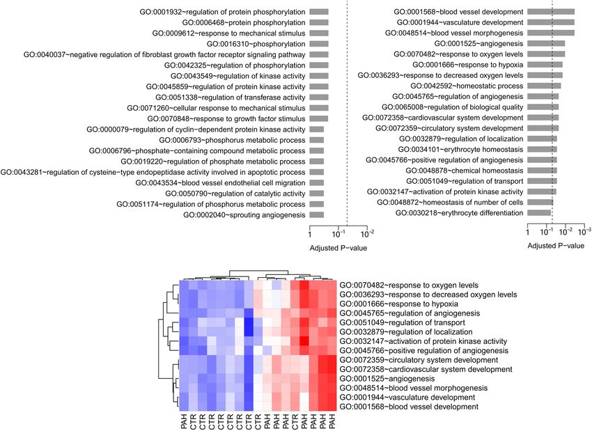
Zhu et al.                                                                                                        HIF2α and Pulmonary Arterial Hypertension

                                            observed between hypoxia and Phd2 deficiency mediated gene set expression profiles.
                                            As expected, 7 of the 19 significantly down-regulated GO terms in Hif2a-KD cells were
                                            found to overlap with the up-regulated GO gene sets in Phd2E C−/− mice compared to
                                            WT controls, suggesting opposing effects of HIF2α and PHD2 on PAH pathogenesis.
                                            Conclusion: HIF2α-mediated gene sets may be used to differentiate pulmonary
                                            arterial hypertension.
                                            Keywords: HIF2α, PHD2, pulmonary arterial hypertension, hypoxia, microarray

INTRODUCTION                                                                      escape degradation, which leads to their nuclear translocation,
                                                                                  heterodimerization with HIF1β, and increased expression of
Pulmonary artery hypertension (PAH) is a progressive and                          hypoxia-induced genes (Bishop and Ratcliffe, 2015). PHD2 is
lethal disease caused by multiple pulmonary vascular disorders.                   the most important isoenzyme under normoxic conditions,
Patients with PAH eventually died from right heart failure.                       and is involved in various hypoxia-affected processes, such
Endothelial dysfunction, sustained vasoconstriction and vascular                  as angiogenesis and cardiac function. We, and others, have
remodeling of pulmonary arterioles are the major characteristics                  already reported that the Phd2EC−/− mice, the endothelial
of PAH, all of which act to narrow the blood vessels and                          conditional knockout mice, developed spontaneous PH with
increase pulmonary vascular resistance (PVR) and pulmonary                        severe pulmonary vascular remodeling and occlusive pulmonary
artery pressure (PAP). Most patients with PAH have an insidious                   vascular lesions under normoxic conditions (Dai et al., 2016;
onset initially with no specific symptoms, however, with the                      Kapitsinou et al., 2016; Tang et al., 2018), while heterozygotes,
further increase of PAP, asthma, chest pain, dizziness occur.                     Phd2EC−/− mice developed mild PH symptoms. Significant
Severe cases of right heart failure include edema of the lower                    progress has also been made on the mechanism of HIF2α in
extremities, enlargement of the liver, and even ascites or pleural                the pathogenesis of PAH and it appears that HIF2α causes
effusion. Once the symptoms of right heart failure appear, the                    PH by inducing endothelial-to-mesenchymal transition (Endo-
prognosis of patients is poor. At present, the drugs targeting PAH                MT), a phenomenon of endothelial cells transforming into
in clinical usage are suboptimal. In general, the median survival                 fibroblast phenotype, which exacerbates pulmonary vascular
rate after diagnosis of idiopathic pulmonary hypertension is                      lesions (Tang et al., 2018).
only 2–3 years. Most importantly, PH cannot be measured by                            Preclinical studies have also begun to evaluate small molecule
conventional sphygmomanometers and most PH is found by                            HIF2α inhibitors as potential candidates for PAH treatment.
echocardiography after patients are already exhibiting severe                     The transcriptional activity of HIF2α is activated by binding
symptoms, which delays treatment. Therefore, it is critical that                  with ARNT while Gardner and his colleagues have found that
earlier methods of PAH diagnosis are developed.                                   the hydrophobic structure in the PAS-B domain of HIF2α
   In pathological conditions such as PAH, one of the common                      that can be inactivated by specific small molecule entry, which
phenomenon is oxygen utilization rate decrease. Pulmonary                         leads to the dissociation of the HIF2α and ARNT heterodimer.
hypertension caused by lung disease and/or hypoxia (WHO                           Ultimately the DNA-binding capacity of HIF2A is abolished
group 3 PH) has a large population, which cannot be ignored.                      (Scheuermann et al., 2009). A series of HIF2α inhibitors
A large amount of evidence supports the crucial role of                           for cancer therapy have been produced using this method,
hypoxia inducible factors (HIFs) in chronic hypoxia induced                       including PT2385, PT2399, and PT2576. The advantages of
PH (Brusselmans et al., 2003; Kim et al., 2013; Cowburn et al.,                   high specificity and oral high bioavailability make them of
2016; Dai et al., 2016). HIFs are a class of transcription factors                high clinical value. These HIF2α inhibitors show high safety
sensitive to oxygen concentration, which are mainly responsible                   and effectiveness in both animal experiments and clinical trials,
for regulating the adaptability of the body to hypoxia (Prabhakar                 which can significantly inhibit tumor growth in patients with
and Semenza, 2012). HIFs, including HIF-1 and HIF-2 are                           metastatic clear cell renal cell carcinoma (ccRCC), with proven
involved in the regulation of cell proliferation, migration, and                  safety and high dose tolerance (Chen et al., 2016; Cho et al.,
pulmonary vascular remodeling in PH (Voelkel and Tuder,                           2016).
2000; Brusselmans et al., 2003; Prabhakar and Semenza, 2012).                         Since HIF2α is an important cause of pulmonary artery
More importantly, HIF1α and HIF2α, particularly HIF2α in                          remodeling during PH, preclinical studies of HIF2α inhibitors
pulmonary artery endothelial cells, plays a major role in                         for PH therapy have been evaluated. Published data shows
the development of PAH (Skuli et al., 2009; Kim et al.,                           that PT2567 significantly reduced hemodynamic parameters
2013; Ball et al., 2014; Dai et al., 2016; Kapitsinou et al.,                     related to the development of PH in Sugen 5416/hypoxia-
2016). HIFs are regulated by HIF-prolyl hydroxylases (PHDs)                       rats (Macias et al., 2020). These results suggest that HIF2α
through the hydroxylation of conserved proline residues.                          could be an early biomarker of PAH and that HIF2α inhibitors
Hydroxylated HIFs are then degraded by the proteasome via                         might provide a promising new approach for the treatment of
the von Hipel-Lindau ubiquitination complex. Under hypoxia,                       PAH. There is evidence that HIF-2α and its target genes were
the hydroxylation reaction is attenuated allowing HIFs to                         upregulated in lung tissue and pulmonary artery endothelial

Frontiers in Cell and Developmental Biology | www.frontiersin.org             2                                     August 2021 | Volume 9 | Article 701247
Zhu et al.                                                                                               HIF2α and Pulmonary Arterial Hypertension

cells (PAECs) from idiopathic PAH patients and a variety                 University and the procedures applied in the studies were
of rodent PH models (Dai et al., 2018; Tang et al., 2018).               carried out in accordance with the National Institutes of Health
These findings suggest that HIF2α plays an important role                guidelines for use of live animals. Right ventricular pressure
not only in hypoxia-induced PH, but also in other types                  (RVP) was collected by MPVS ULTRATM system in real time
of PH. However, due to the heterogeneity of PH it is vital               and right ventricular systolic pressure (RVSP) was calculated in
to specifically utilize the mots efficacious therapeutic agents.         LabChart8 application. The Fulton Index was measured as a key
Therefore, in this study we aim to investigate the gene                  indicator of RV hypertrophy (RVH). Paraffin embedded sections
expression of HIF2α in PAH in order to determine if it was               of mice lung tissues were used for morphological analysis. The
possible to identify a useful diagnostic method to differentiate         sections were stained with hematoxylin and eosin (H&E) to
PAH human subjects.                                                      evaluate the degree of vascular remodeling by calculating the
   Because highly reproducible gene expression pattern can be            thickness of pulmonary artery walls.
acquired from the mechanism-associated gene sets related to
PH development from transcriptional analysis (Abraham et al.,            Transcriptome Data Processing
2010; Yang et al., 2012; Gardeux et al., 2014), We focused on the        The whole-genome gene expression data packages of WT
global gene expression pattern associated with the HIF2α rather          and Hif2a-KD VHL-deficient human cell renal cell carcinoma
than examining the expression abundance of individual genes.             (ccRCC) cell line A498 were acquired from the Gene Expression
We compared the Gene Ontology (Bertout et al., 2009) gene set            Omnibus (GEO) database (Edgar et al., 2002) (GEO accession:
(Ashburner et al., 2000) expression pattern between wild type            GSE16622; Bertout et al., 2009). Affymetrix Mouse Gene 2.0
(WT) and Hif2a knockdown (KD) von Hippel-Lindau (VHL)                    ST Array (Affymetrix, Inc., Santa Clara, CA) has been used
deficient cells. The differentially expressed GO gene sets were          to profile mRNA expression abundance in Phd2EC−/− mice
deemed as HIF2α-mediated. We further validated the potential             lung tissues. Briefly, the Affymetrix arrays were analyzed using
of the HIF2α-mediated gene sets as a possible diagnostic tool for        the Affymetrix Power Tools. A robust multi-array averaging
PAH by analyzing the transcriptome data in lungs and PBMCs of            method was used to summarize the probe set expression signals
PAH patients and healthy controls. To verify the ability of HIF2α-       (Gong et al., 2015) algorithm and log2 -transformed with a
mediated gene sets for the identification of PAH, endothelial            median polish. The transcript was deemed reliably expressed if
cell-specific Phd2 KO (Phd2EC−/− ) mice were generated, and              the Affymetrix implemented detection above ground (DABG)
their lung tissues were analyzed by transcriptome sequencing.            P-value was < 0.01 in all samples from at least one group. We
Comparing the differences and similarities of gene expression            focused on genes with unique annotations. Genes on X and Y
and biological processes among WT, hypoxia and Phd2EC−/−                 chromosomes were discarded to avoid potential confounding
mice lung tissues we found that differentially expressed GO genes        factors caused by sex. Significance analysis of microarrays (SAM)
associated with PH were mediated by HIF2α. Together, these               algorithm, implemented in the samr library of the R Statistical
HIF2α-mediated gene sets were effectively able to differentiate          Package, was applied to identify the differentially expressed
PAH patients from controls.                                              genes between WT and Hif2a-KD cells. Transcripts with a fold-
                                                                         change (FC) > 1.3 and false discovery rate (FDR) < 1% were
                                                                         deemed differentially expressed. Affymetrix Mouse Gene 2.0 ST
MATERIALS AND METHODS                                                    Array has been used to analyze mRNA expression abundance in
                                                                         Phd2EC−/− mice lung tissues.
Generation of Phd2 Gene Endothelial                                         To understand the role of HIF2α in PAH pathogenesis, four
Cell-Specific Knockout Mice                                              transcriptome data sets of human samples were acquired from
The endothelial cell specific-knockout (Gong et al., 2015) mouse         the GEO database: GSE48149, GSE15197, and GSE53408 from
lines were generated by Cre-LOX technique according to the               lung tissue and GSE22356 from PBMCs. These specific data sets
methods previously described (Bishop and Ratcliffe, 2015). The           been selected were dependent on the availability of annotated
Phd2 floxed mice were hybridized with Tie2-Cre mice to produce           patient classification. For all these data sets, we aggregated genes
Tie2-Cre+ /Phd2f lox/flox mice, which were named endothelial cell-       into gene set-level mechanisms using GO (Ashburner et al.,
specific Phd2 knockout mice (Phd2EC−/− ). The expression of Cre          2000) annotations.
recombinase was regulated by the Tie2 promoter.
                                                                         Computing Gene Set Score Using
Western Blotting                                                         Human Transcriptome Data
Western blotting was done as previously described (Tang et al.,          In order to calculate a gene set score for each GO biological
2018). Antibodies that have been used are anti-HIF2α (Cat No.            process term, the Functional Analysis of Individual Microarray
ab199, Abcam), anti-PHD2 (Cat No. 66589-1-Ig, Proteintech),              Expression (FAIME) algorithm was adopted (Yang et al., 2012).
and anti-β-actin (Cat No. 4970, Cell Signaling).                         Gene set scores computed by the FAIME tool made the
                                                                         gene expression of individual samples rank-weighted, which
Hemodynamic Measurements                                                 transferred each sample’s transcriptome profile into pathway-
All animal studies were approved by the Institutional Animal             level data (Yang et al., 2012). A higher gene set score indicates
Care and Use Committee (IACUC) of Guangzhou Medical                      an overall up-regulation of a given GO term.

Frontiers in Cell and Developmental Biology | www.frontiersin.org    3                                     August 2021 | Volume 9 | Article 701247
Zhu et al. HIF2α and Pulmonary Arterial Hypertension
RESULTS (GEO accession: GSE15197; Rajkumar et al., 2010) containing
13 controls and 18 PAH patients and the cohort B (GEO
Deregulated Gene Sets in Hif2a-KD Cells accession: GSE53408; Zhao et al., 2014) with 11 controls
To infer the gene sets potentially mediated by HIF2α, and 12 PAH patients. Principal component analysis (PCA)
we tapped the value of the difference in gene expression indicates that the FAIME score of the HIF2α-mediated GO
pattern between WT and Hif2a-KD cell lines. A microarray gene sets differentiate PAH patients from controls in both
data set containing both WT and Hif2a-KD VHL-deficient the validation cohorts (Figures 2A,B). There was a significant
human ccRCC cells gene expression abundance were obtained difference in the second principal component (PC2) between
from the GEO database (GEO accession: GSE16622; Bertout controls and PAH patients (t-test: P = 2.2 × 10⁻² and
et al., 2009). Within the specified significance level range P = 2.6 × 10⁻² for the cohort A and B respectively) (Figure 2C).
(FDR < 1% and FC > 1.3), 193 genes were found to be Area under the receiver operating characteristic curve (AUC)
up-regulated in Hif2a-KD cells while 353 genes were down- was adopted to evaluate the classification performance of
regulated. These differentially expressed genes were presented PC2 which were 0.722 and 0.772 for the cohorts A and B,
in supplementary material (Supplementary Tables 1, 2). We respectively (Figure 2D).
hold the opinion that these deregulated genes were influenced A resampling test has been conducted to test whether the
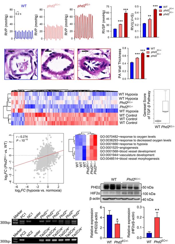
by HIF2α. Meanwhile, the enriched GO biological processes discriminative power of the 14 HIF2α-mediated GO terms
terms (Ashburner et al., 2000) among the HIF2α influenced was significantly better than random gene sets. We obtained
genes were analyzed. No significantly enriched GO terms were 1,000 random set of GO terms by randomly selecting 14 GO
existed in the genes up-regulated in Hif2a-KD cells (Figure 1A). biological process terms (the same size as the 14 HIF2α-
However, 19 GO biological process terms were identified as mediated GO terms) from the GO database and calculated the
significantly associated with genes down-regulated in Hif2a-KD AUC for each random GO term set. Our alternative hypothesis
cells. These included “blood vessel development,” “angiogenesis,” was that the AUCs of the 14 HIF2α-mediated GO terms
“response to decreased oxygen level,” “erythrocyte homeostasis,” ought to be more positive than the accidental expectation if
etc. (Figure 1B), all of which are strongly related to the the predictive power of the 14 HIF2α-mediated GO terms
pathogenesis of PAH. was significantly better than the random GO term sets. Our
analysis shows that the predictive power of null hypothesis
is accidental and should be rejected. The mean of AUC
Expression Profile of HIF2α-Mediated (for cohorts A and B) derived from the 14 HIF2α-mediated
Gene Sets in Lung Tissue Differentiates GO terms was significantly better than that of the random
Between Control and Patients With PAH GO term sets (Right-tailed: P = 0.041) (Figure 3A), which
To further determine the regulatory networks of HIF2α suggests the non-random predictive power of the 14 HIF2α-
in PAH pathogenesis, we assessed the discriminative power mediated GO terms.
of HIF2α-mediated gene sets in PAH human subjects. To
accomplish this we examined a lung tissue transcriptome
data set with nine controls and eight PAH patients (GEO
PBMC Expression Profile of
accession: GSE48149) (Hsu et al., 2011). For each GO biological HIF2α-Mediated Gene Sets Differentiate
process terms, we computed a gene set score by using PAH Patients From Controls
the FAIME algorithm (Yang et al., 2012). The higher the Chronic exposure of PBMCs to a PAH vascular environment
score of gene set, the higher the overall expression level of may be reflected by transcriptome changes in these cells.
specific GO gene set. 14 of the 19 significantly down-regulated Thus, we further tested the hypothesis that PBMC expression
GO terms in Hif2a-KD cells were found to overlap with profile of HIF2α-mediated gene sets may differentiate between
the up-regulated GO gene sets (t-test: P < 0.05) in PAH controls and PAH patients. Accordingly, we looked into a
patients compared to controls, which was statistically significant PBMC transcriptome data set with 10 controls and eight PAH
(cumulative hypergeometric test: P = 9.4 × 10⁻⁴ ). The gene patients (GEO accession: GSE22356; Risbano et al., 2010).
set score heat map of the overlapped GO terms generated Gene sets score was computed for each GO biological process
by unsupervised hierarchical cluster analysis reveals a clear terms based on the PBMC gene expression data, using the
separation between controls and PAH patients (Figure 1C). FAIME algorithm. 13 of the 19 significantly down-regulated
Together our results suggest that HIF2α plays a regulatory GO terms in Hif2a-KD cells were found to overlap with the
role in PAH. Therefore, we deemed the 14 overlapped GO up-regulated GO gene sets (t-test: P < 0.05) in PAH patients
terms “HIF2α-mediated” in lung. Physiologic parameter and vs. controls, which was statistically significant (cumulative
the differentially expressed genes in the citation (Hsu et al., hypergeometric test: P = 7.3 × 10⁻⁷ ). The PBMC based
2011) between IPAH group and Control group were presented gene set score heat map of the 13 overlapped GO terms
in Supplementary Material (Supplementary Tables 3–5 and reveals a clear separation between controls and PAH patients
Supplementary Figure 1). (Figure 3B), which suggests that the differences exist in PBMC
We next tested the classification power of the 14 HIF2α- expression of the HIF2α-mediated gene sets can be used as a
mediated GO terms in distinguishing PAH patients from controls biomarker of PAH to identify high-risk population and promote
in two validation lung transcriptome data sets: the cohort A early diagnosis.
Frontiers in Cell and Developmental Biology | www.frontiersin.org 4 August 2021 | Volume 9 | Article 701247Zhu et al. HIF2α and Pulmonary Arterial Hypertension FIGURE 1 | GO biological process analysis in Hif2a-KD VHL-deficient human ccRCC cells and PAH patients. (A) The top 20 GO biological process terms associated with the genes up-regulated in Hif2a-KD VHL-deficient human ccRCC cells. Fisher’s exact test was adopted to calculate the P-values which was then corrected by Benjamini-Hochberg procedure. The significance level of 0.05 was denoted by the dash line. (B) The top 20 GO biological process terms associated with the genes down-regulated in Hif2a-KD cells. The P-values were calculated by Fisher’s exact test and corrected by Benjamini-Hochberg procedure. The dash line denotes the significance level of 0.05. All the GO gene sets listed are statistically significant except the last term, “GO:0030218∼erythrocyte differentiation.” (C) Gene set score heatmap of the HIF2α-mediated GO biological process terms in human lungs. In total, 14 GO terms up-regulated in lung tissues from PAH patients were listed. Red and blue represent relative up-regulation and down-regulation of gene expression, respectively. FIGURE 2 | The HIF2α-mediated GO terms distinguish PAH patients from controls in the lung validation data sets. (A,B) Principal component analysis on the HIF2α-mediated GO terms in the validation cohorts (A,B), respectively. PC1 and PC2 represent the first principal component and the second principal component, respectively. (C) PC2 differentiates the PAH patients from the controls in the validation cohorts. (D) The ROC curves of PC2 in distinguishing PAH patients from controls in the validation cohorts. CTR, control; PAH, pulmonary arterial hypertension. Frontiers in Cell and Developmental Biology | www.frontiersin.org 5 August 2021 | Volume 9 | Article 701247
Zhu et al.                                                                                                                HIF2α and Pulmonary Arterial Hypertension

  FIGURE 3 | Gene set score heatmap of the HIF2α-mediated GO biological process terms in human PBMCs. (A) Superior predictive power of the HIF2α-mediated
  GO terms compared with randomized pattern. The histogram shows the distribution of the mean of AUC (both the validation cohorts A,B) for the 1,000 resampled
  GO term sets (The same size as the HIF2α-mediated GO terms, i.e., 14 GO terms). The mean of AUC of the 14 HIF2α-mediated GO terms is marked with black
  triangle. Right-tailed P-value was generated from sampling distribution. (B) In total, 13 GO terms up-regulated in PBMCs from PAH patients were listed. Red and
  blue represent relative up-regulation and down-regulation of gene expression, respectively.

HIF2α-Mediated Molecular Signaling                                                   higher than that of WT mice (Figure 4H), which suggesting that
Events Involved in the Development of                                                the activation of the HIF2α and TGF-β signaling pathways in
                                                                                     the Phd2 deficiency mouse endothelial cells presumably resulting
Phd2-KO Mediated PH                                                                  in severe pulmonary hypertension. Meanwhile, 7 of the 19
To further investigate the correlation between HIF2α and                             significantly down-regulated GO terms in Hif2a-KD cells were
the pattern of gene expression in a spontaneous PH model,                            found to significantly (t-test: P < 0.05) overlap with the up-
endothelial cell-specific Phd2 KO mice were generated. Both                          regulated GO gene sets in Phd2EC−/− mice vs. WT (Figure 4G),
Phd2EC+/− and Phd2EC−/− mice developed spontaneous PH                                We genotyped WT (Phd2+/+ ·Tie2-Cre+/− ) and Phd2EC+/−
under normoxia. RVP representative tracings intuitively                              (Phd2f /+ ·Tie2-Cre+/− ) mice (Figure 4I), and then WB assay
represented the changes in right ventricular pressure                                was used to confirm the activation of HIF2α following partial
(Figure 4A). The right ventricular systolic pressure was                             deficiency of PHD2 in the mice lung tissues (Figure 4J). These
20.4869 ± 0.3781 mmHg in wild type mice (Phd2f /f ), but                             findings reveal the opposing impact of HIF2α and PHD2 on
31.6415 ± 0.6030 and 59.2777 ± 4.5342 mmHg in Phd2EC+/−                              PAH development.
and Phd2EC−/− mice, respectively (Figure 4A). Elevated
RVSP was also consistent with significantly aggravated
RV hypertrophy in Phd2EC−/− mice. The Fulton index of                                DISCUSSION
Phd2EC+/− and Phd2EC−/− mice, which was 0.2902 ± 0.0173
and 0.4507 ± 0.0086, significantly increased compared to                             PAH is a highly lethal vascular disease characterized by the
Phd2+/+ , respectively (Figure 4B). In Phd2EC−/− mice, severe                        medial remodeling in pulmonary arterioles and endothelial
remodeling of the pulmonary arterioles characterized by a                            dysfunction, which leads to a progressive increase in PVR
dramatic PA wall thickening was also observed (Figures 4C,D).                        and pulmonary pressure (Rabinovitch, 2012). HIFs, especially
   To investigate the molecular signaling events that promote PH                     HIF2α, has proved to play a significant role in the pathogenesis
development caused by Phd2 knockout, we used gene microarray                         of PH. By deeply analyzing gene expression data from the
to analyze mRNA expression profiles in the lung tissues of                           GEO database, the present study was able to demonstrate
Phd2EC−/− mice. Globally, 459 genes (FDR < 10% and FC > 1.3)                         that Hif2a knockdown regulates multiple biological processes
were identified which were universally deregulated in both                           associated with PAH, including but not limited to “blood vessel
hypoxia induced PH mice and Phd2EC−/− mice (Figure 4E).                              development” and “angiogenesis,” which were further shown
The correlation in log2 -transformed gene expression fold change                     to present among genes up-regulated in PAH patient lung
(log2 FC) between hypoxia vs. normoxic mice and Phd2EC−/−                            tissues. More importantly, the 14 GO terms shared by Hif2a
vs. WT mice was derived at the whole-genome level. The two                           knockdown and up-regulated in PAH patient lung tissues show
sets of fold changes showed a significant positive correlation                       significantly better predictive power for PAH than random
(Pearson correlation test: r = 0.274, P < 10−10 ) (Figure 4F),                       gene sets. Interestingly, 13 of the 19 down-regulated GO terms
demonstrating that the deregulated genes induced by hypoxia                          associated with Hif2a knockdown overlapped with the up-
could be mimicked by Phd2 Knockout. The gene set scores of                           regulated GO terms in the peripheral blood mononuclear cells
the TGF-β signaling pathway (annotated by the KEGG database)                         from PAH patients. By reason of the foregoing, these data indicate
in WT and Phd2EC−/− mice were calculated. The result showed                          that HIF2α plays a critical role in PAH pathogenesis and HIF2α
that the gene set score of the Phd2EC−/− mice was significantly                      mediated gene sets can provide a distinctive and useful diagnostic

Frontiers in Cell and Developmental Biology | www.frontiersin.org                6                                           August 2021 | Volume 9 | Article 701247
Zhu et al. HIF2α and Pulmonary Arterial Hypertension FIGURE 4 | HIF2α-mediated GO terms involved in the development of Phd2-KO mediated spontaneous PH. (A) A representative waveform shows RVP (left) and statistical data (right) represents peak RVSP in WT, Phd2E C+/− and Phd2E C - / - mice (n = 6). Scale bars = 0.2 sec. (B) Statistical data of the RV/(LV + S) ratio shows RV hypertrophy defined by the Fulton index as a ratio [RV/(LV + S)] in WT, Phd2E C+/− and Phd2E C - / - mice (n = 6). (C) Typical H & E images of pulmonary arterioles from WT, Phd2E C+/− and Phd2E C - / - mice. Scale bars = 10 µm. (D) Statistical data shows PA wall thickness that generated by the ratio of wall area to total vessel area in PAs which were restricted less than 100 µm in diameter from WT, Phd2E C+/− or Phd2E C - / - mice (n = 6). (E) Expression heatmap of the genes commonly deregulated in both hypoxia and Phd2E C - / - deficient groups. Red and blue represent relative up-regulation and down-regulation of gene expression, respectively. (F) Correlation in log2 FC between hypoxia vs. normoxia mice (X-axis) and Phd2E C - / - vs. WT mice (Y -axis) The correlation coefficient (r) and P-value were computed by Pearson correlation test. (G) Gene set score heatmap of the HIF2α-mediated GO biological process terms in WT and Phd2E C - / - mice. In total, seven GO terms up-regulated in Phd2E C - / - lungs were listed. Red and blue represent relative up-regulation and down-regulation of gene expression, respectively. (H) Comparison of gene set score of the TGF-β signaling pathway between WT and Phd2E C - / - mice. (I) Genotyping of WT and Phd2E C+/− mice. PC1: positive control 1 (Phd2f /+ ), PC2: positive control 2 (Tie2-Cre+/− ), NC1: negative control 1 (Phd2+/+ ), NC2: negative control 2 (Tie2-Cre- / - ). (J) Relative expression of the PHD2 and HIF2α in mice lung tissues detected by western blotting with β-actin as control (n = 3). Significance levels: *P < 0.05, **P < 0.01 and ***P < 0.001 (t-test). Frontiers in Cell and Developmental Biology | www.frontiersin.org 7 August 2021 | Volume 9 | Article 701247
Zhu et al.                                                                                               HIF2α and Pulmonary Arterial Hypertension

method for distinguishing PAH human subjects. We investigated            (Patel and Simon, 2008). The accumulation of HIF2α in ECs
gene set expression profile rather than evaluating the expression        leads to increased ET-1 production, enhanced HIF1α activity in
level of individual genes for a compelling reason: mechanism-            PASMCs during PAH, and the HIF target genes up-regulation
associated gene sets are highly reproducible and meaningful              (Shimoda and Laurie, 2014). It has been reported that Hif2a+/−
in transcriptome research (Abraham et al., 2010; Yang et al.,            mice exhibit alleviated PAH pathological symptoms, including
2012; Gardeux et al., 2014). Generally, single gene expression           reduced right ventricular pressure and vascular remodeling
information is insufficient to unveil the underlying biological          (Brusselmans et al., 2003). The result of a statistical study showed
mechanisms. More importantly, most individual gene markers               that there is a correlation between HIF2α dysfunction and the
derived from genome-wide screening fail to be reproducible               decreased PAP in Tibetan natives (van Patot and Gassmann,
(Kern, 2012), because different genes jointly participating in           2011). Conversely, HIF2α overexpression is associated with
a mechanism, such as a given GO term, may alternately be                 development of PH in both humans (Gale et al., 2008; Formenti
deregulated in different samples (Gardeux et al., 2014). Thus,           et al., 2011) and mice (Tan et al., 2013). Our GO analysis
the mechanistic gap between gene expression and transcriptome            of PAH patient lung tissues samples further strengthens the
analysis can be effectively narrowed by mechanism-associated             important role of HIF2α in PAH. Comparing to the controls,
gene sets. Outliers appear in gene data sets of human CTR and            14 up-regulated GO gene sets (t-test: P < 0.05) in PAH
PAH groups, which may be due to differences in their genetic             patients overlapped with the down-regulated GO gene sets in
background, or some of them have other underlying diseases.              Hif2a-KD cells (Figures 1, 2). As we can see in Figure 2,
In clinical samples, it is often impossible to control intra group       most of the GO terms up-regulated in PAH patient samples
differences like animal models, which shows the advantages of            are associated with the function of HIF2α. PAH is a disease
animal model experiment.                                                 related to the dysregulation of the lung vascular system and
    HIF1α and HIF2α belong to a group of transcription factors,          we also identified the up-regulation of GO gene sets related
which regulate the transcription of most genes related to                to angiogenesis and blood vessel development (GO: 0001525,
hypoxia adaptation (Hu et al., 2003). The degradation of HIFs            GO: 0045766, GO: 0001568). HIF2α has been shown to control
depends on an oxygen dependent process. HIF1α and HIF2α                  genes involved in the regulation of angiogenesis and vascular
are hydroxylated by PHDs, ubiquitinated and rapidly degraded             development, and we identified two such gene among the
through ubiquitin-proteasome under normoxia (Maxwell et al.,             genes significantly down-regulated in Hif2a-KD cells: vascular
1999). However, under hypoxia, the decay of PHDs activity                endothelial growth factor A (VEGFA) and sirtuin 1 (SIRT1).
directly leads to the increase of HIF-α stability. This will             By binding its receptor VEGFR2, VEGFA regulates angiogenesis
facilitate their nuclear transposition, dimerization with ARNT           and vascular permeability through promoting the proliferation
and activation of their DNA-binding capacity. Then, the                  and survival of ECs, where the downstream signaling pathways
intracellular hypoxia adaptive regulation will be lunched (Hu            include phosphoinositide 3 kinase (PI3K)/Akt, focal adhesion
et al., 2003). Some evidences show that HIF2α can act as an              kinase (FAK), Rho family GTPases, nitric oxide (NO), and
independent pathogenic factor during PAH. A mutation prevents            p38 mitogen−activated protein kinase (MAPK) (Claesson-Welsh
HIF2α hydroxylation by PHDs, which accumulates HIF2α that                and Welsh, 2013). Moreover, a recent study showed HIV
will ultimately contribute to the development of PAH both in             protein transactivator of transcription (TAT) stimulated the
mice and human (Brusselmans et al., 2003; Tan et al., 2013).             proliferation of pulmonary artery SMCs (PASMCs) through
Current studies have shown that PHD2 expression was reduced              increasing the expression VEGFA, and may be a potential
in endothelial cells at the lesion sites of the pulmonary arteries       therapeutic target for the treatment of HIV-associated PAH
in idiopathic PAH patients, and the loss of PHD2 in endothelial          (Guo et al., 2018). SIRT1 is a NAD+ -dependent deacetylase
cells activates HIF2α rather than HIF1α (Dai et al., 2016). This         shown to play important roles in angiogenesis signaling both
is the reason that Phd2 KO mice lung tissues were chosen for             in vitro and in mice through controlling the acetylation
experimental verification. These studies indicate the importance         of the forkhead transcription factor, Foxo1, which is highly
of the PHD2/HIF2α axis in PAH development.                               expressed in ECs and mediates the expression of angiopoietin
    Hypoxia-inducible genes been transcriptional activated               2 (Ang2) (Potente et al., 2005, 2007). In addition, SIRT1 also
include vascular endothelial growth factor-a (VEGF-A),                   regulates the proliferation of PASMCs by activating peroxisome
erythropoietin (EPO) and inducible nitric oxide synthase                 proliferator-activated receptor gamma coactivator 1-alpha (PGC-
(iNOS) (Semenza and Wang, 1992; Melillo et al., 1995; Tuder              1α) (Zurlo et al., 2018). Thus, it is likely that uncontrolled
et al., 1995). The adaptation to hypoxia mediated by HIFs                activation of HIF2α induces the overexpression of its downstream
is mainly achieved by regulating the protein content of α                genes, such as VEGFA and SIRT1, which causes dysregulated
subunit. A hypoxic environment is a common occurrence                    development of the lung vascular system and contributes to the
in the lung in the pathological situation of PAH. As HIF-α               development of PAH.
signaling contributes to the endothelial dysfunction, vascular              In conclusion, by deeply analyzing selected gene expression
smooth muscle cells (VSMCs) proliferation, migration and                 data deposited in the GEO database we were able to demonstrate
tissue remodeling processes, it has the potential to become a            that HIF2α-regulated gene sets significantly overlapped with
therapeutic target.                                                      gene sets up-regulated in PAH patient lung tissue samples and
    HIF1α is widely expressed in the cells of most mammals, while        peripheral blood mononuclear cells. HIF2α, as a key regulatory
HIF2α is restricted and highly expressed in the endothelial cells        molecule, plays a significant role in the pathogenesis of PAH and

Frontiers in Cell and Developmental Biology | www.frontiersin.org    8                                     August 2021 | Volume 9 | Article 701247
Zhu et al. HIF2α and Pulmonary Arterial Hypertension
speculate that this methodology could provide a new application from JW, WL, and SB. All authors contributed to the article and
prospect in identifying PAH patients. approved the submitted version.
DATA AVAILABILITY STATEMENT
FUNDING
The raw data supporting the conclusions of this article will be
made available by the authors, without undue reservation. This work was funded by National Key Research and
Development Program of China (2019YFE0119400), the Natural
Science Foundation of China (81970052 and 81770059),
ETHICS STATEMENT Natural Science Basic Research Program of Shaanxi Province
(2018JC-012), and NIH NHLBI grants (P01HL134610 and P01
The animal study was reviewed and approved by the Institutional HL146369 to SB).
Animal Care and Use Committee (IACUC) of Guangzhou
Medical University.
SUPPLEMENTARY MATERIAL
AUTHOR CONTRIBUTIONS
The Supplementary Material for this article can be found
HT designed the project. JZ, LZ, YH, GC, and CB performed the online at: https://www.frontiersin.org/articles/10.3389/fcell.2021.
experiments. YH, TZ, and HT wrote the manuscript with input 701247/full#supplementary-material
REFERENCES in pulmonary arterial hypertension with a HIF-2alpha inhibitor. Am. J. Respir.
Crit. Care Med. 198, 1423–1434. doi: 10.1164/rccm.201710-2079oc
Abraham, G., Kowalczyk, A., Loi, S., Haviv, I., and Zobel, J. (2010). Prediction of Edgar, R., Domrachev, M., and Lash, A. E. (2002). Gene expression omnibus: NCBI
breast cancer prognosis using gene set statistics provides signature stability and gene expression and hybridization array data repository. Nucleic Acids Res. 30,
biological context. BMC Bioinformatics 11:277. doi: 10.1186/1471-2105-11-277 207–210. doi: 10.1093/nar/30.1.207
Ashburner, M., Ball, C. A., Blake, J. A., Botstein, D., Butler, H., Cherry, J. M., et al. Formenti, F., Beer, P. A., Croft, Q. P., Dorrington, K. L., Gale, D. P., Lappin,
(2000). Gene ontology: tool for the unification of biology. the gene ontology T. R., et al. (2011). Cardiopulmonary function in two human disorders of the
consortium. Nat. Genet. 25, 25–29. hypoxia-inducible factor (HIF) pathway: von Hippel-Lindau disease and HIF-
Ball, M. K., Waypa, G. B., Mungai, P. T., Nielsen, J. M., Czech, L., Dudley, V. J., et al. 2alpha gain-of-function mutation. FASEB J. 25, 2001–2011. doi: 10.1096/fj.10-
(2014). Regulation of hypoxia-induced pulmonary hypertension by vascular 177378
smooth muscle hypoxia-inducible factor-1alpha. Am. J. Respir. Crit. Care Med. Gale, D. P., Harten, S. K., Reid, C. D., Tuddenham, E. G., and Maxwell, P. H. (2008).
189, 314–324. Autosomal dominant erythrocytosis and pulmonary arterial hypertension
Bertout, J. A., Majmundar, A. J., Gordan, J. D., Lam, J. C., Ditsworth, D., Keith, B., associated with an activating HIF2 alpha mutation. Blood 112, 919–921. doi:
et al. (2009). HIF2alpha inhibition promotes p53 pathway activity, tumor cell 10.1182/blood-2008-04-153718
death, and radiation responses. Proc. Natl. Acad. Sci. U.S.A. 106, 14391–14396. Gardeux, V., Achour, I., Li, J., Maienschein-Cline, M., Li, H., Pesce, L., et al. (2014).
doi: 10.1073/pnas.0907357106 ‘N-of-1-pathways’ unveils personal deregulated mechanisms from a single pair
Bishop, T., and Ratcliffe, P. J. (2015). HIF hydroxylase pathways in cardiovascular of RNA-Seq samples: towards precision medicine. J. Am. Med. Inform. Assoc.
physiology and medicine. Circ. Res. 117, 65–79. doi: 10.1161/circresaha.117. 21, 1015–1025. doi: 10.1136/amiajnl-2013-002519
305109 Gong, H., Rehman, J., Tang, H., Wary, K., Mittal, M., Chaturvedi, P., et al.
Brusselmans, K., Compernolle, V., Tjwa, M., Wiesener, M. S., Maxwell, P. H., (2015). HIF2alpha signaling inhibits adherens junctional disruption in acute
Collen, D., et al. (2003). Heterozygous deficiency of hypoxia-inducible factor- lung injury. J. Clin. Invest. 125, 652–664. doi: 10.1172/jci77701
2alpha protects mice against pulmonary hypertension and right ventricular Guo, M. L., Kook, Y. H., Shannon, C. E., and Buch, S. (2018). Notch3/VEGF-A axis
dysfunction during prolonged hypoxia. J. Clin. Invest. 111, 1519–1527. doi: is involved in TAT-mediated proliferation of pulmonary artery smooth muscle
10.1172/jci15496 cells: implications for HIV-associated PAH. Cell Death Discov. 4:22.
Chen, W., Hill, H., Christie, A., Kim, M. S., Holloman, E., Pavia-Jimenez, A., et al. Hsu, E., Shi, H., Jordan, R. M., Lyons-Weiler, J., Pilewski, J. M., and Feghali-
(2016). Targeting renal cell carcinoma with a HIF-2 antagonist. Nature 539, Bostwick, C. A. (2011). Lung tissues in patients with systemic sclerosis have gene
112–117. expression patterns unique to pulmonary fibrosis and pulmonary hypertension.
Cho, H., Du, X., Rizzi, J. P., Liberzon, E., Chakraborty, A. A., Gao, W., et al. Arthritis Rheum. 63, 783–794. doi: 10.1002/art.30159
(2016). On-target efficacy of a HIF-2alpha antagonist in preclinical kidney Hu, C. J., Wang, L. Y., Chodosh, L. A., Keith, B., and Simon, M. C. (2003).
cancer models. Nature 539, 107–111. doi: 10.1038/nature19795 Differential roles of hypoxia-inducible factor 1alpha (HIF-1alpha) and HIF-
Claesson-Welsh, L., and Welsh, M. (2013). VEGFA and tumour angiogenesis. 2alpha in hypoxic gene regulation. Mol. Cell Biol. 23, 9361–9374.
J. Intern. Med. 273, 114–127. doi: 10.1111/joim.12019 Kapitsinou, P. P., Rajendran, G., Astleford, L., Michael, M., Schonfeld, M. P., Fields,
Cowburn, A. S., Crosby, A., Macias, D., Branco, C., Colaco, R. D., Southwood, T., et al. (2016). The endothelial Prolyl-4-hydroxylase domain 2/hypoxia-
M., et al. (2016). HIF2alpha-arginase axis is essential for the development of inducible factor 2 axis regulates pulmonary artery pressure in mice. Mol. Cell
pulmonary hypertension. Proc. Natl. Acad. Sci. U.S.A. 113, 8801–8806. doi: Biol. 36, 1584–1594. doi: 10.1128/mcb.01055-15
10.1073/pnas.1602978113 Kern, S. E. (2012). Why your new cancer biomarker may never work: recurrent
Dai, Z., Li, M., Wharton, J., Zhu, M. M., and Zhao, Y. Y. (2016). Prolyl- patterns and remarkable diversity in biomarker failures. Cancer Res. 72, 6097–
4 hydroxylase 2 (phd2) deficiency in endothelial cells and hematopoietic 6101. doi: 10.1158/0008-5472.can-12-3232
cells induces obliterative vascular remodeling and severe pulmonary arterial Kim, Y. M., Barnes, E. A., Alvira, C. M., Ying, L., Reddy, S., and Cornfield,
hypertension in mice and humans through hypoxia-inducible factor-2alpha. D. N. (2013). Hypoxia-inducible factor-1alpha in pulmonary artery smooth
Circulation 133, 2447–2458. doi: 10.1161/circulationaha.116.021494 muscle cells lowers vascular tone by decreasing myosin light chain
Dai, Z., Zhu, M. M., Peng, Y., Machireddy, N., Evans, C. E., Machado, R., et al. phosphorylation. Circ. Res. 112, 1230–1233. doi: 10.1161/circresaha.112.300
(2018). Therapeutic targeting of vascular remodeling and right heart failure 646
Frontiers in Cell and Developmental Biology | www.frontiersin.org 9 August 2021 | Volume 9 | Article 701247Zhu et al. HIF2α and Pulmonary Arterial Hypertension Macias, D., Moore, S., Crosby, A., Southwood, M., Du, X., Tan, H., et al. (2020). vascular function and tumor angiogenesis. Blood 114, 469–477. doi: 10.1182/ Targeting HIF2alpha-ARNT hetero-dimerisation as a novel therapeutic strategy blood-2008-12-193581 for pulmonary arterial hypertension. Eur. Respir. J. 57, 1902061. doi: 10.1183/ Tan, Q., Kerestes, H., Percy, M. J., Pietrofesa, R., Chen, L., Khurana, T. S., et al. 13993003.02061-2019 (2013). Erythrocytosis and pulmonary hypertension in a mouse model of Maxwell, P. H., Wiesener, M. S., Chang, G. W., Clifford, S. C., Vaux, E. C., human HIF2A gain of function mutation. J. Biol. Chem. 288, 17134–17144. Cockman, M. E., et al. (1999). The tumour suppressor protein VHL targets doi: 10.1074/jbc.m112.444059 hypoxia-inducible factors for oxygen-dependent proteolysis. Nature 399, 271– Tang, H., Babicheva, A., McDermott, K. M., Gu, Y., Ayon, R. J., Song, S., et al. 275. doi: 10.1038/20459 (2018). Endothelial HIF-2alpha contributes to severe pulmonary hypertension Melillo, G., Musso, T., Sica, A., Taylor, L. S., Cox, G. W., and Varesio, L. (1995). due to endothelial-to-mesenchymal transition. Am. J. Physiol. Lung Cell Mol. A hypoxia-responsive element mediates a novel pathway of activation of the Physiol. 314, L256–L275. inducible nitric oxide synthase promoter. J. Exp. Med. 182, 1683–1693. doi: Tuder, R. M., Flook, B. E., and Voelkel, N. F. (1995). Increased gene expression for 10.1084/jem.182.6.1683 VEGF and the VEGF receptors KDR/Flk and Flt in lungs exposed to acute or to Patel, S. A., and Simon, M. C. (2008). Biology of hypoxia-inducible factor-2alpha chronic hypoxia. modulation of gene expression by nitric oxide. J. Clin. Invest. in development and disease. Cell Death Differ. 15, 628–634. 95, 1798–1807. doi: 10.1172/jci117858 Potente, M., Ghaeni, L., Baldessari, D., Mostoslavsky, R., Rossig, L., Dequiedt, F., van Patot, M. C., and Gassmann, M. (2011). Hypoxia: adapting to high altitude et al. (2007). SIRT1 controls endothelial angiogenic functions during vascular by mutating EPAS-1, the gene encoding HIF-2alpha. High Alt. Med. Biol. 12, growth. Genes Dev. 21, 2644–2658. doi: 10.1101/gad.435107 157–167. doi: 10.1089/ham.2010.1099 Potente, M., Urbich, C., Sasaki, K., Hofmann, W. K., Heeschen, C., Aicher, A., Voelkel, N. F., and Tuder, R. M. (2000). Hypoxia-induced pulmonary vascular et al. (2005). Involvement of Foxo transcription factors in angiogenesis and remodeling: a model for what human disease? J. Clin. Investig. 106, 733–738. postnatal neovascularization. J. Clin. Invest. 115, 2382–2392. doi: 10.1172/jci23 doi: 10.1172/jci11144 126 Yang, X., Regan, K., Huang, Y., Zhang, Q., Li, J., Seiwert, T. Y., et al. (2012). Single Prabhakar, N. R., and Semenza, G. L. (2012). Adaptive and maladaptive sample expression-anchored mechanisms predict survival in head and neck cardiorespiratory responses to continuous and intermittent hypoxia mediated cancer. PLoS Comput. Biol. 8:e1002350. doi: 10.1371/journal.pcbi.1002350 by hypoxia-inducible factors 1 and 2. Physiol. Rev. 92, 967–1003. doi: 10.1152/ Zhao, Y., Peng, J., Lu, C., Hsin, M., Mura, M., Wu, L., et al. (2014). Metabolomic physrev.00030.2011 heterogeneity of pulmonary arterial hypertension. PLoS One 9:e88727. doi: Rabinovitch, M. (2012). Molecular pathogenesis of pulmonary arterial 10.1371/journal.pone.0088727 hypertension. J. Clin. Invest. 122, 4306–4313. doi: 10.1172/jci60658 Zurlo, G., Piquereau, J., Moulin, M., Pires Da Silva, J., Gressette, M., Ranchoux, Rajkumar, R., Konishi, K., Richards, T. J., Ishizawar, D. C., Wiechert, A. C., B., et al. (2018). Sirtuin 1 regulates pulmonary artery smooth muscle cell Kaminski, N., et al. (2010). Genomewide RNA expression profiling in lung proliferation: role in pulmonary arterial hypertension. J. Hypertens. 36, 1164– identifies distinct signatures in idiopathic pulmonary arterial hypertension and 1177. doi: 10.1097/hjh.0000000000001676 secondary pulmonary hypertension. Am. J. Physiol. Heart Circ. Physiol. 298, H1235–H1248. Conflict of Interest: The authors declare that the research was conducted in the Risbano, M. G., Meadows, C. A., Coldren, C. D., Jenkins, T. J., Edwards, M. G., absence of any commercial or financial relationships that could be construed as a Collier, D., et al. (2010). Altered immune phenotype in peripheral blood cells potential conflict of interest. of patients with scleroderma-associated pulmonary hypertension. Clin. Transl. Sci. 3, 210–218. doi: 10.1111/j.1752-8062.2010.00218.x Publisher’s Note: All claims expressed in this article are solely those of the authors Scheuermann, T. H., Tomchick, D. R., Machius, M., Guo, Y., Bruick, R. K., and and do not necessarily represent those of their affiliated organizations, or those of Gardner, K. H. (2009). Artificial ligand binding within the HIF2alpha PAS- the publisher, the editors and the reviewers. Any product that may be evaluated in B domain of the HIF2 transcription factor. Proc. Natl. Acad. Sci. U.S.A. 106, this article, or claim that may be made by its manufacturer, is not guaranteed or 450–455. doi: 10.1073/pnas.0808092106 endorsed by the publisher. Semenza, G. L., and Wang, G. L. (1992). A nuclear factor induced by hypoxia via de novo protein synthesis binds to the human erythropoietin gene enhancer Copyright © 2021 Zhu, Zhao, Hu, Cui, Luo, Bao, Han, Zhou, Lu, Wang, Black at a site required for transcriptional activation. Mol. Cell Biol. 12, 5447–5454. and Tang. This is an open-access article distributed under the terms of the Creative doi: 10.1128/mcb.12.12.5447 Commons Attribution License (CC BY). The use, distribution or reproduction in Shimoda, L. A., and Laurie, S. S. (2014). HIF and pulmonary vascular responses to other forums is permitted, provided the original author(s) and the copyright owner(s) hypoxia. J. Appl. Physiol. 116, 867–874. doi: 10.1152/japplphysiol.00643.2013 are credited and that the original publication in this journal is cited, in accordance Skuli, N., Liu, L., Runge, A., Wang, T., Yuan, L., Patel, S., et al. (2009). with accepted academic practice. No use, distribution or reproduction is permitted Endothelial deletion of hypoxia-inducible factor-2alpha (HIF-2alpha) alters which does not comply with these terms. Frontiers in Cell and Developmental Biology | www.frontiersin.org 10 August 2021 | Volume 9 | Article 701247
You can also read