Pulmonary hypertension phenotypes in patients with systemic sclerosis
←
→
Page content transcription
If your browser does not render page correctly, please read the page content below
EUROPEAN RESPIRATORY REVIEW
REVIEW
A. HAQUE ET AL.
Pulmonary hypertension phenotypes in patients with systemic
sclerosis
Ashraful Haque1,2,3,6, David G. Kiely 1,2
, Gabor Kovacs4,5, A.A. Roger Thompson 1,2
and
Robin Condliffe 1,2,6
1
Sheffield Pulmonary Vascular Disease Unit, Royal Hallamshire Hospital, Sheffield, UK. 2Dept of Infection, Immunity and
Cardiovascular Disease, University of Sheffield, Sheffield, UK. 3Dept of Rheumatology, Royal Hallamshire Hospital, Sheffield, UK.
4
Medical University of Graz, Graz, Austria. 5Ludwig Boltzmann Institute for Lung Vascular Research, Graz, Austria. 6Both authors
contributed equally.
Corresponding author: Robin Condliffe (robin.condliffe@nhs.net)
Shareable abstract (@ERSpublications)
Different forms of pulmonary hypertension can be present in patients with systemic sclerosis. In
this article we review the epidemiology, diagnosis, outcomes and treatment of the spectrum of
pulmonary vascular phenotypes associated with systemic sclerosis. https://bit.ly/3xUwrVB
Cite this article as: Haque A, Kiely DG, Kovacs G, et al. Pulmonary hypertension phenotypes in
patients with systemic sclerosis. Eur Respir Rev 2021; 30: 210053 [DOI: 10.1183/16000617.0053-2021].
Abstract
Copyright ©The authors 2021 Pulmonary hypertension (PH) commonly affects patients with systemic sclerosis (SSc) and is associated
with significant morbidity and increased mortality. PH is a heterogenous condition and several different
This version is distributed under
the terms of the Creative
forms can be associated with SSc, including pulmonary arterial hypertension (PAH) resulting from a
Commons Attribution Non- pulmonary arterial vasculopathy, PH due to left heart disease and PH due to interstitial lung disease. The
Commercial Licence 4.0. For incidence of pulmonary veno-occlusive disease is also increased. Accurate and early diagnosis to allow
commercial reproduction rights optimal treatment is, therefore, essential. Recent changes to diagnostic haemodynamic criteria at the 6th
and permissions contact
World Symposium on Pulmonary Hypertension have resulted in therapeutic uncertainty regarding patients
permissions@ersnet.org
with borderline pulmonary haemodynamics. Furthermore, the optimal pulmonary vascular resistance
Received: 28 Feb 2021 threshold for diagnosing PAH and the role of exercise in identifying early disease require further
Accepted: 4 May 2021 elucidation. In this article we review the epidemiology, diagnosis, outcomes and treatment of the spectrum
of pulmonary vascular phenotypes associated with SSc.
Introduction
Systemic sclerosis (SSc) is a multisystem autoimmune disorder characterised by inflammation, excessive
collagen deposition and fibrosis [1]. Limited cutaneous systemic sclerosis (LcSSc) is characterised by skin
thickening distal to the elbows and knees, with or without facial and neck involvement, and the frequent
presence of anti-centromere antibodies, while diffuse cutaneous systemic sclerosis (DcSSc) is characterised
by proximal skin thickening and a predominance of anti-topoisomerase 1 (Scl-70) antibodies and
anti-RNA polymerase III antibodies [2]. Earlier and more frequent organ involvement occurs in DcSSc [3].
A small proportion of patients may present with clinical features of SSc in the absence of skin thickening
(SSc sine scleroderma).
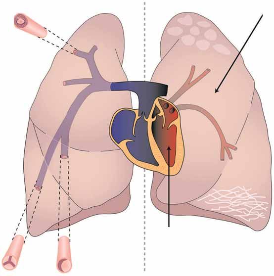
Pulmonary hypertension (PH) describes a heterogenous group of conditions defined by an elevated mean
pulmonary arterial pressure (mPAP). Five classification groups are described: Group 1: pulmonary arterial
hypertension (PAH); Group 2: PH due to left heart disease (PH-LHD); Group 3: PH due to lung diseases
and/or hypoxia (PH-lung); Group 4: PH due to pulmonary artery obstructions; Group 5: PH with unclear
and/or multifactorial mechanisms (figure 1) [5]. PAH is characterised by a progressive pulmonary arterial
vasculopathy. Subsequent increased pulmonary vascular resistance (PVR) and pulmonary arterial pressure
lead to increased right ventricular (RV) afterload with subsequent RV dysfunction, failure and premature
death [6–8]. Despite the availability of specific therapies targeting three main pathways, PAH associated
with SSc (SSc-PAH) is associated with a poor prognosis with 3-year survival of only 52% [9]. Patients
with SSc may also develop other forms of PH (SSc-PH), especially PH-LHD (SSc-PH-LHD) and PH-lung
https://doi.org/10.1183/16000617.0053-2021 Eur Respir Rev 2021; 30: 210053EUROPEAN RESPIRATORY REVIEW PULMONARY HYPERTENSION | A. HAQUE ET AL.
1. Pulmonary arterial 3. PH-lung disease/hypoxia
hypertension COPD
Idiopathic Interstitial lung disease
Heritable Sleep disorder
Drugs Alveolar hypoventilation
Connective tissue disease
HIV
Portal hypertension
Congenital heart disease
Schistosomiasis 5. Multifactorial/unclear
1' Pulmonary veno- Haematological
occlusive disease Chronic haematolytic anaemia
Pulmonary capillary Myeloproliferative disease
haemangiomatosis Splenectomy
Systemic disorders
Sarcoidosis
Langerhans cell histiocytosis
Lymphangioleiomyomatosis
Neurofibromatosis
Vasculitis
Metabolic disorders
Glycogen storage disease
4. Chronic
thromboembolic Gaucher's disease
pulmonary Thyroid disorder
hypertension 2. PH-left heart Others
Systolic dysfunction Tumour obstruction
Operable Diastolic dysfunction Fibrosing mediastinitis
Inoperable Valvular disease Chronic renal failure
FIGURE 1 Potential pulmonary hypertension (PH) classification groups associated with systemic sclerosis. Patients may develop a pulmonary
arterial vasculopathy (group 1, pulmonary arterial hypertension), may develop PH due to left heart disease (group 2) or PH due to lung disease
(group 3; most commonly interstitial lung disease, although the incidence of combined fibrosis and emphysema is also increased). The incidence
of pulmonary veno-occlusive disease (group 1′) also appears to be increased in systemic sclerosis. Chronic thromboembolic PH (group 4) should
be excluded while patients with a previous scleroderma renal crisis who progress to end-stage chronic kidney disease may develop group 5
disease. Reproduced and modified from [4] with permission from the publisher.
(SSc-PH-lung). Pulmonary venous involvement may be relatively common in patients diagnosed with
SSc-PAH while some patients may present with a predominant picture of pulmonary veno-occlusive
disease (PVOD) [10–12]. Patients may also rarely present with group 4 disease, chronic thromboembolic
pulmonary hypertension, as SSc is associated with an increased risk of venous thromboembolism [13].
Diagnostic criteria with regard to the threshold of mPAP used to define the presence of PH, the use or
non-use of a threshold for PVR to diagnose PAH, and the presence or absence of the entity of PH on exercise
(PH-exercise) have changed over recent years (table 1). Furthermore, there are now a group of patients with
elevated mPAP but normal PVR who are unclassifiable according to the most recent World Symposium on
Pulmonary Hypertension (WSPH). Making the correct diagnosis regarding the form of SSc-PH is of critical
importance in informing prognosis and guiding the most appropriate management strategy. Therefore, in this
article we review the different PH phenotypes present in patients with SSc (table 2).
Methods
A PubMed systematic literature search was undertaken using the following search criteria:
((Systemic sclerosis) OR (Scleroderma) OR (Limited cutaneous systemic sclerosis) OR (Diffuse cutaneous
systemic sclerosis)) AND ((Pulmonary hypertension) OR (Pulmonary arterial hypertension)) AND
((Exercise) OR (Borderline) OR (Interstitial lung disease) OR (ILD) OR (Diffusion capacity) OR (Left
heart disease) OR (DLCO) OR (Transfer factor) OR (Phenotype)).
218 search results were all analysed for relevant information. Furthermore, a grey search of the manuscripts
cited within these articles was undertaken together with the inclusion of key legacy papers.
Changing definitions of PH
The first WSPH was organised by the World Health Organization (WHO) in 1973 in response to a
European epidemic of appetite suppressant-induced PH [14]. It defined PH by a mPAP at right heart
https://doi.org/10.1183/16000617.0053-2021 2EUROPEAN RESPIRATORY REVIEW PULMONARY HYPERTENSION | A. HAQUE ET AL.
TABLE 1 Haemodynamic diagnostic criteria of the six World Symposia on Pulmonary Hypertension (WSPH)
First WSPH Second Third Fourth WSPH Fifth WSPH Sixth WSPH
[14] WSPH [15] WSPH [16] [17, 18] [19, 20] [5, 21]
Year 1973 1998 2003 2008 2013 2018
Location Geneva, Evian, Venice, Italy Dana Point, CA, Nice, France Nice, France
Switzerland France USA
mPAP PH diagnostic >25 mmHg Not >25 mmHg ⩾25 mmHg ⩾25 mmHg >20 mmHg
threshold defined
PVR included in PAH No No >3 WU No >3 WU ⩾3 WU
definition
PAWP post-capillary Discussed but not Not >15 mmHg ⩾15 mmHg >15 mmHg >15 mmHg
threshold defined discussed
Isolated post-capillary Not discussed Not Not PVR 30 mmHg No No No
defined discussed
mPAP 21–24 mmHg 20 mmHg as upper Not Not Uncertainty in At-risk patients (e.g. Most now defined as PH;
limit of normal discussed discussed patients with mPAP CTD) should be however, mPAP
recognised 21–24 mmHg followed closely >20 mmHg, PAWP
⩽15 mmHg but PVR
25 mmHg (table 1) [14]. This haemodynamic definition was derived from the
recommendation of a previous WHO report on cor pulmonale, published in 1961 [22]. The diagnostic
threshold of 25 mmHg would remain until the sixth WSPH in 2018 where it was proposed that the
threshold be reduced to >20 mmHg [5]. This change in definition was suggested following a systematic
TABLE 2 Systemic sclerosis (SSc)-pulmonary hypertension (PH) phenotypes
SSc-PAH: post-6th WSPH mPAP ⩾20 mmHg, PAWP ⩽15 mmHg, PVR ⩾3 WU
SSc-PAH: pre-6th WSPH mPAP ⩾25 mmHg, PAWP ⩽15 mmHg, PVR >3 WU
SSc: mPAP >20 mmHg, A group of patients with elevated mPAP who do not fulfil current PH diagnostic
PVR 3 WU on exercise
SSc-PVOD Meets haemodynamic criteria for PAH but radiological and clinical features of
PVOD
SSc-PH-LHD mPAP ⩾20 mmHg, PAWP >15 mmHg
SSc-IpcPH mPAP ⩾20 mmHg, PAWP >15 mmHg, PVR 15 mmHg, PVR ⩾3 WU
SSc-PH-HFpEF SSc-PH-LHD due to heart failure with preserved ejection fraction
SSc-PH-HFrEF SSc-PH-LHD due to heart failure with reduced ejection fraction
SSc-PH-ILD mPAP ⩾20 mmHg, PAWP ⩽15 mmHg, PVR ⩾3 WU in the presence of significant
ILD (often defined as HRCT showing >20% fibrotic lung involvement and/or
FVCEUROPEAN RESPIRATORY REVIEW PULMONARY HYPERTENSION | A. HAQUE ET AL.
review by KOVACS et al. [23] which demonstrated that the mean mPAP within the healthy population was
14.0±3.3 mmHg. In addition, MARON et al. [24] reviewed RHC data from 21 727 patients in the US
Veterans healthcare system and observed increased mortality in patients with a mPAP 19–24 mmHg
compared with 19 mmHg. It is interesting to note that although
the 1961 report proposed a threshold of 25 mmHg, it also commented that the upper limit of normal for
mPAP was 15 mmHg [22]. Furthermore, the first WSPH also stated that the mPAP at rest “never”
exceeded 20 mmHg in healthy individuals [14]. The uncertain nature of patients with mPAP 21–24 mmHg
had been recognised at the fourth and fifth World Symposia, but there were deemed to be insufficient data
to introduce a formal definition of “borderline PH” [18, 19].
The effects of exercise on mPAP were discussed at the first WSPH, but were not included in diagnostic
criteria [13]. By the third WSPH, a diagnostic threshold of mPAP >30 mmHg for the diagnosis of
PH-exercise had been introduced [17, 26]. PH-exercise was, however, dropped from the diagnostic criteria
in the fourth WSPH in 2008 as it was appreciated that mPAP may frequently increase >30 mmHg on
exercise in normal individuals, especially those aged >50 years [17].
PVR was incorporated into the definition of PAH at the third WSPH using a threshold of >3 Wood Units
(WU) [27]. Its inclusion in the definition aimed to prevent patients with flow-related increases in mPAP
being diagnosed with PAH. Although it was temporarily absent from the fourth WSPH, it was reinserted
into the diagnostic criteria for PAH at the fifth WSPH, albeit with the slight change of including patients
with a PVR ⩾3 WU (as opposed to >3 WU) [19].
Left atrial pressure, most commonly assessed by the pulmonary arterial wedge pressure (PAWP), was
introduced in the third WSPH to differentiate between pre-capillary (PAWP ⩽15 mmHg) and post-capillary
PH (PAWP >15 mmHg) [16].
Summary
Haemodynamic criteria for different forms of PH have changed over recent decades. The most recent
WSPH defines pre-capillary PH as mPAP >20 mmHg, PAWP ⩽15 mmHg and PVR ⩾3 WU.
SSc-PAH
Estimates of the prevalence of PAH within the SSc population range between 6.4% and 9% [27, 28]. The
incidence of SSc-PAH in patients with LcSSc and DcSSc is 1.25 and 0.4 cases per 100 patient-years,
respectively [29]. Meta-analysis involving 3818 patients with RHC-confirmed PH identified PAH as the
most common form of PH seen in SSc, comprising 63% of RHC-confirmed cases [27]. There may,
however, be ascertainment bias in these data due to patients with other forms of PH being less likely to
undergo RHC. Historically, SSc-PAH was associated with a poor prognosis with a 3-year survival of 30%
[30]. Mortality remains high despite the availability of PAH-specific therapy. LEFÈVRE et al. [9] observed
1- and 3-year survival rates of 81% and 52%, respectively, in a meta-analysis.
Several studies have demonstrated that, despite having less severe pulmonary haemodynamics, survival of
patients with SSc-PAH is worse than with idiopathic PAH (IPAH) [31–37]. There are several possible
explanations for this including differences in patient characteristics, the underlying pulmonary arterial
vasculopathy, and the ability of the RV to compensate for increased afterload.
Patient characteristics
When compared with IPAH, patients with SSc-PAH are older with a lower coefficient for diffusing
capacity of the lung for carbon monoxide (DLCO). In a multivariate analysis of 375 IPAH and SSc-PAH
patients, RAMJUG et al. [37] identified that higher age and lower DLCO were independent prognostic
markers. The lower DLCO may reflect increased alveolar-capillary block due to overt or covert interstitial
lung disease (ILD), reduced capillary blood volume related to the nature of the pulmonary vasculopathy or
a component of PVOD. The multisystem nature of SSc, with involvement not only of the lungs and heart,
but also the skin, gastrointestinal tract and kidneys, likely also impacts survival [38, 39].
Vasculopathy
OVERBEEK et al. [12] demonstrated intimal fibrosis in histological specimens from all eight patients they
studied with SSc-PAH compared with only three out of 11 patients with IPAH. Plexiform lesions were
much less common than in patients with IPAH. Similarly, DORFMÜLLER et al. [11] observed marked
muscular artery intimal fibrosis in four out of four SSc-PAH patients in contrast with only four out of 29
https://doi.org/10.1183/16000617.0053-2021 4EUROPEAN RESPIRATORY REVIEW PULMONARY HYPERTENSION | A. HAQUE ET AL.
patients with IPAH. In addition to differences in pulmonary arterial histology, there may also be an
increased frequency of pulmonary venous lesions in SSc-PAH (see PVOD section) [40].
Right ventricle
OVERBEEK et al. [41] demonstrated poorer RV contractility in 13 patients with SSc-PAH compared with 17
IPAH patients. Similarly, TEDFORD et al. [42] observed worse RV contractility and coupling of RV
contractility with afterload in seven SSc-PAH patients compared with five IPAH patients. MATHAI et al.
[43] found N-terminal pro-B-type natriuretic peptide (NT-proBNP) levels in 55 SSc-PAH patients to be
significantly higher than in 43 IPAH patients, despite the IPAH group having more severe PH.
Furthermore, although OVERBEEK et al. [41] found no difference in the extent of interstitial fibrosis in the
RVs obtained at autopsy of five SSc-PAH and nine IPAH patients, there was an increased inflammatory
myocardial infiltrate in the SSc-PAH group. Conversely, HSU et al. [44] observed increased interstitial
fibrosis in RV endomyocardial biopsies obtained from 11 SSc-PAH patients when compared with seven
IPAH patients and six SSc patients without PH. Interestingly, when compared with healthy controls,
sarcomere function (as assessed by the maximum calcium-activated force) was significantly lower in
SSc-PAH but significantly higher in IPAH. Sarcomere function in SSc patients without PH was
intermediate between the controls and SSc-PAH.
Medical therapy in SSc-PAH
Current therapies for PAH target three main pathways: nitric oxide, endothelin-1 and prostacyclin [45, 46].
A number of randomised controlled trials (RCTs) have published data on outcomes in patients with
connective tissue disease (CTD), the majority of whom had SSc (table 3) [63]. With the exception of an
unblinded RCT of epoprostenol, short-term monotherapy RCTs have tended to report lower response to
therapy in patients with CTD associated PAH (CTD-PAH) [64, 65]. However, newer data from longer
studies where the majority of patients have received combination therapy have challenged these findings
[54, 56, 61].
Screening
HUMBERT et al. [66] observed milder haemodynamics and superior survival in a cohort of SSc patients
identified in a screening programme as compared to presenting symptomatically. Although lead-time bias
cannot be excluded as a cause of the superior survival, subsequent RCTs of PAH therapies have
demonstrated larger treatment response of patients in functional class (FC) II compared with FC III [67].
There is, therefore, a good rationale for screening asymptomatic SSc patients to enable earlier treatment
[68]. COGHLAN et al. [69] compared the multi-modality 2-step DETECT algorithm with the European
Respiratory Society/European Society of Cardiology (ERS/ESC) approach of echocardiography alone and
observed sensitivity/specificity for identifying PAH (mPAP ⩾25 mmHg and PAWP ⩽15 mmHg) of 96%/
48% and 71%/69%, respectively. The Australian Scleroderma Interest Group (ASIG) studied an approach
using pulmonary function tests and NT-proBNP and reported sensitivity/specificity for identifying PAH of
94%/55% compared with 95%/32% for the ERS/ESC approach [70]. HAO et al. [71] compared all three
approaches in 73 patients and observed that although the DETECT and ASIG approaches performed
similarly, the ASIG algorithm reduced the need for RHC without missing any cases of PAH. A direct
comparison of approaches is, however, difficult due to inclusion criteria used in different studies [72].
Although no difference in the incidence of PAH was identified when comparing the DETECT algorithm
with an earlier approach involving symptoms, DLCO, NT-proBNP and echocardiography, HOFFMANN-VOLD
et al. [73] observed that DETECT identified a significantly higher number of patients with mPAP
21–24 mmHg (31% versus 17%).
Summary
Survival in SSc-PAH is worse than in IPAH which may be related to a number of factors including the
multisystem nature of SSc and the capacity of the RV to accommodate increased afterload. Data from
RCTs of combination therapies with combined morbidity/mortality end-points have, however, reported
equivalent outcomes to those seen in IPAH. Patients with SSc-PAH should therefore receive timely dual
and triple combination therapy. Asymptomatic SSc patients should be entered into screening programmes.
Impact of the sixth WSPH definition
Two studies investigating the effect of lowering the mPAP diagnostic threshold from ⩾25 to >20 mmHg in
patients with SSc have been published. JAAFAR et al. [74] performed a retrospective, single centre analysis
of 268 SSc patients who had undergone RHC [75]. Seven (5%) out of 131 SSc patients without PH
according to the old definition were re-classified to either pre-capillary PH (PAH: n=1; PH-lung: n=3) or
post-capillary PH (n=3) [74]. In those with mPAP 21–24 mmHg but without significant lung or left heart
https://doi.org/10.1183/16000617.0053-2021 5EUROPEAN RESPIRATORY REVIEW PULMONARY HYPERTENSION | A. HAQUE ET AL.
TABLE 3 Key randomised controlled trials and sub-group analyses in systemic sclerosis (SSc)-pulmonary arterial hypertension (PAH)
Study [ref.] Drug Study CTD CTD type Outcome in overall/comparator Outcomes in CTD sub-group
length patients study
n
Nitric oxide
pathway
SUPER-1 Sildenafil 20 mg, 12 weeks 84 45% SSc, Primary: mean placebo-adjusted Primary: 6MWD −13 m (placebo),
[47, 48] 40 mg, 80 mg three 23% SLE, change in 6MWD +45 m* (20 mg), +42 m* (20 mg), +36 m NS (40 mg),
times daily 32% other +46 m* (40 mg), +50 m* (80 mg) +15 m NS (80 mg)
Secondary: improvement in WHO Secondary: improvement in WHO
FC in 7% (placebo), 28%* (20 mg), FC in 5% (placebo), 29%* (20 mg),
36%* (40 mg), 42%* (80 mg) 40%* (40 mg), 42%* (80 mg)
mPAP: −2.1 mmHg* (20 mg), mPAP: −4.6 mmHg* (20 mg),
−2.6 mmHg* (40 mg), −4.7 mmHg* −2.8 mmHg NS (40 mg), −3.2 mmHg
(80 mg) NS (80 mg)
PHIRST-1 Tadalafil 2.5mg– 16 weeks 56 Unknown Primary: mean placebo-adjusted Primary: exact distances not
[49, 50] 40 mg once daily change in 6MWD +27 m* (20 mg), specified but comparable to IPAH
53% background +33 m* (40 mg) Secondary: higher proportion
Bosentan Secondary: no overall significant worsened and lower proportion
effect on WHO FC improved WHO FC in CTD-PAH cf
Time to clinical worsening IPAH
improved in 40 mg dose* Higher rate of clinical worsening in
40 mg dose (11% versus 4% IPAH)
PATENT-1 Riociguat up to 12 weeks 111 59% SSc, Primary: treatment arm 6MWD Primary: SSc treatment arm 6MWD
[51, 52] 2.5 mg three times 16% SLE, +30 m versus placebo −6 m (mean +4 m versus placebo −37 m*
daily 25% other placebo-adjusted change +36 m*) Secondary: SSc placebo:
44% background Secondary: placebo: improvement improvement in WHO FC in 13%
ERA in WHO FC in 14% and worsening and worsening in 27%; treatment:
6% background in 14%; treatment: improvement in improvement in WHO FC in 16%
prostanoid WHO FC in 21% and worsening in and worsening in 2%*
4%* PVR: −79 dyn·s·cm−5 (placebo)
PVR: −9 dyn·s·cm−5 (placebo) versus −132 dyn·s·cm−5 (treatment)
versus −223 dyn·s·cm−5 NT-proBNP: +142 pg·mL−1 (placebo)
(treatment)* versus +98 pg·mL−1 (treatment)*
NT-proBNP: +232 pg·mL−1
(placebo) versus −198 pg·mL−1
(treatment)*
ERA-1
BREATHE-1 Bosentan 16 weeks 63 75% SSc, IPAH treatment arm 6MWD +46 m SSc treatment arm 6MWD +3 m
[53] 125–250 mg twice 25% SLE versus placebo −5 m* versus placebo −40 m
daily
AMBITION Ambrisentan 10 mg Mean 187 63% SSc, 50% risk reduction of combined 56% risk reduction of combined
[54, 55] and/or Tadalafil 74 weeks 12% MCTD, morbidity/mortality end-point* morbidity/mortality end-point*
40 mg once daily 9% SLE
SERAPHIN Macitentan 10 mg Mean 224 63% SSc, 50% risk reduction of combined 56% risk reduction of combined
[56] once daily 115 weeks 12% MCTD, morbidity/mortality end-point* morbidity/mortality end-point*
64% on background 9% SLE
PAH therapy
Prostanoid
[57, 58] Intravenous 12 weeks 111 100% SSc IPAH study treatment arm +47 m Primary: treatment arm 6MWD
epoprostenol versus conventional therapy +46 m versus conventional therapy
−66 m* −48 m*
Secondary: mPAP mean Secondary: mPAP mean
placebo-adjusted difference placebo-adjusted difference
−6.7 mmHg* −6 mmHg*
PVR mean placebo-adjusted PVR mean placebo-adjusted
difference −4.9 WU* difference −5.5 WU*
[59, 60] Subcutaneous 12 weeks 90 50% SSc, Primary: treatment arm 6MWD Primary: treatment arm 6MWD
treprostinil 28% SLE, median +10 m versus placebo +24 m versus placebo +3 m
Background therapy 19% MCTD +0 m* Secondary: mPAP −3 mmHg, PVRi
unclear Secondary: mPAP −2.3 mmHg*, −4 WU·m−2*
PVRi −3.5 WU·m−2*
Continued
https://doi.org/10.1183/16000617.0053-2021 6EUROPEAN RESPIRATORY REVIEW PULMONARY HYPERTENSION | A. HAQUE ET AL.
TABLE 3 Continued
Study [ref.] Drug Study CTD CTD type Outcome in overall/comparator Outcomes in CTD sub-group
length patients study
n
GRIPHON Selexipag 200– Mean 334 51% SSc, 40% risk reduction of combined 41% risk reduction of combined
[61, 62] 1600 µg twice daily 70 weeks 25% SLE, morbidity/mortality end-point* morbidity/mortality end-point*
80% on background 25% MCTD/
PAH therapy other
CTD: connective tissue disease; ERA-1: endothelin receptor antagonist-1; SLE: systemic lupus erythematosus; 6MWD: 6-min walk distance; WHO FC:
World Health Organization functional class; mPAP: mean pulmonary arterial pressure; NS: nonsignificant; IPAH: idiopathic pulmonary arterial
hypertension; PVR: pulmonary vascular resistance; NT-proBNP: N-terminal pro-brain natriuretic peptide; MCTD: mixed connective tissue disease;
PVRi: PVR index; WU: Wood Units. *: pEUROPEAN RESPIRATORY REVIEW PULMONARY HYPERTENSION | A. HAQUE ET AL.
TABLE 4 Key observational studies in systemic sclerosis (SSc) patients with mean pulmonary arterial pressure (mPAP) 21–24 mmHg
First author Year Patients PVR Key findings
[ref.] n
BAE [80]# 2012 28 2.6±1.4 WU 206 patients from the PHAROS registry (35 mPAP ⩽20 mmHg, 28 mPAP 21–24 mmHg, 143
PH with mPAP ⩾25 mmHg)
55% 21–24 mmHg group developed PH (mean follow-up 26 months)
88% 21–24 mmHg group also had an increase in mPAP at exercise which fulfilled the 3rd
WSPH criteria for PH-exercise
VALERIO [81]# 2013 86 2.3±0.9 WU 228 SSc patients (86 mPAP 21–24 mmHg, 142 mPAP ⩽20 mmHg)
19% 21–24 mmHg group developed PAH (mean follow-up 45 months)
In addition, 1 patient developed PH-LHD and 1 patient PH-lung
mPAP 21–24 mmHg (HR 3.7) and TPG ⩾11 mmHg (HR 7.9) predicted development of PAH
(both p30 mmHg during
exercise in athletes or in the elderly [14]. As noted above, the diagnosis of exercise PH was introduced at
the third WSPH in 2003 but, following a systematic review including data from 1187 healthy volunteers, it
became apparent that the normal pressure response to exercise varies with age and exercise level and so
defining an abnormal response by pressure alone was not possible [6]. The diagnosis of exercise PH was
therefore removed at the fourth WSPH [17]. More recently, HERVE et al. [89] studied 169 patients with
resting mPAP ⩽20 mmHg who underwent exercise-RHC. The addition of a total pulmonary resistance
(TPR=mPAP/cardiac output) at maximal exercise of >3 WU to the previous criteria of mPAP >30 mmHg
increased the specificity in identifying patients with pulmonary vascular disease or LHD from 0.77 to 1.0 [89].
https://doi.org/10.1183/16000617.0053-2021 8EUROPEAN RESPIRATORY REVIEW PULMONARY HYPERTENSION | A. HAQUE ET AL.
The ERS statement on pulmonary haemodynamics on exercise consequently suggested that “exercise
pulmonary hypertension may be defined as the presence of resting mPAP 30 mmHg during exercise with total pulmonary resistance >3 WU” [86]. The sixth WSPH, however, did
not recommend the return of a formal exercise PH diagnosis, citing the difficulty in distinguishing
exercise-related changes due to pulmonary vascular disease from those due to exercise-related increases in
PAWP, especially given the practical difficulties in measuring PAWP on exercise and the uncertainty
regarding normal values [5].
A number of studies have investigated the prevalence of SSc-PH-exercise with estimates ranging between
7% and 48%. In the majority of studies, however, exercise echocardiography rather than RHC was
employed and so the true prevalence is not known. CONDLIFFE et al. [90] identified 42 patients in the UK
who met the third WSPH definition of SSc-PH-exercise and observed that 18% of patients developed
resting SSc-PAH during a mean follow-up of 2.3 years. STAMM et al. [91] studied 28 SSc-PH-exercise
patients and noted that mean survival (5.2 years) was similar to that of 17 patients with SSc-PH at rest
(4.4 years). ZEDER et al. [92] recently studied 80 SSc patients with a resting mPAP 30 mmHg plus a TPR >3 WU identifies patients with pulmonary vascular or left heart disease.
Technical difficulties in measuring PAWP and uncertainty regarding its normal value on exercise mean
that distinguishing between these states is currently difficult.
Therapy in patients with mPAP 21–24 mmHg and SSc-PH-exercise
There are limited data on the effects of PAH-specific therapies in patients with mPAP 21–24 mmHg or
SSc-PH-exercise. KOVACS et al. [93] studied 10 patients with mPAP 21–24 mmHg (PVR 2±0.8 WU) who
underwent RHC at baseline, 1-year follow-up and after 6 months of treatment with bosentan. Pulmonary
artery pressures worsened during the first year of observation but were then noted to stabilise during the
6 months of therapy while PVR worsened during the observation period but then significantly improved
following therapy [93]. SAGGAR et al. [94] studied 12 patients with SSc-PH-exercise and noted significant
improvements in both resting and exercise haemodyamics and 6MWD after 6 months of ambrisentan. PAN
et al. [95] randomised 38 patients with mPAP 21–24 mmHg or SSc-PH-exercise to 6 months of ambrisentan
or placebo. Although there was no significant effect on mPAP, ambrisentan was associated with significant
improvements in PVR and cardiac index and a trend towards improved 6MWD.
Summary
Adequately powered RCTs of PAH therapies involving SSc patients with mPAP 21–24 mmHg and
PH-exercise are required.
SSc-PVOD
Pulmonary veno-occlusive disease is a rare form of PH with significant involvement of pulmonary venules
and veins [96]. The incidence of idiopathic disease is 0.5 per million per year [96, 97]. It can be autosomal
recessively transmitted due to mutations in the EIF2AK4 gene [98]. It can also develop following exposure
to alkylating chemotherapy agents or organic solvents [99, 100]. An association with SSc has also been
recognised [11, 40, 101]. It is characterised histologically by occlusive venous intimal fibrous thickening
[102]. Alveolar capillaries are often dilated due to downstream obstruction and angioproliferative lesions
identical to those seen in pulmonary capillary haemangiomatosis are present in the majority of cases [103].
Pulmonary arterial involvement with intimal fibrosis and medial hypertrophy may also be present, although
plexiform lesions are not seen. In the second WSPH, PVOD was classified as pulmonary venous
hypertension (group 2.4). Since pulmonary haemodynamics are indistinguishable from those seen in PAH,
PVOD was moved into group 1 disease (1.4.1) in the third WSPH [16]. A separate grouping of 1′ was
devised for the fourth WSPH [17]. In recognition of the fact that PVOD can involve the pulmonary
arterial, capillary and venous bed, the sixth WSPH defined a new classification (1.6): PAH with overt
features of venous/capillaries (PVOD/PCH) involvement [5]. PVOD is characterised at lung function
testing by significantly reduced DLCO and radiologically by septal lines, centrilobular ground-glass changes
and mediastinal lymphadenopathy [10].
GÜNTHER et al. [10] reviewed high-resolution computed tomography (HRCT) images for 26 SSc patients
with pre-capillary PH and reported septal lines in 89%, centrilobular ground-glass opacities in 46% and
mediastinal lymphadenopathy in 58%. The presence of ⩾2 radiographic signs was associated with
subsequent pulmonary oedema following commencement of PAH-specific therapy. DORFMÜLLER et al. [11]
https://doi.org/10.1183/16000617.0053-2021 9EUROPEAN RESPIRATORY REVIEW PULMONARY HYPERTENSION | A. HAQUE ET AL.
compared tissue samples from eight patients with CTD-PAH to samples from 29 IPAH patients.
Pulmonary vein and venule obstructive lesions were present in 75% of CTD-PAH patients but only 17% of
the IPAH group [11]. In addition, 50% of the CTD patients had developed pulmonary oedema following
commencement of PAH-specific therapy. GUPTA et al. [104] recently reported features of PVOD in 15 out
of 18 patients with SSc-PH-ILD who had undergone lung transplantation. It must be noted that the
incidence of pulmonary oedema in these studies was significantly higher than is seen in routine clinical
practice and that extrapolating the high incidences of PVOD observed in highly selected histopathological
studies to the general SSc population is difficult. Nevertheless, PVOD should be considered if clinical
deterioration occurs following commencement of PAH-specific therapy in patients with SSc. Survival in
PVOD is poor and early transplant referral in suitable patients is recommended [105–107].
Summary
SSc is not only associated with the development of overt PVOD but it is likely that a proportion of patients
with SSc-PAH have a PVOD component to their disease.
SSc-PH-LHD
Primary cardiac involvement in SSc may involve the myocardium, pericardium, conduction system and
valves, with estimates of overall prevalence of clinically overt disease of 7–39% [108, 109]. Myocardial
involvement may result from fibrosis or microvascular disease [110, 111] while LHD may also develop
due to comorbidities such as systemic hypertension and coronary arterial disease. DE LUCA et al. [112]
demonstrated greater levels of fibrosis at endomyocardial biopsy and a greater tendency of heart failure in
12 patients with SSc-associated myocarditis compared with 12 patients with idiopathic myocarditis and 10
patients with myocarditis associated with other forms of autoimmune disease. Cardiac magnetic resonance
imaging (MRI) may demonstrate myocardial abnormalities even in the absence of overt cardiac
dysfunction. For example, POINDRON et al. [113] identified evidence of diffuse myocardial fibrosis using T1
mapping at cardiac MRI in 36 out of 72 unselected SSc patients, despite there being no difference in right
or left ventricular volumes or ejection fraction between those with or without elevated T1. NTUSI et al.
[114] demonstrated increased focal myocardial fibrosis (late gadolinium enhancement) and mycocardial
oedema (using T2 mapping) in addition to higher T1 levels in 19 SSc patients compared with 20 controls.
Although biventricular size and global ventricular function were preserved, impairment of peak systolic
circumferential strain and peak diastolic strain rate, which correlated inversely with the level of diffuse
myocardial fibrosis, were observed in the SSc group. Using echocardiography, TENNØE et al. [115]
observed left ventricular diastolic dysfunction in 17% of 275 SSc patients at baseline and in 29% of
patients after a median of 3.4 years follow-up. ALLANORE et al. [116] identified reduced left ventricular
systolic function at echocardiography in 5.4% of 7073 patients in the European Scleroderma Trials and
Research Group database. Patients without evidence of left ventricular dysfunction using standard
echocardiography may have evidence of early “sub-clinical” disease using newer techniques. GUERRA et al.
[117] compared global longitudinal strain by performing speckle tracking echocardiography in 52 SSc
patients without PH or known LHD and 52 age-matched controls. They observed a 2.5-fold increased risk
of subclinical left ventricular systolic impairment and a 3.3-fold increased risk of subclinical right
ventricular systolic impairment. D’ALTO et al. [118] recently compared the response to fluid challenge in
25 SSc patients without PH and 25 healthy controls and concluded that SSc patients have an increased
frequency of subclinical LV diastolic dysfunction.
Patients diagnosed with SSc-PAH may have co-existing LHD or occult PH-LHD. FISHER et al. [35]
demonstrated LV diastolic dysfunction in 33% of patients who fulfilled haemodynamic diagnostic criteria
for SSc-PAH but in only 10% of patients diagnosed with IPAH. FOX et al. [119] reclassified 11 (48%) out
of 29 SSc-PAH patients with PH-LHD following a fluid challenge; mean left atrial dimension was higher
in the reclassified patients. ROBBINS et al. [120] subsequently performed a fluid challenge in 207 patients
(49% with an underlying CTD) who met haemodynamic diagnostic criteria for PAH. 46 patients (22%)
were reclassified with PH-LHD; body mass index was higher and there was a higher frequency of systemic
hypertension, diabetes and left atrial enlargement when compared with those patients who were not
reclassified [120].
Differentiating between SSc-PAH and SSc-PH-LHD purely on the basis of haemodynamics may be
problematic given the difficulties that can be experienced in obtaining reliable PAWP measurements.
LAMMI et al. [121] observed that 30% of 120 patients in the PHAROS registry with repeat RHC changed
their PH classification group at follow-up. Patients’ pre-test probability for SSc-PAH or SSc-PH-LHD
should therefore be assessed by considering risk factors (such as systemic hypertension, obesity and
diabetes), ECG, left atrial size and markers of diastolic dysfunction on echocardiography [103]. It is
https://doi.org/10.1183/16000617.0053-2021 10EUROPEAN RESPIRATORY REVIEW PULMONARY HYPERTENSION | A. HAQUE ET AL.
recommended that PAWP is measured at end-expiration and that blood oxygen saturation in the wedged
position should be checked to ensure that a reliable wedged position has been achieved [122].
PH-LHD may exist as a purely passive process whereby increased left ventricular filling pressures are
transmitted backwards through the pulmonary circulation [21]. The fifth WSPH introduced the term
isolated post-capillary PH (table 1) to describe the clinical state which is currently defined as mPAP
>20 mmHg, PAWP >15 mmHg and PVR 20 mmHg, PAWP >15 mmHg and PVR ⩾3 WU [21]. Patients with
CpcPH typically also have an elevated transpulmonary gradient (TPG=mPAP–PAWP) >12 mmHg and a
diastolic pulmonary artery pressure to PAWP gradient ⩾7 mmHg.
20% of patients with SSc-PH in a large multicentre cohort were diagnosed with SSc-PH-LHD [27]. The
commonest form of SSc-PH-LHD is that associated with heart failure with preserved ejection fraction
(SSc-PH-HFpEF). BOURJI et al. [124] compared 93 SSc-PAH patients with 24 SSc-PH-HFpEF patients.
Patients with SSc-PH-HFpEF had higher body mass index, mPAP and PAWP and larger left atria but
similar TPG to patients with SSc-PAH. Survival in SSc-HFpEF, when adjusted for haemodynamics, was
inferior [124].
A number of RCTs have assessed the role of PAH-specific therapies in patients with PH-LHD [125–128].
Apart from a small study by GUAZZI et al. [126] involving 44 patients treated with sildenafil which
reported improvements in PVR and cardiopulmonary exercise test parameters, the published studies, to
date, have failed to reach their primary end-points. Only one of these studies, the MELODY-1 trial,
enrolled patients with CpcPH. There are a lack of data assessing response to PAH-specific therapies in
SSc-PH-LHD.
Summary
The incidence of subclinical and overt LHD is increased in patients with SSc. A fluid challenge should be
considered in patients with an increased pre-test probability of PH-LHD who have a PAWP of 13–
15 mmHg. Treatment of underlying LHD should be optimised. Further data are needed regarding response
to PAH therapies, especially in patients with SSc-CpcPH.
SSc-PH-ILD
Interstitial changes are visible on HRCT in up to 80% of SSc patients while clinically overt ILD is present
in up to 40% [129]. The majority of SSc patients with ILD have nonspecific interstitial pneumonia with
usual interstitial pneumonia being present in 20% lung involvement
on HRCT or forced vital capacity (FVC) 5% plus total lung capacity (TLC) or FVCEUROPEAN RESPIRATORY REVIEW PULMONARY HYPERTENSION | A. HAQUE ET AL.
disease and a lower 6MWD, was an independent prognostic factor in a study involving 68 SSc-PH-ILD
and 62 SSc-PAH patients. Response to PAH-specific therapies appears to be reduced in patients with
SSc-PH-ILD. LE PAVEC et al. [144] studied 70 patients with SSc-PH-ILD and demonstrated no
improvements in WHO FC, 6MWD or pulmonary haemodynamics following institution of PAH therapies.
CHAUVELOT et al. [138] observed poorer survival and lower frequency of improvement in WHO FC in
SSc-PH-ILD compared with SSc-PAH patients.
Previous RCTs of PAH therapies in patients with non-CTD associated ILD±PH have either been negative
[145] or associated with adverse outcomes [146, 147]. However, WAXMAN et al. [148] recently published
the results of the INCREASE study which included 72 PH patients with CTD-associated ILD
(CTD-PH-ILD) who were randomised to nebulised treprostinil or placebo for 16 weeks. Receiving
treprostinil was associated with significant benefits in NT-proBNP and clinical deterioration (both pEUROPEAN RESPIRATORY REVIEW PULMONARY HYPERTENSION | A. HAQUE ET AL.
11 Dorfmüller P, Humbert M, Perros F, et al. Fibrous remodeling of the pulmonary venous system in pulmonary
arterial hypertension associated with connective tissue diseases. Hum Pathol 2007; 38: 893–902.
12 Overbeek MJ, Vonk MC, Boonstra A, et al. Pulmonary arterial hypertension in limited cutaneous systemic
sclerosis: a distinctive vasculopathy. Eur Respir J 2008; 34: 371–379.
13 Schoenfeld SR, Choi HK, Sayre EC, et al. Risk of pulmonary embolism and deep venous thrombosis in
systemic sclerosis: a general population-based study. Arthritis Care Res 2016; 68: 246–253.
14 Hatano S, Strasser T, World Health Organization. Primary Pulmonary Hypertension: Report on a WHO
meeting. Geneva, World Health Organization, 1975.
15 Rich S. Primary Pulmonary Hypertension: Executive Summary from the World Symposium – Primary
Pulmonary Hypertension 1998. http://www.wsphassociation.org/wp-content/uploads/2019/04/Primary-
Pulmonary-Hypertension-Evian-1998.pdf
16 Barst RJ, McGoon M, Torbicki A, et al. Diagnosis and differential assessment of pulmonary arterial
hypertension. J Am Coll Cardiol 2004; 43: S40–S47.
17 Badesch DB, Champion HC, Gomez Sanchez MA, et al. Diagnosis and assessment of pulmonary arterial
hypertension. J Am Coll Cardiol 2009; 54: S55–S66.
18 Hoeper MM, Barberà JA, Channick RN, et al. Diagnosis, assessment, and treatment of non-pulmonary
arterial hypertension pulmonary hypertension. J Am Coll Cardiol 2009; 54: S85–S96.
19 Hoeper MM, Bogaard HJ, Condliffe R, et al. Definitions and diagnosis of pulmonary hypertension. J Am Coll
Cardiol 2013; 62: D42–D50.
20 Vachiéry JL, Tedford RJ, Rosenkranz S, et al. Pulmonary hypertension due to left heart disease. Eur Respir J
2019; 53: 1801897.
21 Vachiéry J-L, Adir Y, Barberà JA, et al. Pulmonary hypertension due to left heart diseases. J Am Coll Cardiol
2013; 62: D100–D108.
22 Chronic cor pulmonale. Report of an expert committee. World Health Organ Tech Rep Ser 1961; 213: 35.
23 Kovacs G, Berghold A, Scheidl S, et al. Pulmonary arterial pressure during rest and exercise in healthy
subjects: a systematic review. Eur Respir J 2009; 34: 888–894.
24 Maron BA, Hess E, Maddox TM, et al. Association of borderline pulmonary hypertension with mortality and
hospitalization in a large patient cohort: insights from the Veterans Affairs clinical assessment, reporting,
and tracking program. Circulation 2016; 133: 1240–1248.
25 Kolte D, Lakshmanan S, Jankowich MD, et al. Mild pulmonary hypertension is associated with increased
mortality: a systematic review and meta-analysis. J Am Heart Assoc 2018; 7: e009729.
26 Condliffe R. Unmasking hidden disease: exercise pulmonary haemodynamics in systemic sclerosis. Eur Respir J
2017; 50: 1700885.
27 Avouac J, Airò P, Meune C, et al. Prevalence of pulmonary hypertension in systemic sclerosis in European
Caucasians and metaanalysis of 5 studies. J Rheumatol 2010; 37: 2290–2298.
28 Rubio-Rivas M, Homs NA, Cuartero D, et al. The prevalence and incidence rate of pulmonary arterial
hypertension in systemic sclerosis: systematic review and meta-analysis. Autoimmun Rev 2021; 20: 102713.
29 Hachulla E, De Groote P, Gressin V, et al. The three-year incidence of pulmonary arterial hypertension
associated with systemic sclerosis in a multicenter nationwide longitudinal study in France. Arthritis Rheum
2009; 60: 1831–1839.
30 Koh ET, Lee P, Gladman DD, et al. Pulmonary hypertension in systemic sclerosis: an analysis of 17 patients.
Br J Rheumatol 1996; 35: 989–993.
31 Kawut SM, Taichman DB, Archer-Chicko CL, et al. Hemodynamics and survival in patients with pulmonary
arterial hypertension related to systemic sclerosis. Chest 2003; 123: 344–350.
32 Chung L, Liu J, Parsons L, et al. Characterization of connective tissue disease-associated pulmonary arterial
hypertension from REVEAL: identifying systemic sclerosis as a unique phenotype. Chest 2010; 138:
1383–1394.
33 Clements PJ, Tan M, McLaughlin VV, et al. The pulmonary arterial hypertension quality enhancement
research initiative: comparison of patients with idiopathic PAH to patients with systemic sclerosis-associated
PAH. Ann Rheum Dis 2012; 71: 249–252.
34 Hurdman J, Condliffe R, Elliot CA, et al. ASPIRE registry: Assessing the Spectrum of Pulmonary hypertension
Identified at a REferral centre. Eur Respir J 2012; 39: 945–955.
35 Fisher MR, Mathai SC, Champion HC, et al. Clinical differences between idiopathic and scleroderma-related
pulmonary hypertension. Arthritis Rheum 2006; 54: 3043–3050.
36 Ramjug S, Hussain N, Hurdman J, et al. Long-term outcomes of domiciliary intravenous iloprost in
idiopathic and connective tissue disease-associated pulmonary arterial hypertension. Respirology 2017; 22:
372–377.
37 Ramjug S, Hussain N, Hurdman J, et al. Idiopathic and systemic sclerosis-associated pulmonary arterial
hypertension: a comparison of demographic, hemodynamic, and MRI characteristics and outcomes. Chest
2017; 152: 92–102.
https://doi.org/10.1183/16000617.0053-2021 13EUROPEAN RESPIRATORY REVIEW PULMONARY HYPERTENSION | A. HAQUE ET AL.
38 Coghlan JG, Schreiber B. An update on the evaluation and management of pulmonary hypertension in
scleroderma. Curr Rheumatol Rep 2012; 14: 1–10.
39 Condliffe R, Howard LS. Connective tissue disease-associated pulmonary arterial hypertension. F1000Prime
Rep 2015; 7: 6.
40 Dorfmüller P, Montani D, Humbert M. Beyond arterial remodelling: pulmonary venous and cardiac
involvement in patients with systemic sclerosis associated pulmonary arterial hypertension. Eur Respir J
2010; 35: 6–8.
41 Overbeek MJ, Mouchaers KTB, Niessen HM, et al. Characteristics of interstitial fibrosis and inflammatory cell
infiltration in right ventricles of systemic sclerosis-associated pulmonary arterial hypertension. Int J
Rheumatol 2010; 2010: 604615.
42 Tedford RJ, Mudd JO, Girgis RE, et al. Right ventricular dysfunction in systemic sclerosis-associated
pulmonary arterial hypertension. Circ Heart Fail 2013; 6: 953–963.
43 Mathai SC, Bueso M, Hummers LK, et al. Disproportionate elevation of N-terminal pro-brain natriuretic
peptide in scleroderma-related pulmonary hypertension. Eur Respir J 2010; 35: 95–104.
44 Hsu S, Kokkonen-Simon KM, Kirk JA, et al. Right ventricular myofilament functional differences in humans
with systemic sclerosis-associated versus idiopathic pulmonary arterial hypertension. Circulation 2018; 137:
2360–2370.
45 Humbert M, Lau EMT, Montani D, et al. Advances in therapeutic interventions for patients with pulmonary
arterial hypertension. Circulation 2014; 130: 2189–2208.
46 Sommer N, Ghofrani HA, Pak O, et al. Current and future treatments of pulmonary arterial hypertension.
Br J Pharmacol 2021; 178: 6–30.
47 Galiè N, Ghofrani HA, Torbicki A, et al. Sildenafil citrate therapy for pulmonary arterial hypertension. N Engl
J Med 2005; 353: 2148–2157.
48 Badesch DB, Hill N, Burgess G, et al. Sildenafil for pulmonary arterial hypertension associated with
connective tissue disease. J Rheumatol 2007; 34: 2417–2422.
49 Galiè N, Brundage BH, Ghofrani HA, et al. Tadalafil therapy for pulmonary arterial hypertension. Circulation
2009; 119: 2894–2903.
50 Galiè N, Denton CP, Dardi F, et al. Tadalafil in idiopathic or heritable pulmonary arterial hypertension (PAH)
compared to PAH associated with connective tissue disease. Int J Cardiol 2017; 235: 67–72.
51 Ghofrani HA, Galiè N, Grimminger F, et al. Riociguat for the treatment of pulmonary arterial hypertension.
N Engl J Med 2013; 369: 330–340.
52 Humbert M, Coghlan JG, Ghofrani HA, et al. Riociguat for the treatment of pulmonary arterial hypertension
associated with connective tissue disease: results from PATENT-1 and PATENT-2. Ann Rheum Dis 2017; 76:
422–426.
53 Rubin LJ, Badesch DB, Barst RJ, et al. Bosentan therapy for pulmonary arterial hypertension. N Engl J Med
2002; 346: 896–903.
54 Coghlan JG, Galiè N, Barberà JA, et al. Initial combination therapy with ambrisentan and tadalafil in
connective tissue disease-associated pulmonary arterial hypertension (CTD-PAH): subgroup analysis from
the AMBITION trial. Ann Rheum Dis 2017; 76: 1219–1227.
55 Galiè N, Barberà JA, Frost AE, et al. Initial use of ambrisentan plus tadalafil in pulmonary arterial
hypertension. N Engl J Med 2015; 373: 834–844.
56 Pulido T, Adzerikho I, Channick RN, et al. Macitentan and morbidity and mortality in pulmonary arterial
hypertension. N Engl J Med 2013; 369: 809–818.
57 Barst RJ, Rubin LJ, Long WA. A comparison of continuous intravenous epoprostenol ( prostacyclin) with
conventional therapy for primary pulmonary hypertension. N Engl J Med 1996; 334: 296–301.
58 Badesch DB, Tapson VF, McGoon MD, et al. Continuous intravenous epoprostenol for pulmonary
hypertension due to the scleroderma spectrum of disease: a randomized, controlled trial. Ann Intern Med
2000; 132: 425–434.
59 Simonneau G, Barst RJ, Galie N, et al. Continuous subcutaneous infusion of treprostinil, a prostacyclin
analogue, in patients with pulmonary arterial hypertension: a double-blind, randomized, placebo-controlled
trial. Am J Respir Crit Care Med 2002; 165: 800–804.
60 Oudiz RJ, Schilz RJ, Barst RJ, et al. Treprostinil, a prostacyclin analogue, in pulmonary arterial hypertension
associated with connective tissue disease. Chest 2004; 126: 420–427.
61 Gaine S, Chin K, Coghlan G, et al. Selexipag for the treatment of connective tissue disease-associated
pulmonary arterial hypertension. Eur Respir J 2017; 50: 1602493.
62 Sitbon O, Channick R, Chin KM, et al. Selexipag for the treatment of pulmonary arterial hypertension. N Engl
J Med 2015; 373: 2522–2533.
63 Khanna D, Zhao C, Saggar R, et al. Long-term outcomes in patients with connective tissue
disease-associated pulmonary arterial hypertension in the modern treatment era: meta-analyses of
randomized, controlled trials and observational registries. Br J Pharmacol 2021; 73: 837-847.
https://doi.org/10.1183/16000617.0053-2021 14EUROPEAN RESPIRATORY REVIEW PULMONARY HYPERTENSION | A. HAQUE ET AL.
64 Rhee RL, Gabler NB, Sangani S, et al. Comparison of treatment response in idiopathic and connective tissue
disease-associated pulmonary arterial hypertension. Am J Respir Crit Care Med 2015; 192: 1111–1117.
65 Badesch DB, McGoon MD, Barst RJ, et al. Longterm survival among patients with scleroderma-associated
pulmonary arterial hypertension treated with intravenous epoprostenol. J Rheumatol 2009; 36: 2244–2249.
66 Humbert M, Yaici A, De Groote P, et al. Screening for pulmonary arterial hypertension in patients with
systemic sclerosis: clinical characteristics at diagnosis and long-term survival. Arthritis Rheum 2011; 63:
3522–3530.
67 White RJ, Vonk-Noordegraaf A, Rosenkranz S, et al. Clinical outcomes stratified by baseline functional class
after initial combination therapy for pulmonary arterial hypertension. Respir Res 2019; 20: 208.
68 Condliffe R, Kovacs G. Identifying early pulmonary arterial hypertension in patients with systemic sclerosis.
Eur Respir J 2018; 51: 1800495.
69 Coghlan JG, Denton CP, Grünig E, et al. Evidence-based detection of pulmonary arterial hypertension in
systemic sclerosis: the DETECT study. Ann Rheum Dis 2014; 73: 1340–1349.
70 Thakkar V, Stevens W, Prior D, et al. The inclusion of N-terminal pro-brain natriuretic peptide in a sensitive
screening strategy for systemic sclerosis-related pulmonary arterial hypertension: a cohort study. Arthritis
Res Ther 2013; 15: R193.
71 Hao Y, Thakkar V, Stevens W, et al. A comparison of the predictive accuracy of three screening models for
pulmonary arterial hypertension in systemic sclerosis. Arthritis Res Ther 2015; 17: 7.
72 Weatherald J, Montani D, Jevnikar M, et al. Screening for pulmonary arterial hypertension in systemic
sclerosis. Eur Respir Rev 2019; 28: 190023.
73 Hoffmann-Vold AM, Fretheim H, Midtvedt Ø, et al. Frequencies of borderline pulmonary hypertension before
and after the DETECT algorithm: results from a prospective systemic sclerosis cohort. Rheumatol (Oxford)
2018; 57: 480–487.
74 Jaafar S, Visovatti S, Young A, et al. Impact of the revised haemodynamic definition on the diagnosis of
pulmonary hypertension in patients with systemic sclerosis. Eur Respir J 2019; 54: 1900586.
75 Kovacs G, Olschewski H. Debating the new haemodynamic definition of pulmonary hypertension: much ado
about nothing? Eur Respir J 2019; 54: 1901278.
76 Xanthouli P, Jordan S, Milde N, et al. Haemodynamic phenotypes and survival in patients with systemic
sclerosis: the impact of the new definition of pulmonary arterial hypertension. Ann Rheum Dis 2019; 79:
370–378.
77 Kovacs G, Olschewski A, Berghold A, et al. Pulmonary vascular resistances during exercise in normal
subjects: A systematic review. Eur Respir J 2012; 39: 319–328.
78 Maron BA, Brittan EL, Hess E, et al. Pulmonary vascular resistance and clinical outcomes in patients with
pulmonary hypertension: a retrospective cohort study. Lancet Respir Med 2020; 8: 873–884.
79 Ratwatte S, Anderson J, Strange G, et al. Pulmonary arterial hypertension with below threshold pulmonary
vascular resistance. Eur Respir J 2020; 56: 1901654.
80 Bae S, Saggar R, Bolster MB, et al. Baseline characteristics and follow-up in patients with normal
haemodynamics versus borderline mean pulmonary arterial pressure in systemic sclerosis: results from the
PHAROS registry. Ann Rheum Dis 2012; 71: 1335–1342.
81 Valerio CJ, Schreiber BE, Handler CE, et al. Borderline mean pulmonary artery pressure in patients with
systemic sclerosis: transpulmonary gradient predicts risk of developing pulmonary hypertension. Arthritis
Rheum 2013; 65: 1074–1084.
82 Visovatti SH, Distler O, Coghlan JG, et al. Borderline pulmonary arterial pressure in systemic sclerosis
patients: a post-hoc analysis of the DETECT study. Arthritis Res Ther 2014; 16: 493.
83 Coghlan GJ, Wolf M, Distler O, et al. Incidence of pulmonary hypertension and determining factors in
patients with systemic sclerosis. Eur Respir J 2018; 51: 1701197.
84 Nagel C, Marra AM, Benjamin N, et al. Reduced right ventricular output reserve in patients with systemic
sclerosis and mildly elevated pulmonary artery pressure. Arthritis Rheumatol 2019; 71: 805–816.
85 Kovacs G, Douschan P, Maron BA, et al. Mildly increased pulmonary arterial pressure: a new disease entity
or just a marker of poor prognosis? Eur J Heart Fail 2019; 21: 1057–1061.
86 Kovacs G, Herve P, Barbera JA, et al. An official European Respiratory Society statement: pulmonary
haemodynamics during exercise. Eur Respir J 2017; 50: 1601708.
87 Lau EMT, Chemla D, Godinas L, et al. Loss of vascular distensibility during exercise is an early hemodynamic
marker of pulmonary vascular disease. Chest 2016; 149: 353–361.
88 Maor E, Grossman Y, Balmor RG, et al. Exercise haemodynamics may unmask the diagnosis of diastolic
dysfunction among patients with pulmonary hypertension. Eur J Heart Fail 2015; 17: 151–158.
89 Herve P, Lau EM, Sitbon O, et al. Criteria for diagnosis of exercise pulmonary hypertension. Eur Respir J
2015; 46: 728–737.
90 Condliffe R, Kiely DG, Peacock AJ, et al. Connective tissue disease-associated pulmonary arterial
hypertension in the modern treatment era. Am J Respir Crit Care Med 2009; 179: 151–157.
https://doi.org/10.1183/16000617.0053-2021 15EUROPEAN RESPIRATORY REVIEW PULMONARY HYPERTENSION | A. HAQUE ET AL.
91 Stamm A, Saxer S, Lichtblau M, et al. Exercise pulmonary haemodynamics predict outcome in patients with
systemic sclerosis. Eur Respir J 2016; 48: 1658–1667.
92 Zeder K, Avian A, Bachmaier G, et al. Exercise pulmonary resistances predict long-term survival in systemic
sclerosis. Chest 2021; 159: 781–790.
93 Kovacs G, Maier R, Aberer E, et al. Pulmonary arterial hypertension therapy may be safe and effective in
patients with systemic sclerosis and borderline pulmonary artery pressure. Arthritis Rheum 2012; 64:
1257–1262.
94 Saggar R, Khanna D, Shapiro S, et al. Effect of ambrisentan treatment on exercise-induced pulmonary
hypertension in systemic sclerosis: a prospective single-center, open-label pilot study. Arthritis Rheum 2012;
64: 4072–4077.
95 Pan Z, Marra AM, Benjamin N, et al. Early treatment with ambrisentan of mildly elevated mean pulmonary
arterial pressure associated with systemic sclerosis: a randomized, controlled, double-blind, parallel group
study (EDITA study). Arthritis Res Ther 2019; 21: 217.
96 Montani D, Lau EM, Dorfmüller P, et al. Pulmonary veno-occlusive disease. Eur Respir J 2016; 47: 1518–1534.
97 Montani D, Price LC, Dorfmuller P, et al. Pulmonary veno-occlusive disease. Eur Respir J 2009; 33: 189–200.
98 Eyries M, Montani D, Girerd B, et al. EIF2AK4 mutations cause pulmonary veno-occlusive disease, a recessive
form of pulmonary hypertension. Nat Genet 2014; 46: 65–69.
99 Perros F, Günther S, Ranchoux B, et al. Mitomycin-induced pulmonary veno-occlusive disease: evidence
from human disease and animal models. Circulation 2015; 132: 834–847.
100 Montani D, Lau EM, Descatha A, et al. Occupational exposure to organic solvents: a risk factor for
pulmonary veno-occlusive disease. Eur Respir J 2015; 46: 1721–1731.
101 Johnson SR, Patsios D, Hwang DM, et al. Pulmonary veno-occlusive disease and scleroderma associated
pulmonary hypertension. J Rheumatol 2006; 33: 2347–2350.
102 Pietra GG, Capron F, Stewart S, et al. Pathologic assessment of vasculopathies in pulmonary hypertension.
J Am Coll Cardiol 2004; 16: S25–S32.
103 Lantuéjoul S, Sheppard MN, Corrin B, et al. Pulmonary veno-occlusive disease and pulmonary capillary
hemangiomatosis: a clinicopathologic study of 35 cases. Am J Surg Pathol 2006; 30: 850–857.
104 Gupta S, Gupta A, Rehman S, et al. Pulmonary veno-occlusive disease is highly prevalent in scleroderma
patients undergoing lung transplantation. ERJ Open Res 2019; 5: 00168-2018.
105 Holcomb B Jr, Lloyd J, Johnson E, et al. Pulmonary veno-occlusive disease: a case series and new
observations. Chest 2000; 118: 1671–1679.
106 Montani D, Achouh L, Dorfmüller P, et al. Pulmonary veno-occlusive disease: clinical, functional, radiologic,
and hemodynamic characteristics and outcome of 24 cases confirmed by histology. Medicine (Baltimore)
2008; 87: 220–233.
107 Hadinnapola C, Bleda M, Haimel M, et al. Phenotypic characterization of EIF2AK4 mutation carriers in a
large cohort of patients diagnosed clinically with pulmonary arterial hypertension. Circulation 2017; 136:
2022–2033.
108 Bissell LA, Md Yusof MY, Buch MH. Primary myocardial disease in scleroderma – a comprehensive review of
the literature to inform the UK Systemic Sclerosis Study Group cardiac working group. Rheumatol (Oxford)
2017; 56: 882–895.
109 Fernández-Codina A, Simeón-Aznar CP, Pinal-Fernandez I, et al. Cardiac involvement in systemic sclerosis:
differences between clinical subsets and influence on survival. Rheumatol Int 2017; 37: 177.
110 Bulkley BH, Ridolfi RL, Salyer WR, et al. Myocardial lesions of progressive systemic sclerosis. A cause of
cardiac dysfunction. Circulation 1976; 53: 483–490.
111 Mizuno R, Fujimoto S, Saito Y, et al. Cardiac Raynaud’s phenomenon induced by cold provocation as a
predictor of long-term left ventricular dysfunction and remodelling in systemic sclerosis: 7-year follow-up
study. Eur J Heart Fail 2010; 12: 268–275.
112 De Luca G, Campochiaro C, De Santis M, et al. Systemic sclerosis myocarditis has unique clinical,
histological and prognostic features: a comparative histological analysis. Rheumatol (Oxford) 2020; 59:
2523–2533.
113 Poindron V, Chatelus E, Canuet M, et al. T1 mapping cardiac magnetic resonance imaging frequently detects
subclinical diffuse myocardial fibrosis in systemic sclerosis patients. Semin Arthritis Rheum 2020; 50:
128–134.
114 Ntusi NA, Piechnik SK, Francis JM, et al. Subclinical myocardial inflammation and diffuse fibrosis are
common in systemic sclerosis – a clinical study using myocardial T1-mapping and extracellular volume
quantification. J Cardiovasc Magn Reson 2014; 16: 21.
115 Tennøe AH, Murbræch K, Andreassen JC, et al. Left ventricular diastolic dysfunction predicts mortality in
patients with systemic sclerosis. J Am Coll Cardiol 2018; 72: 1804–1813.
116 Allanore Y, Meune C, Vonk MC, et al. Prevalence and factors associated with left ventricular dysfunction in
the EULAR Scleroderma Trial and Research group (EUSTAR) database of patients with systemic sclerosis.
Ann Rheum Dis 2010; 69: 218–221.
https://doi.org/10.1183/16000617.0053-2021 16You can also read