Antimicrobial activity of epsilon poly l lysine against phytopathogenic bacteria - Nature
←
→
Page content transcription
If your browser does not render page correctly, please read the page content below
www.nature.com/scientificreports
OPEN Antimicrobial activity
of Epsilon‑Poly‑l‑lysine
against phytopathogenic bacteria
Bárbara Rodrigues1, Tâmara P. Morais1,2, Paulo A. Zaini3, Cássio S. Campos1,
Hebréia O. Almeida‑Souza1, Abhaya M. Dandekar3, Rafael Nascimento1 & Luiz R. Goulart1*
Antimicrobial peptides (AMPs) are components of immune defense in many organisms, including
plants. They combat pathogens due to their antiviral, antifungal and antibacterial properties, and
are considered potential therapeutic agents. An example of AMP is Epsilon-Poly-l-lysine (EPL), a
polypeptide formed by ~ 25 lysine residues with known antimicrobial activity against several human
microbial pathogens. EPL presents some advantages such as good water solubility, thermal stability,
biodegradability, and low toxicity, being a candidate for the control of phytopathogens. Our aim was
to evaluate the antimicrobial activity of EPL against four phytobacterial species spanning different
classes within the Gram-negative phylum Proteobacteria: Agrobacterium tumefaciens (syn. Rhizobium
radiobacter), Ralstonia solanacearum, Xanthomonas citri subsp. citri (X. citri), and Xanthomonas
euvesicatoria. The minimum inhibitory concentration (MIC) of the peptide ranged from 80 μg/ml for X.
citri to 600 μg/ml for R. solanacearum and X. euvesicatoria. Two hours of MIC exposure led to pathogen
death due to cell lysis and was enough for pathogen clearance. The protective and curative effects of
EPL were demonstrated on tomato plants inoculated with X. euvesicatoria. Plants showed less disease
severity when sprayed with EPL solution, making it a promising natural product for the control of
plant diseases caused by diverse Proteobacteria.
Phytobacteria constitute an important group of plant pathogens that reduce yields of valuable crops. They are
easily disseminated and can spread quickly, causing severe bacterial infections that are difficult to control1.
Globally, the estimate is that 20–30% of crops are lost annually due to plant d iseases2. Proteobacteria is a diverse
group comprising many important phytopathogenic bacteria that negatively impact agricultural production
worldwide3. In terms of scientific and economic importance, Ralstonia solanacearum, Agrobacterium tumefaciens
and Xanthomonas axonopodis pathovars are considered the second, third and sixth most relevant phytobacteria,
respectively4,5. R. solanacearum is the causative agent of bacterial wilt, the main vascular disease of bacterial etiol-
ogy in the world, and one of the most destructive diseases for over 200 plant species. Yield losses can reach 90% in
tomato and potato crops, and 30% in tobacco5,6. A. tumefaciens (updated scientific name Rhizobium radiobacter)
is responsible for causing crown gall, one of the most important plant diseases of grape, cherry, walnut, woody
and herbaceous perennials, as well as roses and other o rnamentals7–9. Xanthomonas citri subsp. citri is also an
important pathogen that causes citrus canker, one of the most severe diseases of all commercially important
citrus varieties leading to enormous yield loss, increasing production costs due to control and prevention10–12.
Xanthomonas euvesicatoria is the causal agent of bacterial spot and can reduce the yield of tomato and pepper
up to 50%, through reduced photosynthetic capacity and d efoliation13,14.
When disease-resistant commercial cultivars are not available, application of chemical control is commonly
used but with limited efficacy and potential environmental and human health hazards. Crop rotation can suppress
some diseases, but is not always feasible and some pathogens can survive for many years in the soil or unnoticed
in asymptomatic hosts. This scenario warrants an urgent need for agents that effectively control phytobacteria
without selecting for resistance such as when conventional antibiotics are used. Antimicrobial peptides (AMPs)
are efficient against a wide variety of pathogens due to their properties and are gaining increasing attention15–17.
The main mechanism of action of AMPs occurs through electrostatic interactions with the anionic phospholipids
1
Laboratory of Nanobiotechnology, Institute of Biotechnology, Federal University of Uberlândia, Uberlândia,
Brazil. 2Federal Institute of Education, Science and Technology of South of Minas Gerais, Machado,
Brazil. 3Department of Plant Sciences, University of California, Davis, Davis, CA, USA. *email: lrgoulart@ufu.br
Scientific Reports | (2020) 10:11324 | https://doi.org/10.1038/s41598-020-68262-1 1
Vol.:(0123456789)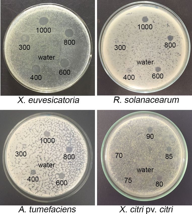
www.nature.com/scientificreports/
Figure 1. Inhibition of bacterial growth by spotting assay. Each bacterium was grown on LB agar plates
and different concentrations of the peptide (70–1,000 μg/ml) were tested in different spots to determine the
minimum inhibitory concentration of EPL able to inhibit bacterial growth. Water was used as negative control.
The plates were placed at 28 °C for two days and evaluated. Three biological replicates were done.
of the lipopolysaccharide layer in cell membranes, compromising their physical integrity, and causing the extrava-
sation of cellular c ontent18.
Epsilon-Poly-l-lysine (EPL) is a linear homopolypeptide, generally composed by 25 to 35 identical l-lysine
residues with a unique structure characterized by the peptidic bond of lysine monomers to gamma-amino
functional groups and carboxyl g roups19. The homopolymer is biodegradable, water soluble, non-toxic and
highly stable at high temperatures. EPL has wide antimicrobial activity, dependent on the molecular weight of
the peptide20. Its application against several animal pathogens is well-documented21–25 and it has been used as a
food preservative since 1980 and considered safe for human c onsumption26. However, there is a lack of studies
that demonstrate its efficacy against phytopathogenic bacteria and the possible control of plant diseases by topi-
cal application. The aim of this work was to investigate the in vitro antimicrobial potential of EPL against the
phytopathogens R. solanacearum, X. euvesicatoria, X. citri and A. tumefaciens and to verify the in vivo action in
the control of bacterial spot on tomato plants.
Results
EPL minimum inhibitory concentration and bacterial clearance dynamics. The antimicrobial
effect of EPL was first evaluated through in vitro spotting assays. The minimum inhibitory concentration (MIC)
was determined based on the lowest concentration of the peptide solution that was able to inhibit any bacterial
growth (Fig. 1). EPL efficiently inhibited the growth of all four tested bacterial species. The MIC of EPL varied
between 80 μg/ml to inhibit X. citri growth, 400 μg/ml for A. tumefaciens and 600 μg/ml for R. solanacearum and
X. euvesicatoria. In order to assess a putative bactericidal effect, further investigation was conducted. Incubating
the cells with the determined MICs of EPL confirmed the antibacterial activity of the peptide in the first 30 min,
when the quantity of colony forming units (CFUs) in the treatment group was already statistically different from
the water-treated control (Fig. 2). The peptide efficiently inhibited the growth of colonies after 30 min of EPL
treatment with a reduction in bacterial growth close to 100%. After two hours, the reduction remained, confirm-
ing the bactericidal activity of EPL (Fig. 2).
The effect of EPL on cell surface integrity and on cell viability of phytopathogenic bacte‑
ria. After confirming the growth inhibition effect of EPL on all four bacterial species, we further investigated
its effect on cell integrity to better understand if there was a bactericidal effect or simply growth suppression
(bacteriostatic effect). To assess this, we used spectroscopy and microscopy methods. First, we measured the
EPL effect on the fluorescent signal coming from cells treated with SYTO 9, a dye that binds DNA (Fig. 3). In all
four bacterial species the fluorescence emission was lower in cells incubated with MIC levels of EPL compared to
controls treated with water. This reduction indicates EPL is effective disrupting the cells instead of simply halting
their multiplication. Next, an additional dye was used, propidium iodide, which only enters cells with damaged
membranes. The combination of both dyes allows for a direct visual inspection of EPL effects, as shown in Fig. 4.
Scientific Reports | (2020) 10:11324 | https://doi.org/10.1038/s41598-020-68262-1 2
Vol:.(1234567890)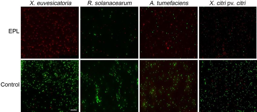
www.nature.com/scientificreports/

                            Figure 2.  Growth inhibition curves in the presence of EPL treatment at MIC. Aliquots were taken in 30-min
                            intervals, during 2 h, serially diluted and plated. Plates were kept for two days at 28 °C when CFU were counted.
                            The numbers of CFU/ml are the averages for three replicates plating from each sample, and the error bars
                            represent the standard deviations. Percentage of cell mortality was calculated as the ratio of cell counts in the
                            treated group with EPL to those in the control group.

                               Fluorescence microscopy images showed that cells emitted red fluorescence due to interaction of propidium
                            iodide with DNA. In the control group without EPL, the number of cells that emitted green fluorescence from
                            intact membranes was considerably higher (Fig. 4). This result suggested that EPL-treated cells had their cell
                            membrane damaged by the peptide.
                               To assess membrane integrity and bacterial morphology in finer detail, the bacterial cells were visualized
                            under scanning electron microscopy (SEM) after 1 h of treatment with EPL at the MIC (Fig. 5). The SEM images
                            revealed the rupture of the cytoplasmic membrane and the extravasation of cellular content due to the membrane
                            integrity disruption effect of EPL. Compared with treated cells, the surface of untreated cells was bright and
                            smooth. Taken together these results provide further support that EPL is actively damaging the bacterial cells
                            and not just simply arresting their proliferation.

                            EPL protects tomato plants against bacterial spot disease. After attesting the in vitro antimicro-
                            bial activity of EPL, we performed in vivo assays in tomato plants infected with X. euvesicatoria to establish the
                            potential of this peptide to control bacterial diseases by topical application.
                                Twenty days post-inoculation of the pathogen the initial symptoms such as spots and irregularly shaped
                            watery patches on the leaves started appearing in all 12 control plants treated with water only. Three out of twelve
                            plants that were treated preventively with EPL (at MIC level) developed initial symptoms. Interestingly, plants
                            that were treated preventively with twofold MIC did not show any symptoms. At thirty days post- inoculation the
                            disease was more severe in the control group (yellowish and bottom leaves death), and some plants were classified
                            as level 5 according to the disease scale proposed by Mello et al.27. Plants that were treated preventively with EPL
                            (onefold MIC) developed some disease symptoms, however no symptom was observed on plants that were treated
                            preventively with twofold MIC, demonstrating the long-lasting effect of preventive EPL treatment (Fig. 6a).
                                We also tested the potential curative effect of EPL after X. euvesicatoria infection. Plant recovery, however,
                            was not as robust as prevention with EPL. When EPL was applied two days after X. euvesicatoria infection all
                            the plants showed initial symptoms of bacterial spot (onefold MIC and twofold MIC), however, by the end of
                            the experiment, no plants were classified as level 5 according to the disease scale, meaning that EPL treatment
                            can slow down the disease progress or alternatively reduce its severity (Fig. 6b).

Scientific Reports |   (2020) 10:11324 |               https://doi.org/10.1038/s41598-020-68262-1                                             3

                                                                                                                                         Vol.:(0123456789)
www.nature.com/scientificreports/
Figure 3. Cell viability measured by fluorescence emitted by bacterial cells treated with EPL at MIC for one
hour. SYTO 9 was added to the bacterial suspensions and incubated for 15 min in the dark. Samples were
excited at 470 nm and emission spectrum (490–700 nm) was recorded. A lower emitted fluorescence near
530 nm indicates a lower number of viable cells. Controls included water-only treatment (non EPL-treated) and
EPL with fluorophore (without bacterial cells). Values shown are the average of three biological replicates.
Figure 4. Overlap of fluorescence images of bacterial cells stained with SYTO 9 and propidium iodide for
15 min in the dark. Bacterial cells were treated with EPL at MIC for one hour or water as control. The red
fluorescence is emitted because of an interaction between propidium iodide and DNA when the cytoplasmic
membrane is damaged, whereas bacteria with intact cell membranes are impermeable to propidium iodide and
display green fluorescence. Images shown are representative of three biological replicates. Scale bar = 10 μm.
Scientific Reports | (2020) 10:11324 | https://doi.org/10.1038/s41598-020-68262-1 4
Vol:.(1234567890)www.nature.com/scientificreports/
Figure 5. The effect of EPL on the bacterial cell surface. Scanning electron microscopy of bacterial cells treated
with EPL at MIC for 1 h. No-EPL (water) treatment was used as control. Scanning electron microscope was
operated at 10 kV. Scale bar = 1 μm.
Figure 6. Enhanced protection against bacterial spot in tomatoes sprayed with EPL solution. Twelve Santa
Cruz-Kada tomato plants were analyzed for each condition after 30 days of X. euvesicatoria inoculation. The
figure shows representative leaves of three tomato plants treated with EPL (onefold MIC or twofold MIC)
2 days before inoculation (a), and tomato plants treated with EPL (onefold MIC or twofold MIC) 2 days post-
inoculation (b).
Discussion
Antimicrobial peptides are widespread in nature, among competing microbial communities and also among plant
and animal hosts as a means to fight off infections. They hold great potential in agriculture, since phytopathogenic
bacteria are still problematic to control due to lack of effective bactericides and the emergence of resistance.
Here we evaluated the potential use of EPL to control some of the most devastating bacterial plant pathogens. Its
effectiveness against bacterial pathogens in mammalian and safety for human consumption has been previously
confirmed, making it a promising candidate for agricultural applications. A pioneering study revealed EPL to be
nontoxic in a dosage level of 10,000 μg/ml and nonmutagenic in usage-relevant concentrations in a two-genera-
tion reproduction study using r ats28. This value is 125-fold higher than what we determined here to be necessary
to control X. citri, 25-fold higher than necessary to control A. tumefaciens, and around 17-fold higher than the
MIC to control R. solanacearum and X. euvesicatoria, again reinforcing its safety at effective levels for bacterial
clearance. Zhao et al. studied the effect of EPL and Epsilon-caprolactone (CPL) copolymer-based nanoparticles
on mammalian cells viability using human breast carcinoma and human umbilical vein endothelial cells as mod-
els, and reported no apparent cytotoxic effects for both cell types even at the concentration of 1,000 μg/ml29, but
studies on acute toxicity are also required before recommendation for commercial application. Some countries
including Japan, South Korea and Canada have already regulated EPL use as a natural preservative for the food
industry19,30. In 2004, the US Food and Drug Administration has given EPL GRAS status (generally recognized
as safe), and approved its use as an antimicrobial agent in cooked or sushi rice at levels up to 50 mg/kg of r ice31.
Scientific Reports | (2020) 10:11324 | https://doi.org/10.1038/s41598-020-68262-1 5
Vol.:(0123456789)www.nature.com/scientificreports/
We demonstrated that the in vitro antibacterial activity of the peptide was able to inhibit growth of all four
tested phytobacteria. These results are correlated with loss of cellular viability verified by SEM and fluorescence
assays and encouraged further investigation on its therapeutic potential in vivo. Previous studies have shown great
variability of microbial sensitivity to EPL, with Gram-negative bacteria consistently showing higher sensitivity
compared to Gram-positive. EPL was tested against the Gram-negative Escherichia coli and Gram-positive Listeria
innocua, for which the MIC was defined as 74 µg/ml for E. coli and 750 µg/ml for L. innocua32. To understand
these differences, it is necessary to correlate the mechanism of action of the peptide, the membrane and cell wall
composition of the pathogens, and the phospholipid stoichiometry of the different microbial targets33,34. The
kill curves obtained in this study confirmed the effective and fast bactericidal effect of EPL. Our results are in
accordance with other studies testing AMPs. For example, the mortality ratio of E. coli O157:H7 treated with
different concentrations of EPL ranged from 5 to 50 µg/ml, and after 15 h was higher than 95%25.
Bacterial cell death is defined as the incapacity of the cell to grow as a viable colony in bacteriological media.
However, there are different ways to evaluate cell viability without culturing cells. One of them is based on fluo-
rescent probes that can be detected through fluorescence microscopy and spectroscopy35. Fluorescence detection
is a fast and reliable technique that is valid to quantify bacteria of different g enera36. A known effect of EPL on
bacterial cells is the damage of the cell wall, compromising cellular integrity. Cell viability can be monitored using
fluorescent dyes that differ in spectral characteristics and in the ability to penetrate bacterial cells. This allows
reliable quantitative distinction between bacteria with intact or damaged citoplasmic membrane, consequently
differentiating living and dead c ells35. The results obtained through fluorescence spectroscopy and SEM are in
accordance with the initial results that showed reduced colony formation following incubation with EPL. The
predominance of red-fluorescent cells confirms that the growth inhibition is due to the bactericidal effect of
the peptide and not only a bacteriostatic effect. In order to study the effects of EPL on S. aureus cell membrane,
Tan et al. showed that after EPL treatment there was a remarkable increase in fluorescence intensity measured
by propidium iodide assay indicating an increase in cell membrane p ermeability37. When S. aureus cells were
treated with 250 μg/ml of EPL, the cells appeared collapsed, lysed and with non-integral cell morphology37.
Similar results were obtained after treating E. coli cells with 50 μg/ml of EPL for 4 h. They observed that the
external membrane and cytoplasm were damaged, surrounded by cell debris and with wrinkled a ppearance25.
In another study the interaction between S. aureus and B. subtilis cells with nanoparticles composed by EPL and
CPL were e valuated29. They observed the effect of EPL treatment on cell structure by SEM and similar to our
findings revealed cells broken in appearance, with rupture of cell wall and membrane, lysis of cellular content,
and extravasation of cytoplasm. All these observations confirm that EPL can adsorb on the surface of the microbe
membrane resulting in physical damage to the cell. The previous reports showed that EPL can affect the cell
membrane permeability and compromise their v iability25, 37, 38. The EPL mechanism of action assures that the
microorganism does not easily develop resistance.
Despite its in vitro efficacy, it is important to understand how EPL performs in vivo under more realistic con-
ditions encountered in the field. In this study, tomato plants that were sprayed with EPL before bacterial infection
were protected against bacterial spot disease. Some spots were observed in some leaves, but the disease did not
fully develop. When a higher concentration was applied (twofold MIC), no symptom was observed. Therefore,
our data shows the higher effectiveness of EPL when it is applied as a preventive method at higher dosages such
as twofold MIC. However, we also observed symptom reduction when EPL was applied after bacterial infection,
even at MIC level.
Here we concluded that EPL, even at low concentrations, has significant in vitro antimicrobial activity against
diverse phytobacteria, attesting to its broad range of activity towards microbial cells. It is known that EPL is
biodegradable, non-toxic, resistant to thermal degradation, and possess antimicrobial activity against a wide
spectrum of microorganisms. Our results confirm that EPL is a promising alternative to control phytobacteri-
osis prophylactically. In light of these findings, more investigations will determine the optimal method for the
application of this peptide in agricultural contexts, and the effect of this peptide in the pathosystem and phyto-
sphere microbiome in general.
Materials and methods
Bacterial strains. EPL antimicrobial activity was tested against the phytopathogenic bacteria Ralstonia sola-
nacearum strain GMI1000, phylotype I, biovar 3, provided by Dr. Carlos Alberto Lopes, Embrapa Hortaliças,
Brazil; Xanthomonas citri subsp. citri 306, obtained from Dr. Shaker Chuck Farah at University of São Paulo,
Brazil; Xanthomonas euvesicatoria EH 2009-130 and Agrobacterium tumefaciens C58, obtained from Dr. Alice
Maria Quezado Duval, Embrapa Hortaliças, Brazil. Bacterial stocks were kept at − 80 °C in LB broth supple-
mented with 50% glycerol (v/v).
Minimum inhibitory concentration assay. To obtain the minimum inhibitory concentration (MIC) of
EPL necessary to prevent bacterial growth, a spotting assay39,40 was performed. Briefly, bacteria were cultured in
LB broth at 28 °C, 200 rpm, for 12 h. Ten microliters of these bacterial suspensions were diluted in LB to 100 μl
and spread onto LB agar plates. Ten microliters of EPL solutions at different concentrations (70–1,000 μg/ml)
were spotted on the agar plates that had previously received the bacterial suspensions. Plates were then incu-
bated at 28 °C until formation of clearance zones where growth inhibition could be clearly seen. MIC values
were registered as the least concentration of EPL that would inhibit bacterial growth. Distilled water was used as
negative control spotted in the center of each plate. All assays were performed in biological triplicates.
Kill‑curves. After EPL MIC values were determined for each bacterial species, the growth inhibition dynam-
ics was further evaluated. Bacteria were cultured overnight in LB broth and OD600nm adjusted to 0.1 with LB
Scientific Reports | (2020) 10:11324 | https://doi.org/10.1038/s41598-020-68262-1 6
Vol:.(1234567890)www.nature.com/scientificreports/
medium (~ 106 CFU/ml). EPL was added to each bacterial suspension at the previously determined MICs and
these were incubated for two hours at 28 °C and 200 rpm. Aliquots were taken at 0, 30, 60, 90, and 120-min inter-
vals, serially diluted with LB broth and plated. Plates were kept for two days at 28 °C and the number of CFUs
was used to determine the efficiency of EPL in clearing the pathogen. Three biological replicas were performed.
Fluorescence spectroscopy and microscopy. Bacterial viability after EPL treatment was assessed by
fluorescence emitted by EPL-treated compared to non-treated bacterial cells. Initially log-phase bacterial cul-
tures were adjusted to OD600nm 0.1, and centrifuged at 10,000×g for 15 min. Supernatant was removed and pellets
suspended in EPL solutions at MIC or distilled water control. Cells were incubated at 28 °C for one hour when
1 µl of SYTO 9 from the Live/Dead BacLight bacterial viability kit (Life Technologies) was added to 1 ml of the
bacterial suspensions. The samples were then incubated in the dark for 15 min and fluorescence measured using
flat bottom black plates and fluorimeter (PerkinElmer). Samples were excited at 470 nm and emission spectrum
(490–700 nm) recorded. Three biological replicas were performed, and two technical replica readings of each
sample were taken. Controls included water-only treatment (non EPL-treated) and EPL + fluorophores (without
bacterial cells). Fluorescence microscopy was used to visualize viable and non-viable cells stained with 3 µL of a
1:1 mixture of SYTO 9 and propidium iodide from the Live/Dead BacLight kit components. Cells were prepared
as described above and imaged on an Evos FL fluorescence microscope (ThermoFisher), in which viable cells
fluoresce in green and non-viable cells in red.
Scanning electron microscopy. Morphological alterations on bacterial cell surface after EPL treatment
were investigated by SEM. Cells were cultured in 24-well plates containing a 12 mm round coverslip and 2 ml
LB in each well, for one day. EPL was then added according to previously determined MICs, or distilled water
as control, and incubated for an additional hour. Coverslips containing attached cells were then fixed with 2.5%
(v/v) glutaraldehyde for 16 h at 4 °C. Next, four 10-min washes were made with 0.1 M cacodylate buffer pH 7.2,
and a secondary fixation in 1% (v/v) osmium tetroxide for one hour. Another three washes in cacodylate buffer
were made and samples incubated in 1% tannic acid for 30 min, followed by two quick washes in water before
dehydration in increasing ethanol concentrations (50–100% [v/v]) for 10 min each step. Critical point drying
was then followed by sputter coating with gold at 50 mA for 120 s before being imaged with an EVO MA Carl
Zeiss (Zeiss, Germany) scanning electron microscope operating at 10 kV.
In vivo EPL protection assay. Tomato plants (Solanum lycopersicum L.) cv. Santa Cruz Kada were grown
in the greenhouse at 25 ± 2 °C to the V2 developmental stage41. Xanthomonas euvesicatoria EH 2009-130 was cul-
tured in LB medium and adjusted to 107 CFU/ml with NaCl 0.85% (w/v). The bacterial suspension was sprayed
on the tomato leaves until dripping. Plants were kept in a humid chamber made with a transparent plastic bag
cover for 24 h before and after inoculation to facilitate infection. To test protective and curative effect of EPL
to bacterial infection, onefold and twofold MIC diluted in water plus 1% Pentra-Bark surfactant were sprayed
on entire tomato plant surfaces (twelve plants for each treatment). Water was used as negative control. Applica-
tions were done two days before inoculation with X. euvesicatoria (prophylactic) or two days after inoculation
(curative). Progress of symptom development was recorded as visual appearance of leaf spots until 30 days post-
inoculation, and scores were given from 1 (1% of leaf area affected) to 5 (50% or more of leaf area affected),
according to the diagrammatic scale proposed by Mello et al.27.
Statistical analysis. The necessary assumptions required for the analysis of variance (ANOVA) were veri-
fied. Normality of errors and variance of homogeneity were evaluated with Shapiro–Wilk and Levene tests. Kill-
curve assays were analyzed in a split-plot arrangement placing the EPL treatment and the incubation time in the
main and sub plots, respectively. Complex variances were applied when significant interactions were observed.
Averages of peptide treatment and incubation periods were compared by Tukey test and polynomial regression,
respectively. All analyses were done considering significance of 0.05.
Received: 14 February 2020; Accepted: 5 May 2020
References
1. Kering, K. K., Kibii, B. J. & Wei, H. Biocontrol of phytobacteria with bacteriophage cocktails. Pest Manag. Sci. 75, 1775–1781. https
://doi.org/10.1002/ps.5324 (2019).
2. He, D. C., Zhan, J. S. & Xie, L. H. Problems, challenges and future of plant disease management: from an ecological point of view.
J. Integr. Agric. 15, 705–715. https://doi.org/10.1016/S2095-3119(15)61300-4 (2016).
3. Pini, F., Galardini, M., Bazzicalupo, M. & Mengoni, A. Plant-bacteria association and symbiosis: are there common genomic traits
in Alphaproteobacteria?. Genes 4, 1017–1032. https://doi.org/10.3390/genes2041017 (2011).
4. Mansfied, J. S. et al. Top 10 plant pathogenic bacteria in molecular plant pathology. Mol. Plant Pathol. 13, 614–629. https://doi.org
/10.1111/j.1364-3703.2012.00804.x (2012).
5. Yuliar Nion, Y. A. & Toyota, K. Recent trends in control methods for bacterial wilt diseases caused by Ralstonia solanacearum.
Microbes Environ. 30, 1–11. https://doi.org/10.1264/jsme2.ME14144 (2015).
6. Hayward, A. C. Biology and epidemiology of bacterial wilt caused by Pseudomonas solanacearum. Annu. Rev. Phytopathol. 29,
65–87 (1991).
7. Young, J. M., Kuykendall, L. D., Martínez-Romero, E., Kerr, A. & Sawada, H. A revision of Rhizobium Frank 1889, with an emended
description of the genus, and the inclusion of all species of Agrobacterium Conn 1942 and Allorhizobium undicola de Lajudie et al.
1998 as new combinations: Rhizobium radiobacter, R. rhizogenes, R. rubi, R. undicola and R. vitis. Int. J. Syst. Evol. Microbiol. 51,
89–103 (2001).
Scientific Reports | (2020) 10:11324 | https://doi.org/10.1038/s41598-020-68262-1 7
Vol.:(0123456789)www.nature.com/scientificreports/
8. Subramoni, S., Nathoo, N., Kilmov, E. & Yuan, Z. C. Agrobacterium tumefaciens responses to plant-derived signaling molecules.
Front Plant Sci. https://doi.org/10.3389/fpls.2014.00322 (2014).
9. Lacroix, B. & Citovsky, C. Transfer of DNA from bacteria to eukaryotes. mBIO https://doi.org/10.1128/mBio.00863-16 (2016).
10. Bock, C. H., Cook, A. Z., Parker, P. E., Gottwald, T. R. & Graham, J. H. Short-distance dispersal of splashed bacteria of Xanthomonas
citri subsp. citri from canker-infected grapefruit tree canopies in turbulent wind. Plan. Pathol. 61, 829–836. https://doi.org/10.11
11/j.1365-3059.2011.02588.x (2012).
11. Stover, E., Randall, D., Richardson, L. M., Hall, G. D. & Duan, Y. Incidence and severity of asiatic citrus canker on diverse citrus
and citrus-related germplasm in a Florida field. HortScience 49, 4–9. https://doi.org/10.21273/HORTSCI.49.1.4 (2014).
12. Ferrarezi, J. H. et al. Extratos de fungos da Antártica: Avaliação da atividade contra Xanthomonas citri subsp. citri. Preprint at
https://docplayer.com.br/42602759-Extratos-de-fungos-da-antartica-avaliacao-da-atividade-contra-xanthomonas-citri-subsp
-citri.html (2016).
13. Potnis, N. et al. Bacterial spot of tomato and pepper: diverse Xanthomonas species with a wide variety of virulence factors posing
a worldwide challenge. Mol. Plant. Pathol. 16, 907–920. https://doi.org/10.1111/mpp.12244 (2015).
14. Larrea-Sarmiento, A. et al. Development of a genome-informed loop-mediated isothermal amplification assay for rapid and specific
detection of Xanthomonas euvesicatoria. Sci. Rep. https://doi.org/10.1038/s41598-018-32295-4 (2018).
15. Guell, I. et al. Design, synthesis and biological evaluation of cyclic peptidotriazoles derived from BPC194 as novel agents for plant
protection. Biopolymers https://doi.org/10.1002/bip.23012 (2017).
16. Almaytaah, A. et al. Peptide consensus sequence determination for the enhancement of the antimicrobial activity and selectivity
of antimicrobial peptides. Infect. Drug Resist. 10, 1–17. https://doi.org/10.2147/IDR.S118877 (2017).
17. Morais, T. et al. The plant-based chimeric antimicrobial protein SlP14a-PPC20 protects tomato against bacterial wilt disease caused
by Ralstonia solanacearum. Plant Sci. 280, 197–205. https://doi.org/10.1016/j.plantsci.2018.11.017 (2019).
18. Jenssen, H., Hamill, P. & Hancock, R. E. W. Peptide antimicrobial agents. Clin. Microbiol. Rev. 19, 491–511. https: //doi.org/10.1128/
CMR.00056-05 (2006).
19. Hiraki, J. ε-Polylysine, its development and utilization. Fine Chem. 29, 18–25 (2001).
20. Shima, S., Matsuoka, H., Iwamoto, T. & Sakai, H. Antimicrobial action of ε-Poly-l-Lysine. J. Antibiot. 37, 1449–1455. https://doi.
org/10.7164/antibiotics.37.1449 (1984).
21. Yoshida, T. & Nagasawa, T. ɛ-Poly-l-lysine: microbial production, biodegradation and application potential. Appl. Microbiol.
Biotechnol. 62, 21–26. https://doi.org/10.1007/s00253-003-1312-9 (2003).
22. Geornaras, I., Yoon, Y., Belk, K. E., Smith, G. C. & Sofios, J. N. Antimicrobial activity of epsilon-polylysine against Escherichia coli
O157:H7 Salmonella Typhimurium, and Listeria monocytogenes in various food extracts. J. Food Sci. 72, 330–334. https://doi.org
/10.1111/j.1750-3841.2007.00510.x (2007).
23. Najjar, M. B., Kashtanov, D. & Chikindas, M. ɛ-Poly-l-lysine and nisin A act synergistically against Gram-positive food-borne path-
ogens Bacillus cereus and Listeria monocytogenes. Lett. Appl. Microbiol. 45, 13–18. https: //doi.org/10.1111/j.1472-765X.2007.02157
.x (2007).
24. Takehara, M., Hibino, A., Saimura, M. & Hirohara, H. High-yield production of short chain length poly (ε-l-lysine) consisting of
5–20 residues by Streptomyces aureofaciens, and its antimicrobial activity. Biotechnol. Lett. 32, 1299–1303. https://doi.org/10.1007/
s10529-010-0294-9 (2010).
25. Ye, R. et al. Antibacterial activity and mechanism of action of ɛ-Poly-l-lysine. Biochem. Biophys. Res. Commun. 439, 148–153.
https://doi.org/10.1016/j.bbrc.2013.08.001 (2013).
26. Hiraki, J. et al. Use of ADME studies to confirm the safety of epsilon-polylysine as a preservative in food. Regul. Toxicol. Pharmacol.
37, 328–340 (2003).
27. Mello, S. C. M., Takatsu, A. & Lopes, C. A. Escala diagramática para avaliação da mancha-bacteriana do tomateiro. Fitopatol. Brasil.
3, 447–448 (1997).
28. Neda, K., Sakurai, T., Stakahashi, M., Shiychi, M. & Ohgushi, M. Two generation reproduction study with teratology test of ε-poly
ε-lysine by dietary administration in rats. Jpn. Pharmacol. Ther. 27, 1139–1159 (1999).
29. Zhao, R. et al. Biodegradable cationic E-Poly-l-lysine-conjugated polymeric nanoparticles as a new effective antibacterial agent.
30. Chheda, A. H. & Vernekar, M. R. A natural preservative ɛ-Poly-l-lysine: fermentative production and applications in food industry.
Sci. Bull. 60, 216–226. https://doi.org/10.1007/s11434-014-0704-9 (2015).
Int. Food Res. J. 22, 23–30 (2015).
31. US Food and Drug Administration (USFDA). GRAS Notice 000135: ε-Polylysine. Office of Food Additive Safety. Preprint at https
://www.fda.gov (2004).
32. Hyldgaard, M. et al. The antimicrobial mechanism of action of Epsilon-Poly-l-Lysine. Appl. Environ. Microbiol. 80, 7758–7770.
https://doi.org/10.1128/AEM.02204-14 (2014).
33. Koebnik, R., Locher, K. P. & Van Gelder, P. Structure and function of bacterial outer membrane proteins: barrels in a nutshell. Mol.
Microbiol. 37, 239–253 (2000).
34. Yeaman, M. R. & Yount, N. Y. Mechanisms of antimicrobial peptide action and resistance. Pharmacol. Rev. 96, 254–260. https://
doi.org/10.1124/pr.55.1.2 (2003).
35. Berney, M., Hammes, F., Bosshard, F., Weilenmann, H. & Egli, T. Assessment and interpretation of bacterial viability by using the
LIVE/DEAD BacLight kit in combination with flow cytometry. Appl. Environ. Microbiol. 73, 3283–3290. https://doi.org/10.1128/
AEM.02750-06 (2007).
36. Guo, R., McGoverin, C., Swift, S. & Vanhoulsbeeck, F. A rapid and low-cost estimation of bacteria counts in solution using fluo-
rescence spectroscopy. Anal. Bioanal. Chem. 409, 3959–3967. https://doi.org/10.1007/s00216-017-0347-1 (2017).
37. Tan, Z. L. et al. The antimicrobial effects and mechanism of E-poly-lysine against Staphylococcus aureus. Bioresour. Bioprocess.
https://doi.org/10.1186/s40643-019-0246-8 (2019).
38. Tan, Z. L., Bo, T., Guo, F. Z., Cui, J. D. & Jia, S. R. Effects of E-poly-l-lysine on the cell wall of Saccharomyces cerevisiae and its
involved antimicrobial mechanism. Int. J. Biol. Macromol. 118, 2230–2236. https://doi.org/10.1016/j.ijbiomac.2018.07.094 (2018).
39. Chakraborty, S. et al. The PDB database is a rich source of alpha-helical anti-microbial peptides to combat disease causing patho-
gens. F1000Res. 3, 295-310, 10.12688/f1000research.5802.2 (2014).
40. Wang, J., Woo, M. & Yan, C. Spot plating assay for the determination of survival and plating efficiency of Escherichia coli in sub-
MIC levels of antibiotics. JEMI Methods 1, 26–29 (2017).
41. Revelo, E., Dorado, G., Lagos, L. E. & Burbano-Figueroa, O. Foliar virulence of isolates of Phytophthora infestans sensu lato on
detached leaves of two Solanum betaceum cultivars. Tropic Plant Pathol. https: //doi.org/10.1590/S1982- 567620 11000 60000 5 (2011).
Acknowledgements
We thank the Scanning electron microscopy multiuser lab staff from the Chemical Engineering Department at
University of Uberlândia for assistance. We also thank Embrapa Hortaliças Brazil for providing some of the bacte-
rial species used in this study. The authors thank the Brazilian funding agencies, National Council for Scientific
and Technological Development (CNPq), Coordination for the Improvement of Higher Education Personnel
(CAPES) and Minas Gerais State Agency for Research and Development (FAPEMIG), for providing financial
Scientific Reports | (2020) 10:11324 | https://doi.org/10.1038/s41598-020-68262-1 8
Vol:.(1234567890)www.nature.com/scientificreports/
support to the National Institute of Science and Technology in Theranostics and Nanobiotechnology – INCT-Ter-
aNano (CNPq/CAPES/FAPEMIG, Grant numbers CNPq-465669/2014-0 and FAPEMIG-CBB-APQ-03613-17).
Author contributions
A.M.D. and L.R.G. conceived the idea. R.N. and L.R.G. obtained funds to carry out the work. B.R., T.P.M.,
P.A.Z., A.M.D. and R.N. designed the experiments. B.R. and C.S.C. performed the experiments. B.R., T.P.M.,
and H.O.A.S. analyzed data. B.R., T.P.M., and P.A.Z. wrote the manuscript. All authors read and approved the
manuscript.
Competing interests
The authors declare no competing interests.
Additional information
Correspondence and requests for materials should be addressed to L.R.G.
Reprints and permissions information is available at www.nature.com/reprints.
Publisher’s note Springer Nature remains neutral with regard to jurisdictional claims in published maps and
institutional affiliations.
Open Access This article is licensed under a Creative Commons Attribution 4.0 International
License, which permits use, sharing, adaptation, distribution and reproduction in any medium or
format, as long as you give appropriate credit to the original author(s) and the source, provide a link to the
Creative Commons license, and indicate if changes were made. The images or other third party material in this
article are included in the article’s Creative Commons license, unless indicated otherwise in a credit line to
the material. If material is not included in the article’s Creative Commons license and your intended use is
not permitted by statutory regulation or exceeds the permitted use, you will need to obtain permission directly
from the copyright holder. To view a copy of this license, visit http://creativecommons.org/licenses/by/4.0/.
© The Author(s) 2020
Scientific Reports | (2020) 10:11324 | https://doi.org/10.1038/s41598-020-68262-1 9
Vol.:(0123456789)You can also read