Comparison of oral and nasal immunization with inactivated porcine epidemic diarrhea virus on intestinal immunity in piglets
←
→
Page content transcription
If your browser does not render page correctly, please read the page content below

1596                             EXPERIMENTAL AND THERAPEUTIC MEDICINE 20: 1596-1606, 2020

   Comparison of oral and nasal immunization with inactivated
 porcine epidemic diarrhea virus on intestinal immunity in piglets
           EN ZHANG, JIALU WANG, YUCHEN LI, LULU HUANG, YONGHENG WANG and QIAN YANG

                       MOE Joint International Research Laboratory of Animal Health and Food Safety,
             College of Veterinary Medicine, Nanjing Agricultural University, Nanjing, Jiangsu 210095, P.R. China

                                        Received July 1, 2019; Accepted February 20, 2020

                                                     DOI: 10.3892/etm.2020.8828

Abstract. Porcine epidemic diarrhea virus (PEDV) has proven         Introduction
to be a major problem for the porcine industry worldwide.
Conventional injectable vaccines induce effective systemic          Porcine epidemic diarrhea (PED) is an acute and highly conta-
immune responses but are less effective in preventing PEDV          gious disease, which is characterized by vomiting, diarrhea,
at mucosal invasion sites, including the nasal or oral mucosa.      dehydration and a high mortality rate in neonatal piglets (1).
Additionally, antigens delivered orally are easily degraded.        PED was first observed in 1971 and caused substantial
Nasal immunization induces intestinal mucosal immune                economic losses to the pig industry (2). As the causative
responses, which can aid in blocking viral invasion, and            agent, PED virus (PEDV) exclusively infects and replicates
requires fewer antigen inoculation doses. Therefore, nasal          in the villous enterocytes of the small intestine (3). Currently
immunizations are considered to be a potential approach to          available PEDV vaccines are administered via intramuscular
overcome viral infections. In the present study, nasal immu-        or subcutaneous injection and induce a systemic immunoglob-
nization of piglets was performed using inactivated PEDV            ulin (Ig)G response (4). However, serum IgG is not effective
combined with Bacillus subtilis as an immunopotentiator and         in preventing PEDV infection in the intestinal mucosa (4,5).
the efficacy of nasal immunization was assessed. The results        Studies have demonstrated that secretory IgA (SIgA), a domi-
demonstrated that compared with oral immunization, piglets          nant antibody in the intestinal mucosal immune responses,
from the nasal immunization group exhibited higher levels of        inhibits the invasion of enteric pathogens (6). Consequently,
neutralizing antibodies (P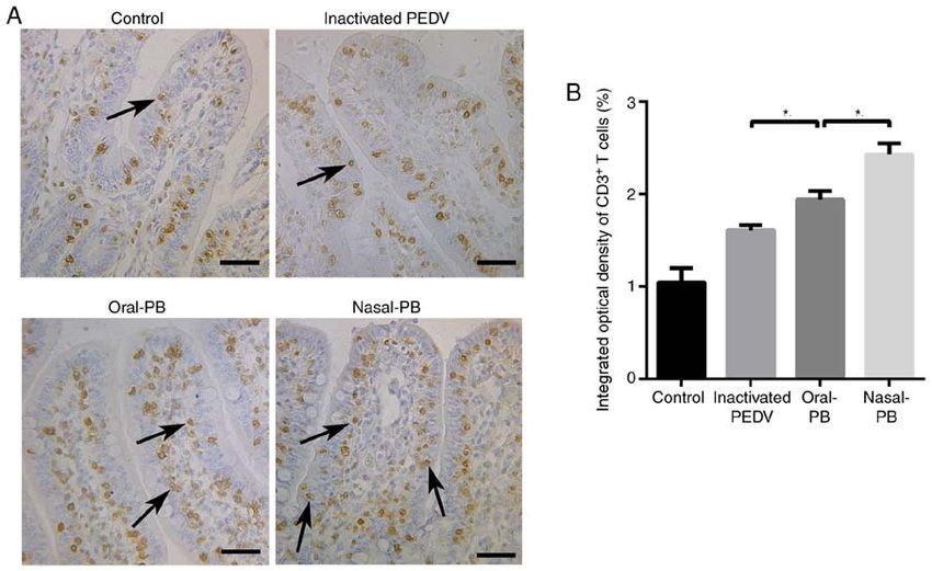
ZHANG et al: COMPARISON OF ORAL AND NASAL IMMUNIZATION ON INTESTINAL IMMUNITY OF PIGLETS                        1597

capable of adequately strengthening the immune response.           Experimental design and collection of samples. A total of 24
Bacillus subtilis (B. subtilis) is a gram‑positive nonpathogenic   SPF DLY piglets, born on the same day, were raised in indi-
and endospore‑forming bacterium species that is prevalent          vidual cages with high sanitary conditions. They were weighed
worldwide (15). B. subtilis has been used as an additive when      and randomly assigned to four groups, each group containing
feeding animals in the pig industry (16). Previous studies have    6 piglets. All piglets were first immunized at 5 days of age
demonstrated that B. subtilis exhibits a significant immunopo-     and were given a booster immunization at 12 days of age.
tentiating effect that leads to an enhanced mucosal immune         The groups established were as follows: i) Control group, oral
response (17,18).                                                  immunization with 1,100 µl phosphate‑buffered saline (PBS);
    In the present study, the immune responses after nasal         ii) inactivated PEDV group, oral immunization with 100 µl
immunization with inactivated PEDV combined with                   inactivated PEDV (100 µg/dose) combined with 1 ml PBS;
B. subtilis were evaluated in piglets. The results revealed that   iii) oral‑PB group, oral immunization with 100 µl inactivated
nasal immunization could induce the immune response in the         PEDV (100 µg/dose) combined with 1x10⁹ colony‑forming
local nasal mucosa and in the intestinal mucosa. Compared          units (CFU) B. subtilis; and iv) nasal‑PB group, nasal immuni-
with oral immunization, piglets receiving nasal immuniza-          zation with 100 µl inactivated PEDV (100 µg/dose) combined
tion exhibited higher SIgA levels and an increased number of       with 1x10⁹ CFU of B. subtilis.
immune cells in the intestine. The results of the present study         The piglets were fasted for 3 h before each sample collec-
demonstrate a convenient and effective strategy for PEDV           tion. Collection of each sample was performed between 10:00
prevention and provide information regarding the common            and 11:00 a.m. before feeding. For the detection of IgG antibody
mucosal immune system in pigs.                                     levels in the serum, 2 ml blood sample was collected from the
                                                                   precaval vein, once a week on days 0, 7, 14, 21 and 28 after the
Materials and methods                                              initial immunization. Serum was collected after centrifugation
                                                                   at 13,000 x g for 20 min at 4˚C and stored at ‑70˚C. Saliva as
Probiotics, virus and animals. The B. subtilis strain was main-    well as nasal and anal secretions were also collected on days
tained at ‑70˚C by the present laboratory. The strains were        0, 7, 14, 21 and 28 after the initial immunization using cotton
grown in Luria‑Bertani broth containing 50 µg/ml kanamycin.        swabs. Piglets were fasted for 3 h before saliva collection.
PEDV Zhejiang08 strain was provided by the Veterinary              Saliva was collected using a cotton swab that was bitten by
Medicine Research Center (Beijing Da Bei Nong Science and          the piglets three times. Nasal secretions were collected using
Technology Group Co., Ltd.). This strain has been successfully     cotton swabs that were inserted 1.5 cm into the nose. Anal
attenuated and can induce a classical cytopathic effect in Vero    samples were collected using cotton swabs that were inserted
E6 cells (19). PEDV was amplified in Vero cells in DMEM            4 cm into the rectum. Subsequently, the collected samples were
(Wisent Biotechnology) containing 2% FBS at 37˚C in 5%             rapidly diluted in 800 µl PBS, vortexed for 30 sec, centrifuged
CO2 for 60 h. And then condensed by high‑speed centrifuga-         at 3,000 x g at 4˚C for 10 min and stored at ‑70˚C for ELISA
tion (100,000 x g at 4˚C for 2.5 h). Protein concentrations        detection. Piglets were euthanized by the intravenous injection
were confirmed using bicinchoninic acid (BCA) assays and           of sodium pentobarbital (100 mg/kg) at 33 days of age. The
the inactivated PEDV was diluted to a concentration of up to       pigs were confirmed dead when the corneal reflex disappeared,
100 µg/100 µl in LB broth. In the present study, the PEDV          breathing stopped and the heart stopped beating. Fresh intes-
was placed in a 6‑cm plate and illuminated for 12 h using          tines were rinsed using 1 ml DMEM (Wisent Biotechnology)
a UV lamp. A total of 24, 0‑day‑old specific pathogen free         prior to analysis in the plaque reduction neutralization test
(SPF) Duroc Landrace Yorkshire (DLY) piglets (12 males;            (PRNT). A portion of small intestine tissue was fixed in
12 females; mean body weight ~1.5 kg) were provided by             Bouin's fluid (picric acid, 4% paraformaldehyde, glacial acetic
the Institute of Veterinary Research, Jiangsu Academy of           acid) at room temperature for 24 h for staining and another
Agricultural Sciences. Colostrum was not provided to the           small intestine portion was collected and stored at ‑70˚C for
piglets. The piglets were kept in individual cages and food and    cytokine detection. All animal experiments were approved by
water were given ad libitum under the same standard condi-         the Ethics Committee of Animal Experiments of the College
tions at 12 h light/dark cycles. The temperature of the cages      of Veterinary Medicine, Nanjing Agricultural University. All
was 30˚C in the first week and kept 26˚C in the subsequent         animal care and use were conducted in strict accordance with
weeks of the experimental period. The relative humidity of the     the Animal Research Committee guidelines of the College of
atmosphere was maintained at 70%.                                  Veterinary Medicine, Nanjing Agricultural University.

Antibodies. Goat anti‑pig IgA (1:100; cat. no. ab112639)           ELISA for PEDV‑specific SIgA in the local mucosa and IgG
and rabbit anti‑pig CD3 (SP7) (1:100; cat. no. ab16669)            in the serum. The protein concentration of serum and the
monoclonal antibodies were purchased from Abcam. The               supernatant of mucosal secretions were measured using a
streptavidin‑biotin complex (SABC) kit (Wuhan Boster               BCA protein assay kit (Thermo Fisher Scientific, Inc.). The
Biological Technology, Ltd), including biotinylated goat           PEDV‑specific antibody levels were detected using ELISA.
anti‑rabbit IgG (cat. no. sa1022), biotinylated rabbit anti‑goat   Briefly, ELISA plates were coated in PEDV, 2 µg purified
IgG antibodies (cat. no. sa1023) and horseradish peroxidase        PEDV/well, at 4˚C overnight. The plates were washed with
(HRP)‑labeled SABC, was purchased from Boster Biological           PBS containing 0.05% Tween‑20 (PBS‑T) to remove the virus.
Technology. The 3,3‑diaminobenzidine (DAB) HRP Color               After virus removal, plates were blocked for 2 h at 37˚C with
Development kit (cat. no. ar1022) was also purchased from          3% BSA (Sigma‑Aldrich; Merck KGaA) dissolved in PBS.
Boster Biological Technology.                                      Subsequently, 1:100 dilutions of serum samples or 1:2 dilutions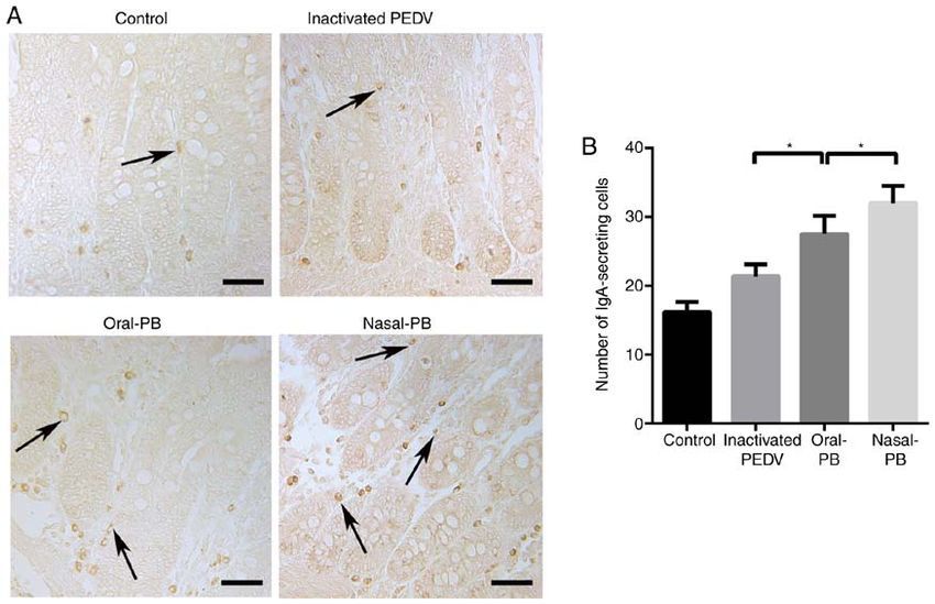
1598                              EXPERIMENTAL AND THERAPEUTIC MEDICINE 20: 1596-1606, 2020

of lavage fluid from small intestines, were added to the plates       patches was measured using a light microscope (CX23;
and incubated at 37˚C for 1.5 h. The plates were washed with          Olympus Corporation; magnification, x10) equipped with a
PBST and then 100 µl of HRP‑conjugated goat anti‑pig IgA              digital camera (MD30; Guangzhou Micro‑shot Technology
antibody (1:2,000; cat. no. ab112746; Abcam) was added and            Co., Ltd.) and image analysis software (Mshot Image Analysis
the plates were incubated at 37˚C for 1 h. Plates were washed         System v1.0; Guangzhou Micro‑shot Technology Co., Ltd.).
5 times and incubated with 3,3'‑ and 5,5'‑tetramethylbenzidine        The area was calculated based on a previous report (21). In the
(TMB) at 37˚C for 15 min. The reaction was then stopped with          present study, 10 images were randomly selected from each
2M sulfuric acid and the absorbance was read at 450 nm using          group to measure the area of Peyer's patches (22).
a microplate reader (Tecan Group, Ltd.).
                                                                      Immunohistochemistry for IgA‑secreting cells and CD3+
PRNT for the PEDV neutralizing antibody. PRNT was                     lymphocytes in the intestine. Preparation of paraffin sections,
performed as described in a previous study (8). Fresh intes-          dewaxing and hydration were conducted as aforementioned.
tinal samples were rinsed three times with 1 ml DMEM.                 The endogenous peroxidase activity was neutralized using
DMEM containing intestine washing liquid was centrifuged at           3% H 2O2 at 37˚C for 30 min and the sections were rinsed
14,600 x g at 4˚C for 15 min to remove the feces. The intestine       three times with PBS for 15 min. Antigens were unmasked
washing liquid was then serially diluted two‑fold in DMEM.            by microwaving (800 W) in citrate buffer (containing 2 mM
PEDV PRNT was performed in monolayers of Vero cells                   citric acid and 10 mM trisodium citrate; pH 6.0) for 15 min.
(1.0x10⁶ cells/ml) in DMEM containing 10% FBS in 12‑well              The sections were then treated with 5% BSA at 37˚C for 1 h
plates. In brief, log dilutions (1:16, 1:32 and 1:64) of intestinal   to block non‑specific binding prior to incubation with rabbit
washing liquid (450 µl) were `incubated with 50 µl 1x10³              anti‑pig CD3 (1:100; cat. no. ab16669; Abcam) or goat anti‑pig
plaque‑forming units of PEDV at 37˚C for 1 h. The 500 µl              IgA (1:100; cat. no. ab112639; Abcam) overnight at 4˚C. The
virus/intestinal sample mixture was then transferred onto             paraffin sections were rinsed three times with PBS for 15 min
Vero cell monolayers and incubated at 37˚C for 1 h. The cell          and then incubated with biotinylated goat anti‑rabbit IgG or
monolayers were washed with DMEM and overlaid with 0.9%               rabbit anti‑goat IgG at 37˚C for 1 h. The paraffin sections were
low‑melting agarose at 37˚C in 5% CO2 for 72 h. Plaques were          then rinsed three times with PBS and incubated with HRP
visualized by staining the monolayer with 0.5% crystal violet.        labeled SABC at 37˚C. After 1 h, the paraffin sections were
Plaque images were acquired under ultraviolet radiation light         rinsed three times with PBS and stained with DAB at room
in Tanon 5200 Automatic Chemiluminescence/Fluorescence                temperature for 2 min. Cells were counterstained with 0.5%
Image Analysis system (Tanon Science and Technology                   hematoxylin for 20 sec at room temperature. After staining,
Co., Ltd,).                                                           sections were dehydrated through increasing concentrations
                                                                      of ethanol (75% for 1 min, 85% for 1 min, 95% for 1 min and
Hematoxylin and eosin (H&E) staining for intestine intraepi‑          100% for 2 min) and xylene (10 min) at room temperature,
thelial lymphocytes (IELs) and Peyer's patches. Intestinal            and finally sealed with a coverslip. All incubations were
tissues were fixed in Bouin's fluid at room temperature for 24 h      performed in a humidified chamber. Control staining was
and then dehydrated through a serial alcohol gradient (75%            carried out simultaneously, in which the primary antibody
overnight, 85% for 1 h, 95% for 1 h and 100% for 2 h), washed         was replaced with PBS. No specific staining was found for
with xylene (5 min) at room temperature and finally embedded          the control staining (without antibody). Sections were sealed
in paraffin. The embedded intestinal tissues were cut into 5‑µm       with glass coverslips. Optical density of CD3+T cells was
serial sections and placed on glass slides. Methods of revealing      measured as previously described (23). The regions containing
intraepithelial lymphocytes by H&E staining have been previ-          SIgA‑secreting cells were counted using an optical micro-
ously reported (17). Before staining, paraffin sections were          scope (24). A total of 20 images were randomly selected from
dewaxed in xylene at room temperature for 15 min, rehydrated          each group and SIgA‑secreting cells were counted in the same
through decreasing concentrations of ethanol (100% for 2 min,         size field of view using the same multiple light microscope
95% for 1 min, 85% for 1 min and 75% for 1 min) and rinsed            (CX23; Olympus Corporation; magnification, x40).
in PBS for 2 min at room temperature. Following the hydra-
tion of paraffin sections through decreasing concentrations of        ELISA for interleukin (IL)‑6 expression level in the intestine.
ethanol, H&E staining was conducted using hematoxylin (0.5%           Small intestine tissue was placed in 1.5‑ml cryogenic vials
for 20 sec) and eosin (0.5% for 5 sec) at room temperature.           containing 500 µl PBS and triturated using a Tissuelyser‑24L
After staining, sections were dehydrated through increasing           multi‑sample tissue grinder (Shanghai Jingxin Industrial
concentrations of ethanol (75% for 1 min, 85% for 1 min, 95%          Development Co., Ltd). The supernatant of the homogenate
for 1 min, and 100% for 2 min) and xylene (10 min) at room            was collected after a 12,000 x g centrifugation for 10 min at
temperature, and finally sealed with a coverslip. IELs were           4˚C for the detection of IL‑6. The protein concentration of
measured by counting ≥500 nuclei (epithelial and lymphocyte)          the supernatant was measured using a BCA protein assay kit
and the results expressed as IEL/100 epithelial cells (20). In        (Thermo Fisher Scientific, Inc.). After a 10‑fold dilution, the
the present study, 10 fields of view were randomly selected           expression levels of IL‑6 were measured using an ELISA kit
from each group and three intestinal villi were randomly              (cat. no. ab100755; Abcam) according to the manufacturer's
selected per field. The numbers of IELs were counted in an            instructions. A total of 100 µl of each standard and sample
area of 100 epithelial cells using a light microscope (CX23;          was added to the antibody‑conjugated wells. The wells were
Olympus Corporation; magnification, x40). The area of Peyer's         covered and incubated overnight at 4˚C with gentle shaking.
patches was measured after H&E staining. The area of Peyer's          The solution was discarded and washed 4 times with 300 µl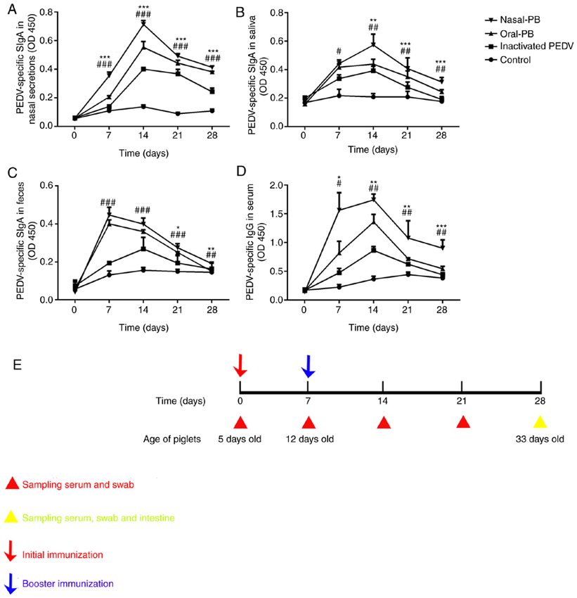
ZHANG et al: COMPARISON OF ORAL AND NASAL IMMUNIZATION ON INTESTINAL IMMUNITY OF PIGLETS                          1599

of 1X Wash Solution using a multi‑channel pipette. After          (Takara Bio, Inc.), 0.4 µl each primer, 2 µl template cDNA and
the final wash, the solution was removed by aspiration or         7.2 µl DDW. The thermocycling conditions used for the PCR
decantation. A total of 100 µl of biotinylated IL‑6 antibody      were as follows: Initial denaturation 95˚C for 5 sec, 40 cycles
(cat. no. ab100755; Abcam) was added to each well. The            of 95˚C for 30 sec and 60˚C for 34 sec. The relative expression
samples were incubated for 1 h at room temperature with gentle    levels were calculated using the 2‑ΔΔCq method (25).
shaking and then the solution was discarded. The washing step
was repeated as previously performed. A total of 100 µl of 1X     Statistical analysis. GraphPad Prism V6.0 (GraphPad
HRP‑Streptavidin solution was added to each well and incu-        Software, Inc.) was used to perform the statistical analyses.
bated for 45 min at room temperature with gentle shaking. The     One‑way ANOVAs and repeated‑measures ANOVAs were used
solution was discarded and 100 µl TMB One‑Step Substrate          to analyze the significance of the differences between means.
Reagent was added to each well. This was incubated for            Bonferroni's correction was used to analyze PEDV‑specific
30 min at room temperature in the dark with gentle shaking. A     titer datasets, while Tukey's multiple comparison tests were
total of 50 µl Stop Solution was added to each well. Data were    used to analyze the other datasets. Values are shown as the
immediately acquired using an automated ELISA plate reader        mean ± SD. P
1600                                  EXPERIMENTAL AND THERAPEUTIC MEDICINE 20: 1596-1606, 2020

Figure 1. Changes in PEDV‑specific SIgA titers in the local mucosa and IgG titers in the serum. ELISA detection of PEDV‑specific SIgA titers in (A) nasal
secretions, (B) saliva and (C) feces. (D) ELISA detection of PEDV‑specific IgG titers in serum. (E) The immunization program and sampling times. Data were
acquired using an automated ELISA plate reader at 450 nm. Data are presented as the mean ± SD. *P
ZHANG et al: COMPARISON OF ORAL AND NASAL IMMUNIZATION ON INTESTINAL IMMUNITY OF PIGLETS                                                   1601

Figure 2. Changes in PEDV neutralizing antibody levels in the intestine. PEDV plaque reduction neutralization tests were performed in monolayers of Vero
cells. (A) Images of plaques formed when the washing liquid was diluted at ratios of 1:16, 1:32 and 1:64. (B) Quantification of the inhibition of plaque formation
at a variety of concentrations of intestinal samples. Data were obtained using the ratio of the number of plaques to the number in the control group. Data are
presented as the mean ± SD. *P
1602                                    EXPERIMENTAL AND THERAPEUTIC MEDICINE 20: 1596-1606, 2020

Figure 3. Changes of IEL numbers and Peyer's patch areas in the intestine. Fixed samples were embedded in paraffin and sectioned at 5‑µm thickness. H&E
staining of the paraffin sections was performed. (A) H&E staining for IELs. The arrows indicate the IELs. Scale bars, 20 µm (inset) and 50 µm. (B) Changes
in IEL numbers. (C) H&E staining for Peyer's patches in the intestine. Peyer's patches are characterized by oval or round lymphoid follicles and were located
in the submucosa layer of the intestine. Scale bars, 200 µm. (D) Changes in the area of Peyer's patches in the intestine. Data are presented as the mean ± SD.
*
 P
ZHANG et al: COMPARISON OF ORAL AND NASAL IMMUNIZATION ON INTESTINAL IMMUNITY OF PIGLETS                                                1603

Figure 4. Changes in IgA‑secreting cell numbers in the intestine. (A) IgA‑secreting cells in the small intestine were detected using immunohistochemistry. The
arrows show IgA secreting cells, with a round morphology. The nuclei are surrounded by a ring of yellow‑brown cytoplasm in the lamina propria of intestinal
villi. Scale bars, 50 µm. (B) Changes in the number of IgA‑secreting cells. Data are presented as the mean ± SD. *P
1604                                  EXPERIMENTAL AND THERAPEUTIC MEDICINE 20: 1596-1606, 2020

Figure 6. Changes in IL‑6 and IFN‑γ expression levels in the intestine. (A) Changes in IL‑6 expression levels in the intestine were detected using ELISAs.
(B) Relative IFN‑γ mRNA expression levels were detected using reverse transcription‑quantitative PCR. (C) The protein expression levels of IFN‑γ were
detected using ELISAs. Data are presented as the mean ± SD. *P
ZHANG et al: COMPARISON OF ORAL AND NASAL IMMUNIZATION ON INTESTINAL IMMUNITY OF PIGLETS                                     1605

immunization a favorable route of mucosal immunization for                 6. Mantis NJ and Forbes SJ: Secretory IgA: Arresting microbial
                                                                              pathogens at epithelial borders. Immunol Invest 39: 383‑406,
the prevention of PEDV infections.                                            2010.
                                                                           7. Poles J, Alvarez Y and Hioe CE: Induction of intestinal immunity
Acknowledgements                                                              by mucosal vaccines as a means of controlling HIV infection.
                                                                              AIDS Res Hum Retroviruses 30: 1027‑1040, 2014.
                                                                           8. Wang J, Huang L, Mou C, Zhang E, Wang Y, Cao Y and Yang Q:
Not applicable.                                                               Mucosal immune responses induced by oral administration
                                                                              recombinant Bacillus subtilis expressing the COE antigen of
                                                                              PEDV in newborn piglets. Biosci Rep 39: BSR20182028, 2019.
Funding                                                                    9. Li Y, Wu Q, Huang L, Yuan C, Wang J and Yang Q: An alterna-
                                                                              tive pathway of enteric PEDV dissemination from nasal cavity to
The current study was supported by grants from the National                   intestinal mucosa in swine. Nat Commun 9: 3811, 2018.
                                                                          10. Guy B: Evaluation of events occurring at mucosal surfaces:
Science Grant of China (grant nos. 31772777 and 31930109)                     Techniques used to collect and analyze mucosal secretions and
and a project funded by the Priority Academic Program                         cells. Clin Diagn Lab Immunol 9: 753‑762, 2002.
Development of Jiangsu Higher Education Institutions (PAPD).              11. Kozlowski PA, Williams SB, Lynch RM, Flanigan TP,
                                                                              Patterson RR, Cu‑Uvin S and Neutra MR: Differential induc-
                                                                              tion of mucosal and systemic antibody responses in women after
Availability of data and materials                                            nasal, rectal, or vaginal immunization: Influence of the menstrual
                                                                              cycle. J Immunol 169: 566‑574, 2002.
                                                                          12. Staats HF, Montgomery SP and Palker TJ: Intranasal immuniza-
The datasets used and/or analyzed during the current study are                tion is superior to vaginal, gastric, or rectal immunization for the
available from the corresponding author on reasonable request.                induction of systemic and mucosal anti‑HIV antibody responses.
                                                                              AIDS Res Hum Retroviruses 13: 945‑952, 1997.
                                                                          13. Lauring AS Jones JO and Andino R: Rationalizing the devel-
Authors' contributions                                                        opment of live attenuated virus vaccines. Nat Biotechnol 28:
                                                                              573‑579, 2010.
EZ, JW, LH and QY conceived the study. EZ and QY partici-                 14. Azegami T, Yuki Y and Kiyono H: Challenges in mucosal
                                                                              vaccines for the control of infectious diseases. Int Immunol 26:
pated in the design of the protocols for the study. EZ and                    517‑528, 2014.
JW performed the experiments. EZ, JW, YL, LH and YW                       15. Amuguni H and Tzipori S: Bacillus subtilis: A temperature
analyzed the data. EZ wrote the manuscript. QY, EZ, JW and                    resistant and needle free delivery system of immunogens. Hum
                                                                              Vaccin Immunother 8: 979‑986, 2012.
YL participated in the revision of the manuscript. All authors            16. Upadhaya SD, Kim SC, Valientes RA and Kim IH: The effect
read and approved the final manuscript.                                       ofbacillus‑based feed additive on growth performance, nutrient
                                                                              digestibility, fecal gas emission, and pen cleanup characteris-
                                                                              tics of growing‑finishing pigs. Asian‑Australas J Anim Sci 28:
Ethics approval and consent to participate                                    999‑1005, 2015.
                                                                          17. Mou C, Zhu L, Xing X, Lin J and Yang Q: Immune responses
All animal experiments were approved by the Ethics Committee                  induced by recombinant bacillus subtilis expressing the spike
                                                                              protein of transmissible gastroenteritis virus in pigs. Antiviral
of Animal Experiments of the College of Veterinary Medicine,                  Res 131: 74‑84, 2016.
Nanjing Agricultural University. All animal care and use were             18. Mou C, Zhu L, Yang J, Xu W, Cheng X and Yang Q: Immune
conducted in strict accordance with the Animal Research                       responses induced by recombinant bacillus subtilis expressing
                                                                              the hemagglutinin protein of H5N1 in chickens. Sci Rep 6:
Committee guidelines of the College of Veterinary Medicine,                   38403, 2016.
Nanjing Agricultural University.                                          19. Li Y, Wang G, Wang J, Man K and Yang QJV: Cell attenuated
                                                                              porcine epidemic diarrhea virus strain Zhejiang08 provides
                                                                              effective immune protection attributed to dendritic cell stimula-
Patient consent for publication                                               tion. Vaccine 35: 7033‑7041, 2017.
                                                                          20. Corazza GR, Frazzoni M and Gasbarrini G: Jejunal intraepi-
Not applicable.                                                               thelial lymphocytes in coeliac disease: Are they increased or
                                                                              decreased? Gut 25: 158‑162, 1984.
                                                                          21. Inoue R, Tsukahara T, Nakatani M, Okutani M, Nishibayashi R,
Competing interests                                                           Ogawa S, Harayama T, Nagino T, Hatanaka H, Fukuta K, et al:
                                                                              Weaning markedly affects transcriptome profiles and peyer's
                                                                              patch development in piglet ileum. Front Immunol 6: 630, 2015.
The authors declare that they have no competing interests.                22. Ryan KA, Daly P, Li Y, Hooton C and O'Toole PW: Strain‑specific
                                                                              inhibition of helicobacter pylori by lactobacillus salivarius and
References                                                                    other lactobacilli. J Antimicrob Chemother 61: 831‑834, 2008.
                                                                          23. Youssef S and Salah M: Differential expression of CD3, TNF‑α,
                                                                              and VEGF induced by olanzapine on the spleen of adult male
 1. Song D, Huang D, Peng Q, Huang T, Chen Y, Zhang T, Nie X,                 albino rats and the possible protective role of vitamin C.
    He H, Wang P, Liu Q and Tang Y: Molecular characterization                Biomedicines 7: E39, 2019.
    and phylogenetic analysis of porcine epidemic diarrhea viruses        24. Lin J, Tu C, Mou C, Chen X and Yang Q: CpG DNA facilitate the
    associated with outbreaks of severe diarrhea in piglets in Jiangxi,       inactivated transmissible gastroenteritis virus in enhancing the
    China 2013. PLoS One 10: e0120310, 2015.                                  local and systemic immune response of pigs via oral administra-
 2. Song D and Park B: Porcine epidemic diarrhoea virus: A                    tion. Vet Immunol Immunopathol 172: 1‑8, 2016.
    comprehensive review of molecular epidemiology, diagnosis, and        25. Livak KJ and Schmittgen TD: Analysis of relative gene expres-
    vaccines. Virus Genes 44: 167‑175, 2012.                                  sion data using real‑time quantitative PCR and the 2(‑Delta Delta
 3. Li W, van Kuppeveld FJM, He Q, Rottier PJM and Bosch BJ:                  C(T)) method. Methods 25: 402‑408, 2001.
    Cellular entry of the porcine epidemic diarrhea virus. Virus          26. Kiyono H and Fukuyama S: NALT‑ versus Peyer's‑patch‑mediated
    Res 226: 117‑127, 2016.                                                   mucosal immunity. Nat Rev Immunol 4: 699‑710, 2004.
 4. Hou X, Jiang X, Jiang Y, Tang L, Xu Y, Qiao X, Min L, Wen C,          27. Sato T, Endoh M, Yoshida H, Yasuo S, Katsuno T, Saito Y,
    Ma G and Li Y: Oral immunization against PEDV with recom-                 Isono K and Koseki H: Mammalian Polycomb complexes are
    binant lactobacillus casei expressing dendritic cell‑targeting            required for Peyer's patch development by regulating lymphoid
    peptide fusing COE Protein of PEDV in Piglets. Viruses 10:                cell proliferation. Gene 379: 166‑174, 2006.
    E106, 2018.                                                           28. Lasa‑Saracíbar B, Aznar MÁ, Lana H, Aizpún I, Gil AG and
 5. Wang D, Fang L and Xiao S: Porcine epidemic diarrhea in China.            Blanco‑Prieto MJ: Lipid nanoparticles protect from edelfosine
    Virus Res 226: 7‑13, 2016.                                                toxicity in vivo. Int J Pharm 474: 1‑5, 2014.
1606 EXPERIMENTAL AND THERAPEUTIC MEDICINE 20: 1596-1606, 2020
29. Kabat AM, Pott J and Maloy KJ: The mucosal immune system 46. Mcghee JR, Fujihashi K, Beagley KW and Kiyono HJ: Role
and its regulation by autophagy. Front Immunol 7: 240, 2016. of interleukin‑6 in human and mouse mucosal IgA plasma cell
30. van Wijk F and Cheroutre H: Mucosal T cells in gut homeostasis responses. Immunol Res 10: 418‑422, 1991.
and inflammation. Expert Rev Clin Immunol 6: 559‑566, 2010. 47. Libbey JE and Fujinami RS: Adaptive immune response to viral
31. Wu RQ, Zhang DF, Tu E, Chen QM and Chen W: The mucosal infections in the central nervous system. Handb Clin Neurol 123:
immune system in the oral cavity‑an orchestra of T cell diversity. 225‑247, 2014.
Int J Oral Sci 6: 125‑132, 2014. 48. Hershberg R and Blumberg RS: The lymphocyte‑epithe-
32. Song D, Moon H and Kang BJ: Porcine epidemic diarrhea: A lial‑bacterial interface. Springer US, 2003.
review of current epidemiology and available vaccines. Clin Exp 49. Li Z, Zhang C, Zhou Z, Zhang J, Zhang J and Tian Z: Small
Vaccine Res 4: 166‑176, 2015. intestinal intraepithelial lymphocytes expressing CD8 and T cell
33. O'Neal CM, Clements JD, Estes MK and Conner ME: Rotavirus receptor γδ are involved in bacterial clearance during Salmonella
2/6 viruslike particles administered intranasally with cholera toxin, enterica serovar Typhimurium infection. Infect Immun 80:
Escherichia coli heat‑labile toxin (LT), and LT‑R192G induce 565‑574, 2012.
protection from rotavirus challenge. J Virol 72: 3390‑3393, 1998. 50. Dharakul T, Rott L and Greenberg HB: Recovery from
34. Van Oirschot JT: Intranasal vaccination of pigs against chronic rotavirus infection in mice with severe combined
Aujeszky's disease: Comparison with one or two doses of attenu- immunodeficiency: Virus clearance mediated by adop-
ated vaccines in pigs with high maternal antibody titres. Res Vet tive transfer of immune CD8+ T lymphocytes. J Virol 64:
Sci 42: 12‑16, 1987. 4375‑4382, 1990.
35. Zhang L, Tian X and Zhou F: Intranasal administration of CpG 51. Vazquez MI, Catalan‑Dibene J and Zlotnik AJC: B cells
oligonucleotides induces mucosal and systemic Type 1 immune responses and cytokine production are regulated by their immune
responses and adjuvant activity to porcine reproductive and microenvironment. Cytokine 74: 318‑326, 2015.
respiratory syndrome killed virus vaccine in piglets in vivo. Int 52. Onyiah JC and Colgan SP: Cytokine responses and epithe-
Immunopharmacol 7: 1732‑1740, 2007. lial function in the intestinal mucosa. Cell Mol Life Sci 73:
36. Schijns VE and Lavelle EC: Trends in vaccine adjuvants. Expert 4203‑4212, 2016.
Rev Vaccines 10: 539‑550, 2011. 53. Steidler L, Robinson K, Chamberlain L, Schofield KM,
37. Blaauboer SM, Mansouri S, Tucker HR, Wang HL, Gabrielle VD Remaut E, Le Page RW and Wells JM: Mucosal delivery of
and Jin L: The mucosal adjuvant cyclic di‑GMP enhances murine interleukin‑2 (IL‑2) and IL‑6 by recombinant strains of
antigen uptake and selectively activates pinocytosis‑efficient Lactococcus lactis coexpressing antigen and cytokine. Infect
cells in vivo. Elife 4: 2015. Immun 66: 3183‑3189, 1998.
38. Setlow P: Spores of Bacillus subtilis: Their resistance to and 54. Weiss ID, Wald O, Wald H, Beider K, Abraham M, Galun E,
killing by radiation, heat and chemicals. J Appl Microbiol 101: Nagler A and Peled A: IFN‑gamma treatment at early stages
514‑525, 2006. of influenza virus infection protects mice from death in a NK
39. Deng J, Li Y, Zhang J and Yang Q: Co‑administration of Bacillus cell‑dependent manner. J Interferon Cytokine Res 30: 439‑449,
subtilis RJGP16 and Lactobacillus salivarius B1 strongly 2010.
enhances the intestinal mucosal immunity of piglets. Res Vet 55. Kak G, Raza M and Tiwari BK: Interferon‑gamma (IFN‑ γ):
Sci 94: 62‑68, 2013. Exploring its implications in infectious diseases. Biomol
40. Yang Y, Jing Y, Yang J and Yang Q: Effects of intranasal Concepts 9: 64‑79, 2018.
administration with Bacillus subtilison immune cells in the nasal 56. Djupesland PG: Nasal drug delivery devices: Characteristics
mucosa and tonsils of piglets. Exp Ther Med 15: 5189‑5198, 2018. and performance in a clinical perspective‑a review. Drug Deliv
41. Jing Y, Liu H, Xu W and Yang Q: Amelioration of the Transl Res 3: 42‑62, 2013.
DSS‑induced colitis in mice by pretreatment with 4,4'‑diapo- 57. El‑Kattan A, Hurst S, Brodfuehrer J and Loi CM: Anatomical
neurosporene‑producingBacillus subtilis. Exp Ther Med 14: and physiological factors affecting oral drug bioavailability in
6069‑6073, 2017. rats, dogs, and humans. John Wiley & Sons, Inc., 2011.
42. Herich RJ: Is the role of IgA in local immunity completely 58. Yen HH, Scheerlinck JP, Gekas S and Sutton P: A sheep cannula-
known? Immunol 28: 1‑15, 2016. tion model for evaluation of nasal vaccine delivery. Methods 38:
43. Mantis NJ, Rol N and Corthésy B: Secretory IgA's complex 117‑123, 2006.
roles in immunity and mucosal homeostasis in the gut. Mucosal
Immunol 4: 603‑611, 2011. This work is licensed under a Creative Commons
44. Holmgren J and Czerkinsky C: Mucosal immunity and vaccines. Attribution-NonCommercial-NoDerivatives 4.0
Nat Med 11 (4 Suppl): S45‑S53, 2005. International (CC BY-NC-ND 4.0) License.
45. Neutra MR and Kozlowski PA: Mucosal vaccines: The promise
and the challenge. Nat Rev Immunol 6: 148‑158, 2006.You can also read