Experimental PCEP-Adjuvanted Swine Influenza H1N1 Vaccine Induced Strong Immune Responses but Did Not Protect Piglets against Heterologous H3N2 ...
←
→
Page content transcription
If your browser does not render page correctly, please read the page content below
Article
Experimental PCEP-Adjuvanted Swine Influenza
H1N1 Vaccine Induced Strong Immune Responses
but Did Not Protect Piglets against Heterologous
H3N2 Virus Challenge
Royford Bundi Magiri 1,2,3,† , Ken John Lai 1,† , George Kiremu Mutwiri 1,2
and Heather Lynne Wilson 1,2, *
 1    Vaccinology & Immunotherapeutic Program, School of Public Health, University of Saskatchewan,
      Saskatoon, SK S7N 2Z4, Canada; royford.magiri@fnu.ac.fj (R.B.M.); ken.lai@usask.ca (K.J.L.);
      george.mutwiri@usask.ca (G.K.M.)
 2    Vaccine & Infectious Disease Organization-International Vaccine Centre (VIDO-InterVac),
      University of Saskatchewan, 120 Veterinary Road, Saskatoon, SK S7N 5E3, Canada
 3    College of Agriculture, Fisheries and Forestry, Fiji National University, Suva 7222, Fiji
 *    Correspondence: heather.wilson@usask.ca
 †    All authors contributed equally to this work.

 Received: 5 March 2020; Accepted: 8 May 2020; Published: 18 May 2020

 Abstract: Vaccination is the most efficient method of protection against influenza infections.
 However, the rapidly mutating viruses and development of new strains make it necessary to
 develop new influenza vaccines annually. Hence, vaccines that stimulate cross-protection against
 multiple influenza subtypes are highly sought. Recent evidence suggests that adjuvants such as PCEP
 that promote Th1-type T cell and Th2-type T cell immune responses and broad-spectrum immune
 responses may confer cross-protection against heterologous influenza strains. In this study, we
 evaluated whether the immunogenic and protective potential of PCEP-adjuvanted inactivated swine
 influenza virus H1N1 vaccine can protect pigs immunized against live H3N2 virus. Piglets were
 vaccinated via the intradermal route with PCEP-adjuvanted inactivated swine influenza virus (SIV)
 H1N1 vaccine, boosted at day 21 with the same vaccines then challenged with infectious SIV H3N2
 virus at day 35 via the tracheobronchial route. The pigs showed significant anti-H1N1 SIV specific
 antibody titres and H1N1 SIV neutralizing antibody titres, and these serum titres remained after
 the challenge with the H3N2 virus. In contrast, vaccination with anti-H1N1 SIV did not trigger
 anti-H3N2 SIV antibody titres or neutralizing antibody titres and these titres remained low until pigs
 were challenged with H3N2 SIV. At necropsy (six days after challenge), we collected prescapular
 lymph nodes and tracheobronchial draining the vaccination sites and challenge site, respectively.
 ELISPOTs from lymph node cells restimulated ex vivo with inactivated SIV H1N1 showed significant
 production of IFN-γ in the tracheobronchial cells, but not the prescapular lymph nodes. In contrast,
 lymph node cells restimulated ex vivo with inactivated SIV H1N1 showed significantly higher IL-13
 and IL-17A in the prescapular lymph nodes draining the vaccination sites relative to unchallenged
 animals. Lung lesion scores show that intradermal vaccination with H1N1 SIV plus PCEP did not
 prevent lesions when the animals were challenged with H3N2. These results confirm previous
 findings that PCEP is effective as a vaccine adjuvant in that it induces strong immune responses and
 protects against homologous swine influenza H1N1 virus, but the experimental H1N1 vaccine failed
 to cross-protect against heterologous H3N2 virus.

 Keywords: polyphosphazene; adjuvant; pig; intradermal; influenza

Vaccines 2020, 8, 235; doi:10.3390/vaccines8020235                            www.mdpi.com/journal/vaccines
Vaccines 2020, 8, 235                                                                               2 of 16

1. Introduction
     Swine influenza virus (SIV) is a highly contagious acute respiratory disease of pigs [1]. There has
been an increase in the genetic diversity of swine influenza A virus with the majority of the SIV
infections in pigs are caused by subtypes H1N1, H1N2 and H3N2 with new reassortments such as
H1N1pdm09 being identified [2–5]. Other subtypes such as H3N8, H4N8, H5N1 and H6N6 have been
identified but they cause a minority of SIV infections. Swine influenza virus is endemic worldwide
and it is responsible for significant economic losses to the swine industry each year [3]. In addition,
SIV infections are a threat to public health since transmission from pigs to humans can occur, hence
a vaccine that stimulates a rapid and long-lasting protective immune response to homologous and
heterologous strains is highly sought. The most cost-effective public health tool available to control
SIV infection is through effective vaccination. Current pig vaccines are comprised of inactivated
H3N2 and H1N1virus [6]. Because subunit vaccines contain highly purified antigens, they are
poorly immunogenic and require the addition of adjuvants to induce protective immune responses.
Additionally, reformulation of the vaccine to include a potent adjuvant may improve the efficacy of
existing SIV vaccines.
     Adjuvants are routinely included in vaccines comprised of inactivated virus and/or subunit
vaccines to augment the magnitude and quality of immune responses and by enhancing onset and
extending duration of immunity. Many adjuvants can improve immune responses and promote
protection against infection with homologous influenza virus strains in humans and animals.
However, cross protection against heterologous virus strains remains a challenge in the development
of influenza vaccines. A recent study by Clegg revealed that the combination adjuvant GLA-SE
(a two-part adjuvant system containing glucopyranosyl lipid adjuvant (GLA), a formulated synthetic
Toll-like receptor 4 agonist, and a stable emulsion (SE) of oil in water), but not the commercial
SE adjuvant, conferred protection against heterosubtypic H5N1 challenge in mice and ferrets [7].
This cross-protection was apparently mediated via induction of Th1-mediated antibody responses [7].
Polyphosphazenes are high-molecular weight, water-soluble polymers that have been shown to promote
and enhance long lasting immune responses with a variety of viral and bacterial antigens [8–13].
Poly (di (sodium carboxylatoethylphenoxy))-phosphazene (PCEP) promotes strong antigen-specific
Th1- and Th2-type immune responses to influenza antigens in mice and pigs [10,13–15]. PCEP induced
significant production of interleukin IL-1β, and IL-13 at the site of injection and IL-1β, and IL-6 at the
draining lymph nodes. and we previously reported that pigs vaccinated via the intradermal route
with PCEP-adjuvanted inactivated SIV H1N1 vaccine were protected against virulent challenge with
homologous H1N1 [13]. PCEP injected intradermally into pigs in the absence of antigen induced
significant production of interleukin IL-1β, and IL-13 at the site of injection and IL-1β, and IL-6 at the
draining lymph nodes, possibly contributing to an immunocompetent environment [15]. In this study,
we evaluated the immunogenicity and cross protective efficacy of PCEP-adjuvanted inactivated SIV
H1N1 vaccine against virulent challenge with heterologous H3N2 in pigs.

2. Materials and Methods

2.1. Swine Influenza Virus Adsorption and Purification
     In separate flasks, swine influenza virus H1N1 A/swine/Saskatchewan/18789/2002 (H1N1) and
the challenge strain H3N2 (A/swine/Texas/4199-2/1998/H3N2 (Tx98)) isolates (swine influenza virus SK
and Texas strain, respectively) were cultured in confluent Madin–Darby canine kidney (MDCK) cells at
a multiplicity of infection (M.O.I) of 0.001 plaque-forming units (PFU)/mL. Cells were grown in tissue
culture media MEM at 37 ◦ C in 5% CO2 with nutation every 15 min. After 1 h, each inoculum was
removed and 10 mL of DMEM with 0.2% heat-inactivated BSA (Sigma-Aldrich# A8806, Oakville, ON,
Canada), 1 µg/mL of N-P-tosyl-phenylalanine chloromethyl ketone (TPCK) (MJS BioLynx, Brockville,
ON, Canada) and 50 µg/mL of gentamicin were added per flask. The cultures were incubated
in a humidified 5% CO2 atmosphere at 37 ◦ C for 3 days or until 90% of cytopathic effect (CPE) was
Vaccines 2020, 8, 235                                                                               3 of 16

observed. Infected cell cultures were centrifuged at 200× g for 10 min at 14 ◦ C. The supernatant was
collected, aliquoted into small volumes, and stored at −70 ◦ C until further purification was performed.
      Virus containing supernatants were subjected to 112,700× g centrifugation for 2.5 h at 4 ◦ C.
The resulting pellet was collected in 500 µL TSE buffer (20 mM Tris, 2 mM EDTA & 150 mM NaCl,
pH 7.4), placed upon a 30%/60% discontinuous sucrose gradient and centrifuged at 107,170× g for
2.5 h at 4 ◦ C. The viral band at the 30%/60% interface was collected using an 18-gauge blunt cannula,
resuspended in TSE buffer and subjected to 210,053× g centrifugation for 1.5 h at 4 ◦ C. The pellet
was collected and resuspended in TSE buffer to an appropriate minimal volume yielding between
108 –109 PFU/mL.

2.2. Formalin Inactivation of Influenza Virus
     The purified virus was inactivated with 10% formalin to a final concentration of 0.1% and incubated
at 37 ◦ C with constant nutation for 48 h. To test and confirm inactivation, an aliquot of the inactivated
virus with PBS mock controls were diluted at 102 , 103 and 104 in 0.1% formalin. Virus inactivation was
confirmed by the inability of the viruses to replicate in MDCK cells as observed by negligible CPE.
The inactivated virus was stored at 4 ◦ C.

2.3. Adjuvant and Vaccine Preparation
     PCEP was synthesized by the Idaho National Laboratory (Idaho Falls, ID, USA) using methods
previously described [9,16] and its endotoxin level was determined to be less than 0.034 ng/mL as
assessed by the Limulus Amebocyte Lysate assay (Biowhittaker, Walkersville, MD, USA). Vaccines
consisted of 4.0 × 10⁴ inactivated SIV HAU (haemagglutination units) alone or plus 4, 20, 100 or 500 µg
PCEP; or 8.0 × 10⁴ inactivated SIV HAU alone or plus 4, 20, 100 or 500 µg PCEP. PBS was injected into
control animals or animals were not immunized (naïve control animals). Inactivated SIV vaccines were
diluted with PBS (pH of 7.4) into 250 µL per injection site. The highest dose of PCEP was chosen based
on the previous experiments using PCEP in pigs [13,17,18].

2.4. Immunization with Inactivated H1N1 and Challenge with H3N2 Virus Experimental Design
     All animal experiments were conducted in VIDO-InterVac (University of Saskatchewan, Saskatoon,
SK, Canada) according to the Guidelines for the Care and Use of Laboratory Animals as indicated
by the Canadian Council on Animal Care and was approved by the Animal Care Committee of the
University of Saskatchewan (The Animal Use Protocol is AUP20170049).
     Commercial cross bred pigs (3–4 weeks of age) were selected from sows prescreened 5 days before
farrowing for negligible H1N1 and H3N2 antibody titers. The piglets were divided into 10 test groups
(n = 6 in each group). Pre-existing H1N1 and H3N2 antibodies and H1N1 and H3N2 neutralizing
antibody levels were quantified on all piglets prior to vaccination as marked by Day 0).
     Please note that four piglets across several groups succumbed to Streptococcus suis infection within
the acclimation period prior to start of the experiment and they were removed from data analysis.
Necropsy was performed on these pigs with S. suis septicaemia determination. An independent
analysis by Prairie Diagnostic Services, Inc. showed no detectable pneumonia or pleural cavity
abnormalities. Two other pigs died by needle venous severance when the pig suddenly and forcibly
moved while extracting blood during serum collection, and another pig died from umbilical hernia.
Upon necropsy of these 3 animals, no detectable pneumonia or pleural cavity abnormalities were
present. All remaining pigs were administered 1.3 mL of 100 mg/mL Baytril® 100 (Bayer, Mississauga,
ON, Canada).
     Pigs were immunized intradermally at the neck on day 0 (left side) and a booster vaccination
given at day 21 (right side). The body temperature, clinical observations and score for local reactions
at both injection sites were taken throughout the study period. The local reaction scores were from
0–3 with 0 = normal, 1 = minor, 2 = moderate and 3 = severe. No local reaction scores were observed
(data not shown) for any vaccine formulation at any time point as described in Figure 1.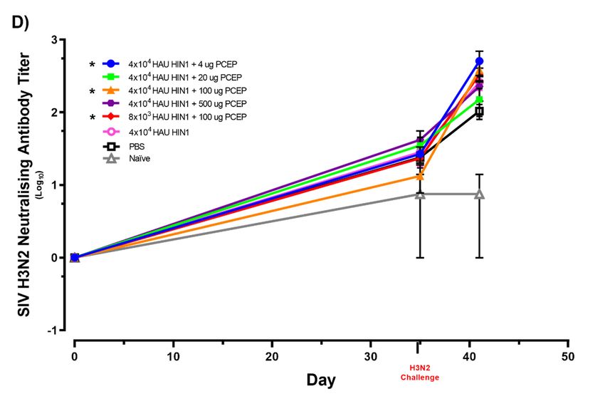
Vaccines 2020, 8, 235 4 of 16
Vaccines 2020, 8, x 4 of 18
Vaccines 2020, 8, x 4 of 18
Figure 1. Schematic presentation of experimental design. Piglets were immunized via the intradermal
Figure 1. Schematic presentation of experimental design. Piglets were immunized via the intradermal
injection with swine influenza virus (SIV) plus PCEP as an adjuvant, on day 0, and a secondary
injection with immunization
swine influenza virus (SIV)
was administered on day 21.plus PCEP
Serum as an for
was collected adjuvant, on titres
serum antibody day assays.
0, and a secondary
immunization Clinical
was administered on day
and injection site markings and21. Serum
scoring was collected
were performed up to day 41for
afterserum antibody titres assays.
initial vaccination.
Clinical and injection site
eachmarkings
Sera fromFigure and scoring
1. Schematic presentation
pig was collected 0,of14, were
experimental
21, and performed
design.
28 days up to day
Piglets were immunized
post-vaccination via the41
and after initial vaccination.
intradermal
the antigen-specific
injection with swine influenza virus (SIV) plus PCEP as an adjuvant, on day 0, and a secondary
antibody response (Figure 2A) and antibody neutralization effect against SIV H1N1 and H3N2
immunization was administered on day 21. Serum was collected for serum antibody titres assays.
Sera from (Figure
each pig was
2B) were
Clinical collected
assessed site 0,
to select
and injection 14,
the 21,scoring
groups
markings and and 28performed
withwere days
optimal post-vaccination
antibody
up responses
to day and the antigen-specific
to vaccination.
41 after initial the challenge.
antibody response (Figure 2A)
Sera from eachand
pig wasantibody neutralization
collected 0, 14, 21, effect
and 28 days post-vaccination andagainst SIV H1N1 and H3N2
the antigen-specific
antibody response (Figure 2A) and antibody neutralization effect against SIV H1N1 and H3N2
(Figure 2B) were assessed to select the groups with optimal antibody responses
(Figure 2B) were assessed to select the groups with optimal antibody responses to the challenge.
to the challenge.
Vaccines 2020, 8, x 5 of 18
Figure 2. Vaccine-induced
Figure 2. Vaccine-induced H1N1 antibody H1N1 antibody responses and
responses andneutralizing antibody responses
neutralizing antibody in pigs. responses in pigs.
Pigs (n = 6 per group) were immunized with 4.0 × 10 or 8.0 × 10 inactivated SIV HAU alone or plus
4 3
Pigs (n = 6 per group) were
4, 20, immunized
100 or 500 μg PCEP. Controlwith were×
animals4.0
4 or 8.0
10injected
either × 10
with PBS
3 immunized (naïve).SIV HAU alone or
or notinactivated
A secondary immunization was performed on day 21 and sera samples were collected over a period
plus 4, 20, 100 or 500ofµg PCEP. Control animals were either injected with PBS or not immunized
28 days. (A) Anti-H1N1 IgG antibodies were assessed by ELISA and (B) anti-H1N1 IgG neutralizing
(naïve). A secondary antibodies
immunization was
were assessed. Data performed onpresented
for animal group are day 21with andmeansera
valuessamples
indicated by were
a collected over
horizontal bar and standard error of the mean are shown. p < 0.05 (*), and p < 0.01 (**).
a period of 28 days. (A) Anti-H1N1 IgG antibodies were assessed by ELISA and (B) anti-H1N1 IgG
Pigs were challenged with 8 × 10⁵ PFU virulent SIV H3N2 via the intratracheal route on day 35
neutralizing antibodies were assessed. Data for animal group are presented with mean values indicated
using Strain A/swine/Texas/4199-2/1998/H3N2 (Tx98) as previously described by [19]. Challenged
by a horizontal barand
and standard
naïve error
pigs were kept of the rooms
in separate meanwith arenoshown. p < 0.05
direct or indirect and p < 0.01 (**).
(*), Antigen-specific
contract.
antibody response (Figure 3A,B) and antibody neutralization effect against SIV H1N1 and H3N2
(Figure 3C,D) were assessed up to day 41 post vaccination. All pigs in all groups were euthanized 6-
days post-infection and samples were collected.
Vaccines 2020, 8, 235                                                                             5 of 16

     Pigs were challenged with 8 × 10⁵ PFU virulent SIV H3N2 via the intratracheal route on day 35 using
Strain A/swine/Texas/4199-2/1998/H3N2 (Tx98) as previously described by [19]. Challenged and naïve
pigs were kept in separate rooms with no direct or indirect contract. Antigen-specific antibody response
(Figure 3A,B) and antibody neutralization effect against SIV H1N1 and H3N2 (Figure 3C,D) were
assessed up to day 41 post vaccination. All pigs in all groups were euthanized 6-days post-infection
and samples Vaccines
             were 2020,
                     collected.
                         8, x                                                                6 of 18

                                             Figure 3. Cont.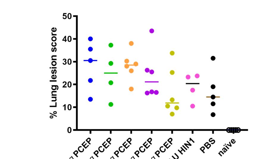
Vaccines 2020, 8, 235                                                                                                             6 of 16
              Vaccines 2020, 8, x                                                                                      7 of 18

      Figure 3. Swine
                  Figure 3.influenza       H1N1
                             Swine influenza   H1N1 and
                                                      andH3N2     ELISA
                                                          H3N2 ELISA    andand   neutralizing
                                                                            neutralizing antibodyantibody
                                                                                                  responses inresponses
                                                                                                                pigs. Pigs in pigs.
      Pigs (n = 6)(nwere
                     = 6) were  immunizedthen
                            immunized         then boosted
                                                    boostedat at
                                                              dayday
                                                                   21 via
                                                                       21intradermal  routes with
                                                                           via intradermal        4 × 10with
                                                                                               routes          4 × 10⁴ or 8 × 10³
                                                                                                        4 or 8 × 10³ HAU

                  H1N1 either alone or with varying doses (4, 20, 100 or 500 μg) of PCEP adjuvant as indicated. Control
      HAU H1N1      either alone or with varying doses (4, 20, 100 or 500 µg) of PCEP adjuvant as indicated.
                  groups were injected with either PBS or were unimmunized (naïve). They were challenged with 8 ×
      Control groups    were injected with either PBS or were unimmunized (naïve). They were challenged with
                  10 PFU virulent H3N2 SIV via the intratracheal route on day 35 and killed 6 days after challenge.
                    5

      8 × 10⁵ PFUSerum
                    virulent    H3N2
                          antibody  titresSIV  via H1N1
                                           for SIV  the intratracheal
                                                         (A) and H3N2 (B)route
                                                                            wereon day 35
                                                                                 assayed  by and
                                                                                             ELISAkilled  6 days
                                                                                                   over time.  Redafter
                                                                                                                   font ischallenge.
                  used fortitres
      Serum antibody        the day
                                 forofSIV
                                       challenge
                                            H1N1  for(A)
                                                      emphasis. Serum SIV
                                                         and H3N2      (B)H1N1
                                                                            were(C)  and H3N2
                                                                                  assayed    by(D) neutralizing
                                                                                                 ELISA           antibody
                                                                                                          over time.    Red font is
      used for the day of challenge for emphasis. Serum SIV H1N1 (C) and H3N2 (D) neutralizing antibody
      titres were assessed up to day 41. Data for animal groups are presented with mean values indicated by
      a horizontal bar and standard error of the mean are shown. p < 0.05 (*), p < 0.01 (**) and p < 0.001 (***).

2.5. Necropsy and Macroscopic Examination of the Lungs
     Animals in all groups were euthanized by intravenous administration of Euthanyl (25 mg sodium
pentobarbital). For all animals, whole lungs were removed at necropsy and evaluated to determine the
percentage of the lungs affected with purple-red, firm lesions that are typical of swine influenza virus
infection [20]. The percentage of areas affected with pneumonia was estimated visually for each lung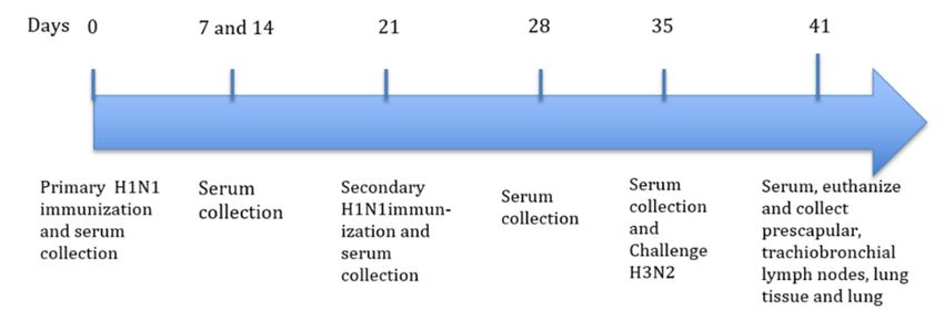
Vaccines 2020, 8, 235                                                                              7 of 16

lobe. The total percentage lesions for the entire lung was calculated based on weight proportions of
each lung lobe to the total lung volume as described by [21].

2.6. Detection of Swine Influenza Virus H1N1 and H3N2 Antibodies in Porcine Serum by Enzyme-Linked
Immunosorbent Assay (ELISA)
      Purified SIV H1N1 and H3N2 were inactivated by mixing one-part virus with one part 5%
N-lauroyl sarcosine sodium salt (Sigma-Aldrich# L5125, Oakville, ON, CAN) and 8 parts PBS + 0.1%
sodium azide and leaving at room temperature for 30 min. The viruses were diluted to 0.5 µg/mL and
1 µg/mL in 0.5 M bicarbonate buffer (coating buffer), respectively. Diluted virus was applied to separate
Immulon 2 96U plates (Thermo Lab systems #3655) at 100 µL per well and the plates were incubated
at 4 ◦ C overnight. Experimental serum samples, and positive and negative serum control standards
were diluted in PBS supplemented with 0.05% Tween-20 (Sigma-Aldrich Canada Co., Oakville, ON,
Canada), transferred into coated-blocked plates, and 4-fold serially diluted. Antigen-specific total
IgG were detected with alkaline phosphatase-conjugated KPL goat anti-swine IgG (H + L) (KPL,
Gaithersburg, MD, USA), developed in PNPP substrate (1 mg p-nitrophenyl phosphate per mL in 1%
diethanolamine with 0.5 mM MgCl2 , pH 9.8) for 2 h, read at λ405 nm with a reference λ490 nm and the
final titers were calculated.

2.7. Enzyme-Linked Immunosorbent SPOT Assay (ELISPOT) for IFN-γ, IL-13 and IL-17A
      Tracheobronchial and prescapular lymph nodes draining the vaccination and challenge sites
were dissected 6-days post-challenge and the cells were isolated and processed to generate a cell
suspension following the protocol detailed in [22] with few modifications. The single-cell suspensions
were restimulated with inactivated SIV H1N1 at the concentration of 25 µg/mL. The frequency
of IFN-γ, IL-13 and IL-17A-producing cells was determined by ELISPOT. Under sterile condition,
separate Millipore MultiScreen HA ELISPOT plates (Fisher Scientific # MAHAS4510, Thermo Fisher
Scientific, Waltham, MA, USA) were coated overnight at 4 ◦ C with Rabbit Anti-Porcine-IL-17A
(Cedarlane# KP0498S-1000 (KingFisher Biotech Inc., St. Paul, MN, USA)), mouse Mab anti-porcine
IFN-γ (ThermoFisher-EN-MP700, Thermo Fisher Scientific, Waltham, MA, USA) and Goat Anti-Po-IL-13
antibody (Cedarlane# PB0094S-100 (KingFisher Biotech Inc.)) diluted in coating buffer to 2, 5 and
3.5 µg/mL respectively. Plates were then washed four times with phosphate-buffered saline
supplemented with 0.05% Tween 20 (PBST; Sigma-Aldrich) then 5 × 10⁵ cells were seeded in 100 µL
AIM-V supplemented with 2% FBS medium per well in triplicate wells. Plates were incubated at 37 ◦ C,
5% CO2 , for 16 h (IFN-γ) or 35 h (IL-17A & IL-13), washed 5 times with PBST, and respective plates were
incubated for 2 h with detection antibodies at either 0.2 µg/well biotinylated rabbit anti-pig-IL-17A
(Cedarlane# KPB0499S-050 (KingFisher Biotech Inc.)), rabbit anti-pig IFN-γ (Fisher# # PP700) or 1 µg/mL
Goat anti -Pig-IL-13 (Cedarlane# PBB0096S-050 (KingFisher Biotech Inc.)). IL-17A and IL-13 plates were
washed 5× with PBST, and 100 µL of streptavidin-alkaline phosphatase was added and incubated for
90 min. After washing 8× in distilled water, 100 µL freshly prepared BCIP/NBT (Sigma-Aldrich# B5655,
Oakville, ON, Canada) was added to each well and incubated for 3–20 min. Plates were washed with
distilled water to stop the reaction and air-dried overnight before spot enumeration on an AID ELISPOT
Reader model ELRO7 (IFL; AID GmbH, Strassberg, Germany) in conjunction with AID EliSpot Reader
software version 7.0. The IFN-γ plates were treated with a secondary antibody, goat anti Rabbit IgG
(H + L) biotin, for 2 h, prior to the 90 min 100 µL of streptavidin-alkaline phosphatase incubation, and
BCIP/NBT developed similarly to the IL-17A and IL-13 plates. The number of IFN-γ and IL-17A and
IL-13-producing cells per million cells from each tissue were determined by multiplying the number of
spots per well by 2 and then averaging values from triplicate wells.

2.8. Swine Influenza Virus Neutralization Assay
     Swine influenza virus neutralization was tested on serum samples that had been heat-inactivated
at 56 ◦ C for 30 min. Sera was tested in 4 replicates per serum dilution. Heat-inactivated serum
Vaccines 2020, 8, 235                                                                                 8 of 16

samples were 10-fold diluted in unsupplemented MEM + 1 µg/mL TPCK-trypsin to a final volume of
60 µL in round bottom 96-well microtiter plates. The test virus was added (i.e., 60 µL) to all wells of
the microtiter plate at 100 TCID50 /50 µL swine influenza virus per well except the negative control.
The virus-serum mixtures were gently agitated and incubated for 2 h at 37 ◦ C in 5% CO2 . Afterwards
100 µL of the virus-serum mixture was placed upon 90% confluent MDCK cells grown on tissue culture
treated 96-well microtiter plates and incubated for 2 h at 37 ◦ C in 5% CO2 . Inoculum was removed,
fresh MEM medium containing 1 µg/mL TPCK-trypsin and 50 µg/mL gentamicin was added and
plates were incubated at 37 ◦ C in 5% CO2 for 2 to 3 days. A corresponding Back Titration plate of
100 TCID50 /50 µL at 12 log10 dilutions to 10−2 (i.e., 0.01 TCID50 ) was set up for each test virus and were
subjected to the same incubation conditions as the serum-virus sample plates. Sample plates were
scored when the back-titration plate for its corresponding virus showed 50% of the wells showing CPE
at 1 TCID50 .

2.9. Statistical Analysis
     Statistical analyses were assessed using Graph-Pad Prism 7.03 software (GraphPad Software,
San Diego, CA, USA). Analysis of gross lesions on lungs, antibody titres, ELISA titres and antibody
neutralization between different treatment groups were compared using non-parametric Kruskal–Wallis
test where Dunn’s multiple comparisons test was used post-hoc to identify statistically significant
differences between different adjuvant test groups. p < 0.05 was considered to be statistically significant.

3. Results

3.1. Immune Responses after Intradermal Vaccination of Pigs with PCEP-Adjuvanted Inactivated SIV H1N1
Swine Influenza Virus Vaccine and Viral Protection Study
      Pigs were injected intradermally with PBS (negative control) or with inactivated SIV H1N1
(4.0 × 10⁴ or 8.0 × 10³ HAU) alone or adjuvanted with 4, 20, 100 or 500 µg PCEP on day 0. A secondary
immunization was administered 21 days later. Serum samples were collected over a period of up to
28 days and antigen-specific serum antibody titres were assayed by ELISA. Clinical analysis showed
that there was no significant different in temperature nor did the pigs show any signs of coughing
(data not shown). Serum collected from each piglet at day 0 show that pre-existing H1N1 and H3N2
antibodies were negligible prior to vaccination (Figure 2A). Further, negligible antibody titres were
detected in piglets from all groups after the primary immunization suggesting that the primary
response was not very robust (Figure 2A). After 28 days (7 days post-second immunization), pigs
immunized with 4.0 × 10⁴ inactivated SIV HAU + 20 µg PCEP, 4.0 × 10⁴ inactivated SIV HAU + 100 µg
PCEP, and 4.0 × 10⁴ inactivated SIV HAU + 500 µg PCEP showed significantly higher serum H1N1
antibody titres relative to the negative controls animals (Figure 2A; p < 0.01 for all), but not H3N2
antibody titres (data not shown).
      Pre-existing H1N1 and H3N2 neutralizing antibody testing was quantified on all piglets prior
to vaccination as marked by Day 0 (Figure 2B) and they were negligible. Piglets immunized with
4.0 × 10⁴ inactivated SIV HAU + 20 µg PCEP (p < 0.01), 4.0 × 10⁴ inactivated SIV HAU + 100 µg
PCEP (p < 0.01) and 4.0 × 10⁴ inactivated SIV HAU + 500 µg PCEP (p < 0.05) showed significantly
higher neutralizing anti-H1N1 serum antibody titres but not anti-H3N2 neutralizing antibody titres
(Figure 2B) in serum after 28 days, which correlated well with the antibody response assayed by
ELISA (Figure 2A) compared to naïve pigs (Figure 2B). Vaccines formulated with 8.0 × 10³ inactivated
SIV HAU alone or plus PCEP failed to promote significant antibody and neutralizing antibody titres
relative to the PBS controls and thus we selected 4.0 × 10⁴ HAU Inactivated SIV H1N1 as our vaccine
dose for subsequent studies.
Vaccines 2020, 8, 235                                                                                 9 of 16

3.2. Immune Responses after Vaccination with Inactivated SIV H1N1 Swine Influenza Virus with PCEP
Adjuvant after Challenge with Live H3N2
     Next, we followed up piglets in the PCEP-adjuvanted 4.0 × 10⁴ HAU inactivated SIV H1N1
vaccinated groups with a challenge study using heterologous live SIV H3N2 to determine if piglets
immunized against inactivated H1N1 were protected against virulent H3N2. Results show that
4.0 × 10⁴ inactivated SIV HAU + 100 µg PCEP and 4.0 × 10⁴ inactivated SIV HAU + 500 µg PCEP
induced significant antibody responses against H1N1 (p < 0.05) and (p < 0.01), respectively) relative to
the naïve unchallenged group at day 41 (Figure 3A). These antibodies were neutralizing against HIN1
in the groups immunized with 4.0 × 10⁴ inactivated SIV HAU + 20 µg PCEP, 4.0 × 10⁴ inactivated
SIV HAU + 100 µg PCEP, 4.0 × 10⁴ inactivated SIV HAU + 500 µg PCEP doses (p < 0.01, p < 0.001,
and p < 0.05, respectively) compared to naïve unchallenged group (Figure 3C).
     When we assessed the H3N2 antibodies at day 41 (six days after challenge with H3N2), the
groups vaccinated with 4.0 × 10⁴ inactivated SIV HAU + 20 µg PCEP, 4.0 × 10⁴ inactivated SIV HAU
+ 100 µg PCEP and 4.0 × 10⁴ Inactivated SIV HAU + 500 µg PCEP showed significantly induced
antibody responses against H3N2 (p < 0.01), p < 0.01 and p < 0.05, respectively) at day 41 relative
to naïve unchallenged group (Figure 3B). Further, groups vaccinated with 4.0 × 10⁴ inactivated SIV
HAU + 4 µg PCEP, 4.0 × 10⁴ inactivated SIV HAU + 100 µg PCEP and 8.0 × 10⁴ inactivated SIV HAU
+ 100 µg PCEP (p < 0.05, p < 0.05 and p < 0.05, respectively) had significantly higher neutralizing
antibody titers against H3N2 relative to naïve unchallenged group (Figure 3D) which can be attributed
to the challenge with virulent H3N2 swine influenza virus six days earlier.

3.3. Cytokine Production from Lymph Nodes Collected from the Injection and Vaccination Site at Time
of Termination
      At necropsy, we collected prescapular and tracheobronchial lymph nodes draining the vaccination
and challenge sites, respectively. The lymph nodes were processed to single-cell suspension, and
restimulated with inactivated SIV H1N1. We assayed for cytokine production by ELISPOT analysis
to assess the antigen-specific cell-mediated immune responses. We observed significantly increased
number of IFNγ-producing cells in tracheobronchial lymph nodes (i.e., the site of viral challenge)
in animals immunized with 4.0 × 10⁴ inactivated SIV HAU + 20 µg PCEP, 4.0 × 10⁴ inactivated SIV
HAU + 100 µg PCEP, 4.0 × 10⁴ inactivated SIV HAU + 500 µg PCEP and 8.0 × 10³ inactivated SIV
HAU + 100 µg PCEP (p < 0.01, p < 0.01, p < 0.01 and p < 0.05, respectively) relative to the naïve
unchallenged pigs. Relatively fewer IFNγ-producing cells were obtained from prescapular lymph
nodes, the site of vaccine injection (Figure 4A). In contrast, pigs immunized with 4.0 × 10⁴ inactivated
SIV HAU + 20 µg PCEP and 4.0 × 10⁴ inactivated SIV HAU + 100 µg PCEP showed significantly
higher IL-13 secreting cells (p < 0.05 and p < 0.01, respectively) from the prescapular lymph nodes
cells relative naïve unchallenged pigs. IL-13-secreting cells from tracheobronchial lymph nodes cells
were not significantly increased in any vaccinated pigs relative to naïve unchallenged pigs (Figure 4B).
Further, in pigs immunized with 4.0 × 10⁴ inactivated SIV HAU + 20 µg PCEP and 4.0 × 10⁴ inactivated
SIV HAU + 100 µg PCEP, we observed significantly higher IL-17A secreting cells (p < 0.01 and p < 0.001,
respectively) from cells from the prescapular lymph nodes cells relative to naïve unchallenged pigs
(Figure 4C). IL-17A-secreting cells were not significantly increased in tracheobronchial lymph nodes of
any vaccinated pigs relative to either control group (Figure 4C).
Vaccines 2020, 8, 235 10 of 16
Vaccines 2020, 8, x 11 of 18
Vaccines 2020, 8, x 12 of 18
Figure 4. Adjuvant PCEP Figure 4.induces
Adjuvant PCEP immune responses
induces immune against
responses against inactivated
inactivated swine
swine influenza influenza virus H1N1.
virus H1N1.
Pigs (n = 6) were immunized then boosted at day 21 via intradermal routes with 4 × 10 or 8 × 10 HAU
4 3
Pigs (n = 6) were immunized
H1N1 plus 0–500thenμgboosted
PCEP adjuvant at asday 21 via
indicated. Theyintradermal routes
were challenged with 58 × 10 with 4 × 10⁴ or 8 × 10³ HAU
PFU virulent
H3N2 SIV via the intratracheal route on day 35 and killed 6-days after challenge. Tracheobronchial
H1N1 plus 0–500 µg and PCEP adjuvant as indicated. They were challenged with 8 × 10⁵ PFU virulent
prescapular lymph nodes were collected and cells were harvested at 41 days post immunization.
H3N2 SIV via the intratracheal route
Individual single-cell on day
suspensions of 5 ×35
5 10 and
lymph killed
node cells6-days after challenge.
were re-stimulated with killed SIV Tracheobronchial
H1N1 for 16 h for IFN-γ (A), and 48 h for IL-13 (B) and IL-17A (C) and measured by ELISPOT analysis.
and prescapular lymph Data nodes
for animal were
groups are collected
presented as and cellswith
a bar graph were harvested
the mean and standard at
error41 days
of the meanpost immunization.
Individual single-cell presented.
suspensions of 5 × 10⁵ lymph
p < 0.05 (*), p < 0.01 (**) and p < 0.001 (***).
node cells were re-stimulated with killed SIV H1N1
for 16 h for IFN-γ 3.4.
(A), and
Lung 48Score
Lesion h for IL-13 (B) and IL-17A (C) and measured by ELISPOT analysis. Data
for animal groups are Previously,
presented we asshowed
a barthat graphpigs immunized
with theintradermally,
mean andwith inactivated
standard H1N1
error ofswine
the mean presented.
influenza virus were protected against homologous challenge [13]. Here, animals were challenged
p < 0.05 (*), p < 0.01 (**) and p < 0.001 (***).
with heterologous SIV H3N2 virus to determine whether the PCEP-adjuvanted H1N1 vaccine was
cross-protective. All challenged animals had lung lesions (Figure 5) indicating that the vaccine did
not protect against heterologous challenge.Vaccines 2020, 8, 235 11 of 16
3.4. Lung Lesion Score
Previously, we showed that pigs immunized intradermally, with inactivated H1N1 swine
influenza virus were protected against homologous challenge [13]. Here, animals were challenged
with heterologous SIV H3N2 virus to determine whether the PCEP-adjuvanted H1N1 vaccine was
cross-protective. All challenged animals had lung lesions (Figure 5) indicating that the vaccine did not
Vaccines 2020, 8, x 13 of 18
protect against heterologous challenge.
Figure 5. Lesion scores
Figure in lungs
5. Lesion scores inof vaccinated
lungs of vaccinatedand challenged
and challenged animals.
animals. Pigs
Pigs (n = 6) were(n = 6) were immunized
immunized
then boosted atthen
day boosted
21 viaat day 21 via intradermal
intradermal routes
routes with 4
with 4 ××1010 48or
4 or × 1083HAU 3 HAU
× 10H1N1 plus H1N1
0–500 μg plus
PCEP 0–500 µg PCEP
adjuvant or PBS as indicated. Naïve animals were neither immunized, nor challenged. The challenge
adjuvant or PBS aswas
dose indicated.
8 × 10⁵ PFU Naïve animals
virulent H3N2 were
SIV via neither immunized,
the intratracheal norallchallenged.
route on day 35 and animals were The challenge
dose was 8 × 10 5 PFU
killed 6 daysvirulent H3N2
after challenge. SIVlesion
The lung via scores
the intratracheal route on
were assessed (as indicated day
in the 35 and
methods) all animals were
up to
6-days post challenge. Each data point indicates an individual animal and the horizontal bar
killed 6 days after challenge. The lung lesion scores were assessed (as indicated
represents the median. The colors of each data point are coordinated with the group colors from
in the methods) up to
6-days post challenge.
Figures 1–3.Each data point indicates an individual animal and the horizontal bar represents
the median. The colors of each data point are coordinated with the group colors from Figures 1–3.
4. Discussion
4. Discussion Swine influenza is widely prevalent in swine herds in North America and Europe causing
significant economic losses to the pig industry and it is a public health threat [23–25]. Pigs can be
infected byis
Swine influenza both avian andprevalent
widely mammalian influenza
in swine viruses and co-infection
herds in North can lead to a reassortment
America and Europe causing
of influenza viruses capable of causing pandemics in humans [26,27]. Should reassortment occur, the
significant economic
resultant influenza virus can be transmitted from person to person, and may cause more [23–25].
losses to the pig industry and it is a public health threat severe Pigs can be
infected by both avian and mammalian influenza viruses and co-infection can lead to a reassortment of
disease in humans than the original viruses [27]. For these reasons, vaccination of pigs would protect
humans from future influenza outbreaks.
influenza viruses Vaccination
capable of causing pandemics in humans [26,27]. Should reassortment occur, the
is the most efficient method of protection against influenza infections [26,28] but the
resultant influenza virus can be transmitted
rapidly mutating virus has resulted in from person
diversity to person,
of contemporary and
swine may cause
influenza more severe disease
virus strains
causing an impediment in the development of new influenza vaccines [29]. Current commercial
in humans than the original viruses [27]. For these reasons, vaccination of pigs would protect humans
vaccines provide satisfactory immunity against homologous viruses but protection against
from future influenza
heterologous outbreaks.
viruses is not adequate [30]. Because of the high mutation rate of SIV and reassortment
in pigs, each
Vaccination is the most year’s efficient
circulating vaccine
method strains
ofcan vary widelyagainst
protection and lead toinfluenza
the sudden emergence
infections of [26,28] but the
substantially different strains which can trigger a human pandemic [31,32]. If this should happen, a
rapidly mutating virus has resulted in diversity of contemporary swine influenza virus strains causing
an impediment in the development of new influenza vaccines [29]. Current commercial vaccines
provide satisfactory immunity against homologous viruses but protection against heterologous viruses
is not adequate [30]. Because of the high mutation rate of SIV and reassortment in pigs, each year’s
circulating vaccine strains can vary widely and lead to the sudden emergence of substantially different
strains which can trigger a human pandemic [31,32]. If this should happen, a vaccine shortage is
likely to occur since traditionally used adjuvants, such as alum, have not shown significant antigen
sparing and are not effective adjuvants for H5N1 and pandemic H1N1 influenza virus vaccines [33,34].Vaccines 2020, 8, 235 12 of 16
Hence, there is a need for a new generation of adjuvant capable of inducing protective immune
response that induces cross protection against different strains of influenza viruses.
Some authors claim that mucosally delivered attenuated virus vaccines have the potential to
provide broad cross-protection [35] which is attributed to the fact that infected cells recognize influenza
virus RNA via pattern-recognition receptors (PRRs). Activation of PRRs stimulates production
of pro-inflammatory cytokines and type I interferons [36]. Additionally, live-attenuated influenza
vaccines have an advantage over inactivated products because they mimic a natural route of infection.
Inactivated vaccines are poor inducers of innate immunity and they generally lead to immediate
protection. In contrast, vaccination with live products provides both humoral and cell-mediated
immunity, and they can induce mucosal IgA responses in the upper respiratory tract thus providing
more comprehensive cross-reactive and longer-lasting immune responses [37,38].
However, despite their capacity for cross-protection, live vaccines do pose significant risks for
reversion to virulence and disease, and also the potential for shedding which creates public health
concerns. In contrast, inactivated vaccines a long track record of safety but they require formulation
with adjuvants to improve their immunogenicity and protection and the potential for cross-protection
against challenge with heterologous influenza strains. Clegg et al. reported that the combination
adjuvant GLA-SE, but not the commercial SE adjuvant, protected against heterosubtypic H5N1
challenge in mice and ferrets [7]. This cross-protection was apparently mediated via induction of
Th1-mediated antibody responses [7]. Thus, adjuvants that induce broad immune responses that
include Th1- and Th2-type immune responses can potentially mediate cross-protection.
We have reported that the experimental adjuvant PCEP promotes strong antigen-specific Th1- and
Th2-type immune responses to influenza antigens in mice and pigs [10,13,14]. Further, intradermal
administration of an inactivated H1N1 SIV vaccine formulated with PCEP in pigs induced both
systemic (both humoral and cell-mediated) and mucosal immune responses as indicated by reduced
lung viral titres in pigs challenged with homologous H1N1 virus [13]. The broad-spectrum immune
responses induced by PCEP-adjuvanted influenza vaccine suggested to us that this adjuvant can
confer cross-protection. Thus, in the present study, we investigated the immunogenicity and protective
potential of the same vaccine and assessed its protective efficacy against a tracheobronchial challenge
with the heterologous H3N2 virus strain. The current vaccine induced strong SIV H1N1-and
H3N2-specific systemic antibody and neutralizing antibody production. Further, lymph nodes
draining the site of challenge (tracheobronchial) but not the site of H1N1 immunization, had significant
induction of cells that secreted IFN-γ at the site of heterologous challenge which is consistent with
our previous results [13]. The lymph nodes draining the site of injection had significant induction
of cells that secreted IL-13 and Il-17A six days after the heterologous challenge when re-stimulated
with SIV H1N1. Similar to our recent findings, other study shows that subcutaneous immunization
in mice utilizing LPS as an adjuvant resulted in a CD4 T cell mixed response in the sense that cells
capable of secreting IFN-γ, IL-4 or IL-17 were induced to varying degrees [39]. Effective influenza
vaccination is currently assessed by anti-influenza antibody levels due to the accepted and established
importance of humoral immunity for protection [40]. However, studies have shown that cellular
responses play an important role in protection, with associations drawn between pre-existing elevated
IFN-γ -producing influenza-specific CD4+ and CD8+ T-cells and less severe disease [41,42] which is
probably the reason for increased IFN-γ production at the site of challenge. These results suggest that
the vaccines induced humoral immunity as well as a mixed Th1/Th2 type T cell response. In agreement
with our current findings is another study that established that immunization of piglets with live or
attenuated swine influenza virus primed both the CD4+ CD8+ and CD4+ CD8− T-cell populations for
early IFN-γ recall responses [35]. In our study, animals with high antibody and neutralizing antibody
responses against H1N1 had high lung lesion score when challenged with H3N2 suggesting that no
cross protection occurred and that over stimulation of immune system with one antigen may lead to
a more severe infection with none cross protective strains. As observed in our studies, the level of
lung lesion score after heterologous H3N2 challenge is higher in vaccinated pigs than unvaccinatedVaccines 2020, 8, 235 13 of 16
pigs. However, non-neutralizing or low-affinity neutralizing antibodies following vaccination or
infection have also been correlated with augmenting influenza disease post-infection in other animal
models and several human observational studies [43,44]. Interestingly, in our study the H1N1-induced
antibodies did not neutralize H3N2 after challenge nor did the H1N1-adjuvanted vaccine induce
antibodies specific for H3N2 targets. Further, the percentage of lung lesion scores after heterologous
H3N2 challenge was not significantly higher in vaccinated groups than unvaccinated (PBS injected)
pigs (Figure 5). However, others studies have reported that the lack of neutralizing antibodies or
low-affinity neutralizing antibodies following vaccination or infection correlates with augmenting
influenza disease post infection or vaccination in other animal models [43,45,46]. This condition
is referred to as antibody-dependent enhancement (ADE) of influenza disease and pathology [45].
The frequency and severity of ADE depends on the levels of vaccine-induced non-neutralizing
antibodies in the lower respiratory tract, and their interaction with various innate cells via FcR and
activation of complement [47].
In contrast to our results, others showed that vaccination of pigs intranasally with NS1-truncated
H3N2 swine influenza virus primed T cells and conferred cross-protection against an H1N1 heterologous
challenge [35]. Our studies show that polyphosphazene are potent immunostimulants in mice
and pigs that enhance the magnitude of the immune response as well as alter the quality of
immune response when administered intranasally and intradermally with influenza antigens [13,14].
However, as postmortem lung scores did not differ between vaccinated and control piglets, we
must conclude that this intradermal vaccine formulation did not cross protect against experimental
heterologous H3N2 challenge.
5. Conclusions
The adjuvant PCEP induced a variety of antigen-specific immune responses against H1N1
including, neutralizing antibodies, and IFN-γ, IL-13, IL-17A in the draining lymph nodes in pigs.
These cellular and humoral antigen-specific immune response against H1N1 were protective against
challenge with homologous virus (H1N1), but were not cross protective against the heterologous
H3N2 virus.
Author Contributions: Conceptualization, G.K.M.; data curation, R.B.M. and K.J.L.; funding acquisition, G.K.M.;
investigation, K.J.L.; methodology, R.B.M., K.J.L. and H.L.W.; supervision, G.K.M. and H.L.W.; writing—original
draft, R.B.M.; writing—review and editing, K.J.L., G.K.M. and H.L.W. All authors have read and agreed to the
published version of the manuscript.
Funding: Financial support for this work was provided by Saskatchewan Agriculture Development Fund
(20150263) and Alberta Livestock and Meat Agency (2012F055R).
Acknowledgments: R.B.M. was involved in design, planning of experiments, data analysis and wrote the first
draft of the manuscript. K.J.L. was involved in design and planning of experiments, performance of the laboratory
work, data analysis, he wrote sections of the Materials and Methods and he performed some editing. H.L.W. and
G.K.M. were involved in conception of the ideas design, planning of experiments, data analysis, editing of the
manuscript and they supervised the study. We greatly appreciate the technical assistance from the animal care
personnel at VIDO-InterVac. This manuscript is published with permission from the Director of VIDO-InterVac as
journal series number #890.
Conflicts of Interest: The authors declare no conflict of interest.
References
1. Dandagi, G.L.; Byahatti, S.M. An insight into the swine-influenza A (H1N1) virus infection in humans.
Lung India Off. Organ Indian Chest Soc. 2011, 28, 34. [CrossRef] [PubMed]
2. Vincent, A.L.; Ma, W.; Lager, K.M.; Janke, B.H.; Richt, J.A. Swine influenza viruses: A North American
perspective. Adv. Virus Res. 2008, 72, 127–154. [PubMed]
3. Anderson, T.K.; Nelson, M.I.; Kitikoon, P.; Swenson, S.L.; Korslund, J.A.; Vincent, A.L. Population dynamics
of cocirculating swine influenza A viruses in the United States from 2009 to 2012. Influenza Other Resp. Virus.
2013, 7, 42–51. [CrossRef] [PubMed]Vaccines 2020, 8, 235 14 of 16
4. Brown, I.H. History and Epidemiology of Swine Influenza in Europe. In Swine Influenza. Current Topics
in Microbiology and Immunology; Richt, J., Webby, R., Eds.; Springer: Berlin/Heidelberg, Germany, 2011;
Volume 370.
5. Smith, G.J.; Vijaykrishna, D.; Bahl, J.; Lycett, S.J.; Worobey, M.; Pybus, O.G.; Ma, S.K.; Cheung, C.L.;
Raghwani, J.; Bhatt, S.; et al. Origins and evolutionary genomics of the 2009 swine-origin H1N1 influenza
A epidemic. Nature 2009, 459, 1122–1125. [CrossRef] [PubMed]
6. Rahn, J.; Hoffmann, D.; Harder, T.; Beer, M. Vaccines against influenza a viruses in poultry and swine:
Status and future developments. Vaccine 2015, 33, 2414–2424. [CrossRef] [PubMed]
7. Clegg, C.H.; Roque, R.; Van Hoeven, N.; Perrone, L.; Baldwin, S.L.; Rininger, J.A.; Reed, S.G. Adjuvant solution
for pandemic influenza vaccine production. Proc. Natl. Acad. Sci. USA 2012, 109, 17585–17590. [CrossRef]
8. McNeal, M.M.; Rae, M.N.; Ward, R.L. Effects of different adjuvants on rotavirus antibody responses and
protection in mice following intramuscular immunization with inactivated rotavirus. Vaccine 1999, 17,
1573–1580. [CrossRef]
9. Mutwiri, G.; Benjamin, P.; Soita, H.; Townsend, H.; Yost, R.; Roberts, B.; Andrianov, A.K.; Babiuk, L.A.
Poly[di(sodium carboxylatoethylphenoxy)phosphazene] (PCEP) is a potent enhancer of mixed Th1/Th2
immune responses in mice immunized with influenza virus antigens. Vaccine 2007, 25, 1204–1213. [CrossRef]
10. Mutwiri, G.; Benjamin, P.; Soita, H.; Babiuk, L.A. Co-administration of polyphosphazenes with CpG
oligodeoxynucleotides strongly enhances immune responses in mice immunized with Hepatitis B virus
surface antigen. Vaccine 2008, 26, 2680–2688. [CrossRef]
11. Eng, N.F.; Garlapati, S.; Gerdts, V.; Potter, A.; Babiuk, L.A.; Mutwiri, G.K. The potential of polyphosphazenes
for delivery of vaccine antigens and immunotherapeutic agents. Curr. Drug Deliv. 2010, 7, 13–20. [CrossRef]
12. Awate, S.; Wilson, H.L.; Lai, K.; Babiuk, L.A.; Mutwiri, G. Activation of adjuvant core response genes by the
novel adjuvant PCEP. Mol. Immunol. 2012, 51, 292–303. [CrossRef] [PubMed]
13. Magiri, R.; Lai, K.; Chaffey, A.; Zhou, Y.; Pyo, H.-M.; Gerdts, V.; Wilson, H.L.; Mutwiri, G. Intradermal
immunization with inactivated swine influenza virus and adjuvant polydi (sodium carboxylatoethylphenoxy)
phosphazene (PCEP) induced humoral and cell-mediated immunity and reduced lung viral titres in pigs.
Vaccine 2018, 36, 1606–1613. [CrossRef] [PubMed]
14. Mutwiri, G.; Gerdts, V.; Lopez, M.; Babiuk, L.A. Innate immunity and new adjuvants. Rev. Sci. Technol. 2007,
26, 147–156. [CrossRef]
15. Magiri, R.; Lai, K.; Huang, Y.; Mutwiri, G.; Wilson, H.L. Innate immune response profiles in pigs injected
with vaccine adjuvants polydi (sodium carboxylatoethylphenoxy) phosphazene (PCEP) and Emulsigen.
Vet. Immunol. Immunopathol. 2019, 209, 7–16. [CrossRef] [PubMed]
16. Andrianov, A.K.; Svirkin, Y.Y.; LeGolvan, M.P. Synthesis and biologically relevant properties of
polyphosphazene polyacids. Biomacromolecules 2004, 5, 1999–2006. [CrossRef]
17. Dar, A.; Lai, K.; Dent, D.; Potter, A.; Gerdts, V.; Babiuk, L.A.; Mutwiri, G.K. Administration of poly[di(sodium
carboxylatoethylphenoxy)]phosphazene (PCEP) as adjuvant activated mixed Th1/Th2 immune responses
in pigs. Vet. Immunol. Immunopathol. 2012, 146, 289–295. [CrossRef]
18. Magiri, R.; Lai, K.; Chaffey, A.; Wilson, H.; Berry, W.; Szafron, M.; Mutwiri, G. Response of immune
response genes to adjuvants poly [di (sodium carboxylatoethylphenoxy) phosphazene](PCEP), CpG
oligodeoxynucleotide and emulsigen at intradermal injection site in pigs. Vet. Immunol. Immunopathol. 2016,
175, 57–63. [CrossRef]
19. Bikour, M.H.; Cornaglia, E.; Elazhary, Y. Evaluation of a protective immunity induced by an inactivated
influenza H3N2 vaccine after an intratracheal challenge of pigs. Can. J. Vet. Res. 1996, 60, 312–314.
20. Ma, W.; Vincent, A.L.; Gramer, M.R.; Brockwell, C.B.; Lager, K.M.; Janke, B.H.; Gauger, P.C.; Patnayak, D.P.;
Webby, R.J.; Richt, J.A. Identification of H2N3 influenza A viruses from swine in the United States. Proc. Natl.
Acad. Sci. USA 2007, 104, 20949–20954. [CrossRef]
21. Richt, J.A.; Lager, K.M.; Janke, B.H.; Woods, R.D.; Webster, R.G.; Webby, R.J. Pathogenic and antigenic
properties of phylogenetically distinct reassortant H3N2 swine influenza viruses cocirculating in the United
States. J. Clin. Microbiol. 2003, 41, 3198–3205. [CrossRef]
22. Bordet, E.; Frétaud, M.; Crisci, E.; Bouguyon, E.; Rault, S.; Pezant, J.; Pleau, A.; Renson, P.; Giuffra, E.;
Larcher, T.; et al. Macrophage-B cell interactions in the inverted porcine lymph node and their response to
Porcine Reproductive and Respiratory Syndrome Virus. Front. Immunol. 2019, 10, 953. [CrossRef] [PubMed]Vaccines 2020, 8, 235 15 of 16
23. Brown, I.H. The epidemiology and evolution of influenza viruses in pigs. Vet. Microbiol. 2000, 74, 29–46.
[CrossRef]
24. Ma, W.; Gramer, M.; Rossow, K.; Yoon, K.-J. Isolation and genetic characterization of new reassortant H3N1
swine influenza virus from pigs in the midwestern United States. J. Virol. 2006, 80, 5092–5096. [CrossRef]
[PubMed]
25. Simon, G.; Larsen, L.E.; Dürrwald, R.; Foni, E.; Harder, T.; van Reeth, K.; Markowska-Daniel, I.; Reid, S.M.;
Dan, A.; Maldonado, J. European surveillance network for influenza in pigs: Surveillance programs,
diagnostic tools and Swine influenza virus subtypes identified in 14 European countries from 2010 to 2013.
PLoS ONE 2014, 9, e115815. [CrossRef] [PubMed]
26. Thacker, E.; Janke, B. Swine influenza virus: Zoonotic potential and vaccination strategies for the control of
avian and swine influenzas. J. Infect Dis. 2008, 197 (Suppl. 1), S19–S24. [CrossRef]
27. Ma, W.; Kahn, R.E.; Richt, J.A. The pig as a mixing vessel for influenza viruses: Human and veterinary
implications. J. Mol. Genet. Med. 2009, 3, 158. [CrossRef]
28. Nichol, K.L.; Treanor, J.J. Vaccines for seasonal and pandemic influenza. J. Infect Dis. 2006, 194 (Suppl. 2),
S111–S118. [CrossRef]
29. Lorusso, A.; Vincent, A.L.; Harland, M.L.; Alt, D.; Bayles, D.O.; Swenson, S.L.; Gramer, M.R.; Russell, C.A.;
Smith, D.J.; Lager, K.M.; et al. Genetic and antigenic characterization of H1 influenza viruses from United
States swine from 2008. J. Gen. Virol. 2011, 92, 919–930. [CrossRef]
30. Lee, J.H.; Gramer, M.R.; Joo, H.S. Efficacy of swine influenza A virus vaccines against an H3N2 virus variant.
Can. J. Vet. Res. 2007, 71, 207.
31. Schnitzler, S.U.; Schnitzler, P. An update on swine-origin influenza virus A/H1N1: A review. Virus Genes
2009, 39, 279. [CrossRef]
32. Girard, M.P.; Tam, J.S.; Assossou, O.M.; Kieny, M.P. The 2009 A (H1N1) influenza virus pandemic: A review.
Vaccine 2010, 28, 4895–4902. [CrossRef] [PubMed]
33. Ehrlich, H.J.; Müller, M.; Oh, H.M.; Tambyah, P.A.; Joukhadar, C.; Montomoli, E.; Fisher, D.; Berezuk, G.;
Fritsch, S.; Löw-Baselli, A.; et al. A clinical trial of a whole-virus H5N1 vaccine derived from cell culture.
N. Engl. J. Med. 2008, 358, 2573–2584. [CrossRef] [PubMed]
34. Zhu, F.C.; Wang, H.; Fang, H.H.; Yang, J.G.; Lin, X.J.; Liang, X.F.; Zhang, X.F.; Pan, H.X.; Meng, F.Y.; Hu, Y.M.;
et al. A novel influenza A (H1N1) vaccine in various age groups. N. Engl. J. Med. 2009, 361, 2414–2423.
[CrossRef] [PubMed]
35. Kappes, M.A.; Sandbulte, M.R.; Platt, R.; Wang, C.; Lager, K.M.; Henningson, J.N.; Lorusso, A.; Vincent, A.L.;
Loving, C.L.; Roth, J.A. Vaccination with NS1-truncated H3N2 swine influenza virus primes T cells and
confers cross-protection against an H1N1 heterosubtypic challenge in pigs. Vaccine 2012, 30, 280–288.
[CrossRef] [PubMed]
36. Iwasaki, A.; Pillai, P.S. Innate immunity to influenza virus infection. Nat. Rev. Immunol. 2014, 14, 315.
[CrossRef]
37. Loving, C.L.; Vincent, A.L.; Pena, L.; Perez, D.R. Heightened adaptive immune responses following
vaccination with a temperature-sensitive, live-attenuated influenza virus compared to adjuvanted,
whole-inactivated virus in pigs. Vaccine 2012, 30, 5830–5838. [CrossRef]
38. Hoft, D.F.; Lottenbach, K.R.; Blazevic, A.; Turan, A.; Blevins, T.P.; Pacatte, T.P.; Yu, Y.; Mitchell, M.C.;
Hoft, S.G.; Belshe, R.B. Comparisons of the humoral and cellular immune responses induced by live
attenuated influenza vaccine and inactivated influenza vaccine in adults. Clin. Vaccine Immunol. 2017, 24,
e00414–e00416. [CrossRef]
39. O’Donnell, H.; Pham, O.H.; Li, L.-X.; Atif, S.M.; Lee, S.-J.; Ravesloot, M.M.; Stolfi, J.L.; Nuccio, S.-P.; Broz, P.;
Monack, D.M. Toll-like receptor and inflammasome signals converge to amplify the innate bactericidal
capacity of T helper 1 cells. Immunity 2014, 40, 213–224. [CrossRef]
40. Baylor, N.W.; Houn, F. Considerations for Licensure of Influenza Vaccines with Pandemic and Prepandemic Indications;
Vaccines for Pandemic Influenza Springer: Berlin, Heidelberg, 2009; pp. 453–470.
41. Wilkinson, T.M.; Li, C.K.; Chui, C.S.; Huang, A.K.; Perkins, M.; Liebner, J.C.; Lambkin-Williams, R.; Gilbert, A.;
Oxford, J.; Nicholas, B. Preexisting influenza-specific CD4+ T cells correlate with disease protection against
influenza challenge in humans. Nat. Med. 2012, 18, 274. [CrossRef]You can also read