Chemogenomic screening identifies the Hsp70 co chaperone DNAJA1 as a hub for anticancer drug resistance
←
→
Page content transcription
If your browser does not render page correctly, please read the page content below
www.nature.com/scientificreports
OPEN Chemogenomic screening identifies
the Hsp70 co‑chaperone DNAJA1
as a hub for anticancer drug
resistance
Nitika1, Jacob S. Blackman1, Laura E. Knighton1, Jade E. Takakuwa1, Stuart K. Calderwood2 &
Andrew W. Truman1*
Heat shock protein 70 (Hsp70) is an important molecular chaperone that regulates oncoprotein
stability and tumorigenesis. However, attempts to develop anti-chaperone drugs targeting molecules
such as Hsp70 have been hampered by toxicity issues. Hsp70 is regulated by a suite of co-chaperone
molecules that bring “clients” to the primary chaperone for efficient folding. Rather than targeting
Hsp70 itself, here we have examined the feasibility of inhibiting the Hsp70 co-chaperone DNAJA1 as
a novel anticancer strategy. We found DNAJA1 to be upregulated in a variety of cancers, suggesting
a role in malignancy. To confirm this role, we screened the NIH Approved Oncology collection for
chemical-genetic interactions with loss of DNAJA1 in cancer. 41 compounds showed strong synergy
with DNAJA1 loss, whereas 18 dramatically lost potency. Several hits were validated using a DNAJA1
inhibitor (116-9e) in castration-resistant prostate cancer cell (CRPC) and spheroid models. Taken
together, these results confirm that DNAJA1 is a hub for anticancer drug resistance and that DNAJA1
inhibition is a potent strategy to sensitize cancer cells to current and future therapeutics. The large
change in drug efficacy linked to DNAJA1 suggests a personalized medicine approach where tumor
DNAJA1 status may be used to optimize therapeutic strategy.
Hsp70 is a molecular chaperone that plays important roles in protein quality control processes such as protein
folding, transport, degradation, and the prevention of protein a ggregation1. Hsp70 levels are elevated in various
cancers and overexpression correlates with poor prognosis for survival and response to cancer t herapy2. The
elevated levels of Hsp90 and Hsp70 chaperones in cancer and their role in fostering multiple oncogenic pathways
has made these proteins attractive drug targets with numerous anti-chaperone compounds having been developed
so far3. Problematically, Hsp70 is required for cell survival and protein homeostasis, and thus its inhibition is
detrimental to the viability of both normal and cancer cells, with dubious selectivity for tumor cells4.
Hsp70 performs all its functions in association with a large spectrum of helper proteins known as co-chaper-
ones that include J-proteins, tetratricopeptide repeat (TPR) domain-containing proteins and nucleotide exchange
factors (NEFs) which fine-tune Hsp70 specificity and activity in the cell. The J-proteins recruit the protein
substrates or clients and interact with such clients at the interface of NBD and SBDβ of Hsp70. This interaction
leads to increased Hsp70-mediated ATP turnover and activation of protein folding. J-proteins have a highly
conserved 70 amino acid motif containing Histidine, Proline and Aspartic acid amino acid residues known as
HPD motif which is essential for stimulating ATPase activity of H sp705. In humans, the J-protein family has
about 50 members which are further divided into three groups based on the localization of J-domain within a
protein6. The Hsp40 DNAJA1 (more commonly referred to as DNAJA1) associates with unfolded polypeptide
chains, preventing their aggregation6. Several Hsp70 inhibitors have failed in clinical trials due to their toxicity.
More recently, alternative strategies have focused on sensitizing cells to anticancer agents by either manipulating
post-translational modification of chaperones or their interaction with specific co-chaperones4,7–11.
DNAJA1 (mammalian homolog of yeast Ydj1) is an interesting possible anticancer target as a key mediator of
Hsp70 function that appears to regulate specific features of tumorigenesis8,12. A recent study demonstrated that
1
Department of Biological Sciences, University of North Carolina Charlotte, Charlotte, NC 28223,
USA. 2Department of Radiation Oncology, Beth Israel Deaconess Medical Center, Harvard Medical School, 330
Brookline Ave, Boston, MA 02215, USA. *email: atruman1@uncc.edu
Scientific Reports | (2020) 10:13831 | https://doi.org/10.1038/s41598-020-70764-x 1
Vol.:(0123456789)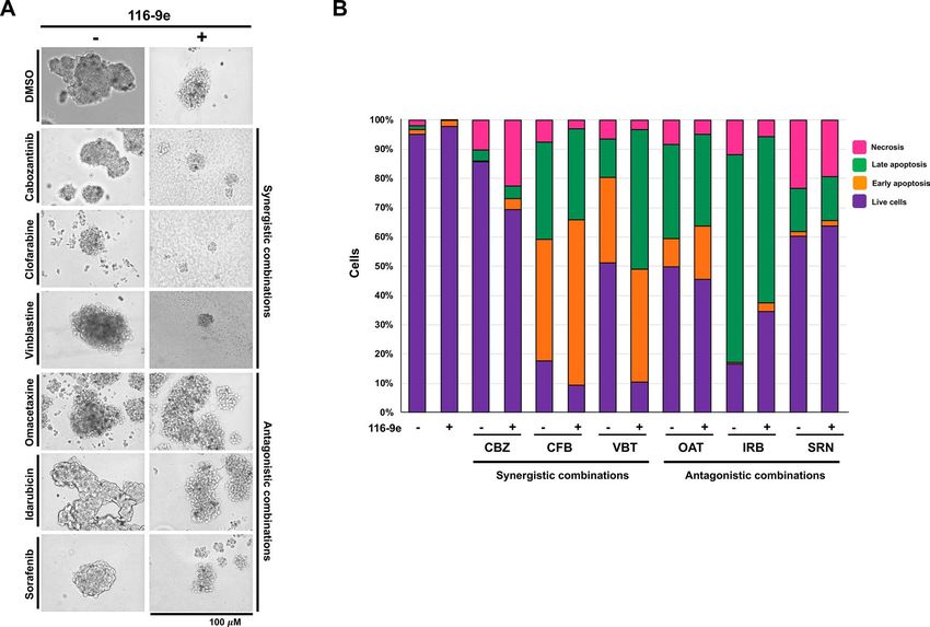
www.nature.com/scientificreports/
CRPCs expressing ARv7 are insensitive to Hsp90 inhibitors but are sensitive to Hsp40 i nhibition13. In addition,
we have shown that targeting specific oncoprotein complexes (ribonucleotide reductase) with a combination of
traditional as well as a DNAJA1 inhibitor produces highly synergistic e ffects8. We propose that targeting DNAJA1
in cancer may offer an attractive alternative to the toxicity induced by full Hsp90/Hsp70 inhibition.
Anticancer monotherapies using broadly active cytotoxic or molecularly targeted drugs are limited in their
ability to demonstrate a reliable clinical response. This is due to redundant signaling pathways, feedback loops
and resistance mechanisms in cancer c ells14. Thus, combination anticancer therapies have been used clinically
for over 50 years to improve the responses achieved by monotherapies alone. Cancer cell line-based models
for these combination therapies are easy and inexpensive to perform using high-throughput drug screening
protocols (HTS) to identify the most effective drug combination15,16. HTS helps to explore the relationship
between the cell line characteristics and drug specific dose r esponses15. Chemogenomics is one such HTS-based
approach where a large collection of anticancer chemical drugs are screened to identify biological targets. These
screening sets often contain small molecules that are well annotated and have defined molecular targets. Such
an approach is particularly beneficial for cancer research because malignant cells often contain multiple aber-
rations that require targeted therapy to inactivate cancer driver activities and mitigate deleterious effects of the
drugs to normal c ells14.
Here, we performed an unbiased screen of the NIH Approved Oncology Drug set containing 131 anti-cancer
drugs in combination with HAP1 cancer cell lines depleted of J-protein DNAJA1. We identified 41 compounds
showing strong synergy with the loss of DNAJA1, and in contrast 18 molecules that displayed reduced potency
in the knockout cell line. We validated three drugs (cabozantinib, clofarabine and vinblastine) in combination
with a unique DNAJA1 inhibitor (116-9e) for synergy in the LNCaP cancer cell lines and confirmed omacetaxine
mepesuccinate, idarubicin and sorafenib for antagonism (i.e. with reduced potency after DNAJA1 inhibition).
This study demonstrates the validity of developing Hsp70 co-chaperone inhibitors to sensitize cells to current
anticancer therapies and suggests that determining DNAJA1 status of a tumor may be beneficial in selecting the
most appropriate course of treatment.
Results
DNAJA1 is mutated and overexpressed in a variety of cancers. While the roles of Hsp90 and
Hsp70 in cancer have been thoroughly studied, much less is known of the role that regulatory co-chaperone
proteins such as DNAJA1 play in tumorigenesis. As a first step, we queried the cBioPortal cancer genomic data-
base (cbioportal.org) to determine the incidence of DNAJA1 alterations in cancer. Analysis of data from 176
non-redundant studies representing 44,347 patient samples revealed that DNAJA1 was altered at a frequency of
greater than 1% in 35 cancer types (Fig. 1A). Although the majority of alterations in DNAJA1 occur at a relatively
low frequency (< 5% of cancers) DNAJA1 is significantly amplified in prostate neuroendocrine cancer (PNC)
and castration-resistant prostate cancer at a frequency of 17.31% and 17.14% respectively (Fig. 1A). Hsp70 and
Hsp90 are often overexpressed in tumors2. To determine whether the DNAJA1 expression is also overexpressed
in cancer, we analyzed DNAJA1 mRNA expression in samples from the TGCA PAN-CAN Atlas. Interestingly,
DNAJA1 mRNA was expressed at significantly higher levels in these samples, with a median expression in cancer
over 3,000 × relative to WT reference samples (Fig. 1B). To determine if this dramatic overexpression of DNAJA1
was a result of amplification, we plotted DNAJA1 expression vs amplification (Fig. 1C). Interestingly, there was
minimal correlation between amount of amplification and DNAJA1 expression (r = 0.45) suggesting that while
DNAJA1 may be an important marker in cancer it is not caused by gene amplification.
Characterizing the role of DNAJA1 in anticancer drug resistance. The existing literature is contra-
dictory as to whether DNAJA1 may possess tumor suppressor or driver p roperties12,17. To clarify whether silenc-
ing of DNAJA1 could be beneficial in the treatment of cancer, we screened wildtype HAP1 cells and HAP1 cells
lacking DNAJA1 (HAP1DNAJA1 KO) for comparative resistance against the NIH NCI Approved Oncology Collec-
tion (Fig. 2A) (https://dtp.cancer.gov/organization/dscb/obtaining/available_plates.html). Prior to screening, we
validated the status of the DNAJA1 knockout cell line by Western blotting for DNAJA1 and other major chaper-
ones and co-chaperones (Hsp70, Hsc70, Hsp90, Bag-3 and Hsp110). As expected, we confirmed loss of DNAJA1
and interestingly did not observe any compensatory effects on the levels of the other chaperones/co-chaperones
studied (Fig. S1). According to pharmacologic action, the compounds in the library have been divided into
seven categories: protein synthesis inhibitors, proteasome inhibitors, epigenetic modifiers, metabolic inhibi-
tors, cytoskeletal inhibitors, signal transduction inhibitors and DNA synthesis/repair inhibitors. Further fold
enrichment of each drug category was calculated for the drugs whose potency increased or decreased with loss
of DNAJA1. To monitor the screening quality, each screening plate contained control wells treated with vehicle
(1% DMSO). The final concentration of the screening compounds was 50 μmol/L. Positive hits (synergistic) or
negative hits (antagonistic) were determined by normalizing the l og2 ratio of viability of DNAJA1 knockout cells
over wildtype cells. A full list of the screening results is shown in Supplementary Table T1 and the sorted data
are graphically plotted in Fig. 2B. The effectiveness of a large proportion of anticancer molecules in the collection
were impacted, with 41 of (31%) showing increased potency and 18 (14%) showing reduced potency upon loss
of DNAJA1 (Fig. 2C). Drug target analysis was carried out by calculating fold enrichment of positive hits (syner-
gistic) or negative hits (antagonistic) over the total number of drugs in that category. Drug target analysis of the
synergistic drug hits revealed significant enrichment in DNA synthesis and repair inhibitors, signal transduction
inhibitors as well as cytoskeletal inhibitors (Fig. 2D). In contrast, drug target analysis of antagonistic drug hits
revealed a higher enrichment in categories such as epigenetic modifiers, protein synthesis inhibitors, cytoskeletal
inhibitors and proteasome inhibitors (Fig. 2E). For a full list of drugs in each category and raw data from screen,
please see supplemental Table T1.
Scientific Reports | (2020) 10:13831 | https://doi.org/10.1038/s41598-020-70764-x 2
Vol:.(1234567890)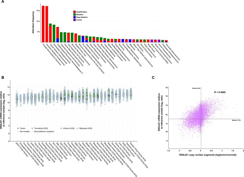
www.nature.com/scientificreports/
Figure 1. DNAJA1 is altered in cancer. (A) Prevalence of DNAJA1 alterations in various cancer genomes
analyzed via the cBioPortal. (B) DNAJA1 mRNA expression in cancers (TGCA PanCan) obtained from
cBioPortal. mRNA expression value is log2 ratio of expression seen in cancer vs. reference cells (please see www.
cbioportal.org/faq for more information). (C) increased DNAJA1 expression is not driven by copy number
increase. DNAJA1 copy number vs DNAJA1 expression was plotted and Pearson’s correlation coefficient
(R-value) was calculated. Median of both variables is marked by dotted line on the graph.
Strikingly, a small number of compounds with supposedly related function showed dissimilar alteration of
potency upon loss of DNAJA1 function, potentially caused by off-target drug effects (see discussion).
Validation of anticancer drugs significantly altered for potency upon loss of DNAJA1. Many
anticancer compounds have low potency, poor therapeutic index or suffer from the development of resistance .
Monotherapy is rarely efficient and instead drug cocktails are widely used in the c linic16. Establishing these com-
binations can enhance the scope of preclinical studies and inform the design of future clinical trials. Although
knockout of DNAJA1 substantially increased the potency of a number of anticancer molecules, it remained to
be determined whether small-molecule inhibition of DNAJA1 could produce a similar result. Our previous
bioinformatics analysis indicated that a large proportion of prostate cancer cells contain either amplification or
mutation of DNAJA1 (approximately 18%, see Fig. 1). To validate the results of our initial screen, we analyzed
the effect of treating prostate cancer cells (LNCaP) with a combination of 116-9e, a small molecule inhibitor
of DNAJA118 and selected hits from our screen. We decided to focus on three synergistic drugs discovered in
the screen: cabozantinib (receptor tyrosine kinase inhibitor), clofarabine (an RNR inhibitor)19 and vinblastine
(microtubule inhibitor/G2 arresting agent)20,21. We also validated three drugs that demonstrated a significant
loss of potency in cells lacking DNAJA1: sorafenib (a VEGFR-2 inhibitor)22, omacetaxine mepesuccinate (more
commonly known as homoharringtonine, a protein translation inhibitor)23 and idarubicin (topoisomerase II
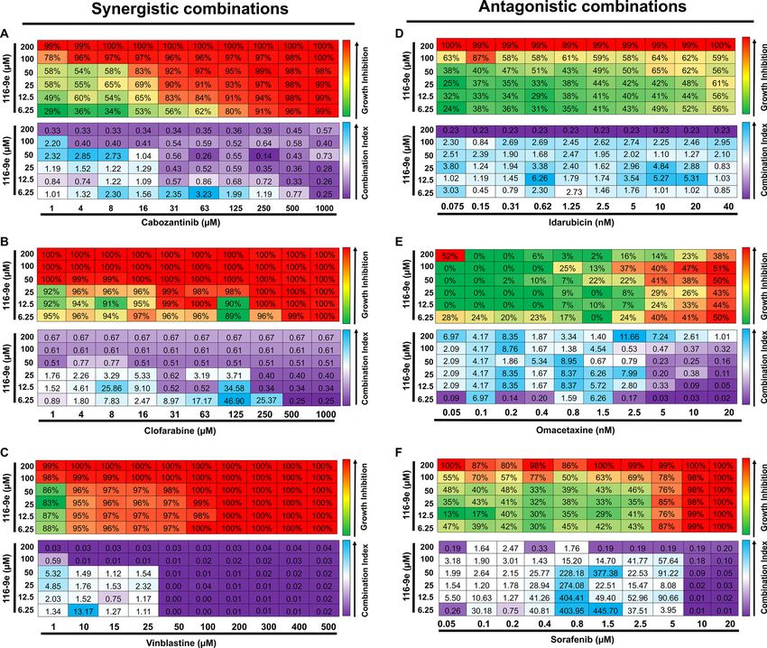
inhibitor)24. To determine synergy in a quantitative manner, we calculated drug synergy (Combination Index
values, CI) between 116-9e and either synergistic or antagonistic drugs hits across a broad range of concen-
trations using the Chou-Talalay m ethod25 (for effects of individual drugs, please see Fig. S2). For three hits
identified in our screen (cabozantinib, clofarabine and vinblastine) we confirmed significant synergy (CI < 1)
with 116-9e across a range of doses (Fig. 3A–C). In contrast, idarubicin, omacetaxine and sorafenib displayed a
significantly antagonistic interaction (CI > 1) across a range of doses (Fig. 3D–F). These data suggest that while
DNAJA1 inhibition is a promising strategy to sensitize cells to a number of currently used anticancer drugs, the
loss of DNAJA1 can significantly decrease the potency of a small subset of inhibitors.
Scientific Reports | (2020) 10:13831 | https://doi.org/10.1038/s41598-020-70764-x 3
Vol.:(0123456789)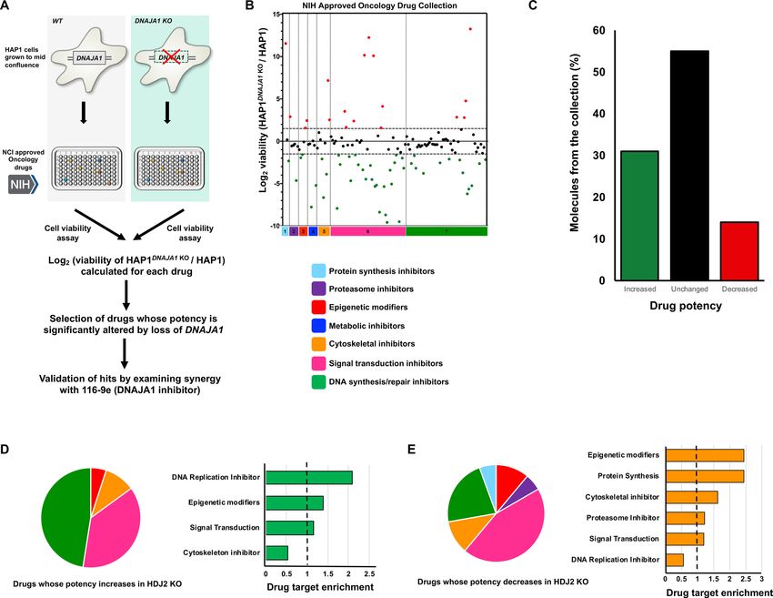
www.nature.com/scientificreports/
Figure 2. Sensitivity of WT and DNAJA1 knockout cells to the NIH Approved Oncology Collection. (A)
Workflow of high-throughput cell-based screen. (B) A collection of 132 drugs were screened at 50 μmol/L with
Wild-type and DNAJA1 KO cells. Results are the average of at least triplicates and error is SEM. The dotted lines
represent a potency change of L og2 > 1.5 or Log2www.nature.com/scientificreports/
Figure 3. Drug interaction between 116-9e (DNAJA1 inhibitor) and selected hits. LNCaP cells were treated
with different concentrations of cabozantinib (A), clofarabine (B), vinblastine (C), idarubicin (D), omacetaxine
(E) and sorafenib (F) with or without 116-9e for 72 h in RPMI-1640 medium containing 10% FBS. Each point
is the mean ± SD for three independent experiments. Growth inhibition was determined using Cell Titer-Glo
assay. Combination Index (CI, measure of drug synergy) was determined using Chou-Talalay method via
Compusyn software. CI values are as follows: < 0.1 (very strongly synergistic), 0.1–0.3 (strongly synergistic), < 0.9
(synergistic), 0.9–1.1 (additive), 1.1–3.3 (antagonistic), 3.3–10 (strongly antagonistic), > 10 (very strongly
antagonistic).
Discussion
Although inhibitors of Hsp70 and Hsp90 have been developed for research purposes, the conversion of these
molecules for use in patient treatment have been hampered by toxicity issues4. We undertook this study to
resolve conflicting literature on whether inhibiting DNAJA1, a co-chaperone of Hsp70 may be useful as a novel
anticancer strategy. Our bioinformatic analysis of DNAJA1 expression and mutation clearly identify DNAJA1 as
being highly altered in a range of cancers, particularly in prostate cancer. Interestingly, DNAJA1 despite being
substantially overexpressed in a range of cancers, there was minimal correlation between DNAJA1 copy number
and level of expression. While beyond the scope of this study, it is possible that the high levels of DNAJA1 expres-
sion observed may be a result of increased transcription brought on hyperactive signaling pathways common
in cancer cells. This data in conjunction with a recent finding that Hsp40 is involved in regulation of A Rv13 and
p5328 makes DNAJA1 inhibition an ideal choice as a novel therapeutic target in Prostate Cancer.
In this study, loss of DNAJA1 increased the potency of a substantial number (31%) of clinically used antican-
cer drugs. This increased potency may be related to the destabilization of clients that are the target of these small
molecules. For example, Hsp70 activates many proteins involved in the DNA damage response and DNA repair
pathways (DDR), including ATM, APE1, PARP1, X RCC129. Recently, studies from our group have established
roles for both Hsp70 and DNAJA1 in stability of the RNR c omplex8,29,30. It is unsurprising then that many of
the anticancer agents displaying synergy with loss of DNAJA1 are connected to inhibition of the DNA damage
Scientific Reports | (2020) 10:13831 | https://doi.org/10.1038/s41598-020-70764-x 5
Vol.:(0123456789)www.nature.com/scientificreports/
Figure 4. Effect of combination treatments on prostate cancer spheroids. (A) Cells were plated on Matrigel-
coated 24 well plates. Six drugs (cabozantinib, clofarabine, vinblastine, idarubicin, omacetaxine and sorafenib)
were tested on prostate cancer spheroids. These experiments were performed in triplicate and are average of
3 replicates from 3 different wells of a cell culture plate. The pictures are representative images as acquired
using an EVOS cell imager. (B) Proliferation of spheroids treated with cabozantinib (CBZ), clofarabine (CFB),
vinblastine (VBT), idarubicin (IRB), omacetaxine (OAT) and sorafenib (SRN) measured using AnnexinV/PI
staining.
response/repair. These include molecules such as 5-fluorouracil (5-FU), premetrexed, clofarabine, olaparib and
niraparib etoposide, teniposide and valrubicin. Here we validated synergy with the RNR inhibitor clofarabine.
Clofarabine is phosphorylated intracellularly to form cytotoxic active 5′-triphosphate metabolite, which inhibits
the enzymatic activities of RNR and DNA polymerase, resulting in inhibition of DNA synthesis and repair31.
While most DDR inhibitors displayed increased potency with DNAJA1 depletion, four of them were antago-
nistic to loss of DNAJA1. These include topoisomerase inhibitors and nucleic acid synthesis inhibitors such as
trifluridine, irinotecan, epirubicin (4′-epi-isomer of the antibiotic doxorubicin) and idarubicin (4-demethoxy
analogue of daunorubicin)32. While at first these results seem paradoxical, it is worth noting that inrinotecan is
a type I topoisomerase inhibitor, whereas Etoposide (synergistic with loss of DNAJA1) is a type II topoisomerase
inhibitor. It may be that Hsp70 and DNAJA1 play opposing regulatory roles in the stabilization and activation
of these related proteins.
In addition to DDR, DNAJA1 is also involved in signal transduction, with previous reports indicating that the
yeast homolog of DNAJA1 (Ydj1) is critical for supporting the integrity of kinase signaling n etworks33. DNAJA1 is
mobilized to specific sites within the nucleus in response to inappropriate targeting or folding of specific mutant
receptors. DNAJA1 overexpression ameliorates the defective transactivation and trans-repression activity of
mutant Glucocorticoid receptors34. In line with the previous studies, we found that a handful of Receptor Tyrosine
kinase inhibitors were synergistic with DNAJA1 depletion. These included Vascular endothelial growth factor
receptor (VEGFR) inhibitors such as sunitinib, cabozantinib, lenvatinib and pazopanib. Interestingly, randomized
phase III clinical trials are being conducted to validate the efficacy of Cabozantinib in heavily pretreated prostate
cancer patients35. One implication from our study is that DNAJA1 inhibition might significantly enhance the
effect of cabozantinib monotherapy.
Strikingly, some of the kinase inhibitors were antagonistic to DNAJA1 depletion. These include VEGFR
inhibitors such as regorafenib and sorafenib. This disparity can be explained by the different target receptors and
mechanisms of action of these drugs. Interestingly, recent studies indicated that these small molecule inhibitors
exhibit off-target effects. Some of these drugs are misidentified and mischaracterized for their target specific
inhibition, which has contributed to the high failure rate of these drugs in the treatment of cancer patients36.
In addition to its role in signal transduction, DNAJA1 is also important for maintaining the cellular cytoskel-
eton. Previous studies have suggested that YDJ1 (the yeast homolog of DNAJA1) is important for the proper
assembly of m icrotubules37. Another report showed that DNAJA1 depletion causes relocation of N-cadherin and
enhanced activity of metalloproteinases. This leads to changes in the actin cytoskeleton indicating that DNAJA1
Scientific Reports | (2020) 10:13831 | https://doi.org/10.1038/s41598-020-70764-x 6
Vol:.(1234567890)www.nature.com/scientificreports/
is important for prevention of the amoeboid-like transition of tumor cells38. These studies indicated the involve-
ment of DNAJA1 in maintaining cytoskeletal organization. We found 3 anticancer drugs targeting the cytoskel-
eton to be synergistic with DNAJA1 depletion, including vinblastine sulfate (cytoskeletal inhibitor that disrupts
microtubule formation during mitosis and interferes with glutamic acid metabolism), estramustine (binds to
microtubule-associated proteins (MAPs) and inhibits microtubule dynamics) and ixabepilone (promotes tubulin
polymerization and microtubule stabilization, thereby arresting cells in the G2-M p hase39. Strikingly, two of the
tubulin inhibitors were found to be antagonistic to DNAJA1 depletion. These include paclitaxel and ixabepilone.
Paclitaxel inhibits the disassembly of microtubules resulting in the inhibition of cell division whereas Ixabepilone
promotes tubulin polymerization and microtubule stabilization, arresting cells in the G2-M phase of the cell
cycle39. This apparent discrepancy may be explained by off-target effects of these molecules (see below).
Epigenetic modifying drugs display substantially modified potency depending on cellular DNAJA1 status.
While previous studies have indicated the association between proteomic changes and histone PTMs in response
to Hsp90 inhibitor treatment in bladder carcinoma cells, no such association has been shown for DNAJA1 and
Histone PTMs40. Interestingly, vorinostat was the only drug that was synergistic to DNAJA1 inhibition. It is a
histone deacetylase inhibitor that binds to the catalytic domain of the histone deacetylases (HDACs). However,
we also identified two histone deacetylase inhibitor drugs to be antagonistic to DNAJA1 depletion, panobinostat
and romidepsin. These inhibit histone deacetylase (HDAC) which may impact cell cycle protein expression, cell
cycle arrest in the G2/M phase and a poptosis41. Excitingly, our data suggest a functional link between histones,
their modifications and DNAJA1. While these findings require further investigation, it is possible that DNAJA1
may regulate the stability of histones themselves or histone chaperones. Interestingly, bortezomib (a protea-
some inhibitor) lost potency when DNAJA1 was either inhibited with 116-9e or knocked out with CRISPR.
Interestingly, a similar phenomenon has been observed in B16F10 melanoma cells. While treatment of these
cells with 10 nM bortezomib was cytotoxic, this effect was not observed in cells treated with a combination of
both quercetin (an Hsp70 inhibitor) and bortezomib42. This apparent antagonism may be explained by their
mechanism of action on the heat shock transcription factor, HSF1. While bortezomib acts to trigger the heat
shock response in some cancers, the Hsp70/co-chaperone system maintains HSF1 in an less active immature
form43–46. It is interesting to note that while there are clear classes of drugs that are made more potent by loss of
DNAJA1 function (DNA damage response, cytoskeletal function etc.), there are a small number of drugs in these
classes that are not impacted at all or even made less potent. This apparent discrepancy implies that some of these
inhibitors might have multiple cellular targets in addition to their proposed primary mechanism of action. This
theory has been validated in fascinating studies comparing effects of small molecule therapies, gene knockout
and knockdowns that theoretically target the same g enes36.
As in the case of any chemogenomic screen, care must be taken to validate screening results with other
methods. While CRISPR KO cell lines offer the advantage of complete, specific and permanent gene silencing as
compared to transient inhibition seen with small molecules or siRNA, the authors acknowledge that permanent
knockout lines may respond by overpexressing/suppressing other genes to compensate, leading to non-specific
effects. In this study, we took the approach of following up our intitial screen with small molecule validation
in 2D and 3D cell culture models. Going forward, we intend to validate several of these hits in vivo (mouse)
model systems. 116-9e is a relatively new and unexplored molecule; while it clearly alters JDP binding, the exact
impact on all Hsp70-JDPs has not been c haracterized8,18. Interestingly, the DNAJA1 knockout cell line grows
effectively the same as WT and our examination of key chaperone protein levels do note reveal any major altera-
tions. However, we do acknowledge that it is possible that the DNAJA1 knockout cell line may have adapted by
overexpressing other JDPs such as DNAJA2, DNAJB1 or DNAJB6. In future studies, we hope to investigate this
further by quantitating the proteome in HAP1 cells (WT and DNAJA1 knockout) in both untreated and 116-9e
treated conditions. Given the essential nature of Hsp70/Hsc70 in cancer cells, if 116-9e truly inhibited all JDP
interactions it would be highly toxic to cells which we do not observe, suggesting there must be some selectivity
in JDP inhibition.
Recent studies from our group and other have described the clear impact of Hsp70/JDP inhibition on indi-
vidual oncoprotein client stability and prostate cancer cell survival8,13. Overall, this study demonstrates the larger
feasibility of inhibiting Hsp70 co-chaperones such as DNAJA1 as a novel anticancer therapy, acting to fine-tune
Hsp70 function rather than completely abolishing it. Nearly a third of the anticancer compounds screened
demonstrated increased potency in DNAJA1 knockout cells. Rather than attempting to develop co-chaperone
inhibitors as a monotherapy, we believe their strength lies as sensitizing agents to existing therapies. Moreover,
our data imply that overexpression of DNAJA1 in patient tumors may impact the effectiveness of a number of
commonly used anticancer drugs. While further experiments characterizing (1) the specific DNAJA1-mediated
effects in cancer proliferation and (2) the specificity of drugs such as 116-9e are required, our studies suggest
perhaps a future precision medicine approach that uses tumor DNAJA1 status to guide treatment strategy.
Materials and methods
Cell culture. The HAP1 Chronic Myelogenous Leukemia cancer cell line and DNAJA1 knockout cell line
was purchased from Horizon Discovery and were cultured in Iscove’s Modified Eagle Medium (Invitrogen) with
10% fetal bovine serum (Gibco), 100 units/ml penicillin, and 100 μg/ml streptomycin at 5% CO2 and 37° C. The
LNCaP cancer cell line was purchased from ATCC and were cultured in RPMI-1640 medium (Invitrogen) with
10% fetal bovine serum (FBS, Clontech), 100 units/ml penicillin, and 100 μg/ml streptomycin at 5% C O2 and
37° C.
Drug screening. Approved Oncology Drug plates consisting of the most current FDA approved anticancer
drugs were obtained from the National Cancer Institute (NCI). For experiments delineating the synergy between
Scientific Reports | (2020) 10:13831 | https://doi.org/10.1038/s41598-020-70764-x 7
Vol.:(0123456789)www.nature.com/scientificreports/
the loss of DNAJA1 and approved anticancer drug, HAP1 cells and HAP1 (DNAJA1 KO) cells were plated in
growth media at 20% confluency 1 day prior to drug treatment. On Day 1 of treatment, cells were treated with
DMSO (control), Approved oncology anticancer drugs at 50 µM for 72 h. Following drug treatments, Cell Titer-
Glo reagent was added directly to the wells according to the manufacturer’s instructions. The luminescence was
measured on Bio-Tek Plate reader. Luminescence reading was normalized to and expressed as a relative percent-
age of the plate averaged DMSO control. The data shown are the mean and SEM of three independent biological
replicates.
Combination index (CI) calculations. For IC50 calculations, LNCaP cells were seeded in triplicates in
96-well white bottom Nunc plates in growth media at 20% confluency 1 day prior to initiation of drug treatment.
On Day 1 of treatment, cells were treated with DMSO (control) and ten folds serial dilution of anti-cancer drugs
cabozantinib, clofarabine, vinblastine, sorafenib, idarubicin and omacetaxine mepesuccinate and 116-9e. After
72 h, cell viability was measured using Promega Cell Titer-Glo cell viability assay on Bio-Tek plate reader. The
combination index was calculated using the Chou-Talalay method using CompuSyn s oftware47.
Spheroid generation. Single-cell suspensions (5,000/well) were plated in one well of 24-well plates in a 1:1
mixture of RPMI medium and Matrigel (BD Bioscience CB-40324). Cells in Matrigel were kept cold at all times
and under continuous agitation. Warm PBS was added to all empty wells, if any. Plates were incubated at 37 °C
with 5% CO2 for 15 min to solidify the gel before addition of 100 µl of pre-warmed RPMI to each well. Two
days after seeding, the media was fully aspirated and replaced with fresh RPMI containing the indicated drugs.
The same procedure was repeated daily on two consecutive days. Twenty-four hours after the last treatments,
the media was aspirated and the wells were washed with 100 µl of pre-warmed PBS. To prepare for downstream
assays, spheroids were released from the Matrigel by incubating at 37 °C for 40 min in 100 µl of 10 mg/mL Dis-
pase (Sigma).
Apoptosis assay. Apoptosis of LNCaP spheroids was detected by the Annexin V–FITC/propidium iodide–
binding assay. Cells were treated with either 0.1% DMSO (dimethyl sulfoxide),116-9e, cabozantinib, clofarabine,
vinblastine, sorafenib, idarubicin, omacetaxine mepesuccinate and sorafenib alone or in combination with 116-
9e for 48 h at the IC50 concentrations, and then stained with Annexin V–FITC and propidium iodide. The rate of
apoptosis was determined using a BD LSR Fortessa flow cytometer, and the collected data were analyzed using
FlowJo software. Apoptosis was reported as the mean ± SD. The results are representative of three independent
experiments.
Bioinformatics. Cancer genome data and Cancer Cell Line Encyclopedia data were accessed from the cBio-
Portal (www.cbioportal.org) for Cancer Genomics 48. Total patient numbers and detailed information regarding
published datasets and associated publications are indicated in Fig. 1A,B.
Statistical analysis. Data were analyzed using GraphPad Prism built-in statistical tests indicated in rel-
evant figure legends. The following asterisk system for P-value was used: P < 0.05; P < 0.01; 0.001; and P < 0.0001.
Western blotting. Protein extracts were made as d escribed8. 30 μg of protein was separated by 4–12%
NuPAGE SDS-PAGE (Thermo). Proteins were detected using the following antibodies; anti-DNAJA1/HDJ2
(Thermo # MA5-12748), anti-Actin (CST # 9774), Anti-Hsc70 (Santa Cruz, # sc-7298), anti-Hsp70 (Enzo #
C92F3A5), anti-Hsp90 ⍺/ℬ (Santa Cruz # sc-13119), anti-Bag3 (Santa Cruz # sc-136467), anti-Hsp110 (Stress
Marq, # SPC-195),) at 1:4,000 dilution in TBST + 1% BSA. The secondary antibody (StarBright Blue 700 Fluo-
rescent Secondary Mouse) was used at 1:3,000 dilution in TBST + 1% BSA. Blots were imaged on a Chemi Doc
MP imaging system (Bio-Rad).
Received: 29 February 2020; Accepted: 29 July 2020
References
1. Rosenzweig, R., Nillegoda, N. B., Mayer, M. P. & Bukau, B. The Hsp70 chaperone network. Nat. Rev. Mol. Cell Biol. 20, 665–680.
https://doi.org/10.1038/s41580-019-0133-3 (2019).
2. Ciocca, D. R. & Calderwood, S. K. Heat shock proteins in cancer: diagnostic, prognostic, predictive, and treatment implications.
Cell Stress Chaperones 10, 86–103. https://doi.org/10.1379/csc-99r.1 (2005).
3. Gestwicki, J. E. & Shao, H. Inhibitors and chemical probes for molecular chaperone networks. J. Biol. Chem. 294, 2151–2161. https
://doi.org/10.1074/jbc.TM118.002813 (2019).
4. Evans, C. G., Chang, L. & Gestwicki, J. E. Heat shock protein 70 (hsp70) as an emerging drug target. J. Med. Chem. 53, 4585–4602.
https://doi.org/10.1021/jm100054f (2010).
5. Kampinga, H. H. & Craig, E. A. The HSP70 chaperone machinery: J proteins as drivers of functional specificity. Nat. Rev. Mol. Cell
Biol. 11, 579–592. https://doi.org/10.1038/nrm2941 (2010).
6. Craig, E. A. & Marszalek, J. How do J-Proteins get Hsp70 to do so many different things?. Trends Biochem. Sci. 42, 355–368. https
://doi.org/10.1016/j.tibs.2017.02.007 (2017).
7. Nitika & Truman, A. W. Cracking the Chaperone Code: cellular roles for Hsp70 phosphorylation. Trends. Biochem. Sci . 42,
932–935. https://doi.org/10.1016/j.tibs.2017.10.002 (2017).
Scientific Reports | (2020) 10:13831 | https://doi.org/10.1038/s41598-020-70764-x 8
Vol:.(1234567890)www.nature.com/scientificreports/
8. Sluder, I. T., Nitika, Knighton, L. E. & Truman, A. W. The Hsp70 co-chaperone Ydj1/HDJ2 regulates ribonucleotide reductase
activity. PLoS Genet 14, e1007462, https://doi.org/10.1371/journal.pgen.1007462 (2018).
9. Truman, A. W. et al. CDK-dependent Hsp70 phosphorylation controls G1 cyclin abundance and cell-cycle progression. Cell 151,
1308–1318. https://doi.org/10.1016/j.cell.2012.10.051 (2012).
10. Woodford, M. R. et al. Impact of posttranslational modifications on the anticancer activity of Hsp90 inhibitors. Adv. Cancer Res.
129, 31–50. https://doi.org/10.1016/bs.acr.2015.09.002 (2016).
11. Dushukyan, N. et al. Phosphorylation and ubiquitination regulate protein phosphatase 5 activity and its prosurvival role in kidney
cancer. Cell Rep. 21, 1883–1895. https://doi.org/10.1016/j.celrep.2017.10.074 (2017).
12. Stark, J. L. et al. Structure and function of human DnaJ homologue subfamily a member 1 (DNAJA1) and its relationship to pan-
creatic cancer. Biochemistry 53, 1360–1372. https://doi.org/10.1021/bi401329a (2014).
13. Moses, M. A. et al. Targeting the Hsp40/Hsp70 chaperone axis as a novel strategy to treat castration-resistant prostate cancer.
Cancer Res. 78, 4022–4035. https://doi.org/10.1158/0008-5472.CAN-17-3728 (2018).
14. Palmer, A. C. & Sorger, P. K. (2017) Combination cancer therapy can confer benefit via patient-to-patient variability without drug
additivity or synergy. Cell 171, 1678–1691. doi: https://doi.org/10.1016/j.cell.2017.11.009 (2017).
15. Ding, K. F. et al. Nonlinear mixed effects dose response modeling in high throughput drug screens: application to melanoma cell
line analysis. Oncotarget 9, 5044–5057. https://doi.org/10.18632/oncotarget.23495 (2018).
16. Koh, S. B. et al. Mechanistic distinctions between CHK1 and WEE1 inhibition guide the scheduling of triple therapy with gem-
citabine. Cancer Res. 78, 3054–3066. https://doi.org/10.1158/0008-5472.CAN-17-3932 (2018).
17. Wang, C. C. et al. HDJ-2 as a target for radiosensitization of glioblastoma multiforme cells by the farnesyltransferase inhibitor
R115777 and the role of the p53/p21 pathway. Cancer Res. 66, 6756–6762. https: //doi.org/10.1158/0008-5472.CAN-06-0185 (2006).
18. Wisen, S. et al. Binding of a small molecule at a protein-protein interface regulates the chaperone activity of hsp70-hsp40. ACS
Chem. Biol. 5, 611–622. https://doi.org/10.1021/cb1000422 (2010).
19. Wisitpitthaya, S. et al. Cladribine and fludarabine nucleotides induce distinct hexamers defining a common mode of reversible
RNR inhibition. ACS Chem. Biol. 11, 2021–2032. https://doi.org/10.1021/acschembio.6b00303 (2016).
20. Wu, C. C. et al. Structural basis of type II topoisomerase inhibition by the anticancer drug etoposide. Science 333, 459–462. https
://doi.org/10.1126/science.1204117 (2011).
21. Senra, J. M. et al. Inhibition of PARP-1 by olaparib (AZD2281) increases the radiosensitivity of a lung tumor xenograft. Mol. Cancer
Ther. 10, 1949–1958. https://doi.org/10.1158/1535-7163.MCT-11-0278 (2011).
22. Dudgeon, C. et al. Inhibiting oncogenic signaling by sorafenib activates PUMA via GSK3beta and NF-kappaB to suppress tumor
cell growth. Oncogene 31, 4848–4858. https://doi.org/10.1038/onc.2011.644 (2012).
23. Gandhi, V., Plunkett, W. & Cortes, J. E. Omacetaxine: a protein translation inhibitor for treatment of chronic myelogenous leukemia.
Clin. Cancer Res. 20, 1735–1740. https://doi.org/10.1158/1078-0432.CCR-13-1283 (2014).
24. Hevener, K., Verstak, T. A., Lutat, K. E., Riggsbee, D. L. & Mooney, J. W. Recent developments in topoisomerase-targeted cancer
chemotherapy. Acta Pharm. Sin. B 8, 844–861. https://doi.org/10.1016/j.apsb.2018.07.008 (2018).
25. Chou, T. C. Drug combination studies and their synergy quantification using the Chou-Talalay method. Cancer Res. 70, 440–446.
https://doi.org/10.1158/0008-5472.CAN-09-1947 (2010).
26. Vlachogiannis, G. et al. Patient-derived organoids model treatment response of metastatic gastrointestinal cancers. Science 359,
920–926. https://doi.org/10.1126/science.aao2774 (2018).
27. Pickl, M. & Ries, C. H. Comparison of 3D and 2D tumor models reveals enhanced HER2 activation in 3D associated with an
increased response to trastuzumab. Oncogene 28, 461–468. https://doi.org/10.1038/onc.2008.394 (2009).
28. Parrales, A. et al. DNAJA1 controls the fate of misfolded mutant p53 through the mevalonate pathway. Nat. Cell Biol. 18, 1233–1243.
https://doi.org/10.1038/ncb3427 (2016).
29. Knighton, L. E., Delgado, L. E. & Truman, A. W. Novel insights into molecular chaperone regulation of ribonucleotide reductase.
Curr. Genet. 65, 477–482. https://doi.org/10.1007/s00294-018-0916-7 (2019).
30. Truman, A. W. et al. Quantitative proteomics of the yeast Hsp70/Hsp90 interactomes during DNA damage reveal chaperone-
dependent regulation of ribonucleotide reductase. J. Proteom. 112, 285–300. https://doi.org/10.1016/j.jprot.2014.09.028 (2015).
31. Huguet, F. et al. Clofarabine for the treatment of adult acute lymphoid leukemia: the Group for Research on Adult Acute Lympho-
blastic Leukemia intergroup. Leuk Lymphoma 56, 847–857. https://doi.org/10.3109/10428194.2014.887708 (2015).
32. Deng, S. et al. Dexrazoxane may prevent doxorubicin-induced DNA damage via depleting both topoisomerase II isoforms. BMC
Cancer 14, 842. https://doi.org/10.1186/1471-2407-14-842 (2014).
33. Gillies, A. T., Taylor, R. & Gestwicki, J. E. Synthetic lethal interactions in yeast reveal functional roles of J protein co-chaperones.
Mol. Biosyst. 8, 2901–2908. https://doi.org/10.1039/c2mb25248a (2012).
34. Tang, Y., Ramakrishnan, C., Thomas, J. & DeFranco, D. B. A role for HDJ-2/HSDJ in correcting subnuclear trafficking, transactiva-
tion, and transrepression defects of a glucocorticoid receptor zinc finger mutant. Mol. Biol. Cell. 8, 795–809 (1997).
35. Grullich, C. Cabozantinib: a MET, RET, and VEGFR2 tyrosine kinase inhibitor. Recent Results Cancer Res. 201, 207–214. https://
doi.org/10.1007/978-3-642-54490-3_12 (2014).
36. Lin, A. et al. Off-target toxicity is a common mechanism of action of cancer drugs undergoing clinical trials. Sci. Transl. Med. https
://doi.org/10.1126/scitranslmed.aaw8412 (2019).
37. Oka, M. et al. Loss of Hsp70-Hsp40 chaperone activity causes abnormal nuclear distribution and aberrant microtubule formation
in M-phase of Saccharomyces cerevisiae. J. Biol. Chem. 273, 29727–29737. https://doi.org/10.1074/jbc.273.45.29727 (1998).
38. Meshalkina, D. A. et al. Knock-down of Hdj2/DNAJA1 co-chaperone results in an unexpected burst of tumorigenicity of C6
glioblastoma cells. Oncotarget 7, 22050–22063. https://doi.org/10.18632/oncotarget.7872 (2016).
39. Cobham, M. V. & Donovan, D. Ixabepilone: a new treatment option for the management of taxane-resistant metastatic breast
cancer. Cancer Manag. Res. 1, 69–77 (2009).
40. Li, Q. Q. et al. Proteomic analysis of proteome and histone post-translational modifications in heat shock protein 90 inhibition-
mediated bladder cancer therapeutics. Sci. Rep. 7, 201. https://doi.org/10.1038/s41598-017-00143-6 (2017).
41. Eckschlager, T., Plch, J., Stiborova, M. & Hrabeta, J. Histone Deacetylase Inhibitors as Anticancer Drugs. Int J Mol Sci . doi:https
://doi.org/10.3390/ijms18071414 (2017).
42. Yerlikaya, A., Okur, E., Eker, S. & Erin, N. Combined effects of the proteasome inhibitor bortezomib and Hsp70 inhibitors on the
B16F10 melanoma cell line. Mol. Med. Rep. 3, 333–339. https://doi.org/10.3892/mmr_00000262 (2010).
43. Mitsiades, N. et al. Molecular sequelae of proteasome inhibition in human multiple myeloma cells. Proc. Natl. Acad. Sci. USA 99,
14374–14379. https://doi.org/10.1073/pnas.202445099 (2002).
44. Shah, S. P. et al. Bortezomib-induced heat shock response protects multiple myeloma cells and is activated by heat shock factor 1
serine 326 phosphorylation. Oncotarget 7, 59727–59741. https://doi.org/10.18632/oncotarget.10847 (2016).
45. Abravaya, K., Myers, M. P., Murphy, S. P. & Morimoto, R. I. The human heat shock protein hsp70 interacts with HSF, the transcrip-
tion factor that regulates heat shock gene expression. Genes Dev. 6, 1153–1164. https://doi.org/10.1101/gad.6.7.1153 (1992).
46. Baler, R., Welch, W. J. & Voellmy, R. Heat shock gene regulation by nascent polypeptides and denatured proteins: hsp70 as a
potential autoregulatory factor. J. Cell Biol. 117, 1151–1159. https://doi.org/10.1083/jcb.117.6.1151 (1992).
47. Chou, T. C. Theoretical basis, experimental design, and computerized simulation of synergism and antagonism in drug combina-
tion studies. Pharmacol. Rev. 58, 621–681. https://doi.org/10.1124/pr.58.3.10 (2006).
Scientific Reports | (2020) 10:13831 | https://doi.org/10.1038/s41598-020-70764-x 9
Vol.:(0123456789)www.nature.com/scientificreports/
48. Cerami, E. et al. The cBio cancer genomics portal: an open platform for exploring multidimensional cancer genomics data. Cancer
Discov. 2, 401–404. https://doi.org/10.1158/2159-8290.CD-12-0095 (2012).
49. Nitika et al. Chemogenomic screening Identifies the Hsp70 Co-chaperone HDJ2 as a Hub for Anticancer Drug Resistance. bioRxiv,
818427, doi:https://doi.org/10.1101/818427 (2019).
Acknowledgements
The authors thank NIH for providing materials used in this study and J. Gestwicki, D. Dreau and T. Erick for
helpful comments. We thank all reviewers for their helpful and constructive comments. This manuscript has
ioRxiv49. This project was supported by NCI R15CA208773 and Grant-In-Aid
been released as a pre-print at B
of Research from Sigma Xi (G2018031591887158), The Scientific Research Society.
Author contributions
N. performed all main experiments and wrote manuscript drafts; J.S.B. assisted with the chemogenomic screen;
J.E.T. and L.E.K. helped with data analysis, figure drawing and manuscript editing; S.K.C. and A.W.T. conceived
the experiments and edited the manuscript.
Competing interests
The authors declare no competing interests.
Additional information
Supplementary information is available for this paper at https://doi.org/10.1038/s41598-020-70764-x.
Correspondence and requests for materials should be addressed to A.W.T.
Reprints and permissions information is available at www.nature.com/reprints.
Publisher’s note Springer Nature remains neutral with regard to jurisdictional claims in published maps and
institutional affiliations.
Open Access This article is licensed under a Creative Commons Attribution 4.0 International
License, which permits use, sharing, adaptation, distribution and reproduction in any medium or
format, as long as you give appropriate credit to the original author(s) and the source, provide a link to the
Creative Commons licence, and indicate if changes were made. The images or other third party material in this
article are included in the article’s Creative Commons licence, unless indicated otherwise in a credit line to the
material. If material is not included in the article’s Creative Commons licence and your intended use is not
permitted by statutory regulation or exceeds the permitted use, you will need to obtain permission directly from
the copyright holder. To view a copy of this licence, visit http://creativecommons.org/licenses/by/4.0/.
© The Author(s) 2020
Scientific Reports | (2020) 10:13831 | https://doi.org/10.1038/s41598-020-70764-x 10
Vol:.(1234567890)You can also read