Interferon-γ/Interleukin-27 Axis Induces Programmed Death Ligand 1 Expression in Monocyte-Derived Dendritic Cells and Restores Immune Tolerance in ...
←
→
Page content transcription
If your browser does not render page correctly, please read the page content below
ORIGINAL RESEARCH
                                                                                                                                             published: 26 October 2020
                                                                                                                                       doi: 10.3389/fimmu.2020.576752

                                            Interferon-γ/Interleukin-27 Axis
                                            Induces Programmed Death Ligand 1
                                            Expression in Monocyte-Derived
                                            Dendritic Cells and Restores Immune
                                            Tolerance in Central Nervous System
                                            Autoimmunity
                              Edited by:
                    Dipyaman Ganguly,       Giacomo Casella 1 , Javad Rasouli 1 , Rodolfo Thome 1 , Hélène C. Descamps 2 ,
            Indian Institute of Chemical    Asrita Vattikonda 1 , Larissa Ishikawa 1 , Alexandra Boehm 1 , Daniel Hwang 1 ,
                           Biology, India   Weifeng Zhang 1 , Dan Xiao 1 , Jeongho Park 3,4 , Guang-Xian Zhang 1 , Jorge I. Alvarez 3 ,
                          Reviewed by:      Abdolmohamad Rostami 1 and Bogoljub Ciric 1*
                          Sin-Hyeog Im,
                                            1
     Pohang University of Science and         Department of Neurology, Thomas Jefferson University, Philadelphia, PA, United States, 2 Department of Microbiology,
              Technology, South Korea       Perelman School of Medicine, University of Pennsylvania, Philadelphia, PA, United States, 3 Department of Pathobiology,
                           Sylvaine You,    School of Veterinary Medicine, University of Pennsylvania, Philadelphia, PA, United States, 4 College of Veterinary Medicine &
   Institut National de la Santé et de la   Institute of Veterinary Science, Kangwon National University, Chuncheon, South Korea
           Recherche Médicale, France
                 *Correspondence:           Antigen (Ag)-specific tolerance induction by intravenous (i. v.) injection of high-dose auto-
                        Bogoljub Ciric
          bogoljub.ciric@jefferson.edu      Ags has been explored for therapy of autoimmune diseases, including multiple sclerosis
                                            (MS). It is thought that the advantage of such Ag-specific therapy over non-specific
                    Specialty section:      immunomodulatory treatments would be selective suppression of a pathogenic immune
          This article was submitted to
         Immunological Tolerance and
                                            response without impairing systemic immunity, thus avoiding adverse effects of
                             Regulation,    immunosuppression. Auto-Ag i.v. tolerance induction has been extensively studied in
                a section of the journal
                                            experimental autoimmune encephalomyelitis (EAE), an animal model of MS, and limited
               Frontiers in Immunology
                                            clinical trials demonstrated that it is safe and beneficial to a subset of MS patients.
           Received: 26 June 2020
      Accepted: 02 September 2020           Nonetheless, the mechanisms of i.v. tolerance induction are incompletely understood,
        Published: 26 October 2020          hampering the development of better approaches and their clinical application. Here, we
                              Citation:     describe a pathway whereby auto-Ag i.v. injected into mice with ongoing clinical EAE
        Casella G, Rasouli J, Thome R,
          Descamps HC, Vattikonda A,
                                            induces interferon-gamma (IFN-γ) secretion by auto-Ag-specific CD4+ T cells, triggering
      Ishikawa L, Boehm A, Hwang D,         interleukin (IL)-27 production by conventional dendritic cells type 1 (cDC1). IL-27 then,
 Zhang W, Xiao D, Park J, Zhang G-X,
                                            via signal transducer and activator of transcription 3 activation, induces programmed
     Alvarez JI, Rostami A and Ciric B
(2020) Interferon-γ/Interleukin-27 Axis     death ligand 1 (PD-L1) expression by monocyte-derived dendritic cells (moDCs) in the
Induces Programmed Death Ligand 1           central nervous system of mice with EAE. PD-L1 interaction with programmed cell death
      Expression in Monocyte-Derived
 Dendritic Cells and Restores Immune
                                            protein 1 on pathogenic CD4+ T cells leads to their apoptosis/anergy, resulting in disease
 Tolerance in Central Nervous System        amelioration. These findings identify a key role of the IFN-γ/IL-27/PD-L1 axis, involving T
                         Autoimmunity.
                                            cells/cDC1/moDCs in the induction of i.v. tolerance.
           Front. Immunol. 11:576752.
    doi: 10.3389/fimmu.2020.576752          Keywords: peripheral tolerance, monocytes, experimental autoimmune encephalitis, PD-L1, cytokines

Frontiers in Immunology | www.frontiersin.org                                       1                                         October 2020 | Volume 11 | Article 576752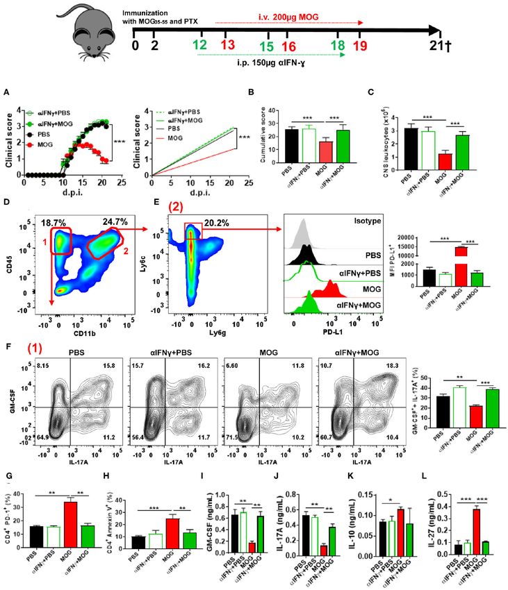
Casella et al.                                                                                       IFN-γ/IL-27 Restore Immune Tolerance in EAE

INTRODUCTION                                                             develop atypically severe EAE, with enhanced T cell proliferation
                                                                         and increased production of inflammatory cytokines (14, 15).
Autoimmune diseases develop due to a break in immune                     Genetic deficiency in PD-L2 did not lead to more severe EAE
tolerance toward certain auto-antigens (auto-Ags). It follows            (14), suggesting that PD-L1 is a dominant inhibitory PD-1 ligand
that the healthy state could be achieved by restoring peripheral         in EAE development. In contrast, blockade of PD-L2, but not
immune tolerance toward those auto-Ags. Devising therapies               PD-L1, in advanced EAE in C57BL/6 mice led to worsening
based on the restoration of Ag-specific immune tolerance                 of disease, indicating that these two PD-1 ligands, or possibly
induction has been a long-standing goal for treatment of                 cell types that express them, have distinct roles in regulating
autoimmune diseases, including multiple sclerosis (MS) (1).              different stages of EAE (2, 16, 17). In regard to i.v. tolerance
One such approach relies on intravenous (i.v.) injection of              induction in EAE, it has been shown that tolerization induces
free myelin-derived auto-Ags that are targets of autoimmune              PD-L1 expression by APCs and that PD-1 blockade abrogates
response. This approach, and variants thereof, have been proven          tolerance induction (2, 18).
beneficial in experimental autoimmune encephalomyelitis                     IFN-γ is a cytokine released by almost all activated immune
(EAE), an animal model of MS (1). Some clinical trials have              cells, with natural killer (NK) and T cells being its major
confirmed that repeatedly i.v. injecting large doses of myelin           sources (19). Although IFN-γ has been traditionally considered
auto-Ag can be safe and beneficial to a subset of MS patients (1).       a pro-inflammatory cytokine, it is now clear that IFN-γ also
In comparison with non-specific immunomodulatory therapies               has prominent anti-inflammatory roles that balance its possibly
currently in use, the principal advantage of Ag-specific therapy         damaging inflammatory effects (20). Numerous studies have
would be that it suppresses harmful autoimmune response while            firmly established that IFN-γ suppresses EAE; mice lacking
sparing the rest of the immune system. This would eliminate side         IFN-γ signaling develop severe EAE, and mouse strains resistant
effects and adverse events due to systemic immunosuppression             to EAE become susceptible (21–23). Consistent with this, IFN-
caused by non-specific immunomodulation.                                 γ production by myelin-specific CD4+ T cells is not required
    Even though certain key players in i.v. tolerance induction,         for their encephalitogenicity (24), IFN-γ-deficient CD4+ T cells
such as interleukin (IL)-10, IL-27, and programmed death ligand          could be notably more pathogenic than their IFN-γ-sufficient
1 (PD-L1), have been identified (2, 3), its complete mechanisms          counterparts (25), and IFN-γ production by encephalitogenic T
have not been elucidated. This lack of specific knowledge also           cells in the CNS is required for recovery from EAE (26). Further,
includes the cell types involved, sequence of their interactions,        an increase in IFN-γ levels in the CNS of mice with EAE leads
and relative relevance of the periphery vs. the central nervous          to disease suppression (27, 28). Taken together, these findings
system (CNS) in tolerance induction and maintenance over time.           indicate that IFN-γ could be important in EAE suppression
A more thorough understanding of these mechanisms will be                by i.v. tolerance induction as well, a possibility that has not
helpful in developing better Ag-specific therapies for MS and            been explored.
possibly other autoimmune diseases.                                         Here we show that i.v. administration of auto-Ag (free
    Consistent with the suppressive role of IL-10 in EAE                 encephalitogenic peptide) halts EAE progression by inducing
development (4), i.v. tolerance induction in EAE requires                PD-L1 expression in CNS monocyte-derived dendritic cells
IL-10. Tolerization by i.v. injection of an auto-Ag elicits IL-10        (moDCs) via an IFN-γ/IL-27-dependent mechanism. Blockade
production, and blockade of IL-10 signaling precludes tolerance          of PD-L1, but not PD-L2, or the lack of PD-1 in CD4+ T cells
induction (2, 5, 6).                                                     precluded i.v. tolerance induction. The lack of IFN-γ in CD4+ T
    The lack of IL-27 signaling leads to the development of              cells, or IFN-γR in conventional DCs type 1 (cDC1), abrogated
more severe EAE (7), and treatment with recombinant IL-27                IL-27 production and PD-L1 expression by moDCs. Collectively,
suppresses EAE (8–10), demonstrating its anti-inflammatory               our data reveal a mechanism of Ag-dependent induction of PD-
role in EAE. The anti-inflammatory effects of IL-27 encompass            L1 expression in moDCs that in turn suppresses Ag-specific Th
inhibition of Th17 cell development; suppression of granulocyte-         cell responses and ameliorates CNS autoimmunity.
macrophage colony-stimulating factor (GM-CSF) expression;
induction of PD-L1, CD39, and IL-10 expression; and
enhancement of Treg development and function (8–10).
                                                                         MATERIALS AND METHODS
We have shown that IL-27 is necessary for induction of i.v.
tolerance in EAE (3); in particular, IL-27 signaling in DCs was          Mice
required for tolerance induction, whereas its signaling in T             C57BL/6, B6.Ly5.1 (CD45.1+ ), RAG1−/− , PD-1−/− , 2D2,
cells was not. IL-27-dependent tolerance induction relied on             Zbtb46-iDTR, IFN-γ−/− , IFN-γRα−/− , GREAT (IFN-γ
cooperation of distinct subsets of spleen DCs with the ability to        reporter), Ccr2−/− , Wsx−/− , and Stat3mut mice were purchased
induce T cell-derived IL-10 and interferon-gamma (IFN-γ) (3).            from The Jackson Laboratory (Bar Harbor, ME, USA). IL-27p28
    Programmed cell death protein 1 (PD-1) and its ligands, PD-          reporter mice were a gift of Dr. Ross M. Kedl (University of
L1 and PD-L2, regulate the balance between T cell activation             Colorado). Mice were kept in specific pathogen-free conditions
and immune tolerance (11, 12). The majority of CD4+ T                    with a maximum of 5 mice per cage, in 12/12 h of light/dark
cells in the CNS of mice with EAE express PD-1, while PD-                cycles and food ad libitum throughout the experimental
L1 and PD-L2 are differentially expressed by populations of              procedures. Every effort was made to minimize suffering of
Ag-presenting cells (APC) (13). PD-1−/− and PD-L1−/− mice                mice. Experimental protocols using mice were approved by

Frontiers in Immunology | www.frontiersin.org                        2                                  October 2020 | Volume 11 | Article 576752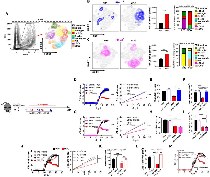
Casella et al.                                                                                            IFN-γ/IL-27 Restore Immune Tolerance in EAE

the Institutional Animal Care and Use Committee of Thomas                    DT Ablation
Jefferson University.                                                        Diphtheria toxin (DTX; Sigma-Aldrich) was administered i.p. at
                                                                             1 µg/20 g of mouse weight in 200 µl of PBS, 1 day before i.v.
                                                                             injection of MOG35−55 . Mice received three injections of DTX
Generation of BMDCs                                                          once every 3 days.
STAT1−/− , STAT3−/− , and WT BMDCs were generated
according to a previously described protocol (3). Briefly, BM                PD-L1, PD-L2, and IFN-γ Blockade
cells were seeded at 2 × 10⁶ cells/mL in Petri dish in complete              Mice with EAE were i.p. injected with 200 µg/mouse of αPD-
IMDM supplemented with 100 ng/mL of recombinant mouse Flt-                   L1 MAb (clone 10F.9G2, BioXCell), or with 200 µg/mouse of
3 (R&D Systems, Minneapolis, MN, USA). Culture medium was                    αPD-L2 MAb (clone TY25, BioXCell), or with 150 µg/mouse of
changed every 3 days. Maturation of the DCs was induced with                 αIFN-γ MAb (clone R4-6A2, BioXCell) 1 day before i.v. injection
LPS (100 ng/mL) for 16 h. At day 9 after starting the culture, DCs           of MOG35−55 . Mice received two MAb injections, 3 days apart in
were enriched by anti-Flt-3-biotin Ab and anti-biotin microbeads             each treatment.
(Miltenyi Biotec, CA, USA), and CD11c+ MHCII+ cells were
then FACS-sorted.                                                            Ag-Specific Recall Response
    IFN-γRα−/− and WT cDCs were generated from BM cells                      Spleens of mice with EAE were dissociated through a 70 µm
following a protocol described in (29) with slight modifications.            strainer to prepare single-cell suspensions in complete IMDM,
                                                                             containing 10% heat-inactivated fetal bovine serum, penicillin
Ag-presentation Assays                                                       (100 U), streptomycin (10 µg/mL), L-glutamine (0.3 mg/mL),
Naive CD4+ T cells from spleens of 2D2 mice were isolated                    and 2-mercaptoethanol (55 µM). After treatment with RBC lysis
using magnetic beads (Naive CD4+ T cell isolation kit, Miltenyi              buffer (Biolegend, CA, USA), cells were extensively washed with
Biotec, CA, USA). 2 × 10⁵ naive CD4+ T cells were added to                   complete IMDM by centrifugation at 1,300 rpm for 5 min at 4◦ C
each well of the cell culture plate containing moDCs, cDC1, or               and the cell density was adjusted to 2 × 10⁶ /mL. One hundred
BMDCs (ratio of 1 DC: 10 T cells) and plates were incubated                  microliter of adjusted cell suspension was added to each well of
at 37◦ C in the presence of MOG35−55 peptide (20 µg/mL) and                  a 96-well plate. MOG35−55 was added to a final concentration of
anti-PD-L1 MAb (1 µg/mL; clone 10F.9G2, BioXCell). Cells                     20 µg/mL. Cells were incubated at 37◦ C for 3 days. As negative
were collected after 72 h and analyzed by flow cytometry, while              control, cells were cultured without MOG35−55 . Cell culture
cytokine concentrations in culture supernatants were measured                supernatants were collected and stored at −20◦ C until use, and
by ELISA.                                                                    cells were analyzed for proliferation and cytokine production by
                                                                             flow cytometry.

EAE and i.v. Tolerance Induction                                             Reconstitution of WT and RAG1–/– Mice
Anesthetized mice were subcutaneously injected with 200 µL                   WT mice with EAE received i.v. 2 × 10⁶ FACS-sorted CD11b+
of an emulsion containing 200 µg of MOG35−55 peptide                         CD11c+ Ly6chigh MHCII+ cells from the CNS of WT mice with
(MEVGWYRSPFSRVVHLYRNGK, Genscript, NJ, USA) in PBS                           EAE previously i.v. injected with MOG35−55 or PBS. CD45.1+
and equal volume of Complete Freund’s adjuvant supplemented                  mice reconstituted with CD45.2+ BM cells from WT, or Zbtb46-
with 10 mg/mL of heat-killed Mycobacterium tuberculosis                      iDTR donors, received i.v. 2 × 10⁶ of in vitro Flt-3-differentiated
H37Ra. Additionally, mice were intraperitoneal (i.p.) injected               cDCs. RAG1−/− mice were i.v. reconstituted with 5 × 10⁶
with 200 ng of pertussis toxin at immunization time and 48 h                 magnetic bead-isolated total CD4+ T cells from spleens of
later. Mice were weighed and scored for clinical signs daily.                WT, PD1−/− , or IFN-γ−/− mice. After 72 h of adoptive transfer,
Clinical assessment of EAE was performed according to the                    mice were immunized for EAE induction.
following scoring criteria: 0, healthy; 1, limp tail; 2, ataxia and/or
paresis of hindlimbs; 3, paralysis of hindlimbs and/or paresis of            Isolation of CNS Infiltrating Leukocytes
forelimbs; 4, tetraparalysis; and 5, moribund or death (30).                 Brain and spinal cord tissues were incubated for 30 min at
    i.v. tolerance was induced in mice after onset of clinical disease       37◦ C with 0.4 mg/mL type IV collagenase (Sigma-Aldrich) and
by injections of 200 µg MOG35−55 in PBS every third day, 3 times             dissociated by passing through a 19-gouge needle. Cells were
in total. Control mice received PBS only (3).                                enriched by centrifugation on a Percoll gradient as previously
                                                                             described (31).

Bone Marrow Chimeras                                                         Flow Cytometry and Cell Sorting
B6.Ly5.1 (CD45.1+ ) congenic mice were lethally irradiated with              Flow cytometry was performed using a FACSaria II (Becton
2 × 2.5 Gy with an 8 h interval between irradiation and were                 Dickinson) and analyzed with FlowJo software (Tree Star).
then i.v. injected with 5 × 10⁶ CD45.2+ BM cells from WT, or                 Fluorochrome-conjugated MAbs specific for: CD45 (clone 30-
Zbtb46-DTR donors. Recipient mice were in other experiments                  F11), CD45.1 (A20), CD11b (M1/70), CD3 (17A2), CD8α (53-
reconstituted with 1:1 mixture (total 1 × 10⁷ cells) of BM                   6.7) CD4 (RM4-5), CD19 (1D3/CD19), CD11c (N418), CD26
cells from Wsx−/− and Ccr2−/− mice, or with mixture of BM                    (H194-112), CD88 (20/70), CD172α (P84), PDCA1 (927), Ly6c
cells from Stat3mut and Ccr2−/− mice. Mice were allowed to                   (AL-21), F4/80 (MB8), Ly6g (1A8), MHC-II (M5/114.15.2), PD-
reconstitute for 6–8 weeks prior to use.                                     1 (29F.1A12), PD-L1 (10F.9G2), PD-L2 (TY25), Caspase 3, and

Frontiers in Immunology | www.frontiersin.org                            3                                   October 2020 | Volume 11 | Article 576752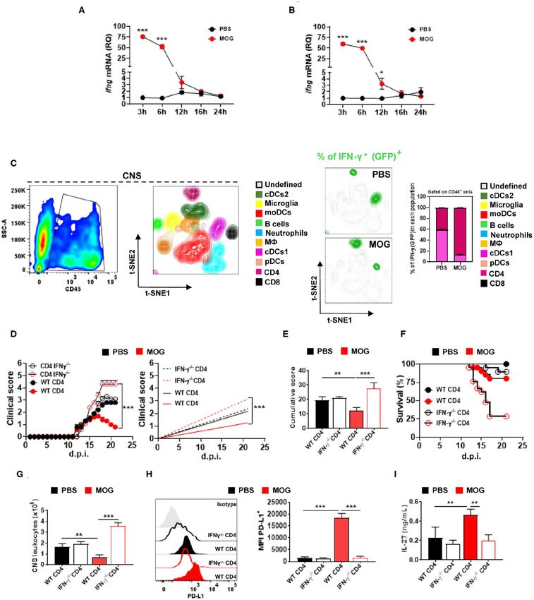
Casella et al.                                                                                         IFN-γ/IL-27 Restore Immune Tolerance in EAE

Annexin V were purchased either from BD Biosciences, R&D,                  MOG35−55 in phosphate-buffered saline (PBS), or with PBS, at
Biolegend, Santa Cruz, or Abcam.                                           the onset of clinical disease; two more doses of MOG35−55 and
   For intracellular staining, cells were stimulated for 4 h               PBS were injected 3 days apart (Supplementary Figures 1A,B).
with phorbol 12-myristate 13-acetate (PMA; 50 ng/ml, Sigma-                Mice were sacrificed at 21 days post immunization (d.p.i.), and
Aldrich) and ionomycin (500 ng/ml, Sigma-Aldrich) in the                   cells isolated from their CNS were analyzed by flow cytometry.
presence of GolgiPlug (1:1,000, BD Pharmigen), permeabilized               t-distributed stochastic neighbor embedding (t-SNE) analysis
using a Cytofix/Cytoperm Plus kit (BD Bioscience) and stained              (33) identified eight populations of CD45+ MHCII+ cells:
with the following fluochrome-conjugated MAbs: GM-CSF                      cDC1, cDC2, microglia, plasmacytoid DCs (pDCs), moDCs,
(MP1-22E9), IL-17A (TC11-18H10.1), and IFN-γ (XMG1.2)                      macrophages (M8), neutrophils, and B cells (Figure 1A).
from Biolegend and BD Pharmingen. Dead cells were excluded                 MOG35−55 -treated mice had increased numbers of PD-L1+
using L/D stain (BD Pharmingen). Data were acquired on a                   and PD-L2+ cells compared with PBS-treated mice. Next, we
FACSAria Fusion (BD Biosciences) and analyzed using FlowJo                 investigated which of these cells expressed PD-L1 and PD-L2.
software (TreeStar).                                                       moDCs (75%) were the bulk of PD-L1+ cells, whereas in control
                                                                           mice, cDC1 (58%) and cDC2 (33%) were the most abundant
ELISA                                                                      PD-L1+ cells (Figure 1B). In mice that received MOG35−55 , PD-
Supernatants from cell cultures were kept at −20◦ C until                  L2 was mostly expressed by neutrophils (50%) and M8 (35%),
use. Cytokine concentrations in culture supernatants were                  whereas in control mice, PD-L2 was expressed by microglia
measured with sandwich enzyme-linked immunosorbent assay                   (64%) and cDC2 (34%) (Figure 1C). These data show that
(ELISA) using commercial kits, following the manufacturer’s                i.v. delivery of auto-Ag induces a robust PD-L1 expression by
recommendation (R&D Systems, Minneapolis, MN, USA).                        moDCs and PD-L2 expression by neutrophils and M8. We also
                                                                           found increased numbers of PD-L1+ and PD-L2+ cells among
qPCR                                                                       lymph node and splenic cells of MOG35−55 -tolerized mice (data
Total RNA was extracted from moDCs, CD4+ T cells, and cDC1                 not shown).
with RNeasy Mini Kit (Qiagen), whereas from total CNS and                      We then tested the role of PD-L1 and PD-L2 in tolerance
spleen with Trizol (Invitrogen). Genomic DNA was removed                   induction by i.p. injecting anti-PD-L1 or anti-PD-L2 MAbs 24 h
by treatment with DNAse I type (Qiagen). cDNA synthesis was                before i.v. injecting MOG35−55 . Treatment with anti-PD-L1 MAb
performed using ThermoscriptTM RT-PCR system (Invitrogen).                 precluded tolerance induction (Figures 1D,E) and led to an
Apoptosis (cat# 4413255), Jak/Stat signaling (cat# 4391524)                increase in the number of leukocytes in the CNS (Figure 1F),
arrays, Il27ra, (Mm00497259_m1 ), and Gapdh (4352339E).                    whereas treatment with anti-PD-L2 MAb did not have an effect
mRNA levels were measured by real-time RT-PCR (Applied                     (Figures 1G–I). Further, we transplanted PD-1−/− or wild-type
Biosystems, Invitrogen). The 2–11CT method was used to                     (WT) CD4+ T cells into RAG1−/− mice and induced EAE in
calculate relative changes in gene expression (32).                        them. MOG35−55 /i.v. treatment suppressed EAE in mice with
                                                                           WT CD4+ T cells, but not in mice with PD-1−/− CD4+ T cells
Statistical Analysis                                                       (Figures 1J–L). These data demonstrate that PD-L1 and PD-1
Statistical analysis was performed by GraphPad Prism 8 software.           are critical for i.v. tolerance induction in ongoing EAE, whereas
Statistical evaluations are expressed as mean ± s.d. or mean ±             PD-L2 does not play a role.
s.e.m., as appropriate. Results were analyzed using Two- or One-
way ANOVA and posttested with Bonferroni, and with unpaired,               Monocyte-Derived Dendritic Cells From
two-tailed Student’s t-test. Statistical significance was ranked ∗ p
                                                                           the Central Nervous System of Tolerized
< 0.05; ∗∗ p < 0.001; ∗∗∗ p < 0.0001; ∗∗∗∗ p < 0.00001.
                                                                           Mice With Experimental Autoimmune
RESULTS                                                                    Encephalomyelitis Are Suppressive via
                                                                           Programmed Death Ligand 1
Intravenous Tolerance Induction in                                         Given that tolerized mice had increased numbers of apoptotic
Experimental Autoimmune                                                    annexin V+ CD4+ T cells (Supplementary Figures 1C,D) and
Encephalomyelitis Is Dependent on                                          PD-L1+ moDCs in the CNS (Figure 1B), we investigated a
                                                                           correlation between their numbers. There was a robust positive
Programmed Death Ligand 1 and                                              correlation of apoptotic CD4+ T cells with PD-L1+ moDCs
Programmed Cell Death Protein 1, but Not                                   (Supplementary Figure 1E), but not with PD-L2+ moDCs
on Programmed Death Ligand 2                                               (Supplementary Figure 1F) or PD-L2+ neutrophils (data not
We and others have reported that i.v. delivery of auto-Ag in mice          shown). This suggests that PD-L1+ moDCs mediate apoptosis of
with EAE induces expression of PD-L1 by APCs (2, 3). Given                 T cells.
the importance of PD-1 and its ligands in immune tolerance,                   We next evaluated the effect of moDCs on myelin-specific
we investigated their role in i.v. tolerance induction in EAE.             CD4+ T cells ex vivo. MHCII+ Ly6chigh moDCs were sorted
Mice were immunized with myelin oligodendrocyte glycoprotein               from the CNS of mice with EAE treated with MOG35−55 /i.v. and
(MOG)35−55 for EAE induction and i.v. injected with 200 µg of              co-cultured with naive CD4+ T cells expressing a transgenic T

Frontiers in Immunology | www.frontiersin.org                          4                                  October 2020 | Volume 11 | Article 576752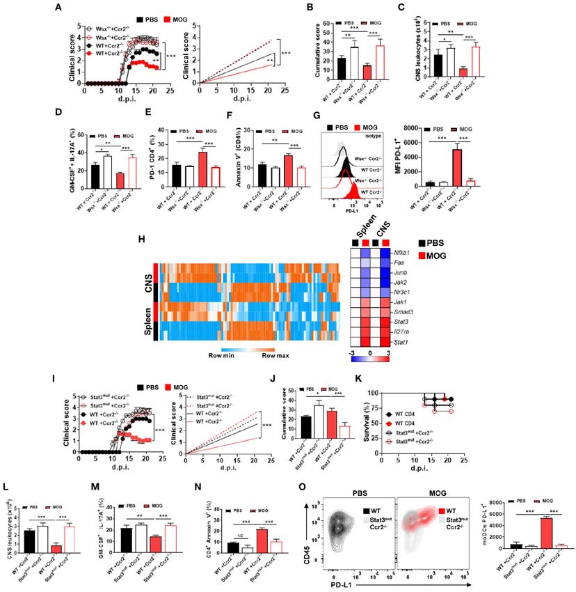
Casella et al.                                                                                                           IFN-γ/IL-27 Restore Immune Tolerance in EAE

 FIGURE 1 | Intravenous administration of myelin oligodendrocyte glycoprotein (MOG)35−55 induces programmed death ligand 1 (PD-L1) expression in central nervous
 system (CNS) monocyte-derived dendritic cells (moDCs) and suppresses experimental autoimmune encephalomyelitis (EAE) in a programmed cell death protein 1
 (PD-1)/PD-L1 manner. C57BL/6 wild-type (WT) mice (n = 10/group each experiment) were immunized with MOG35−55 for EAE induction and starting from disease
 onset i.v. injected with 200 µg of MOG35−55 in PBS every 3 days. Mice were sacrificed 21 days post immunization (d.p.i.) and CD45+ MHCII+ cells from the CNS were
 analyzed by flow cytometry. (A) Eight populations were identified: microglia (CD11b+ CD45low ), monocytes (CD11b+ CD11c+ Ly6chigh Ly6g− ), macrophages (CD11b+
 Ly6clow Ly6g− F4/80high ), cDC1 (CD11b+ CD11c+ Ly6cmed CD26+ ), cDC2 (CD11b+ CD11c+ Ly6cmed CD172α+ ), plasmacytoid DCs (pDCs; Lin− PDCA1high ), and B
 cells (CD19+ ). (B,C) t-SNE graphs showing the expression and percentage of PD-L1+ (B) and PD-L2 (C) cells from the CNS of PBS- or MOG35−55 -treated EAE mice.
 (D) Mice with EAE (n = 5–7/group in each experiment) were i.p. injected with blocking anti-PD-L1 MAb (200 µg/mouse/injection; clone 10F.9G2), or isotype control
 MAb, on 11, 14, and 17 d.p.i. MOG35−55 or PBS was i.v. injected on 12, 15, and 18 d.p.i. (E) Cumulative disease score for mice described in (D). (F) Mice described
 in (D) were sacrificed at day 21 p.i. and the numbers of CD45+ leukocytes obtained from their CNS were determined by flow cytometry. (G) Mice with EAE (n =
 5–7/group in each experiment) were i.p. injected with blocking anti-PD-L2 MAb (200 µg/mouse/injection; clone TY25), or isotype control MAb, as described in (D). (H)
 Cumulative disease score for mice described in (G). (I) Mice described in (G) were sacrificed at day 21 p.i. and the numbers of CD45+ leukocytes obtained from their
 CNS were determined by flow cytometry. (J) RAG1−/− mice were reconstituted with 5 × 10⁶ total CD4+ T cells from WT or PD-1−/− mice (n = 10/group in each
 experiment); 72 h post reconstitution, recipient mice were immunized for EAE induction and MOG35−55 or PBS was injected i.v. three times, starting from EAE onset.
 (K) Cumulative disease score for mice described in (J). (L) Mice described in (J) were sacrificed at day 21 p.i. and the numbers of CD45+ leukocytes obtained from
 their CNS were determined by flow cytometry. (M) Recipient WT mice with EAE were transplanted at the peak of disease with 2 × 10⁶ moDCs from the CNS of donor
 mice with EAE that were previously i.v. tolerized with MOG35−55 or PBS (n = 5/group in each experiment). Groups of recipient mice with EAE received moDCs that
 were pretreated for 1 h with anti-PD-L1 MAb (1 µg/ml). All data are representative of at least two experiments and symbols depict mean ± SEM. Analyses between
 two groups were carried out by Student’s t-test and between four groups by one-way ANOVA with Bonferroni post-test (E,F,H,I,K,L). EAE experiments were analyzed
 by two-way ANOVA with Bonferroni’s multiple comparison. Values of ** P < 0.001, ***P < 0.0001, and ****P < 0.00001 were considered significant.

Frontiers in Immunology | www.frontiersin.org                                     5                                         October 2020 | Volume 11 | Article 576752
Casella et al.                                                                                      IFN-γ/IL-27 Restore Immune Tolerance in EAE

cell receptor for MOG35−55 from 2D2 mice. Anti-PD-L1 MAb               that IFN-γ plays a critical role in the induction of i.v.
was added in some co-cultures. In comparison with moDCs                tolerance in EAE.
from control mice, moDCs from MOG35−55 -tolerized mice                    To identify the cellular sources of MOG35−55 /i.v.-induced
induced lower T cell proliferation and lower GM-CSF and                IFN-γ, we induced EAE in IFN-γ reporter mice (express GFP
IL-17A production, but greater IFN-γ and PD-1 expression and           from the IFN-γ gene), injected them with MOG35−55 /i.v. at
annexin V staining by T cells (Supplementary Figures 1G–I).            disease onset, and 3 h after the injection analyzed their CNS. IFN-
We also measured a larger quantity of regulatory cytokines,            γ was primarily produced by CD4+ T cells, whereas in control
IL-27 and IL-10, in supernatants of these co-cultures                  mice injected with PBS, both cDCs and CD4+ T cells produced
compared with controls (Supplementary Figure 1J). In the               IFN-γ (Figure 3C). We then investigated whether CD4+ T cells
presence of anti-PD-L1 MAb, moDCs did not reduce T cell                are a relevant source of IFN-γ in tolerance induction. To this
proliferation or induce their apoptosis, demonstrating that            end, we reconstituted RAG1−/− mice with WT or IFN-γ−/−
PD-L1 expressed by moDCs limits myelin-specific CD4+ T cell            CD4+ T cells and immunized them for EAE induction; mice
responses in vitro.                                                    received PBS or MOG35−55 /i.v. at disease onset. MOG35−55 /i.v.
   To validate the immunosuppressive phenotype of                      significantly suppressed disease in mice with WT CD4+ T cells,
MOG35−55 /i.v.-induced moDCs in vivo, we transplanted                  whereas it exacerbated the disease in mice with IFN-γ−/− T
moDCs from the CNS of tolerized mice with EAE into mice                cells (70% of mice died) (Figures 3D,E). Consistent with clinical
with ongoing EAE. Control mice received either moDCs from              outcome, mice with IFN-γ−/− T cells had reduced PD-L1+
PBS/i.v. mice, PBS, or moDCs from MOG35−55 /i.v. mice that             expression in CNS moDCs compared with mice with WT T cells
were pretreated for 1 h with anti-PD-L1 MAb. The transfer of           (Figure 3H). Moreover, splenocytes of mice with IFN-γ−/− T
moDCs from MOG35−55 -treated donors, but not from those                cells upon in vitro activation produced significantly less IL-27
that were PBS-treated, led to recovery from the disease, and           compared with control mice (Figure 3I). These data show that
anti-PD-L1 MAb pretreatment of moDCs from MOG35−55 /i.v.               IFN-γ secretion by CD4+ T cells is required for EAE suppression
donors precluded their suppressive effect on EAE in recipient          by MOG35−55 /i.v. treatment.
mice (Figure 1M). These data show that moDCs of MOG35−55 -
treated mice with EAE are suppressive via PD-L1 in vitro               The Lack of Interferon-γ Signaling in
and in vivo.                                                           Conventional Dendritic Cells Type 1
                                                                       Precludes Their Interleukin-27 Expression
                                                                       in Intravenous Tolerance Induction in
Interferon-γ Secreted by CD4+ T Cells Is
                                                                       Experimental Autoimmune
Necessary for Experimental Autoimmune
                                                                       Encephalomyelitis
Encephalomyelitis Suppression Upon                                     Blocking IFN-γ resulted in reduced IL-27 production, prompting
Myelin Oligodendrocyte                                                 us to search for the cellular source of IL-27 upon MOG35−55 /i.v.
Glycoprotein35−55 /Intravenous Treatment                               treatment. We induced EAE in IL-27 reporter mice (express GFP
Given that IFN-γ suppresses EAE (28, 34) and induces PD-               from the IL-27p28 gene) and injected them with MOG35−55 at
L1 expression (35), we tested whether IFN-γ plays a role               disease onset. IL-27 production in the spleen and CNS peaked
in tolerance induction. We injected anti-IFN-γ MAb into                at 16 h after the injection (Figures 4A,B), and among cells from
mice after EAE onset, 24 h before MOG35−55 /i.v. injection             the CNS, the primary IL-27-producing cells were cDC1 (∼80%
(Figure 2A). Anti-IFN-γ-treated mice developed a severe disease        of GFP+ cells), while in PBS-treated mice, moDCs also produced
that did not respond to MOG35−55 /i.v. treatment (Figures 2B,C).       IL-27 (Figure 4C).
These mice had markedly increased numbers of leukocytes                   We next investigated whether the lack of IFN-γ signaling
in the CNS compared with control MOG35−55 /i.v.-treated                in cDC1 affects IL-27 production and compromises tolerance
mice (Figure 2D). Anti-IFN-γ-treated mice also had lower               induction. We generated a Zbtb46-DTR (CD45.2+ )→ CD45.1+
expression of PD-L1+ in moDCs, reduced numbers of apoptotic            bone marrow (BM) chimera mice in which cDC1 can be depleted
CD4+ T cells, and greater frequencies of GM-CSF+ IL-                   with diphtheria toxin (DTX) (36) (Supplementary Figure 2A).
17A+ CD4+ T cells (Figures 2E–I). Consistent with this                 WT (CD45.2+ )→ CD45.1+ BM chimeras served as control.
observation, we found higher concentrations of GM-CSF and              We immunized chimera mice for EAE induction and depleted
IL-17A in culture supernatants from MOG35−55 -stimulated               cDC1 by administering DTX, starting at disease onset and
splenocytes of the above anti-IFN-γ-treated mice compared              then every other day. We then transplanted in vitro Flt3-
with controls (Figures 2J,K); however, while MOG35−55 /i.v.            differentiated WT or IFN-γR−/− cDC1 into these DTX-
treatment resulted in an increase of IL-10, and especially IL-         treated chimera mice with EAE (Figure 4D). Mice were treated
27 production, treatment with anti-IFN-γ MAb precluded these           with DTX and transplanted with cDC1 twice. Moreover,
increases (Figures 2L,M). We next determined the kinetics              24 h post cDC1 transplantation, mice were injected with
of IFN-γ expression upon MOG35−55 injection into mice                  PBS or MOG35−55 , three times, 3 days apart. MOG35−55 /i.v.
with EAE. Ifng mRNA levels, both in the CNS and spleen,                treatment had no effect in mice that received IFN-γR−/−
were highest at 6 h after the injection and declined to base           cDC1, whereas in mice with WT cDC1, it significantly
levels by 12 h post injection (Figures 3A,B). These data show          suppressed disease (Figure 4D). We also found a reduced

Frontiers in Immunology | www.frontiersin.org                      6                                   October 2020 | Volume 11 | Article 576752
Casella et al. IFN-γ/IL-27 Restore Immune Tolerance in EAE FIGURE 2 | IFN-γ is critical for i.v. tolerance induction in EAE. (A) Mice with EAE (n = 5–7/group in each experiment) were i.p. injected with blocking anti-IFN-γ MAb (150 µg/mouse/injection; clone H22), or isotype control MAb, on 12, 15, and 18 d.p.i. MOG35−55 or PBS was i.v. given on 13, 16, and 19 d.p.i. (B) Cumulative disease score for mice described in (A). (C) Mice described in (A) were sacrificed on day 21 p.i. and the numbers of CD45+ leukocytes in their CNS were determined by flow cytometry. (D) Flow cytometry plot showing lymphoid (1) and infiltrating myeloid (2) cells from the CNS of mice shown in (A). (E) MFI of PD-L1+ moDCs from the CNS of mice described in (A). Frequencies of GM-CSF+ IL-17A+ (F), PD-1+ (G), and annexin V+ (H) CD4+ T cells from the CNS of mice described in (A). (J–M) Cytokine concentrations in supernatants from cultures of spleen cells from mice described in (A) stimulated for 72 h with MOG35−55 . Cytokine concentrations were measured by ELISA. All data are representative of at least two experiments, and symbols depict mean ± SEM. Analysis between four groups was carried out by one-way ANOVA with Bonferroni post-test. EAE experiments in (A) were analyzed by two-way ANOVA with Bonferroni’s multiple comparison. Values of *P < 0.05, **P < 0.001, and ***P < 0.0001 were considered significant. Frontiers in Immunology | www.frontiersin.org 7 October 2020 | Volume 11 | Article 576752
Casella et al.                                                                                                           IFN-γ/IL-27 Restore Immune Tolerance in EAE

 FIGURE 3 | CD4+ T cell-derived IFN-γ is critical for i.v. tolerance induction in EAE. Time course of Ifng mRNA, analyzed by qPCR, in the spleen (A) and CNS (B) after
 treating mice with EAE i.v. with MOG35−55 peptide (200 µg) or PBS (n = 5/group in each experiment). (C) GFP/IFN-γ reporter mice were immunized for EAE induction
 and i.v. injected with MOG35−55 or PBS at disease onset (n = 5/group in each experiment). Mice were sacrificed 3 h post injection and CD45+ cells from the CNS
 were analyzed by flow cytometry. t-SNE graphs showing the expression and percentage of IFN-γ+ (GFP+ ) among these cells. Ten populations were identified:
 microglia (CD11b+ CD45low ), monocytes (CD11b+ CD11c+ Ly6chigh MHCIIhigh Ly6g− ), macrophages (CD11b+ Ly6clow Ly6g− MHCII+ F4/80high ), cDC1 (CD11b+
 CD11c+ Ly6cmed MHCIIhigh CD26+ ), cDC2 (CD11b+ CD11c+ Ly6cmed MHCII+ CD172α+ ), pDCs (Lin− PDCA1high ), B cells (CD19+ MHCII+ ), CD8+ T cells (CD11b−
 CD3+ CD8α+ ), and CD4+ T cells (CD11b− CD3+ CD4+ ). (D) RAG1−/− mice were reconstituted with 5 × 10⁶ total CD4+ T cells from WT or IFN-γ−/− mice (n =
 10/group in each experiment); 72 h post reconstitution, recipient mice were immunized for EAE induction and MOG35−55 or PBS was given i.v. three times, starting at
 EAE onset. (E) Cumulative disease score for mice shown in (D). (F) Survival (%) of EAE mice treated described in (D). (G) Mice described in (D) were sacrificed 21
 d.p.i. and the numbers of CD45+ leukocytes in their CNS were determined by flow cytometry. (H) MFI of PD-L1+ moDCs from the CNS of mice shown in (D). (I)
 Splenocytes from mice described in (D) were stimulated with MOG35−55 for 72 h. Concentrations of IL-27 in culture supernatants were measured by ELISA. All data
 are representative of at least two experiments, and symbols depict mean ± SEM. Analyses between two groups were carried out by Student’s t-test, and analyses
 between four groups by one-way ANOVA with Bonferroni post-test. EAE experiments in (D) were analyzed by two-way ANOVA with Bonferroni’s multiple comparison.
 Values of *P < 0.05, **P < 0.001, and ***P < 0.0001 were considered significant.

Frontiers in Immunology | www.frontiersin.org                                     8                                          October 2020 | Volume 11 | Article 576752
Casella et al. IFN-γ/IL-27 Restore Immune Tolerance in EAE
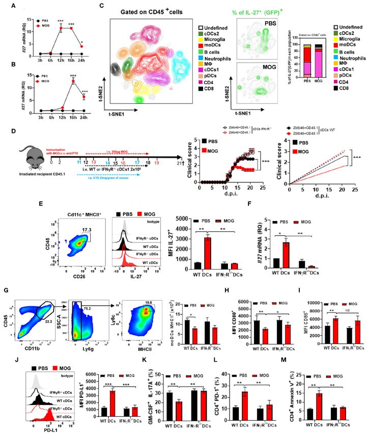
FIGURE 4 | The lack of IFN-γ signaling in cDC1 abrogates their IL-27 production upon i.v. tolerance induction in EAE. Time course of Il27 mRNA expression analyzed
by qPCR, in the spleen (A) and CNS (B) 24 h after treating mice with EAE i.v. with MOG35−55 or PBS (n = 5/group in each experiment). (C) GFP/IL-27p28 reporter
mice were immunized with MOG35−55 for EAE induction and i.v. injected with MOG35−55 or PBS at disease onset (n = 5/group in each experiment). Mice were
sacrificed 16 h post injection and cells from the CNS analyzed by flow cytometry. Ten CD45+ cell populations were identified: microglia (CD11b+ CD45low ), monocytes
(CD11b+ CD11c+ Ly6chigh MHCIIhigh Ly6g− ), macrophages (CD11b+ Ly6clow Ly6g− MHCII+ F4/80high ), cDC1 (CD11b+ CD11c+ Ly6cmed MHCIIhigh CD26+ ), cDC2
(CD11b+ CD11c+ Ly6cmed MHCII+ CD172α+ ), pDCs (Lin− PDCA1high ), B cells (CD19+ MHCII+ ), CD8+ T cells (CD11b− CD3+ CD8α+ ), and CD4+ T cells (CD11b−
(Continued)
Frontiers in Immunology | www.frontiersin.org 9 October 2020 | Volume 11 | Article 576752Casella et al. IFN-γ/IL-27 Restore Immune Tolerance in EAE
FIGURE 4 | CD3+ CD4+ ). t-SNE graphs show the expression and percentage of IL-27p28+ (GFP+ ) cells. (D) CD45.2+ mice were irradiated and transplanted with
Zbtb46-iDTR or CD45.1+ BM and 6–8 weeks later immunized with MOG35−55 (n = 7–10/group in each experiment). cDC depletion (Zbtb46+ MHCII+ CD11c+ ) was
accomplished by i.p. injecting DTX (20 ng/g of mouse weight) every third day after EAE onset. In vitro Flt-3-differentiated BMDCs, IFN-γRα−/− , or WT was i.v.
transferred 1 day post DTX injection, in total twice. MOG35−55 or PBS was i.v. injected at 13, 16, and 19 d.p.i. (E) At 21 d.p.i, cDC1 (CD11b+ CD11c+ CD45+
Ly6cmed MHCIIhigh CD26+ ) were isolated from the CNS of EAE mice described in (D) and their IL-27p28 production was analyzed by flow cytometry and (F) by qPCR.
The numbers of MHCII+ (G), CD80+ (H), CD86+ (I), and MFI of PD-L1+ (J) in moDCs from the CNS of mice described in (D). Frequencies of GM-CSF+ IL-17A+ (K),
PD-1+ (L), and annexin V+ (M) CD4+ T cells from the CNS of mice described in (D). All data are representative of at least two experiments, and symbols depict mean
± SEM. Analysis between two groups was carried out by Student’s t-test, whereas analysis between four groups was carried out by one-way ANOVA with Bonferroni
post-test. EAE experiments in (D) were analyzed by two-way ANOVA with Bonferroni’s multiple comparison. Values of *P < 0.05, **P < 0.001, and ***P < 0.0001
were considered significant.
IL-27 production (both mRNA and protein) by CNS IFN- treated with MOG35−55 /i.v. at disease onset. Mice with Il27ra−/−
γR−/− cDC1 (Figures 4E,F), suggesting that IFN-γ induces BM developed severe disease, compared with mice with WT
IL-27 expression in cDC1. We next investigated whether BM, and MOG35−55 /i.v. treatment failed to suppress disease
reduced IL-27 production by IFN-γR−/− cDC1 affected PD- (Figures 5A,B). Worsening of disease and treatment failure
L1 expression by moDCs and encephalitogenic CD4+ T cells. were associated with a higher number of CNS-infiltrating
CNS moDCs from IFN-γR−/− cDC1-transplanted mice did not leukocytes (Figure 5C) and greater frequencies of Th1 and Th17
differ in MHCII, CD80, and CD86 expression from moDCs cells, whereas the frequency of apoptotic CD4+ T cells was
of control mice, but they had significantly fewer PD-L1+ reduced (Figures 5D–F). Moreover, the lack of IL-27 signaling
moDCs (Figures 4G–J). Failure of tolerance induction in IFN- in CNS moDCs precluded upregulation of PD-L1 on them upon
γR−/− cDC1-transplanted mice was associated with increased MOG35−55 /i.v. treatment (Figure 5G). Taken together, these data
frequencies of IL-17+ and GM-CSF+ CD4+ T cells in the CNS show that MOG35−55 /i.v.-induced IL-27 in turn induces PD-L1
(Figure 4K) and reduced numbers of annexin V+ and PD- expression in CNS moDCs.
1+ CD4+ T cells (Figures 4L,M) compared with WT cDC1- CNS and spleen moDCs upregulated PD-L1 under
transplanted mice. MOG35−55 /i.v. treatment, expressing high levels of genes
We then investigated whether, upon MOG35−55/ i.v. injection, related to Jak–Stat signaling (Figure 5H), especially signal
CNS and splenic cDC1 uptake MOG35−55 , enabling them transducer and activator of transcription-1 (STAT1) and STAT3.
to directly interact with MOG35−55 -specific T cells. Two We therefore investigated whether they were involved in
hours post MOG35−55 or PBS i.v. injection into mice IL-27-mediated PD-L1 induction in moDCs. WT, Stat1−/− ,
with ongoing EAE, cDC1 were FACS-sorted from the CNS and Stat3mut [mutant STAT3 gene with impaired activity (38)]
(Supplementary Figures 2B,C) and co-cultured with 2D2 CD4+ BM-derived DCs (BMDCs) were treated with IL-27 for 24 h, and
T cells. cDC1 from MOG35−55 -injected mice induced greater PD-L1 expression was evaluated. While IL-27 treatment induced
T cell proliferation and IFN-γ production, compared with PD-L1 expression in WT and Stat1−/− moDCs, Stat3mut moDCs
cDC1 from PBS-injected mice (Supplementary Figures 2D–G). failed to upregulate PD-L1. Next, when cultured with naive
Overall, these data show that cDC1 acquire i.v. injected 2D2 T cells, IL-27-treated Stat3mut BMDCs were less suppressive
MOG35−55 , which enables them to activate T cells and induce to T cell proliferation and GM-CSF production, compared with
IFN-γ secretion. IFN-γ signaling in cDC1 is required to induce IL-27-treated WT BMDCs.
their IL-27 production, PD-L1 expression by moDCs, and i.v. To test whether STAT3 is necessary for PD-L1 induction
tolerance in EAE. in moDCs in vivo, we generated mixed BM chimera mice in
which recipient mice (CD45.1) received BM cells from Ccr2−/−
Interleukin-27 Induces Programmed Death mice and from Stat3mut mice. In these mice, virtually all
Ligand 1 Expression in Monocyte-Derived monocytes and monocyte-derived cells (e.g., moDCs) outside of
Dendritic Cells BM are Stat3mut , whereas other immune cells are approximately
Given that IL-27 signaling is critical for i.v. tolerance induction 1:1 mixture of Stat3mut and Stat3WT cells. Control chimera
(3) and that Il27ra−/− (Wsx-1−/− ) mice with EAE had reduced mice received BM cells from Ccr2−/− and WT mice; in
numbers of PD-L1+ moDCs in the CNS upon MOG35−55 /i.v. these mice, monocytes and moDCs outside of the BM are
treatment, we investigated whether the lack of IL-27 signaling in Stat3WT . Chimera mice were immunized for EAE induction
moDCs affects their PD-L1 expression and tolerance induction. and, after onset of clinical disease, treated with MOG35−55 /i.v.
We generated a mixed BM chimeras in which recipient mice Mice with Stat3mut BM developed severe EAE and did not
(CD45.1) received half BM from Ccr2−/− mice and another respond to MOG35−55 /i.v. treatment (Figures 5I,J). Treatment
half from Il27ra−/− (CD45.2) mice. In these chimera mice, failure was associated with an increased number of CNS-
virtually all monocytes outside of the BM are Il27ra−/− Ccr2+/+ , infiltrating leukocytes (Figure 5L), higher frequencies of CNS-
as Ccr2−/− monocytes fail to leave the BM (37). As a control, we infiltrating Th1 and Th17 cells (Figure 5M), reduced frequency
generated mixed chimeras with BMs (1:1) from WT and Ccr2−/− of apoptotic CD4+ T cells (Figure 5N), and reduced PD-L1+
mice. Chimera mice were immunized for EAE induction and expression in moDCs (Figure 5O). Moreover, although Ccr2−/−
Frontiers in Immunology | www.frontiersin.org 10 October 2020 | Volume 11 | Article 576752Casella et al. IFN-γ/IL-27 Restore Immune Tolerance in EAE FIGURE 5 | IL-27 induces PD-L1 expression in moDCs via STAT3. (A) CD45.1+ mice were irradiated and transplanted with 1:1 Wsx−/− and Ccr2−/− BM or WT and Ccr2−/− BM and 6–8 weeks later immunized with MOG35−55 (n = 7–10/group in each experiment). Starting from disease onset, mice were i.v. injected with 200 µg of MOG35−55 every 3 days. (B) Cumulative disease score for mice shown in (A). (C) Mice described in (A) were sacrificed at day 21 p.i. and the numbers of CD45+ leukocytes in their CNS were determined by flow cytometry. Frequencies of GM-CSF+ IL-17A+ (D), PD-1+ (E), and annexin V+ (F) CD4+ T cells from the CNS of mice shown in (A). (G) MFI of PD-L1+ moDCs from the CNS of mice shown in (A). (H) Splenic and CNS Ly6chigh MHCII+ monocytes were FACS-sorted from WT mice with EAE at 21 d.p.i. and Jak/Stat signaling gene array analysis was performed by qPCR. Heat map showing the expression levels of the top 10 genes. Gene expression levels are row centered, log2 transformed, and saturated at −3 and +3 for visualization. (I) CD45.1+ mice were irradiated and transplanted with 1:1 Stat3mut and Ccr2−/− BM, or WT and Ccr2−/− BM, and 6–8 weeks later immunized with MOG35−55 (n = 7–10/group in each experiment). Starting from disease onset, mice were i.v. injected with 200 µg of MOG35−55 every 3 days. (J) Cumulative disease score for mice shown in (I). (K) Survival (%) of mice treated as described in (I) (n = 10/group in each experiment). (L) Mice were sacrificed at day 21 p.i. and the numbers of CD45+ leukocytes from the CNS were determined by flow cytometry. Frequencies of GM-CSF+ IL-17A+ (M) and annexin V+ (N) CD4+ T cells in the CNS of mice shown in (I). (O) Numbers of PD-L1+ moDCs from the CNS of mice shown in (I). All data are representative of at least two experiments, and symbols depict mean ± SEM. Analysis between four groups was carried out by one-way ANOVA with Bonferroni post-test. EAE experiments (A,I) were analyzed by two-way ANOVA with Bonferroni’s multiple comparison. Values of *P < 0.05, **P < 0.001, and ***P < 0.0001 were considered significant. Frontiers in Immunology | www.frontiersin.org 11 October 2020 | Volume 11 | Article 576752
Casella et al.                                                                                          IFN-γ/IL-27 Restore Immune Tolerance in EAE

Stat3mut chimera mice developed severe EAE, we did not find               50) and that IL-27 signaling is beneficial in EAE (10, 51), we
statistical differences in survival compared with Ccr2−/− WT              investigated whether the absence of IL-27 signaling in moDCs
mice (Figure 5K). These findings show that IL-27 induces PD-L1            affects tolerance induction. We show that the absence of IL-27R
expression in moDCs via the STAT3 pathway.                                in CNS moDCs abrogates the expression of PD-L1 and EAE
                                                                          recovery upon auto-Ag i.v. treatment. In contrast, neutrophils do
                                                                          not upregulate PD-L1 upon injection of auto-Ag; instead, they
DISCUSSION                                                                upregulate PD-L2, which is dispensable for tolerance induction.
                                                                              It is well-known that IL-27 induces IL-10 expression by T cells
Extinguishing harmful immune responses by restoring                       and other types of immune cells (10). We and others have shown
peripheral tolerance toward auto-Ags has been a long-standing             that i.v. tolerance induction in EAE induces IL-10 production
goal in the search for therapies for autoimmune diseases (1, 39).         (2, 5), which was also the case in this study. Further, the essential
Although depletion of autoreactive T cells and induction of Tregs         role of IL-10 in i.v. tolerance induction in EAE has been clearly
and tolerogenic DCs are well-known mechanisms of peripheral               demonstrated (2, 3, 41). We therefore did not test its role again
tolerance (5, 40, 41), our study defines an interplay between             here; however, in future studies, it would be interesting to define
molecular and cellular factors that leads to the development of           a pathway by which IL-10 mediates i.v. tolerance, such as its
tolerogenic DCs and depletion of autoreactive T cells. Auto-Ag            relevant cellular sources and targets, and to determine which
administered i.v. is acquired by cDC1 and presented to auto-Ag-           effects of IL-27, if any, are not reliant on IL-10 induction.
specific T cells, leading to their activation and IFN-γ secretion,            Mice lacking PD-L1 develop exacerbated EAE, with PD-L1 on
which in turn induces IL-27 secretion from cDC1. IL-27 acts               CD11c+ DCs playing an important role in limiting self-reactive
on moDCs to induce PD-L1 expression, which then promotes                  CD4+ T cells (52). However, the lack of PD-L2, also a PD-
apoptosis of PD-1+ autoreactive T cells and disease amelioration.         1 ligand, did not worsen EAE (53), demonstrating that PD-L1
    It has been established that IFN-γ plays a protective role in         has a dominant role in regulating EAE severity. In agreement
EAE through multiple mechanisms (22, 25, 42, 43); it is therefore         with these findings, our data reveal that PD-L1 is required
not surprising that it also mediates disease suppression in i.v.          for tolerance induction, whereas PD-L2 is dispensable. This is
tolerance induction. We have shown that i.v. tolerized mice with          seemingly at odds with studies showing that blockade of PD-
EAE have higher frequencies of IFN-γ+ CD4+ T cells compared               L1 with MAb at chronic stage EAE in C57BL/6 mice does not
with controls (3). We show here that i.v. injection of auto-Ag in         worsen the disease, whereas blockade of PD-L2 does (16, 53).
mice with EAE induces a robust and rapid production of IFN-               A possible reason for this inconsistency is that we induced i.v.
γ by CD4+ T cells and that blockade of IFN-γ inhibits IL-27               tolerance while clinical disease was still developing, whereas the
production and PD-L1 expression in CNS moDCs and abrogates                abovementioned studies started PD-L1 and PD-L2 blockade later,
tolerance induction. This is in agreement with the findings               in the chronic phase of disease. Taken together, these findings
that IFN-γ prevents accumulation of activated CD4+ T cells in             suggest that the relative importance of cell types and factors they
response to Ag stimulation by both inhibiting proliferation and           express in regulating disease does change over the disease course.
inducing apoptosis of CD4+ T cells (25). Rapid in vivo/in situ                Studies have shown that IL-27 induces PD-L1 expression (8)
IFN-γ secretion by peptide-specific effector memory CD4+ T                and that STAT3, which together with STAT1 mediates IL-27R
cells upon i.v. injection of the peptide has been demonstrated            signaling (3, 10), is required for PD-L1 expression (12, 54).
(44), a finding fully applicable to our system. Thus, our results         However, the intracellular pathways downstream of IL-27 in i.v.
demonstrate that IFN-γ derived from CD4+ T cells is critical for          tolerance induction in EAE are still unclear. Consistent with
i.v. tolerance induction in ongoing EAE.                                  our previous finding that STAT1 is not necessary for IL-27-
    Several studies have reported that distinct DC subpopulations         induced DC modulation (3), we show here that IL-27 from
can uptake Ags and induce immune tolerance by the induction               cDC1 induces PD-L1 expression in moDCs via STAT3. Indeed,
of IL-27 production and Tregs (3, 10, 45). We have shown                  BM chimera mice with impaired STAT3 signaling in moDCs
that CD11b+ CD103− DCs are the major source of IL-27 in                   failed to upregulate PD-L1 and to recover from EAE upon
i.v. tolerance induction in EAE (3). The engagement of IFN-               MOG35−55 /i.v. treatment.
γR on DCs induces their expression of IL-27 and several other                 Our findings define the regulatory pathway that suppresses
regulatory molecules, and IFN-γ-modified DCs modulate EAE                 auto-Ag-specific immune response. The prerequisite for
severity in an IL-27-dependent manner (42). DCs treated with              activation of this pathway is the existence of a large pool
IFN-γ in vitro and injected into mice with EAE suppress disease           of auto-Ag-specific effector T cells that secrete IFN-γ upon
(46). Consistent with this, we show here that mice lacking IFN-           activation with auto-Ag presented by APCs. Injection of a large
γ signaling in cDC1 fail to recover from EAE upon auto-Ag                 quantity of auto-Ag induces a burst of IFN-γ secretion from
i.v. treatment. We also show that cDC1 uptake i.v. injected               auto-Ag-specific effector T cells, eliciting IL-27 and PD-L1
myelin Ag activate CD4+ T cells and their IFN-γ expression and            expression by APCs, which then in turn suppress immune
induce tolerance.                                                         response by causing anergy/apoptosis of the T cells. This is
    The role of monocytes in EAE is viewed as solely pro-                 a regulatory feedback mechanism for dampening strong and
inflammatory (47, 48). However, there is evidence that CNS                possibly damaging immune responses. It is likely that this
moDCs can acquire regulatory phenotype and facilitate tissue              mechanism regulates myelin-specific autoimmune responses in
repair (49). Given that IL-27 induces PD-L1 in moDCs (8,                  EAE throughout its course, not only after i.v. tolerance induction.

Frontiers in Immunology | www.frontiersin.org                        12                                    October 2020 | Volume 11 | Article 576752
Casella et al. IFN-γ/IL-27 Restore Immune Tolerance in EAE
The existence of this pathway provides a unifying explanation performed some of the flow cytometry experiments and revised
for more severe disease in IFN-γ-, IL-27-, and PD-L1-deficient the manuscript. RT, HD, AV, LI, and AB performed some in
animals. It is likely that additional molecules participate in this vivo experiments and helped with flow cytometry analysis. DH,
pathway, such as IL-10, which is induced in and essential to WZ, and DX revised the manuscript. JP helped with the in
i.v. tolerance induction in EAE (2, 3, 5, 55); upregulation of vivo experiments and revised the manuscript. G-XZ revised the
TGF-β has also been noted (55), but its significance not explored. manuscript. JA conducted some in vivo experiments and revised
Even though we have defined it in the context of i.v. tolerance the manuscript. AR and BC supervised the studies. All authors
induction in EAE, this regulatory pathway is certainly relevant contributed to the article and approved the submitted version.
in other contexts, being either beneficial or detrimental in them.
In addition to CD4+ T cells, the source of IFN-γ could be, for FUNDING
example, pathogen-specific CD8+ T cells or NK cells as well.
This work was supported by a grant from the National Institutes
DATA AVAILABILITY STATEMENT of Health (5R01AI106026) to AR.
The raw data supporting the conclusions of this article will be ACKNOWLEDGMENTS
made available by the authors, without undue reservation.
The authors would like to thank Katherine Regan for editing the
ETHICS STATEMENT manuscript. They would also like to thank Dr. Ross M. Kedl at
the University of Colorado and Dr. Cristopher Hunter at the
The animal study was reviewed and approved by Thomas University of Pennsylvania for providing IL-27p28 reporter mice.
Jefferson University.
SUPPLEMENTARY MATERIAL
AUTHOR CONTRIBUTIONS
The Supplementary Material for this article can be found
GC and BC designed the concept and experiments and wrote online at: https://www.frontiersin.org/articles/10.3389/fimmu.
the manuscript. GC performed most of the experiments. JR 2020.576752/full#supplementary-material
REFERENCES 9. Fitzgerald DC, Ciric B, Touil T, Harle H, Grammatikopolou J, Das Sarma
J, et al. Suppressive effect of IL-27 on encephalitogenic Th17 cells and the
1. Serra P, Santamaria P. Antigen-specific therapeutic approaches effector phase of experimental autoimmune encephalomyelitis. J Immunol.
for autoimmunity. Nat Biotechnol. (2019) 37:238–51. (2007) 179:3268–75. doi: 10.4049/jimmunol.179.5.3268
doi: 10.1038/s41587-019-0015-4 10. Mascanfroni ID, Yeste A, Vieira SM, Burns EJ, Patel B, Sloma I, et al. IL-27
2. Getts DR, Turley DM, Smith CE, Harp CT, McCarthy D, Feeney EM, acts on DCs to suppress the T cell response and autoimmunity by inducing
et al. Tolerance induced by apoptotic antigen-coupled leukocytes is expression of the immunoregulatory molecule CD39. Nat Immunol. (2013)
induced by PD-L1+ and IL-10-producing splenic macrophages and 14:1054–63. doi: 10.1038/ni.2695
maintained by T regulatory cells. J Immunol. (2011) 187:2405–17. 11. Francisco LM, Sage PT, Sharpe AH. The PD-1 pathway in
doi: 10.4049/jimmunol.1004175 tolerance and autoimmunity. Immunol Rev. (2010) 236:219–42.
3. Thome R, Moore JN, Mari ER, Rasouli J, Hwang D, Yoshimura S, et al. doi: 10.1111/j.1600-065X.2010.00923.x
Induction of peripheral tolerance in ongoing autoimmune inflammation 12. Diskin B, Adam S, Cassini MF, Sanchez G, Liria M, Aykut B, et al.
requires interleukin 27 signaling in dendritic cells. Front Immunol. (2017) PD-L1 engagement on T cells promotes self-tolerance and suppression of
8:1392. doi: 10.3389/fimmu.2017.01392 neighboring macrophages and effector T cells in cancer. Nat Immunol. (2020)
4. Bettelli E, Das MP, Howard ED, Weiner HL, Sobel RA, Kuchroo VK. IL-10 is 21:442–54. doi: 10.1038/s41590-020-0620-x
critical in the regulation of autoimmune encephalomyelitis as demonstrated 13. Schreiner B, Bailey SL, Shin T, Chen L, Miller SD. PD-1 ligands expressed
by studies of IL-10- and IL-4-deficient and transgenic mice. J Immunol. (1998) on myeloid-derived APC in the CNS regulate T-cell responses in EAE. Eur
161:3299–306. doi: 10.1016/S0165-5728(98)91500-4 J Immunol. (2008) 38:2706–17. doi: 10.1002/eji.200838137
5. Getts DR, Martin AJ, McCarthy DP, Terry RL, Hunter ZN, Yap WT, et al. 14. Carter LL, Leach MW, Azoitei ML, Cui J, Pelker JW, Jussif J, et al.
Microparticles bearing encephalitogenic peptides induce T-cell tolerance PD-1/PD-L1, but not PD-1/PD-L2, interactions regulate the severity
and ameliorate experimental autoimmune encephalomyelitis. Nat Biotechnol. of experimental autoimmune encephalomyelitis. J Neuroimmunol. (2007)
(2012) 30:1217–24. doi: 10.1038/nbt.2434 182:124–34. doi: 10.1016/j.jneuroim.2006.10.006
6. Wang L, Li Z, Ciric B, Safavi F, Zhang GX, Rostami A. Selective depletion 15. Keir ME, Butte MJ, Freeman GJ, Sharpe AH. PD-1 and its ligands
of CD11c(+) CD11b(+) dendritic cells partially abrogates tolerogenic effects in tolerance and immunity. Annu Rev Immunol. (2008) 26:677–704.
of intravenous MOG in murine EAE. Eur J Immunol. (2016) 46:2454–66. doi: 10.1146/annurev.immunol.26.021607.090331
doi: 10.1002/eji.201546274 16. Zhu B, Guleria I, Khosroshahi A, Chitnis T, Imitola J, Azuma M, et al.
7. Batten M, Li J, Yi S, Kljavin NM, Danilenko DM, Lucas S, et al. Differential role of programmed death-ligand 1 [corrected] and programmed
Interleukin 27 limits autoimmune encephalomyelitis by suppressing the death-ligand 2 [corrected] in regulating the susceptibility and chronic
development of interleukin 17-producing T cells. Nat Immunol. (2006) 7:929– progression of experimental autoimmune encephalomyelitis. J Immunol.
36. doi: 10.1038/ni1375 (2006) 176:3480–9. doi: 10.4049/jimmunol.176.6.3480
8. Casella G, Finardi A, Descamps H, Colombo F, Maiorino C, Ruffini F, et al. IL- 17. Garnier L, Laffont S, Lelu K, Yogev N, Waisman A, Guery JC.
27, but not IL-35, inhibits neuroinflammation through modulating GM-CSF Estrogen signaling in bystander Foxp3(neg) CD4(+) T cells suppresses
expression. Sci Rep. (2017) 7:16547. doi: 10.1038/s41598-017-16702-w cognate Th17 differentiation in trans and protects from central
Frontiers in Immunology | www.frontiersin.org 13 October 2020 | Volume 11 | Article 576752You can also read