Incense smoke induced oxidative stress disrupts tight junctions and bronchial epithelial barrier integrity and induces airway hyperresponsiveness ...
←
→
Page content transcription
If your browser does not render page correctly, please read the page content below
www.nature.com/scientificreports
OPEN Incense smoke‑induced oxidative
stress disrupts tight junctions
and bronchial epithelial barrier
integrity and induces airway
hyperresponsiveness in mouse
lungs
Norio Yamamoto1, Keiko Kan‑o1,2*, Miyoko Tatsuta1,3, Yumiko Ishii1, Tomohiro Ogawa1,
Seiji Shinozaki1, Satoru Fukuyama1, Yoichi Nakanishi1 & Koichiro Matsumoto1
Recent clinical studies have suggested that inhalation of incense smoke (IS) may result in impaired
lung function and asthma. However, there is little experimental evidence to link IS with airway
hyperresponsiveness (AHR) and bronchial epithelial barrier function. Using mouse and cell culture
models, we evaluated the effects of IS exposure on AHR, expression of multiple epithelial tight
junction (TJ)- and adherens junction-associated mRNAs and proteins in the lungs, and the barrier
function of bronchial epithelial cells assessed by transepithelial electronic resistance (TEER). Exposure
of BALB/c mice to IS increased AHR and inflammatory macrophage recruitment to BALF; reduced
claudin-1, -2, -3, -7, -10b, -12, -15, and -18, occludin, zonula occludens-1 [ZO-1], and E-cadherin
mRNA expression; and caused discontinuity of claudin-2 and ZO-1 protein immunostaining in lung
tissue. IS extract dose-dependently decreased TEER and increased reactive oxygen species production
in bronchial epithelial cell cultures. Treatment with N-acetyl-l-cysteine, but not glucocorticosteroids
or long-acting β2-agonists, prevented the detrimental effects of IS. IS exposure can be problematic
for respiratory health, as evidenced by AHR, increased recruitment of inflammatory macrophages and
disruption of TJ proteins in the lung, and damage to epithelial barrier function. However, antioxidants
may be useful for the treatment of IS-induced airway dysfunction.
Burning of incense in temples and homes is a common religious and cultural practice in many Asian and Mid-
dle Eastern countries. Incense smoke (IS) contains particulate matter of varying sizes; gases such as carbon
monoxide, nitrogen dioxide and sulfur dioxide; and volatile organic compounds such as benzene, aldehydes
and polycyclic aromatic h ydrocarbons1. Compared with cigarette smoking, incense burning has been reported
to generate larger quantities of particulate matter of ≤ 2.5 μm diameter and particles that remain in the air for
hours2,3. Increasing evidence suggests that ambient air pollution from IS can cause health problems, especially
airway dysfunction. Recent studies have reported that indoor exposure to IS increases the risk of wheezing and
asthma and is associated with impaired lung function in adolescents4–7. Lin et al. showed that incense smoke
exposure during pregnancy is a risk factor for elevated serum immunoglobulin E (IgE) in human umbilical
cord blood8. Despite a number of clinical studies supporting increased risk of asthma, however, there is little
experimental evidence that inhalation of IS causes airway dysfunction.
Bronchial epithelial cells form a barrier against a wide range of inhaled substances and are at the front line of
mucosal innate immunity. The epithelial barrier function is maintained by apical junctional protein complexes,
composed of apical tight junctions (TJs) and underlying adherens junctions (AJs), that form between neigh-
bouring cells9. TJ-associated proteins, which include members of the occludin, claudin, and junctional adhesion
1
Research Institute for Diseases of the Chest, Graduate School of Medical Sciences, Kyushu University, 3‑1‑1
Maidashi, Higashi‑ku, Fukuoka 812‑8582, Japan. 2Department of Endoscopic Diagnostics and Therapeutics,
Kyushu University Hospital, Fukuoka, Japan. 3Department of Respiratory Medicine, National Hospital Organization
Omuta National Hospital, Fukuoka, Japan. *email: kan-o.keiko.641@m.kyushu-u.ac.jp
Scientific Reports | (2021) 11:7222 | https://doi.org/10.1038/s41598-021-86745-7 1
Vol.:(0123456789)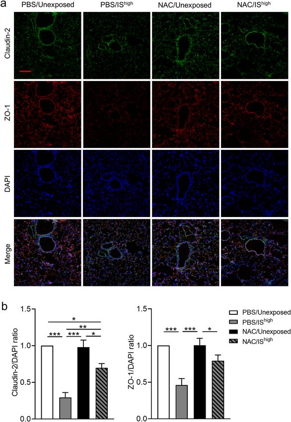
www.nature.com/scientificreports/
molecule families, and AJ-associated proteins such as E-cadherin are the major constituents of junctional com-
plexes in the bronchial e pithelium10. Members of the zonula occludens (ZO) protein family act as scaffolds that
link the intracellular domains of TJ and AJ-associated proteins with the cytoskeleton. There is increasing evidence
that reduced junctional protein expression and airway barrier function are observed in patients with asthma that
may contribute to the development of asthma by enabling greater penetration of inhaled allergen and noxious
particles11,12. We recently reported that exposure of human bronchial epithelial cells to cigarette smoke in vitro
disrupted epithelial barrier function and simultaneously downregulated the expression of multiple TJ and AJ-
associated proteins13. However, the effects of inhalation of harmful substances on multiple junctional protein
expression in animal models is unknown.
In this study, we evaluated the effect of a single exposure of mice to IS on airway hyperresponsiveness (AHR),
inflammation and multiple TJ and AJ-associated protein expression in the lung, and we additionally analysed the
effect of exposure to IS extract (ISE) on epithelial barrier function in human bronchial epithelial cells in air–liquid
interface (ALI) cultures. The benefits of inhaled glucocorticosteroids (GCSs) and long-acting β2-agonists (LABAs)
in asthma are widely r ecognized14. Therefore, we also investigated whether treatment with GCSs, LABAs, or the
antioxidant N-acetyl-l-cysteine (NAC) could protect against IS-induced airway dysfunction in vitro and in vivo.
Methods
Mice. Six-week-old female BALB/c mice were purchased from Japan SLC (Shizuoka, Japan) and housed
under specific pathogen-free conditions. The study protocol was approved by The Kyushu University Animal
Care and Use Committee (A19-200-0). All experiments were performed in accordance with our institutional
guidelines and carried out in compliance with the ARRIVE guidelines.
Mouse experiments. The most commonly used incense sticks in Japan were used for the study (0.4 g/stick,
Nippon Kodo brand). Mice were randomly assigned to two or three groups depending on the experimental
protocol: (i) unexposed, (ii) exposed to a high dose of IS ( IShigh), or (iii) exposed to a low dose of IS ( ISlow). The
mouse groups were housed separately to avoid cross-exposure to IS. Mice were placed in 44-L chambers and
exposed to IS generated by burning of 3.2 g (IShigh) or 1.6 g (ISlow) of incense sticks for 60 min. Incense sticks
were burnt in a separate 8-L chamber connected by a tube to the 44-L exposure chamber, and IS was drawn into
the exposure chamber with fresh air at 4 L/min. Mice from the unexposed group were maintained for the same
period in fresh air. The ratio of the exposure chamber volume to the mean body weight of mice was designed to
recapitulate the ratio of a standard living room volume to the standard body weight of humans.
In some experiments, mice received intraperitoneal injections of NAC (320 mg/kg, Sigma-Aldrich, St. Louis,
MO) at 6 h before and 6 h after the 1-h exposure to IS. NAC was dissolved in phosphate buffer saline (PBS) and
neutralised to pH 7.4 with NaOH.
ISE preparation. ISE for in vitro experiments was prepared by bubbling IS from 1.6 g of incense sticks
(Nippon Kodo brand) through 20 mL of culture medium. After adjustment of the pH to 7.4, the ISE was sterile-
filtered (0.22-μm pore size, 33-mm diameter Millex GV; Merck Millipore, Billerica, MA). This solution was
considered to be 100% ISE and was diluted in medium containing 10% foetal bovine serum (FBS) before use. ISE
preparations were standardized based on absorbance at 320 nm and were freshly prepared for each experiment.
Measurement of AHR. Airway responsiveness was measured according to our previously described
rotocol15. Briefly, mice were anesthetized with a mixture of ketamine and sodium pentobarbital intraperito-
p
neally, and then their tracheas were cannulated via tracheostomy. Mice were ventilated mechanically (tidal vol-
ume, 0.3 mL; frequency, 120 breaths/min) after a paralytic agent was administered. The airway opening pressure
was measured with a differential pressure transducer and recorded continuously. Stepwise increase in acetylcho-
line dose (1.25–20 mg/mL) were given with an ultrasonic nebulizer (NE-U07; OMRON Co., Kyoto, Japan) for
one minute. The data were expressed as the provocative concentration 200 (PC200), the concentration at which
airway pressure was 200% of its baseline value. PC200 was calculated by log-linear interpolation for individual
animals as described previously15. Values of PC200 were expressed as log (100 × PC200).
Collection of bronchoalveolar lavage fluid (BALF), flow cytometric analysis. After measurement
of AHR, mice were euthanised by administration of pentobarbital. For collection of BALF, the lungs were gently
lavaged twice with 1 mL of 0.9% saline via a tracheal cannula. BALF was centrifuged at 250 × g for 10 min and
reviously16.
then total and differential cell counts in BALF were performed as described p
For flow cytometry analysis, lungs not subjected to bronchoalveolar lavage were minced and single-cell
suspensions were prepared. Macrophages and inflammatory macrophages were identified and enumerated by
flow cytometry (Becton Dickinson, Franklin Lakes, NJ), as described in detail in the Supplementary Methods.
Fluorometric TUNEL assay. Apoptotic cells in lung tissue sections were detected using a DeadEnd Fluo-
rometric TUNEL System (Promega, Madison, WI) according to the manufacturer’s instructions and observed by
confocal laser microscopy (LSM700; Zeiss, Jena, Germany). Negative and positive control slides were prepared
by omitting the TdT enzyme from the nucleotide mix and by treating tissue sections with DNase I, respectively.
Quantitative reverse‑transcription PCR (qRT‑PCR). Total RNA was extracted from mouse lungs,
reverse-transcribed, and subjected to qPCR as described in the Supplementary Methods. Primer sequences are
provided in Supplementary Table S1.
Scientific Reports | (2021) 11:7222 | https://doi.org/10.1038/s41598-021-86745-7 2
Vol:.(1234567890)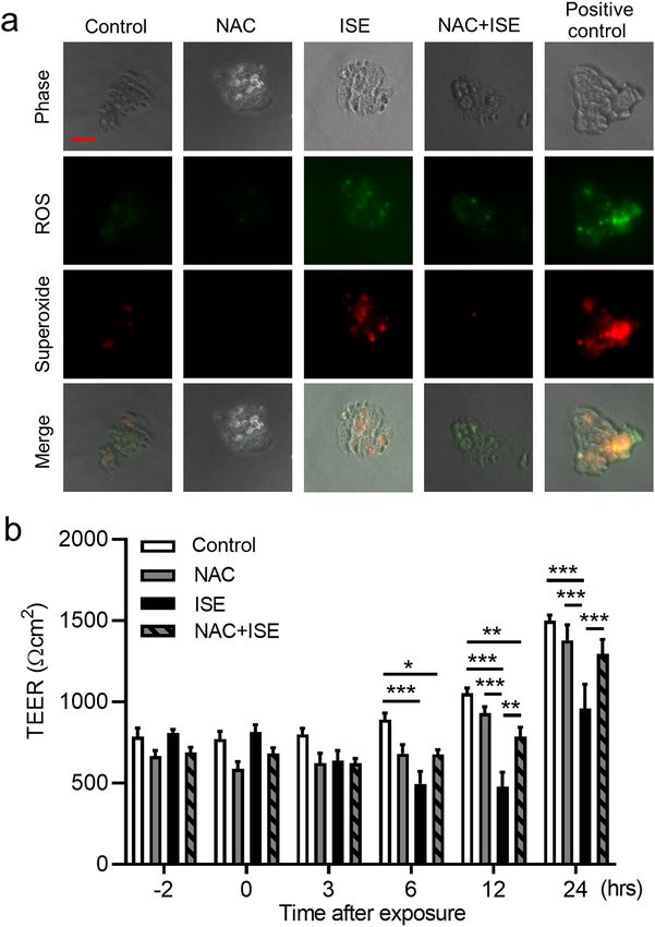
www.nature.com/scientificreports/
Immunofluorescence staining. Immunofluorescence staining were performed according to modified
protocol as described p reviously17. Briefly, freshly isolated lungs were washed in PBS and then lung tissue was
embedded using optimal cutting temperature compound and frozen. Four μm tissue sections on microscope
slides were fixed with cold methanol for 10 min at − 20 °C and then blocked with PBS containing 1% BSA for
30 min at room temperature. The tissue sections were incubated with each primary antibody prepared in PBS
containing 1% BSA at 4 °C overnight, followed by incubation with Alexa Fluor 488-conjugated goat anti-rabbit
IgG antibody (diluted 1:500; Abcam, Cambridge, UK), Alexa Fluor 568-conjugated goat anti-rat IgG antibody
(diluted 1:500; Abcam) and nuclear staining with 4′,6-diamidino-2-phenylindole (DAPI). Images of the stained
tissue sections were obtained with a confocal laser microscope (LSM700; Zeiss). The primary antibodies were
as follows: rabbit anti-claudin-1 polyclonal antibody (diluted 1:200; Thermo Fisher Scientific, Waltham, MA);
rabbit anti-claudin-2 polyclonal antibody (diluted 1:200; Abcam); rabbit anti-claudin-10 polyclonal antibody
(diluted 1:200, Thermo Fisher Scientific); rabbit anti-claudin-15 polyclonal antibody (diluted 1:200; Thermo
Fisher Scientific); rabbit anti-occludin polyclonal antibody (diluted 1:200; Thermo Fisher Scientific); rat anti-
ZO-1 monoclonal antibody (diluted 1:200; Santa Cruz Biotechnology, Inc., Santa Cruz, CA); and rabbit anti-E-
cadherin polyclonal antibody (diluted 1:200; Thermo Fisher Scientific). Immunofluorescence intensity of TJ and
AJ-associated proteins and DAPI was quantified using ImageJ and the relative quantity of TJ and AJ-associated
proteins was plotted against DAPI and normalized by unexposed control.
Cell culture and treatment. Calu-3 cells, a sub-bronchial human epithelial cell line (HTB-55; American
Type Culture Collection, Manassas, VA) were cultured in Dulbecco’s modified Eagle’s medium/F-12 (Thermo
Fisher Scientific) supplemented with 10% FBS and 1% penicillin–streptomycin and maintained at 37 °C in a
humidified atmosphere of 5% CO2 in air. The methods for ALI cultures are described in the Supplementary
Methods. Preliminary experiments demonstrated that transepithelial electronic resistance (TEER) of the cells
cultured under ALI conditions reached a plateau at about day 8 post-seeding and then decreased to about half-
maximal levels at day 2113.
For the present study, cells were cultured for 8 days, and on day 9, 200 μL of control medium, ISE, and/or
pharmacological reagents diluted to appropriate final concentration in culture medium supplemented with 10%
FBS were added to the apical chamber and the cells were cultured for the indicated times. For experiments with
GCSs, LABAs, or NAC, cells were pretreated with 10 nM fluticasone propionate (FP; Tocris, Minneapolis, MN),
10 nM salmeterol (SAL; Tocris), 10 nM budesonide (BUD; Sigma-Aldrich), 10 nM formoterol (FOR; Sigma-
Aldrich), or 1 mM NAC (Sigma-Aldrich) by addition to the apical and basal chambers for 2 h before addition of
ISE. Drugs were present throughout the remainder of the incubation. The concentration of GCSs used reflects
the estimated therapeutic levels achieved in the human lung during i nhalation18.
Measurement of TEER. Bronchial epithelial cell layer integrity was evaluated by TEER measurements
using a Millicell-ERS 2V-Ohmmeter (Millipore Co., Bedford, MA). Medium was added to the apical chamber
1 h prior to TEER measurement. The electrode was soaked in 70% ethanol and rinsed with culture medium prior
to use. TEER was calculated by the following equation19: TEER (Ω cm2) = (Rsample − Rblank) × effective membrane
area (cm2).
Permeability assay. Calu-3 cell permeability was assessed using a fluorescein isothiocyanate (FITC)-dex-
tran (4 kDa, Sigma-Aldrich) assay as described in the Supplementary Methods.
Viability assay. rotocol13. After 24 h of
Cell viability was assessed according to our previously described p
ISE exposure, the medium in the apical chamber was removed and Calu-3 cells were washed with PBS, detached
with trypsin–EDTA, and stained with 0.4% trypan blue solution. Non-viable cells were counted in a LUNA
Automated Cell Counter (Logos Biosystems, Annandale, VA).
Reactive oxygen species (ROS) assay. Calu-3 cells were grown in monolayer culture to approximately
50% confluency, incubated with 1 mM NAC or vehicle for 2 h and then incubated with or without 50% ISE for
1 h. Positive control cells were exposed to 200 μM of the ROS inducer pyocyanin (10 mM stock in anhydrous
dimethylformamide; Enzo Life Sciences, Farmingdale, NY). Total ROS and superoxide production were evalu-
ated using a ROS-ID Total ROS/Superoxide detection kit (Enzo Life Sciences) according to the manufacturer’s
instructions. The cells were then visualised by fluorescence microscopy (BZ-X800; KEYENCE, Osaka, Japan).
Statistical analysis. Unless otherwise stated, data are expressed as the mean ± standard error (SEM). The
Mann–Whitney U-test was used to compare data between two groups and one- or two-way analysis of variance
(ANOVA) followed by Tukey’s multiple comparisons test were used to compare three or more datasets. Statis-
tical analyses were conducted with Prism 8 software (GraphPad Software, San Diego, CA). Differences were
considered statistically significant at p < 0.05.
Results
IS exposure induces airway hyperresponsiveness and inflammation in mouse lungs. To deter-
mine the effects of a single exposure period to IS on airway function, groups of mice were exposed to fresh
air (unexposed) or high or low doses of IS for 1 h, and AHR, recruitment of inflammatory cells to BALF, and
apoptosis of lung cells were assessed over the following 24 h. IS exposure increased AHR, as reflected by P
C200
values, in an acetylcholine dose-dependent manner (Fig. 1a). The number of macrophages, but not neutrophils,
Scientific Reports | (2021) 11:7222 | https://doi.org/10.1038/s41598-021-86745-7 3
Vol.:(0123456789)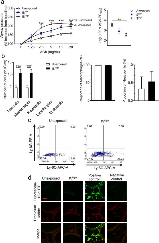
www.nature.com/scientificreports/

                                            Figure 1.  Effects of IS exposure on airway responsiveness, inflammation, and cell apoptosis in mouse lungs. Mice were
                                            unexposed or exposed for 1 h to high or low doses of IS and evaluated 24 h later. (a) Acetylcholine-induced airway
                                            hyperresponsiveness and (b) Total and differential cell counts and percentage of cells in BALF. Data are presented as the
                                            mean ± SEM (n = 7–14 per group) and were pooled from three independent experiments. *p < 0.05, **p < 0.01, ***p < 0.001 by
                                            the Mann–Whitney U-test or one- or two-way ANOVA as appropriate. (c) Flow cytometry analysis of macrophage subsets in
                                            lung-tissue-derived cells and (d) Fluorometric TUNEL assay of apoptotic (green) cells in lung sections. DNA was stained with
                                            propidium iodide (red). Scale bar, 20 μm. Results are representative of two independent experiments (n = 3 in each group per
                                            experiment). ACh acetylcholine, IS incense smoke, PC200 provocative concentration causing 200% response.

          Scientific Reports |   (2021) 11:7222 |                  https://doi.org/10.1038/s41598-021-86745-7                                                          4

Vol:.(1234567890)
www.nature.com/scientificreports/
lymphocytes or eosinophils, in BALF was also significantly increased in IS-exposed compared with unexposed
mice (Fig. 1b). There was no significant difference in the proportion of macrophages and neutrophils in BALF
between the two groups (Fig. 1b). Lymphocytes and eosinophils were observed only in BALF from IS-exposed
mice (mean percentage of cells: 0.13%, 0.08%, respectively). Flow cytometry analysis of lung-derived cells
showed that IS exposure specifically increased the Ly-6Glow/Ly-6Chigh population of inflammatory macrophages,
but not neutrophils (Fig. 1c and Supplementary Fig. S1). Analysis of apoptosis in lung sections using a fluoro-
metric TUNEL assay revealed no increase in cell apoptosis in the lungs of IS-exposed compared with unexposed
mice (Fig. 1d).
IS exposure decreases expression of TJ‑ and AJ‑associated genes in mouse lungs. Next, we
performed qRT-PCR analysis of lung tissues to determine the effects of IS inhalation on transcription of TJ-
associated (claudins, occludin and ZO-1) and AJ-associated (E-cadherin) genes in mouse lungs. IS exposure sig-
nificantly decreased the mRNA levels of claudin-1, claudin-2, claudin-10b and claudin-12 at 6 h post-exposure;
claudin-3, claudin-7, claudin-18, E-cadherin and ZO-1 at 9 h post-exposure; and claudin-15 and occludin at
24 h post-exposure (Fig. 2). Claudin-2, claudin-3 and claudin-7 mRNA levels returned to unexposed control
levels by 24 h (Fig. 2).
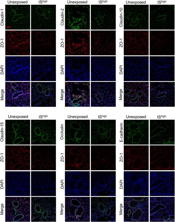
Expression of claudin‑2 and ZO‑1 protein in mouse lungs is disrupted by IS exposure. To deter-
mine whether disruption of TJ- and AJ-associated genes was also observed at the protein level, we performed
immunofluorescence staining of lung sections from unexposed and I Shigh-exposed mice. Confocal microscopy
revealed predominant staining of claudin-1, claudin-10 and E-cadherin in the bronchial epithelium of unex-
posed mice, while claudin-2, claudin-15, occludin and ZO-1 proteins were expressed in both bronchial epithelial
and alveolar cells (Fig. 3 and Supplementary Fig. S2). Notably, lung sections from I Shigh mice showed discontinu-
ous or reduced staining of claudin-2 and ZO-1 at 24 h after IS exposure, indicating disruption of TJs (Fig. 3).
Immunofluorescence intensity of claudin-2 and ZO-1 was significantly decreased in IShigh-exposed compared
with unexposed mice (Supplementary Fig. S3).
IS impairs bronchial epithelial barrier integrity and increases epithelial permeability. TEER
measurements and permeability assays were performed to assess the effects of IS on epithelial barrier function.
Calu-3 sub-bronchial epithelial cells were differentiated in ALI cultures for 9 days, and then incubated with 0%,
25%, or 50% ISE concentrations for an additional 24 h. Treatment with 50% ISE caused a reduction in TEER that
was maintained for 24 h post-exposure (Fig. 4a). In contrast to the significant reduction in TEER and increases
in permeability induced by 50% ISE (Fig. 4b,c), the viability of Calu-3 cells was not influenced at 24 h by expo-
sure to 50% ISE (Fig. 4d). Thus, treatment with 50% ISE was selected for outcome measurements in subsequent
experiments.
ISE‑induced impairment of bronchial epithelial barrier integrity is not affected by GCS or
LABA treatment. We previously demonstrated that GCSs protect against epithelial barrier dysfunction
induced by cigarette smoke extract13. Therefore, we investigated the effects on ISE-induced TEER reduction in
Calu-3 cells after pretreatment with vehicle, 10 nM GCSs (FP or BUD), and/or 10 nM LABAs (SAL or FOR) for
2 h before exposure to 50% ISE (plus GCS/LABA) for an additional 24 h. However, we found no effects of GCSs
and LABAs, alone or in combination, on ISE-induced reduction in TEER (Fig. 5a–c).
The antioxidant NAC protects against ISE‑induced impairment of bronchial epithelial barrier
integrity. Immunofluorescence staining of Calu-3 cells confirmed that generation of total ROS, including
superoxide, could be detected within 1 h of exposure to 50% ISE, and pretreatment of cells with 1 mM NAC for
2 h before ISE exposure diminished ROS generation (Fig. 6a). Similarly, NAC pretreatment strongly suppressed
the decrease in TEER observed at 12 and 24 h after Calu-3 cells exposure to 50% ISE (Fig. 6b), suggesting that
ROS production was directly involved in ISE-induced disruption of epithelial barrier function. Treatment of
ISE-exposed or unexposed cells with NAC alone did not affect cell viability (Supplementary Fig. S4).
NAC treatment prevents disruption of TJ‑associated proteins in mouse lungs exposed to
IS. To determine whether NAC can also protect lungs against IS exposure in vivo, we examined TJ-associated
protein expression in lung tissues. Immunofluorescence staining of claudin-2 and ZO-1 was performed on lung
sections collected at 24 h after exposure. As noted earlier, IShigh exposure caused discontinuous or attenuated
staining of claudin-2 and ZO-1 in both bronchial epithelial and alveolar cells (Fig. 3 and Supplementary Fig. S2).
Although 24 h of NAC treatment alone did not affect the pattern or intensity of protein expression in unexposed
mice, NAC treatment effectively prevented the IShigh-induced disruption of claudin-2 and ZO-1 immunostaining
patterns (Fig. 7a). IS exposure-induced reduction in immunofluorescence intensity of claudin-2 and ZO-1 was
significantly attenuated by NAC treatment (Fig. 7b).
NAC treatment prevents IS‑induced AHR and inflammation in mouse lungs. Finally, we exam-
ined the effects of NAC on IS-induced AHR and accumulation of inflammatory macrophages in BALF of mice
at 24 h after IS exposure. NAC treatment of IShigh mice reversed the effect of IS on acetylcholine-induced AHR to
the same levels detected in unexposed mice (Fig. 8a). Similarly, NAC treatment completely abrogated both the
increase in macrophage abundance and the proportion of inflammatory macrophages in BALF after exposure
to IShigh (Fig. 8b, c).
Scientific Reports | (2021) 11:7222 | https://doi.org/10.1038/s41598-021-86745-7 5
Vol.:(0123456789)www.nature.com/scientificreports/
Figure 2. Effects of IS exposure on TJ- and AJ-associated gene expression in mouse lungs. qRT-PCR analyses of
the indicated mRNAs in lung tissues was performed at up to 24 h after a 1-h exposure to fresh air (unexposed)
or IShigh. mRNA levels were normalised to GAPDH mRNA levels. Data represent the mean ± SEM (n = 6–9
per group) and were pooled from two independent experiments. *p < 0.05, **p < 0.01, by two-way ANOVA. IS
incense smoke.
Discussion
In the present study, we showed that a single exposure of mice to IS aggravated AHR, provoked an influx of
inflammatory macrophages into the lungs, and disrupted the expression and location of TJ-associated proteins
in bronchial epithelium. In vitro experiments confirmed that exposure of bronchial epithelial cells to ISE induced
ROS production and dose-dependently reduced TEER through a mechanism resistant to clinically relevant
concentrations of GCSs and/or LABAs. Finally, NAC treatment ameliorated IS-induced effects on AHR, mac-
rophage recruitment and claudin-2 and ZO-1 expression in the murine airway and reversed the effect of ISE
on TEER in vitro.
Scientific Reports | (2021) 11:7222 | https://doi.org/10.1038/s41598-021-86745-7 6
Vol:.(1234567890)www.nature.com/scientificreports/
Figure 3. IS exposure-induced changes in TJ and AJ-associated protein expression in mouse lungs. Confocal
immunofluorescence microscopy of ZO-1 (red) and additional AJ and TJ-associated proteins (green) in lung
sections was performed at 24 h after a 1-h exposure to fresh air (unexposed) or I Shigh. DAPI staining of nuclei
is shown in blue. Scale bar, 100 μm. Results are representative of two independent experiments (n = 3 in each
group per experiment). DAPI 4′,6-diamidino-2-phenylindole, IS incense smoke.
Scientific Reports | (2021) 11:7222 | https://doi.org/10.1038/s41598-021-86745-7 7
Vol.:(0123456789)www.nature.com/scientificreports/
Figure 4. Effects of ISE on airway epithelial barrier function. (a) Time course of TEER in Calu-3 cells cultured
under ALI conditions for the indicated times after exposure to vehicle (0%) or 50%ISE at the indicated
concentrations. (b) TEER measured at 24 h after exposure of Calu-3 cells to 0–50% ISE for 24 h. (c) FITC-
dextran permeability assay of Calu-3 cells after exposure to 0–50% ISE for 24 h. (d) Trypan blue viability assay of
Calu-3 cells after exposure to 0–50% ISE for 24 h. Data represent the mean ± SEM (n = 6–10 per group) and were
pooled from three independent experiments. *p < 0.05, ** p < 0.001 by one- or two-way ANOVA as appropriate.
ISE incense smoke extract, TEER transepithelial electrical resistance.
Our results suggest that even a brief exposure to IS can disrupt epithelial barrier function and potentially
exacerbate asthma in a manner resistant to treatment with inhaled GCSs. Other studies have demonstrated that
long-term exposure to IS induces histological changes in the lungs in various animal models. Exposure of male
rats to IS for 14 weeks at a rate of 4 g/day induced ultrastructural changes in alveolar pneumocytes, neutrophil
infiltration in pulmonary alveoli, degenerative and necrotic changes, and deposition of collagen fibrils in alveolar
wall20. However, Gaschler et al. showed that mice exposed to cigarette smoke (5 days a week for 8 weeks) have an
increase in the number of cells in BALF, with macrophages representing greater than 95% of cells, similar to our
observations21. Rabah et al. reported that repetitive exposure to IS caused histological changes in murine lungs
in a dose- and duration-dependent m anner22. These data suggest that induction of neutrophilic inflammation
in lungs depends on the duration of exposure, and this may be why no increase in inflammatory cells in BALF
except for macrophages was observed in our study.
The results in the present study indicate that oxidative stress may be responsible for IS-induced respiratory
complications by inducing AHR, disassembly of TJ proteins, and epithelial barrier integrity. Other investigators
also showed that exposure to IS induced oxidative stress and inflammatory response in vivo and in vitro23–27.
Previous reports suggest that increased ROS may be associated with aggravated AHR and impaired epithelial bar-
rier function. Using animal models, ROS has been shown to contribute directly to AHR via damage to oxidant-
sensitive β-adrenergic receptors and increases in vagal tone, and this effect was enhanced when the epithelium
was injured28–30. Studies of cigarette smoke exposure in bronchial epithelium suggest that ROS production after
exposure may induce disassembly of TJ proteins and impair epithelial barrier function through epidermal growth
factor receptor (EGFR)-extracellular signal-regulated kinase 1/2 signaling p athway31,32. Furthermore, Heijink
et al. reported that the protective effect of GCSs against cigarette smoke induced-epithelial barrier dysfunction
was mediated by affecting EGFR-downstream target glycogen synthase kinase-3β33. We also showed that cigarette
smoke-induced reduction in TEER (approximately 35% compared with untreated control) was significantly
attenuated in Calu-3 cells treated with 10 nM BUD (15.6% TEER reduction compared with untreated control) or
10 nM FP (9.7% TEER reduction compared with untreated control)13. However, in the present study, IS-induced
reduction in TEER (approximately 35% compared with untreated control) was affected by neither BUD nor FP.
Scientific Reports | (2021) 11:7222 | https://doi.org/10.1038/s41598-021-86745-7 8
Vol:.(1234567890)www.nature.com/scientificreports/
Figure 5. Effects of GCS and/or LABA treatment on ISE-induced reduction in TEER in Calu-3 cells. Cells were
cultured under ALI conditions and pretreated with vehicle, 10 nM GCS and/or 10 nM LABA for 2 h prior to
addition of vehicle or 50% ISE for 24 h. (a) Cells incubated with or without 10 nM of the GSCs FP and BUD.
(b) Cells incubated with or without 10 nM of the LABAs SAL and FOR. (c) Cells incubated with the indicated
combinations of 10 nM GCSs and 10 nM LABAs. Data represent the mean ± SEM (n = 5–10 per group) and
were pooled from three independent experiments. *p < 0.05, **p < 0.01, ***p < 0.001 by one-way ANOVA. BUD
budesonide, FP fluticasone propionate, FOR formoterol, ISE incense smoke extract, SAL salmeterol, TEER
transepithelial electrical resistance.
Together with those observations, our present results suggest that IS-induced ROS may impair epithelial barrier
integrity through a different signal transduction pathway activated by cigarette smoke exposure and IS-induced
barrier dysfunction might be resistant to treatment with inhaled GCSs.
In this study, we showed that IS exposure disrupted claudin-2 and ZO-1 and simultaneously downregulated
the gene expression of multiple TJ-associated proteins in the lung. Recent findings suggest a broad defect in
adhesion mechanisms in asthma. Bronchial epithelium from patients with asthma has been shown to exhibit
an irregular ZO-1 and occludin staining pattern and reduced barrier function that is further compromised by
exposure to the Th2 cytokines IL-4 and IL-1334,35. Recent studies have identified an association between claudins
and the development of asthma, which are expressed in a tissue- and cell type-selective manner and are essential
for TJ f ormation9. Claudin-18 was found to be expressed at lower levels in the bronchial epithelium of asthma
patients in a manner that correlated inversely with Th2 inflammation m arkers12. Plasma levels of claudin-4 and
claudin-5 were reported to be elevated in asthma patients, particularly during exacerbations, and the levels were
found to correlate negatively with lung function and positively with total IgE level and the percentage of blood
eosinophils36,37. Loss of claudin-18 also induced epithelial barrier dysfunction in murine lungs, increased sensi-
tisation to aspergillus and increased acetylcholine-induced AHR12. Although claudin-2 is expressed in healthy
human bronchiolar and alveolar cells, its precise function in lung physiology is unknown38. A limitation of our
study is that the consequences of claudin-2 and ZO-1 disruption by IS exposure remain unclear. Further studies
are needed to clarify the impact of these changes on the maintenance of airway responsiveness and epithelial
barrier function. Another limitation is that only female mice were utilized in this study since we previously
reported AHR in an ovalbumin (OVA) model of asthma using female mice39,40. Matsubara et al. showed sex dif-
ferences in induction of AHR in OVA-exposed mice41. It reminds unclear whether exposure to IS contributes to
the development of AHR and disruption of TJ-associated proteins in lungs regardless of sex.
Scientific Reports | (2021) 11:7222 | https://doi.org/10.1038/s41598-021-86745-7 9
Vol.:(0123456789)www.nature.com/scientificreports/
Figure 6. Effects of NAC on ISE-induced ROS generation and TEER reduction in Calu-3 cells. Cells were
pretreated with 1 mM NAC or vehicle for 2 h and then incubated with vehicle or 50% ISE. (a) Detection of
total ROS (green) or superoxide (red) in Calu-3 cells at 1 h after exposure to ISE. Scale bar, 50 μm. Images are
representative of two independent experiments (n = 3 in each group per experiment). (b) Analysis of TEER at
the indicated times before and after exposure to ISE. Data represent the mean ± SEM (n = 8–15 per group) and
were pooled from four independent experiments. *p < 0.05, **p < 0.01, ***p < 0.001 by one- or two-way ANOVA
as appropriate. ISE incense smoke extract, NAC N-acetyl-l-cysteine, ROS reactive oxygen species, TEER
transepithelial electrical resistance.
Scientific Reports | (2021) 11:7222 | https://doi.org/10.1038/s41598-021-86745-7 10
Vol:.(1234567890)www.nature.com/scientificreports/
Figure 7. Effects of NAC on IS-induced changes in TJ-associated protein expression in mouse lungs. (a) Confocal
immunofluorescence microscopy of ZO-1 (red) and claudin-2 (green) proteins in lung sections prepared 24 h after a 1-h exposure to
fresh air (unexposed) or IShigh. Mice were administered NAC (320 mg/kg) or PBS (vehicle) by intraperitoneal injection 6 h prior to and
6 h after the 1-h IS exposure. DAPI staining of nuclei is shown in blue. Scale bar, 100 μm. Images are representative of two independent
experiments (n = 3 in each group per experiment). (b) Immunofluorescence intensity of Claudin-2, ZO-1 and DAPI was quantified
using ImageJ and the relative quantity of Claudin-2 or ZO-1 was plotted against DAPI and normalized by unexposed control. Data
represent the mean ± SEM (n = 8 per group) and were pooled from two independent experiments. *p < 0.05, **p < 0.01, ***p < 0.001 by
one-way ANOVA. DAPI 4′,6-diamidino-2-phenylindole, IS incense smoke, NAC N-acetyl-l-cysteine, PBS phosphate-buffered saline.
Scientific Reports | (2021) 11:7222 | https://doi.org/10.1038/s41598-021-86745-7 11
Vol.:(0123456789)www.nature.com/scientificreports/
Figure 8. Effects of NAC on IS-induced AHR and inflammation in mouse lungs. Mice were administered NAC
(320 mg/kg) or vehicle intraperitoneally 6 h prior to and 6 h after a 1-h exposure to fresh air (unexposed) or
IShigh and evaluated 24 h later. (a) Acetylcholine-induced AHR. (b) Total and differential cell counts in BALF. (c)
Flow cytometry analysis of macrophage subsets in the lungs. Data represent the mean ± SEM (n = 5–7 per group)
and were pooled from two independent experiments. *p < 0.05, **p < 0.01, ***p < 0.001 by one- or two-way
ANOVA as appropriate. ACh acetylcholine, AHR airway hyperresponsiveness, IS incense smoke, NAC N-acetyl-
l-cysteine, PBS phosphate-buffered saline, PC200 provocative concentration causing 200% response.
In conclusion, our results suggest that inhalation of IS might be harmful to respiratory health, as evidenced by
AHR, increased recruitment of inflammatory macrophages and disruption of TJ-associated proteins in the lung,
and damage to epithelial barrier function. However, treatment with NAC reversed many of the detrimental effects
of IS exposure, suggesting that antioxidants may be beneficial for the treatment of IS-related airway dysfunction.
Data availability
The data that support the findings of this study are available from the corresponding author, KK, upon reason-
able request.
Received: 18 December 2020; Accepted: 19 March 2021
References
1. Lin, T. C., Krishnaswamy, G. & Chi, D. S. Incense smoke: Clinical, structural and molecular effects on airway disease. Clin. Mol.
Allergy 6, 3. https://doi.org/10.1186/1476-7961-6-3 (2008).
2. Mannix, R. C., Nguyen, K. P., Tan, E. W., Ho, E. E. & Phalen, R. F. Physical characterization of incense aerosols. Sci. Total Environ.
193, 149–158. https://doi.org/10.1016/s0048-9697(96)05343-0 (1996).
3. Zhou, R. et al. Higher cytotoxicity and genotoxicity of burning incense than cigarette. Environ. Chem. Lett. 13, 465–471. https://
doi.org/10.1007/s10311-015-0521-7 (2015).
4. Al-Rawas, O. A., Al-Maniri, A. A. & Al-Riyami, B. M. Home exposure to Arabian incense (bakhour) and asthma symptoms in
children: A community survey in two regions in Oman. BMC Pulm. Med. 9, 23. https://doi.org/10.1186/1471-2466-9-23 (2009).
5. Wang, I. J., Tsai, C. H., Chen, C. H., Tung, K. Y. & Lee, Y. L. Glutathione S-transferase, incense burning and asthma in children.
Eur. Respir. J. 37, 1371–1377. https://doi.org/10.1183/09031936.00137210 (2011).
6. Norback, D. et al. Sources of indoor particulate matter (PM) and outdoor air pollution in China in relation to asthma, wheeze,
rhinitis and eczema among pre-school children: Synergistic effects between antibiotics use and PM10 and second hand smoke.
Environ. Int. 125, 252–260. https://doi.org/10.1016/j.envint.2019.01.036 (2019).
7. Chen, Y. C., Ho, W. C. & Yu, Y. H. Adolescent lung function associated with incense burning and other environmental exposures
at home. Indoor Air 27, 746–752. https://doi.org/10.1111/ina.12355 (2017).
Scientific Reports | (2021) 11:7222 | https://doi.org/10.1038/s41598-021-86745-7 12
Vol:.(1234567890)www.nature.com/scientificreports/
8. Lin, Y. C., Wen, H. J., Lee, Y. L. & Guo, Y. L. Are maternal psychosocial factors associated with cord immunoglobulin E in addition
to family atopic history and mother immunoglobulin E?. Clin. Exp. Allergy 34, 548–554. https://d oi.o
rg/1 0.1 111/j.1 365-2 222.2 004.
1928.x (2004).
9. Tsukita, S., Tanaka, H. & Tamura, A. The Claudins: From tight junctions to biological systems. Trends Biochem. Sci. 44, 141–152.
https://doi.org/10.1016/j.tibs.2018.09.008 (2019).
10. Rezaee, F. & Georas, S. N. Breaking barriers. New insights into airway epithelial barrier function in health and disease. Am. J.
Respir. Cell Mol. Biol. 50, 857–869. https://doi.org/10.1165/rcmb.2013-0541RT (2014).
11. Georas, S. N. & Rezaee, F. Epithelial barrier function: at the front line of asthma immunology and allergic airway inflammation.
J. Allergy Clin. Immunol. 134, 509–520. https://doi.org/10.1016/j.jaci.2014.05.049 (2014).
12. Sweerus, K. et al. Claudin-18 deficiency is associated with airway epithelial barrier dysfunction and asthma. J. Allergy Clin. Immu-
nol. 139, 72–81. https://doi.org/10.1016/j.jaci.2016.02.035 (2017).
13. Tatsuta, M. et al. Effects of cigarette smoke on barrier function and tight junction proteins in the bronchial epithelium: protective
role of cathelicidin LL-37. Respir. Res. 20, 251. https://doi.org/10.1186/s12931-019-1226-4 (2019).
14. Pauwels, R. A. et al. Effect of inhaled formoterol and budesonide on exacerbations of asthma. Formoterol and Corticosteroids
Establishing Therapy (FACET) International Study Group. N. Engl. J. Med. 337, 1405–1411. https://doi.org/10.1056/NEJM199711
133372001 (1997).
15. Kibe, A. et al. Differential regulation by glucocorticoid of interleukin-13-induced eosinophilia, hyperresponsiveness, and goblet
cell hyperplasia in mouse airways. Am. J. Respir. Crit. Care Med. 167, 50–56. https://doi.org/10.1164/rccm.2110084 (2003).
16. Fukuyama, S. et al. A zinc chelator TPEN attenuates airway hyperresponsiveness and airway inflammation in mice in vivo. Allergol.
Int. 60, 259–266. https://doi.org/10.2332/allergolint.09-OA-0167 (2011).
17. Barth, K. et al. P2X7R-dependent regulation of glycogen synthase kinase 3beta and claudin-18 in alveolar epithelial type I cells of
mice lung. Histochem. Cell Biol. 146, 757–768. https://doi.org/10.1007/s00418-016-1499-3 (2016).
18. Kelly, M. M. et al. Corticosteroid-induced gene expression in allergen-challenged asthmatic subjects taking inhaled budesonide.
Br. J. Pharmacol. 165, 1737–1747. https://doi.org/10.1111/j.1476-5381.2011.01620.x (2012).
19. Sekiyama, A. et al. Glucocorticoids enhance airway epithelial barrier integrity. Int. Immunopharmacol. 12, 350–357. https://doi.
org/10.1016/j.intimp.2011.12.006 (2012).
20. Alarifi, S. A., Mubarak, M. M. & Alokail, M. S. Ultrastructural changes of pneumocytes of rat exposed to Arabian incense (Bak-
hour). Saudi Med. J. 25, 1689–1693 (2004).
21. Gaschler, G. J. et al. Cigarette smoke exposure attenuates cytokine production by mouse alveolar macrophages. Am. J. Respir. Cell
Mol. Biol. 38, 218–226. https://doi.org/10.1165/rcmb.2007-0053OC (2008).
22. Rabah, S., Hadad, S. & Albani, F. Histological changes of Mice lungs after daily exposure to different concentration of Incense
smoke. Life Sci. J. 10, 552–560 (2013).
23. Hussain, T. et al. Induction of CYP1A1, CYP1A2, CYP1B1, increased oxidative stress and inflammation in the lung and liver tis-
sues of rats exposed to incense smoke. Mol. Cell Biochem. 391, 127–136. https://doi.org/10.1007/s11010-014-1995-5 (2014).
24. Chuang, H. C., Jones, T., Chen, T. T. & BéruBé, K. Cytotoxic effects of incense particles in relation to oxidative stress, the cell cycle
and F-actin assembly. Toxicol. Lett. 220, 229–237. https://doi.org/10.1016/j.toxlet.2013.05.004 (2013).
25. Chuang, H. C., Jones, T. P., Lung, S. C. & BéruBé, K. A. Soot-driven reactive oxygen species formation from incense burning. Sci.
Total Environ. 409, 4781–4787. https://doi.org/10.1016/j.scitotenv.2011.07.041 (2011).
26. Lin, L. Y. et al. Effects of temple particles on inflammation and endothelial cell response. Sci. Total Environ. 414, 68–72. https://
doi.org/10.1016/j.scitotenv.2011.08.050 (2012).
27. Cohen, R., Sexton, K. G. & Yeatts, K. B. Hazard assessment of United Arab Emirates (UAE) incense smoke. Sci. Total Environ.
458–460, 176–186. https://doi.org/10.1016/j.scitotenv.2013.03.101 (2013).
28. Adam, L., Bouvier, M. & Jones, T. L. Nitric oxide modulates beta(2)-adrenergic receptor palmitoylation and signaling. J. Biol.
Chem. 274, 26337–26343. https://doi.org/10.1074/jbc.274.37.26337 (1999).
29. Owen, S., Pearson, D., Suarez-Mendez, V., O’Driscoll, R. & Woodcock, A. Evidence of free-radical activity in asthma. N. Engl. J.
Med. 325, 586–587. https://doi.org/10.1056/nejm199108223250816 (1991).
30. Hulsmann, A. R. et al. Oxidative epithelial damage produces hyperresponsiveness of human peripheral airways. Am. J. Respir. Crit.
Care Med. 149, 519–525. https://doi.org/10.1164/ajrccm.149.2.8306055 (1994).
31. Petecchia, L. et al. Bronchial airway epithelial cell damage following exposure to cigarette smoke includes disassembly of tight
junction components mediated by the extracellular signal-regulated kinase 1/2 pathway. Chest 135, 1502–1512. https://doi.org/
10.1378/chest.08-1780 (2009).
32. Khan, E. M., Lanir, R., Danielson, A. R. & Goldkorn, T. Epidermal growth factor receptor exposed to cigarette smoke is aberrantly
activated and undergoes perinuclear trafficking. FASEB J. 22, 910–917. https://doi.org/10.1096/fj.06-7729com (2008).
33. Heijink, I. H. et al. Budesonide and fluticasone propionate differentially affect the airway epithelial barrier. Respir. Res. 17, 2. https://
doi.org/10.1186/s12931-015-0318-z (2016).
34. Xiao, C. et al. Defective epithelial barrier function in asthma. J. Allergy Clin. Immunol. 128(549–556), e512–e541. https://doi.org/
10.1016/j.jaci.2011.05.038 (2011).
35. Wawrzyniak, P. et al. Regulation of bronchial epithelial barrier integrity by type 2 cytokines and histone deacetylases in asthmatic
patients. J. Allergy Clin. Immunol. 139, 93–103. https://doi.org/10.1016/j.jaci.2016.03.050 (2017).
36. Moon, K. Y. et al. Claudin 5 in a murine model of allergic asthma: Its implication and response to steroid treatment. J. Allergy Clin.
Immunol. 136, 1694-1696.e1695. https://doi.org/10.1016/j.jaci.2015.08.004 (2015).
37. Lee, P. H. et al. Alteration in Claudin-4 contributes to airway inflammation and responsiveness in asthma. Allergy Asthma Immunol.
Res. 10, 25–33. https://doi.org/10.4168/aair.2018.10.1.25 (2018).
38. Zou, J. et al. Idiopathic pulmonary fibrosis is associated with tight junction protein alterations. Biochim. Biophys. Acta Biomembr.
1862, 183205. https://doi.org/10.1016/j.bbamem.2020.183205 (2020).
39. Matsumoto, K. et al. Essential role of B7–H1 in double-stranded RNA-induced augmentation of an asthma phenotype in mice.
Am. J. Respir. Cell Mol. Biol. 45, 31–39. https://doi.org/10.1165/rcmb.2009-0450OC (2011).
40. Matsumoto, K. et al. IL-6 induced by double-stranded RNA augments allergic inflammation via suppression of Foxp3+ T-cell/
IL-10 axis. Am. J. Respir. Cell Mol. Biol. 46, 740–747. https://doi.org/10.1165/rcmb.2010-0479OC (2012).
41. Matsubara, S. et al. Estrogen determines sex differences in airway responsiveness after allergen exposure. Am. J. Respir. Cell Mol.
Biol. 38, 501–508. https://doi.org/10.1165/rcmb.2007-0298OC (2008).
Acknowledgements
The authors would like to acknowledge Mikiko Nakano, Kayoko Ono, and the staff of The Research Support
Center, Research Center for Human Disease Modeling, Kyushu University Graduate School of Medical Sciences
for technical assistance. We also thank Anne M. O’Rourke, PhD, from Edanz Group (https://en-author-services.
edanzgroup.com/ac) for editing a draft of this manuscript.
Scientific Reports | (2021) 11:7222 | https://doi.org/10.1038/s41598-021-86745-7 13
Vol.:(0123456789)www.nature.com/scientificreports/
Author contributions
K.K., Y.I., S.F., Y.N. and K.M. conceived the study design and supervised the scientific work. N.Y., M.T., T.O.
and S.S. performed the experiments. N.Y. and K.K. analysed the data. All authors contributed to and approved
the final manuscript.
Funding
This work was partly funded by a Novartis Pharma research grant in 2016.
Competing interests
The authors declare no competing interests.
Additional information
Supplementary Information The online version contains supplementary material available at https://doi.org/
10.1038/s41598-021-86745-7.
Correspondence and requests for materials should be addressed to K.K.
Reprints and permissions information is available at www.nature.com/reprints.
Publisher’s note Springer Nature remains neutral with regard to jurisdictional claims in published maps and
institutional affiliations.
Open Access This article is licensed under a Creative Commons Attribution 4.0 International
License, which permits use, sharing, adaptation, distribution and reproduction in any medium or
format, as long as you give appropriate credit to the original author(s) and the source, provide a link to the
Creative Commons licence, and indicate if changes were made. The images or other third party material in this
article are included in the article’s Creative Commons licence, unless indicated otherwise in a credit line to the
material. If material is not included in the article’s Creative Commons licence and your intended use is not
permitted by statutory regulation or exceeds the permitted use, you will need to obtain permission directly from
the copyright holder. To view a copy of this licence, visit http://creativecommons.org/licenses/by/4.0/.
© The Author(s) 2021
Scientific Reports | (2021) 11:7222 | https://doi.org/10.1038/s41598-021-86745-7 14
Vol:.(1234567890)You can also read


