Uncovering the Roles of Clocks and Neural Transmission in the Resilience of Drosophila Circadian Network
←
→
Page content transcription
If your browser does not render page correctly, please read the page content below
ORIGINAL RESEARCH
                                                                                                                                                 published: 26 May 2021
                                                                                                                                        doi: 10.3389/fphys.2021.663339

                                                Uncovering the Roles of Clocks and
                                                Neural Transmission in the Resilience
                                                of Drosophila Circadian Network
                                                Edouard Jaumouillé, Rafael Koch and Emi Nagoshi*
                                                Department of Genetics and Evolution, Institute of Genetics and Genomics of Geneva (iGE3), University of Geneva, Geneva,
                                                Switzerland

                                                Studies of circadian locomotor rhythms in Drosophila melanogaster gave evidence
                                                to the preceding theoretical predictions on circadian rhythms. The molecular
                                                oscillator in flies, as in virtually all organisms, operates using transcriptional-
                                                translational feedback loops together with intricate post-transcriptional processes.
                                                Approximately150 pacemaker neurons, each equipped with a molecular oscillator,
                                                form a circuit that functions as the central pacemaker for locomotor rhythms. Input
                            Edited by:          and output pathways to and from the pacemaker circuit are dissected to the level
                          Ezio Rosato,          of individual neurons. Pacemaker neurons consist of functionally diverse subclasses,
                University of Leicester,
                      United Kingdom
                                                including those designated as the Morning/Master (M)-oscillator essential for driving
                       Reviewed by:
                                                free-running locomotor rhythms in constant darkness and the Evening (E)-oscillator
                        Sheeba Vasu,            that drives evening activity. However, accumulating evidence challenges this dual-
             Jawaharlal Nehru Centre            oscillator model for the circadian circuit organization and propose the view that multiple
    for Advanced Scientific Research,
                                  India         oscillators are coordinated through network interactions. Here we attempt to provide
                    Angelique Lamaze,           further evidence to the revised model of the circadian network. We demonstrate
       University of Münster, Germany
                          Taishi Yoshii,
                                                that the disruption of molecular clocks or neural output of the M-oscillator during
           Okayama University, Japan            adulthood dampens free-running behavior surprisingly slowly, whereas the disruption
                 *Correspondence:               of both functions results in an immediate arrhythmia. Therefore, clocks and neural
                        Emi Nagoshi             communication of the M-oscillator act additively to sustain rhythmic locomotor output.
               Emi.Nagoshi@unige.ch
                                                This phenomenon also suggests that M-oscillator can be a pacemaker or a downstream
                   Specialty section:           path that passively receives rhythmic inputs from another pacemaker and convey output
         This article was submitted to
                                                signals. Our results support the distributed network model and highlight the remarkable
                       Chronobiology,
               a section of the journal         resilience of the Drosophila circadian pacemaker circuit, which can alter its topology to
               Frontiers in Physiology          maintain locomotor rhythms.
        Received: 02 February 2021
                                                Keywords: circadian rhythms, Drosophila, pacemaker, tetanus toxin light chain, circuit, locomotor behavior,
           Accepted: 03 May 2021
                                                circadian clock
           Published: 26 May 2021
                              Citation:
             Jaumouillé E, Koch R and
         Nagoshi E (2021) Uncovering
                                                INTRODUCTION
       the Roles of Clocks and Neural
         Transmission in the Resilience
                                                Circadian oscillators across the evolutionary tree operate using transcriptional-translational
     of Drosophila Circadian Network.           feedback loops (Hurley et al., 2016). In Drosophila, the transcriptional activators CLOCK/CYCLE
            Front. Physiol. 12:663339.          (CLK/CYC) drive the expression of the period (per) and timeless (tim) genes. The PER-containing
     doi: 10.3389/fphys.2021.663339             complexes inhibit the activity of CLK/CYC, thereby forming a principal negative feedback loop.

Frontiers in Physiology | www.frontiersin.org                                         1                                           May 2021 | Volume 12 | Article 663339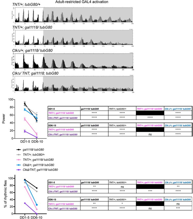
Jaumouillé et al.                                                                                            The Resilient Drosophila Circadian Network

Furthermore, positive- and negative- feedback loops created                      The puzzle that per rhythms in the M-cells are not necessary
by PAR DOMAIN PROTEIN 1 (PDP-1) and VRILLE (VRI)                             but clocks in the LN-EO are not sufficient for driving morning
on Clk expression are, respectively, coupled with the main                   activity peak was solved in the studies focusing on the role of
negative-feedback loop to ensure the generation of 24 h rhythms              DN1ps (Zhang L.Y. et al., 2010; Zhang Y. et al., 2010). Strikingly,
(Hardin, 2011). Circadian pacemaker neurons are classified into              per expression in the DN1ps alone is sufficient to restore morning
anatomically and functionally diverse subclasses: small and large            peak in per0 flies (Zhang Y. et al., 2010). The ability of DN1ps
lateral ventral neurons (s- and l-LNvs), lateral dorsal neurons              to drive morning activity depends on the PDF signaling (Zhang
(LNds), lateral posterior neurons (LPNs) and three groups of                 L.Y. et al., 2010). Furthermore, under dim light conditions,
dorsal neurons (DN1s, DN2s, DN3s) (Helfrich-Forster et al.,                  per expression in the DN1ps alone can also drive evening
2007). The s-LNvs are further divided into four neurons that                 anticipatory activity (Zhang Y. et al., 2010). Taken together, these
express the neuropeptide pigment dispersing factor (PDF) and                 findings have shown that DN1ps are the major output route of the
one PDF-negative neuron (5th s-LNv) (Figure 1A).                             M-cells and can also serve as M- and E- oscillators, depending on
   Previously many studies have posited that the PDF-positive                the environmental conditions (Lamaze and Stanewsky, 2019).
s-LNvs as the Morning-oscillator (M-oscillator; M-cells) that                    Manipulating the speed of the M-cells’ clocks changes the
anticipates dawn (Helfrich-Forster, 1998; Grima et al., 2004;                pace of subsets of the E-cells and free-running period in DD,
Stoleru et al., 2004, 2005). Additionally, these classical studies           within a limited temporal range (Guo et al., 2014; Yao and
showed that the M-cells are required for the generation and                  Shafer, 2014). The pace of DN1ps is strongly controlled by the
setting the pace of free-running locomotor rhythms in constant               speed of clocks in the M-cells in DD (Chatterjee et al., 2018).
darkness (DD). A separate group of pacemaker neurons named                   Among the LN-EO, the pace of two LNds co-expressing CRY
Evening (E)-oscillator (E-cells), including the PDF-negative 5th             and the short neuropeptide F (sNPF) (E-1 group, Figure 1B)
s-LNv, LNds and some of the DN1s (DN1s), controls evening                    is controlled by the clocks in the M-cells, whereas the 5th
bout of activity. PDF released from the M-cells functionally                 LNv and one LNd co-expressing CRY and the ITP ITP do not
couples M- and E-oscillators to generate coherent behavioral                 follow M-cell’s rhythms (E-2 group, Figure 1B; Yao and Shafer,
output (Grima et al., 2004; Stoleru et al., 2004; Picot et al.,              2014; Chatterjee et al., 2018). When the discrepancy of the
2007). Therefore, loss of PDF or PDF receptor (PDFR) results                 periods between the M-cells and PDF-negative clock neurons is
in the absence of the morning anticipation, advancing the                    larger than ∼ 2.5 h, the M-cells no longer dictates the coherent
evening peak, short period locomotor rhythms with very weak                  behavioral rhythms (Yao and Shafer, 2014). Moreover, electrical
rhythmicity (Renn et al., 1999; Hyun et al., 2005; Lear et al.,              silencing or disruption of clocks of non-LNv pacemaker neurons
2005; Shafer and Taghert, 2009; Yoshii et al., 2009; Choi et al.,            deteriorates locomotor rhythms without affecting clockwork in
2012).                                                                       the M-cells (Bulthuis et al., 2019). Behavioral period in DD can
   However, this somewhat simplistic view on the circadian                   be also modified by manipulating the pace of non-M cells (Dissel
network organization and the dominant role of the PDF-positive               et al., 2014). CRISPR knockout of per or tim in the M-cells
s-LNvs has been challenged by accumulating evidence. Works                   do not affect free-running rhythms, whereas ablation of per or
that characterized the property of the E-cells in response to                tim in both M and E-cells render flies arrhythmic (Delventhal
light have redefined the 5th s-LNv, three Cryptochrome (CRY)-                et al., 2019; Schlichting et al., 2019). Collectively, these works
positive LNds and 6–8 posterior subgroup of DN1s (DN1ps) as                  have demonstrated that behavioral period and rhythmicity are
the E-cells (Rieger et al., 2006; Picot et al., 2007; Figure 1A). More       determined by the action of multiple independent oscillators
precisely, since the clocks restricted only in the CRY-positive,             coordinated by network interaction, rather than by a single
PDF-negative 4 Lateral Neurons, i.e., the 5th s-LNvs and 3 CRY-              dominant oscillator.
positive LNds, are able to drive evening activity peak, they were                In this paper, we attempt to provide further evidence to the
defined as the Lateral Neuron-Evening oscillator (LN-EO) (Picot              revised model of the circadian circuit organization. To this end,
et al., 2007). Morning anticipation requires the presence of the             we exclusively use conditional approaches to disrupt molecular
M-cells (Renn et al., 1999; Stoleru et al., 2004) and per expression         clocks or neural communication in adulthood, in order to
only in the M-cells is sufficient to restore morning anticipation            distinguish the outcome caused by the effects during adulthood
in per null (per0 ) mutants (Cusumano et al., 2009) at least under           from any process during development. We find that disruption
the 12 h:12 h LD cycle and constant temperature of around 25◦ C              of molecular clocks in the M-oscillator or both M-oscillator and
(Menegazzi et al., 2020). However, per0 flies expressing per in all          part of the LN-EO only gradually weakens locomotor rhythmicity
neurons except the LNvs exhibit morning anticipation (Stoleru                in DD, which contrasts the immediate loss of morning activity
et al., 2004). Genetic rescue of per in per0 flies with the Mai179-          peak. Suppressing neuronal output of the M-cells in adulthood
GAL4 driver, which is expressed in the s- and l-LNvs, the 5th                reduces the power of the locomotor rhythmicity in DD also
LNv and 3 CRY-positive LNds (Cusumano et al., 2009) or with                  gradually. However, disruption of both molecular clockwork
the DvPdf-GAL4 driver expressed in the s- and l-LNvs, the 5th                and neural output of the M-oscillator leads to an immediate
LNv, three CRY-negative LNd and one LNd co-expressing CRY                    arrhythmia. These results indicate that the M-oscillator can be
and the ion transport peptide (ITP) (Figure 1B; Guo et al., 2014)            a master pacemaker or an output pathway of other pacemakers,
restores morning anticipation. However, per rescue only in the               thus largely support the emerging consensus that circadian circuit
LN-EO with the combination of Pdf-GAL80 and Mai179-GAL4                      is composed of multiple oscillators that can flexibly change the
does not restore morning anticipation (Cusumano et al., 2009).               network topology.

Frontiers in Physiology | www.frontiersin.org                            2                                       May 2021 | Volume 12 | Article 663339
Jaumouillé et al.                                                                                                              The Resilient Drosophila Circadian Network

  FIGURE 1 | Current model of the circadian pacemaker circuit organization in Drosophila melanogaster. (A) Four PDF-positive s-LNvs constitute the Morning (M)-cells
  and the Evening (E)-cells consist of the 5th s-LNv, three CRY-positive LNds and 6–8 DN1ps. Left, in LD and constant temperature, the M-cells control the morning
  activity peak through PDF signaling onto the DN1ps. The E-cells drive the evening activity peak. In certain environmental conditions, the DN1ps are able to drive both
  morning and evening peaks. Right, in DD and constant temperature, the M-cells determine the pace of the locomotor rhythms via PDF signaling to the E-cells.
  However, the coupling between the M- and E-cells are within a limited temporal range. Among the E-cells, CRY- and sNPF-positive, ITP-negative 2 LNds [E-1, see
  (B)] are strongly coupled to the oscillation of the M-cells, whereas the 5th s-LNv and one ITP-positive LNd (E-2) are weakly coupled. One of the locomotor output
  circuits is found downstream of the DN1ps, which are connected to neuroendocrine cells in the pars intercerebralis (PI). (B) The identity of the M- and E-cells and the
  expression patterns of the GAL4 drivers used in this study. Pdf-GAL4 is expressed in the l- and s- LNvs (Renn et al., 1999). gal1118 is expressed in the l- and
  s-LNvs and weakly in the LNds. However, expression in the LNds is detectable only in the flies homozygous for gal1118 (Blanchardon et al., 2001). DvPdf-GAL4 is
  expressed in all the LNvs and one CRY-positive, ITP-positive LNd and three CRY-negative LNds (Bahn et al., 2009; Schubert et al., 2018). (n) indicates the number
  of cells per hemisphere.

MATERIALS AND METHODS                                                                   were performed at 29◦ C in the experiments with adult-restricted
                                                                                        conditional GAL4 induction and at 18◦ C for developmental
Fly Strains                                                                             GAL4 induction. For adult-restricted GAL4 induction, flies were
Drosophila were reared at 25◦ C on a corn-meal medium under                             crossed and raised at 18◦ C until 2 days after eclosion. Male
12 h:12 h light-dark (LD) cycles. UAS-CLK1 (Tanoue et al.,                              flies of appropriate genotypes were then collected and placed in
2004) was a gift from Jadwiga Giebultowicz. UAS-TNT (UAS-                               the DAM monitors and assayed for locomotor activity at 29◦ C.
TNT-G) (Kaneko et al., 2000) was a gift from Jeff Hall. UAS-per                         Flies were first entrained in 12 h:12 h-LD cycles for 4 days and
RNAi (perCt-IR) (Martinek and Young, 2000), Pdf-GAL4 (Park                              then released in DD for 10–12 days. The light intensity of the
et al., 2000), gal118 (Blanchardon et al., 2001), DvPdf-Gal4 (Bahn                      incubator was approximately 1000 lux. For experiments with
et al., 2009), and tubulin-GAL80ts (McGuire et al., 2004) were                          developmental GAL4 expression, flies were raised at 29◦ C until
described previously.                                                                   2 days after eclosion, and then the collected flies were assayed at
                                                                                        18◦ C. In both sets of experiments, behavioral data were analyzed
                                                                                        from the second day in LD. Two to four independent experiments
Behavioral Assays                                                                       were performed for each genotype. The numbers of flies used in
The locomotor behavior assay was performed as described in                              the behavioral assays are indicated in Table 1. The behavioral data
Beuchle et al. (2012) using the Drosophila Activity Monitoring                          were analyzed using FaasX software (Blanchardon et al., 2001).
(DAM) System (Trikinetics, Waltham, MA), except that assays                             The flies with power over 20 and width over 2.5 h according

Frontiers in Physiology | www.frontiersin.org                                       3                                              May 2021 | Volume 12 | Article 663339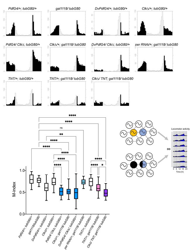
Jaumouillé et al.                                                                                                    The Resilient Drosophila Circadian Network

TABLE 1 | Free-running locomotor rhythms in flies with adult-restricted genetic manipulations.

Genotype                                           n              DD1-5                                            DD6-10

                                                            Period ± SEM (h)           Power ± SEM     %R      Period ± SEM (h)       Power ± SEM         %R

Pdf-GAL4/+; tub-GAL80ts/+                         124           23.9 ± 0.3              95.2 ± 4.3     95.2       24.3 ± 0.1            79.1 ± 2.7        88.1
gal1118/tub-GAL80ts                               60            23.4 ± 0.2              89.4 ± 4.3     96.6       23.4 ± 0.3            46.6 ± 4.3        72.7
UAS-Clk1/+; tub-GAL80ts/+                         56            23.4 ± 0.2             105.2 ± 13.1    91.1       23.5 ± 0.3           61.95 ± 9.3        55.0
DvPdf-GAL4/+; tub-GAL80ts                         25            23.6 ± 0.1              66.3 ± 8.4     76.0       24.1 ± 0.8            35.6 ± 3.7        52.6
UAS-Clk1/Pdf-GAL4; tub-GAL80ts/ +                 59            24.4 ± 0.8              74.9 ± 2.31    98.3       24.0 ± 1.1            37.7 ± 2.0        49.1
UAS-Clk1/+; gal1118/tub-GAL80ts                   63            23.9 ± 0.3              74.25 ± 2.8    95.2       23.3 ± 0.2            52.6 ± 7.9        25.4
UAS-Clk1/DvPdf-GAL4; tub-GAL80ts/ +               26            24.0 ± 0.5              72.2 ± 12.9    76.9       25.1 ± 0.8           34.6 ± 13.9        16.7
UAS-per RNAi/Pdf-GAL4; tub-GAL80ts/ +             59            23.4 ± 0.1              97.6 ± 15.8    98.3       23.6 ± 0.1           73.2 ± 14.3        75.4
UAS-TNT/+; tub-GAL80ts/+                          59            23.5 ± 0.0              84.7 ± 1.3     88.1       23.5 ± 0.1            61.0 ± 5.4        63.2
UAS-TNT/+; gal1118/tub-GAL80ts                    29            23.6 ± 0.1              50.1 ± 0.0     75.9       22.5 ± 0.0            11.1 ± 3.6        12.5
UAS-Clk1/UAS-TNT; gal1118/tub-GAL80ts             40            23.4 ± 0.1              18.7 ± 9.0     3.1            –                 2.98 ± 1.2          0

n, number of flies; %R, % of rhythmic flies.

to the χ2 periodogram analysis were defined as rhythmic. The                           for normality with D’Agostino-Pearson K2 test, and normally
significance threshold was set to 5%. Morning anticipation                             distributed data sets were analyzed using parametric tests
index (M-index) was calculated as described in Im and Taghert                          (ANOVA and unpaired t-test with Welch’s correction) and non-
(2010) with minor modifications. The M-index was calculated for                        normally distributed data were analyzed with non-parametric
individual flies as (sum of activity over 3 h before lights on)/(sum                   tests (the Kruskal-Wallis test and Mann-Whitney U-test).
activity over 6 h before lights on) at each day from LD2 to LD4,                          Welch’s t-test and Mann-Whitney U-test were used for
and the 3 values were averaged to obtain the mean M-index of                           pairwise comparisons of the behavioral rhythmicity between
an individual fly. The mean M-indices were pooled per genotype                         control and test genotype groups, depending on the distribution
and presented as boxplots.                                                             of each data set. Morning anticipation indices were compared
                                                                                       using the Kruskal-Wallis test with Dunn’s multiple comparisons
Immunohistochemistry, Microscopy, and                                                  test. Signal intensities of immunofluorescence images were
                                                                                       compared using the multiple unpaired t-test with Welch’s
Image Analysis                                                                         correction or ANOVA with Sidak’s multiple comparison’s test.
Anti-PER and PDF immunostaining of fly brains was performed
as described previously (Shafer et al., 2002). The brains were
imaged using a Leica SP5 confocal microscope and images
                                                                                       RESULTS
were analyzed using Fiji/Image J software (National Institutes
of Health). To quantify PER staining intensity, sum slices
projections were generated from 2 µm z-section confocal images,
                                                                                       Disruption of Molecular Clocks in Adult
and the mean pixel value of each cell and background pixel value                       Morning/Master-Oscillator Has a Modest
was measured. The mean pixel value of each cell in a given                             Effect on Free-Running Locomotor
subgroup was calculated by subtracting the mean pixel value of                         Rhythms
the background and plotted as relative intensity normalized to                         To conditionally eliminate molecular clockwork only during
the value of the control group at CT0. PDF levels in the s-LNv                         adulthood in the LNvs, we drove the expression of CLK1,
dorsal terminals were measured as described in Kozlov et al.                           a dominant-negative mutant of CLK (Tanoue et al., 2004),
(2017). Briefly, the region of interest (ROI) for the axonal termini                   using the combination of Pdf-GAL4 and temperature sensitive
(from the tip of the s-LNv dorsal termini until where the terminal                     GAL80 expressed ubiquitously under the tubulin promoter (tub-
arbors first branch) was specified manually with the polygon                           GAL80t s ). The flies were raised at 18◦ C until 2 days after
selection tool in Fiji and the intensity sum within each ROI was                       eclosion and adult male flies were maintained at 29◦ C during
measured. The representative confocal images were maximum                              the subsequent experiments (McGuire et al., 2004). To verify
projections generated from the same confocal z-series used for                         if this manipulation effectively blocks molecular clockwork,
the quantification.                                                                    the brains of flies were immunolabeled with anti-PER and
                                                                                       anti-PDF antibodies every 4 h on the third day in constant
Statistical Analysis                                                                   darkness (DD3) following an entrainment to 12 h:12 h light-
Statistical analysis and data visualization were performed using                       dark (LD) cycles for 4 days (Supplementary Figure 1). As
GraphPad Prism (9.0). A p-value < 0.05 is considered a                                 expected, PER levels in the s-LNvs were significantly reduced and
statistically significant test result. Asterisks indicate p-values,                    arrhythmic on DD3. In DN1s, PER levels peaked at CT12 but
where ∗ p < 0.05, ∗∗ p < 0.01, ∗∗∗ p < 0.001, and ∗∗∗∗ p < 0.0001.                     did not show 24 h rhythmicity. This observation is congruent
ns indicates a non-significant test result. Data were first tested                     with the notion that molecular rhythms in the s-LNvs affect

Frontiers in Physiology | www.frontiersin.org                                      4                                      May 2021 | Volume 12 | Article 663339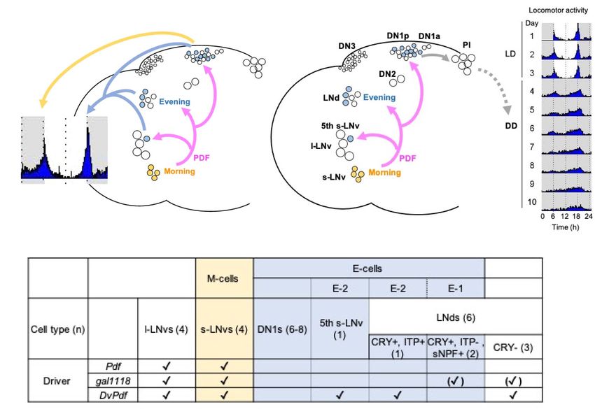
Jaumouillé et al. The Resilient Drosophila Circadian Network
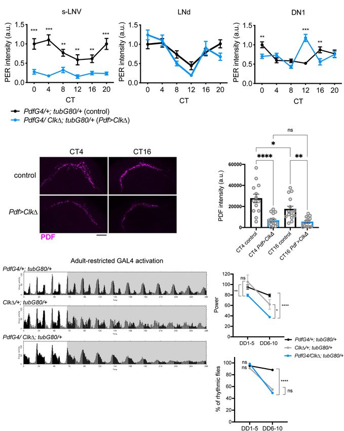
rhythmicity of the DN1s (Nitabach et al., 2006; Zhang L.Y. immediate behavioral arrhythmia in DD (Figures 4A,B). This
et al., 2010; Beuchle et al., 2012; Jaumouille et al., 2015). PER finding verifies that conditional expression of CLK1 is an
levels and oscillations in the LNds were not different between effective tool to disrupt molecular clocks and highlights the
control and CLK1-expressing flies (Figure 2A). Moreover, PDF differential requirements for LNvs’ clocks during development
signal at the s-LNv dorsal termini was reduced and arrhythmic and adulthood in the functioning of the pacemaker circuit.
in CLK1-expressing flies (Figures 2B,C). This observation Previous studies have shown that inactivating per in pacemaker
confirms the previous finding that Clk controls PDF levels and neurons during development does not affect behavioral rhythms
accumulation rhythms via regulating vri expression in adult of adult flies but depleting CYC during development abolishes
s-LNvs (Gunawardhana and Hardin, 2017). locomotor rhythms in adults (Ewer et al., 1990; Goda et al., 2011).
Having validated that adult-restricted expression of CLK1 Our results are in agreement with these observations and support
in the LNvs effectively abolishes molecular clockwork, we next that CLK/CYC activity has non-clock roles during development.
assayed locomotor behavior of these flies. Unexpectedly, CLK1 How could locomotor rhythms persist several days
driven with Pdf-GAL4 and tub-GAL80ts had a modest effect under constant conditions while the M-cells are molecularly
on free-running locomotor rhythms at least up to DD10. Most arrhythmic? As proposed by a number of other studies (Guo
(98.3%) of the Pdf-GAL4/CLK1; tub-GAL80ts/+ flies were et al., 2014; Yao and Shafer, 2014; Bulthuis et al., 2019; Delventhal
rhythmic until DD5, despite with a reduced rhythm power. Their et al., 2019; Schlichting et al., 2019), we hypothesized that other
rhythms damped significantly after DD6 compared to control pacemaker neurons compensate arrhythmic LNvs to drive free-
flies (Figures 2D,E and Table 1). To verify the effect of adult- running rhythms. In particular, recent studies have described
restricted CLK1 expression in the LNvs, we next turned to the that CRISPR-mediated ablation of molecular clocks in the LNvs
gal1118 driver (Malpel et al., 2004). Gal1118 is expressed in the s- does not cause strong behavioral phenotypes in DD, whereas
and l-LNvs and weakly expressed in 5 LNds, but the expression in clock knockout in the M- and all or part of LN-EO cells using
the LNds is detectable only in the flies homozygous for gal1118 Mai179-GAL4 or DvPdf-GAL4 significantly reduces behavioral
(Malpel et al., 2004; Figure 1B). Therefore, we used one copy rhythmicity (although the data of the DvPdf-GAL4-mediated
of gal1118 in combination with tub-GAL80ts to express CLK1 clock knockout are not displayed) (Delventhal et al., 2019;
only during adulthood. These flies remained highly rhythmic Schlichting et al., 2019). Therefore, LN-EO cells are the likely
during the first 5 days in DD. After DD6, a high proportion candidates of the surrogate main pacemaker. To test this idea,
of these flies became arrhythmic (Table 1 and Supplementary we next expressed CLK1 with the DvPdf-GAL4 driver during
Figures 2A,B). These results indicate that behavioral rhythms in adulthood. The majority of these flies remained rhythmic until
DD only gradually dampen over several days when clocks in the DD5, thereafter became arrhythmic (Figures 5A,B and Table 1).
LNvs are disrupted in adulthood. However, the average rhythm power of DvPdf-GAL4/CLK1;
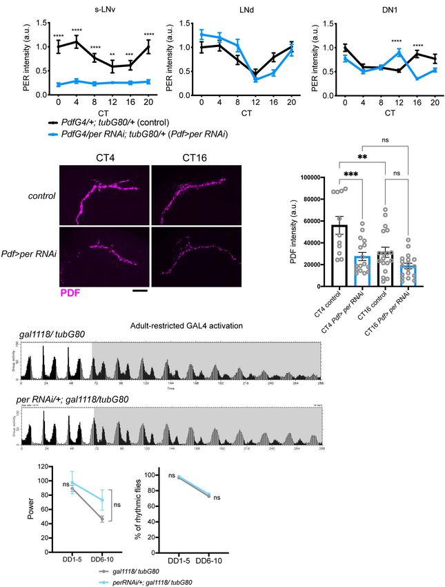
To corroborate these findings, we also used UAS-RNAi against tub-GAL80ts/+ flies were approximately the same as that of
PER as an alternative method to temporarily block molecular two control groups until DD10. Since DvPdf-GAL4 is expressed
clockwork. UAS-PER RNAi was driven in the LNvs with Pdf- in the M-cells and two out of four LN-EO cells (Figure 1B),
GAL4 or gal1118 and tub-GAL80ts and its expression was these observations suggest the possibility that clocks in PDF-
induced by the temperature shift from 18 to 29◦ C 2 days negative pacemaker neurons, including two LNds that do not
after eclosion. For behavioral assays we used ga1118, because express DvPdf-GAL4 (i.e., CRY-positive, sNPF-positive LNds),
many flies carrying Pdf-GAL4, tub-GAL80ts, and UAS-PER RNAi compensate the loss of clocks in the M-cells and maintain
did not survive until the end of behavioral recording for rhythmic locomotor output for several days.
unknown reasons. As with conditional CLK1 expression, this
treatment eliminated PER expression and rhythms in the s-LNvs,
verified by anti-PER and anti-PDF immunostaining on DD3
Neural Output and Clocks of the
(Supplementary Figure 1 and Figure 3A). Levels and rhythms M-Oscillator Are Additively Required to
of PDF accumulation at the dorsal termini of the s-LNvs were Maintain Robust Locomotor Rhythms
also reduced (Figures 3B,C). This observation is congruent with Our results thus far are in line with previous works and suggest
a previous report that CLK represses pdf transcription (Mezan that non-M-cells can output behavioral rhythms independently
et al., 2016) because PER knockdown should increase CLK/CYC- of the M-cells, or they input signals to the M-cells, which then
transcriptional activity (Yu and Hardin, 2006). Free-running output behavioral rhythms without the need of molecular clocks
locomotor rhythmicity of these flies were not disrupted compared in the M-cells. The possibility that non-M-cells output behavioral
to controls at least up to DD10 (Figures 3D,E and Table 1), rhythms via M-cells is backed by the evidence that presence of
consistent with the results of adult-restricted, LNv-targeted the s-LNvs is essential for locomotor rhythms (Helfrich-Forster,
CLK1 expression (Figures 2D,E, Table 1, and Supplementary 1998; Stoleru et al., 2004). To test this hypothesis further, we
Figures 2A,B). sought to block neuronal output of the M-cells with or without
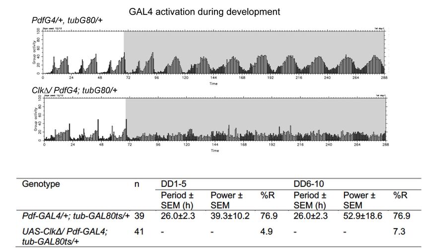
The presence of functional clocks in developing LNvs is disrupting molecular clocks in the M-cells. We first blocked
required for driving normal locomotor rhythms in adulthood output of the LNvs during adulthood by conditionally expressing
(Goda et al., 2011; Beuchle et al., 2012). Consistent with tetanus toxin light chain (TNT) (Sweeney et al., 1995) using
this notion, conditional expression of CLK1 only during the combination of gal1118 and tub-GAL80t s . Gal1118 was used
development until eclosion in the LNvs resulted in an instead of Pdf-GAL4 due to the ease in establishing the desired
Frontiers in Physiology | www.frontiersin.org 5 May 2021 | Volume 12 | Article 663339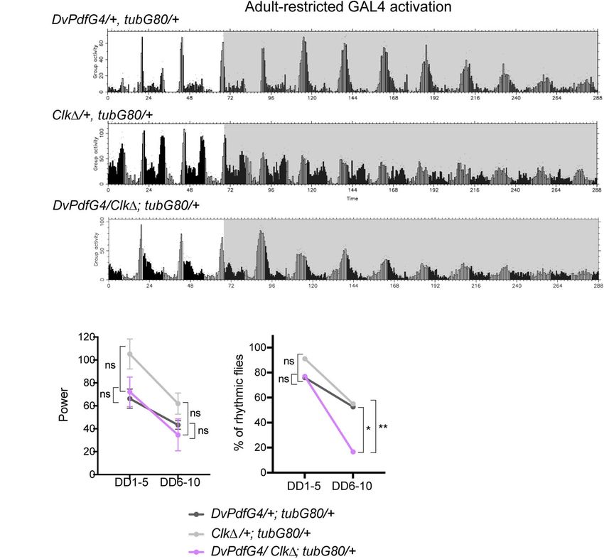
Jaumouillé et al.                                                                                                            The Resilient Drosophila Circadian Network

  FIGURE 2 | Blocking M-oscillator clocks by conditional expression of CLK1 has little effects on free-running locomotor rhythms. UAS-Clk1 was expressed in the
  LNvs only during adulthood with Pdf-GAL4, tub-GAL80t s and a temperature shift from 18to 29◦C. (A) The brains were stained for PER and PDF on DD3 and the
  levels of PER in the s-LNvs, DN1s and LNds were quantified. n = 20–27 hemispheres per group. **p < 0.01, ***p < 0.001 by multiple unpaired t-test with Welch’s
  correction, comparing two genotypes at each timepoint. (B,C) PDF levels in the s-LNv dorsal terminals at two timepoints on DD3. Adult-restricted Clk1 in the LNvs
  significantly reduced PDF levels and disrupted its rhythm. Scale bar, 20 µm. n = 12–15 per group. *p < 0.05, **p < 0.01, and ***p < 0.00 by 2-way ANOVA with
  Sidak’s multiple comparison test. (D) Group average locomotor activities of the flies in LD (white background) and DD (gray background). (E) The power of rhythmicity
  and the percentage of rhythmic flies during the first 5 days in DD (DD1-5) and from the 6th to 10th days in DD (DD6-10) in the flies expressing Clk1 (PdfG4/Clk1;
  tubG80/ + stands for Pdf-GAL4/UAS- Clk1; tub-GAL80t s / +) and in two control groups (PdfG4/+; tubG80/+ indicates Pdf-GAL4/+; tub-GAL80ts /+ and Clk1/+;
  tubG80/ + indicates UAS-Clk1; tub-GAL80t s /+). The difference in the rhythm power was tested using the unpaired t-test with Welch’s correction and Mann-Whitney
  U-test *p < 0.05, **p < 0.01, and ****p < 0.0001. The percentages of rhythmic flies were compared using Fisher’s exact test. ****p < 0.0001. ns, not significant.

Frontiers in Physiology | www.frontiersin.org                                      6                                             May 2021 | Volume 12 | Article 663339
Jaumouillé et al.                                                                                                          The Resilient Drosophila Circadian Network

  FIGURE 3 | PER knockdown in adult M-oscillators disrupts molecular clocks but has little effects on free-running locomotor rhythms. (A) UAS-per RNAi was
  expressed during adulthood with a combination of Pdf-GAL4, tub-GAL80ts . PER levels were quantified on DD3 in the s-LNvs, DN1s and LNds. n = 14–19 per
  group. **p < 0.01, ****p < 0.0001 comparing two genotypes at each timepoint by multiple unpaired t-test with Welch’s correction. (B,C) PDF levels in the s-LNv
  dorsal terminals at two timepoints on DD3. Representative confocal images (B) and quantification (C) of indicated genotypes. Scale bar, 20 µm. n = 11–18
  hemispheres per group. **p < 0.01, ***p < 0.001 by 2-way ANOVA with Sidak’s multiple comparison test. (D) Group average locomotor activities of the flies with
  adult-restricted expression of per RNAi driven with the combination of gal1118 and tub-GAL80ts and the control group carrying only the driver in LD and DD.
  (E) Left, power of rhythmicity in DD1-5 and DD6-10 in the indicated genotypes. No significant differences were found between two groups by the unpaired t-test
  with Welch’s correction. Right, percentage of rhythmic flies in DD1-5 and DD6-10. No significant differences were found by Fisher’s exact test.

Frontiers in Physiology | www.frontiersin.org                                     7                                            May 2021 | Volume 12 | Article 663339
Jaumouillé et al.                                                                                                      The Resilient Drosophila Circadian Network

  FIGURE 4 | CLK1 expression during development in the M-cells irreversibly disrupts locomotor rhythms in adulthood. (A,B) UAS-Clk1 was expressed with
  Pdf-GAL4, tub-GAL80ts only during development. (A) Group average locomotor rhythms assayed at 18◦ C following the developmental CLK1 expression.
  (B) Percentage of rhythmic flies in DD1-5 and DD6-10 in the flies following developmental CLK1 expression and controls.

genotypes in co-expression experiments. TNT is a protease that                     in the M-cells, two scenarios are possible: a surrogate master
cleaves n-synaptobrevin, syntaxin or SNAP-25, thereby inhibits                     pacemaker bypasses the M-cells to control locomotor output;
synaptic transmission (Sweeney et al., 1995) and neuropeptide                      or the M-cells still produce output signals via TNT-insensitive
release (Ding et al., 2019). These flies exhibited locomotor                       transmitters/peptides or by electrical coupling via gap junctions
rhythms with a significantly reduced power already during the                      (Schneider and Stengl, 2006; Ramakrishnan and Sheeba, 2020).
first 5 days in DD. The power of the rhythmicity was further
reduced after DD6 (Figures 6A,B). Approximately 25% of these
flies were arrhythmic before DD5, and thereafter approximately
                                                                                   Morning Anticipation Is Independent of
80% of them became arrhythmic (Figure 6C and Table 1). This                        PER Rhythms but Requires CLK in the
finding is largely congruent with the results presented in Kaneko                  M-Oscillator in Adulthood
et al. (2000), where the same UAS-TNT (UAS-TNT-G) line used                        It has been shown that morning anticipatory activity requires
in the present study was constitutively driven with Pdf-GAL4.                      the presence of the M-cells and PDF neuropeptide (Renn
In contrast, one study reported that constitutive expression of                    et al., 1999; Grima et al., 2004; Shafer and Taghert, 2009)
TNT with Pdf-GAL4 had no effect on both LD and DD behavior,                        but can be observed in the absence of per rhythms within
using another UAS-TNT insert, UAS-TNT-E (Umezaki et al.,                           the M-cells (Stoleru et al., 2004). We examined whether
2011). This discrepancy is likely attributed to the difference                     morning anticipation is impaired when molecular clocks are
in expression levels, as UAS-TNT-G renders a higher level of                       disrupted during adulthood in the LNvs. Adult-restricted
expression than UAS-TNT-E (Kaneko et al., 1997).                                   PER RNAi in the LNvs, which abolishes PER rhythms
    We next expressed both CLK1 and TNT in the LNvs                                and reduces PDF levels and rhythms in the s-LNv axonal
using gal1118 during adulthood. Strikingly, these flies became                     termini (Figures 3A–C), did not impair morning anticipation
immediately arrhythmic in DD (Figures 6A–C and Table 1). In                        (Figures 7A,B, compare perRNAi/+; gal1118/tubG80 and
other words, loss of neural transmission and loss of molecular                     gal1118/tubG80). These results are congruent with the report
clockwork in adult LNvs cumulatively cause the rapid decline of                    that per expression in the M-cells is not required for
rhythmic behavioral output. This finding further suggests that                     morning anticipation (Stoleru et al., 2004). However, the
molecular clocks and neural output function of the M-cells are                     morning anticipation index (M-index) (Sheeba et al., 2010)
independent components that add up to enable robust rhythmic                       was significantly reduced when CLK1 was conditionally driven
behavioral output in DD. Even when clocks are disrupted, the                       in adulthood with Pdf-GAL4 or gal1118 (Figures 7A,B, see
M-cells can receive inputs from other pacemakers and transmit                      PdfG4/Clk1; tubG80/+, Clk1/+; gal1118/tubG80, PdfG4/+;
output signals to the output circuit. When TNT is expressed                        tubG80/+, gal1118/tubG80, and Clk1/+; tubG80/+). Similarly,

Frontiers in Physiology | www.frontiersin.org                                  8                                           May 2021 | Volume 12 | Article 663339
Jaumouillé et al. The Resilient Drosophila Circadian Network FIGURE 5 | Adult-restricted expression of CLK1 with the DvPdf-GAL4 driver has little effects on free-running locomotor rhythms. (A) Group average locomotor activity in LD and DD in the flies expressing CLK1 with the DvPdf-GAL4 driver during adulthood and their controls. (B) Left, the rhythm power of flies in DD1-5 and DD6-10. Right, percentage of rhythmic flies. *p < 0.05, and **p < 0.01 by Fisher’s exact test. adult-restricted CLK1 expression using Dvpdf-GAL4 reduced CLK1 and TNT were co-expressed (Figures 7A,B, compare morning anticipation compared with controls (Figures 7A,B, TNT/+; gal1118/tubG80, CLK1/TNT; gal1118/tubG80, TNT/+; compare DvPdfG4/Clk1; tubG80/+, DvPdfG4/+; tubG80/+, tubG80/+, and gal1118/tubG80). As TNT is known to block and Clk1/+; tubG80/+). CLK1 expression in adult LNvs both neurotransmitter and neuropeptide release (Sweeney et al., reduces the levels and diurnal rhythms of PDF accumulation 1995; Ding et al., 2019), the results suggest the possibility that, in in the s-LNv dorsal projections (Figures 2B,C) as is the addition to the PDF, neurotransmitter, such as glycine (Frenkel case with adult-restricted PER RNAi. These results validate et al., 2017) or the short Neuropeptide F (sNPF) (Johard et al., that adult-restricted expression of CLK1 using GAL4/GAL80ts 2009), may be involved in the normal morning anticipation. is immediately in effect and further suggest that loss of We also noticed that evening activity peak was not apparently PER rhythms and reduction in axonal PDF levels in the advanced in all the genotypes tested (Figure 7A). Free- s-LNvs during adulthood do not necessarily impair morning running rhythms were not shortened either (Table 1). This anticipation. These data instead suggest the involvement was surprising because loss of PDF advances evening peak of other factors that are regulated by CLK in generating in a 12 h:12 h LD cycle at the temperature near 25◦ C morning anticipation. (Renn et al., 1999). The lack of effect on evening activity In this regard, it is noteworthy that morning anticipation even in the flies that have reduced morning anticipation was reduced in the flies expressing TNT in adult LNvs is probably because their PDF levels are only partially compared with controls and even further reduced when reduced (Figures 2B,C, 3B,C). Additionally, the fact that Frontiers in Physiology | www.frontiersin.org 9 May 2021 | Volume 12 | Article 663339
Jaumouillé et al.                                                                                                              The Resilient Drosophila Circadian Network

  FIGURE 6 | Intact neuronal output of the M-oscillator is required for sustaining robust free-running locomotor rhythms. (A) Group average locomotor activity of the
  flies expressing TNT, CLK1 or TNT and CLK1 in the LNvs with the combination of gal1118 and tub-GAL80ts during adulthood and a control group (UAS-TNT/ + ;
  tub-GAL80ts / +). The activity of another control group, gal1118/tub-GAL80ts , is displayed in Figure 3D. (B) Power of rhythmicity of the flies of indicated genotypes
  in DD1-5 and DD6-10. The unpaired t-test with Welch’s correction and Mann-Whitney U-test were used to compare two genotypes at the same time point. The
  tables show the statistical test results, in which groups labeled in the row are compared with those in the column. ∗∗∗∗ p < 0.0001. ns, not significant. (C) The
  percentage of rhythmic flies in DD1-5 and DD6-10. The genotypes are as in (B). Results of Fisher’s exact test for pairwise comparisons are shown in the table.
  ∗ p < 0.05, ∗∗ p < 0.01, ∗∗∗ p < 0.001, and ∗∗∗∗ p < 0.0001. ns, not significant.

all the behavioral experiments were performed at 29◦ C, the                              DISCUSSION
temperature that suppresses daytime activity (Majercak et al.,
1999; Parisky et al., 2016), likely masked the effects on the                            In this study, we disrupted molecular clocks or neural
evening activity peak.                                                                   transmission only in adulthood in restricted subgroups of

Frontiers in Physiology | www.frontiersin.org                                       10                                             May 2021 | Volume 12 | Article 663339
Jaumouillé et al.                                                                                                                 The Resilient Drosophila Circadian Network

  FIGURE 7 | LD-entrained activities of flies with the adult-restricted disruption of clocks or neural output of the M-cells. (A) Average activities of flies of the indicated
  genotypes in LD. White bars indicate activity levels during the light period and black bars represent the dark period. (B) M-indices of the flies of indicated genotypes.
  Whiskers indicate 5th and 95th percentile and the line inside the box indicates the median. ∗ p < 0.05, ∗∗ p < 0.01, ∗∗∗ p < 0.001, and ∗∗∗∗ p < 0.0001 by the
  Kruskal-Wallis test with Dunn’s multiple comparisons test. (C) A schematic summarizing the finding of this study. Circadian pacemaker network consists of diverse
  classes of clock-containing pacemaker neurons, including the M-oscillator (yellow) and the E-oscillator (blue). The traditional model postulates that the M-cells
  (yellow) drive free-running locomotor rhythms via synchronizing the pace of the E-cells. In this study, we show that locomotor rhythms can free-run several cycles
  while clocks in the M-cells and part of the E-cells are disrupted during adulthood, supporting the emerging view that the circadian circuit can flexibly assign different
  neuronal subgroups the pacemaking role to maintain rhythmic locomotor output.

pacemaker neurons to better understand the network property                                morning anticipation does not require PER cycling but requires
of the circadian circuit. Our results are summarized in three                              intact CLK; and (iii) disruption of M-oscillator’s neuronal
main points: (i) free-running locomotor rhythms are maintained                             output dampens free-running rhythms, and the disruption of
for several days while molecular clocks are disrupted in the                               both clocks and neural output of the M-cells results in an
M-oscillator or in both M- and part of E-oscillators; (ii)                                 immediate behavioral arrhythmia under constant conditions.

Frontiers in Physiology | www.frontiersin.org                                         11                                               May 2021 | Volume 12 | Article 663339
Jaumouillé et al.                                                                                           The Resilient Drosophila Circadian Network

These results are largely in line with previous findings, with             are clockless. Intriguingly, changing the pace of the LN-EO does
minor deviations.                                                          not alter period or power of locomotor rhythms in DD when
    Recent studies used cell-specific CRISPR knockout of per               clocks in the M-cells are intact (Chatterjee et al., 2018). Therefore,
or tim and showed that the absence of clocks in the M-cells                role switching from the M-cells to LN-EO seems to occur only
does not impair locomotor rhythms (Delventhal et al., 2019;                when clocks in the M-cells are dysfunctional. DN1p, on the
Schlichting et al., 2019). Adult-specific expression of CLK1 or            other hand, is strongly coupled to the M-cells and are the major
PER RNAi in the M-cells recapitulates their findings. Whereas              output route of the M-cells (Chatterjee et al., 2018). When M-cells
CRISPR-mediated clock knockout in both the M-cells and LN-                 are clockless, the LN-EO or other pacemaker neurons excluding
EO causes an immediate behavioral arrhythmia (Delventhal et al.,           DN1ps, might also output locomotor rhythms without passing
2019; Schlichting et al., 2019), adult-restricted expression of            through the M-cells, since the electrical silencing and TNT
CLK1 with DvPdf-GAL4, which is expressed in the M-cells and                expression of the M-cells alone does not immediately disrupt
two out of four LN-EO cells, gradually dampens the locomotor               locomotor rhythms. There is also a possibility that M-cells’ clocks
rhythmicity over several days. This difference is probably due             may control locomotor output in a manner resistant to TNT, such
to the fact that clocks in the two CRY- and sNPF-positive LNds             as via gap junctions (Schneider and Stengl, 2006; Ramakrishnan
are not disrupted in DvPdf > CLK1 flies and can contribute                 and Sheeba, 2020) or via TNT-insensitive transmitters/peptides.
to maintaining rhythmic locomotor output. Therefore, CRY-                     Previous works have shown that the M-cells can function as
positive LNds, which play a crucial role in driving evening                the cell-autonomous driver of the morning anticipation when
activity in LD, might also promote free-running rhythms in DD              harboring functional clocks but its function can be modulated by
(Rieger et al., 2009). Additionally, the onsets of clock disruption        the DN1p or other pacemakers (Stoleru et al., 2004; Cusumano
differ between our study and clock knockout studies; the exact             et al., 2009; Zhang Y. et al., 2010; Menegazzi et al., 2020). Our
onset of gene deletion may not be reliably determined when                 results of the adult-restricted PER knockdown in the M-cells are
using the GAL4-driven CRISPR knockout strategy, especially                 congruent with these conclusions. However, CLK1 expression
because many GAL4s are also expressed during development.                  in adult M-cells significantly reduces the morning anticipation,
Despite subtle differences, these two results are not mutually             suggesting that intact CLK within the M-cells is required
contradictory and both support that clocks in the M-oscillator             for morning anticipation behavior. Both PER knockdown and
are dispensable during adulthood for maintaining free-running              CLK1 expression reduce PDF levels and rhythms in the dorsal
locomotor rhythms as long as clocks in other pacemakers,                   termini of the M-cells; therefore, factors other than PDF are
including CRY-positive LN-EO, are intact (Figure 7C).                      involved in controlling the morning anticipation. The report
    Adult-restricted electrical silencing of the M-cells does              that M-cell-specific ablation of vrille reduces PDF expression
not affect M-cells’ clocks but leads to a gradual dampening                and rhythms via post-transcriptional regulations but does not
of behavioral rhythms and eventual arrhythmia (Depetris-                   affect morning anticipation (Gunawardhana and Hardin, 2017)
Chauvin et al., 2011). We show that TNT expression in                      also supports this interpretation. Taken together with our finding
adult M-cells recapitulates this phenotype. Importantly, adult-            that expression of TNT in adult M-cells reduces the morning
restricted expression of both TNT and CLK1 in the M-cells                  anticipation, these results suggest that a certain neurotransmitter
results in an immediate behavioral arrhythmia. Therefore, lack of          or sNPF is under the control of CLK and plays an important
molecular clockwork is compensated as long as the M-oscillator             role in driving morning anticipation behavior. Additionally, it is
can produce synaptic and/or peptidergic output. Conversely, lack           noteworthy that both CLK1 expression and per knockdown in
of neural transmission from the M-oscillator can be overcome as            the M-cells reduce PDF levels and rhythms in the dorsal termini
long as their internal clocks are functional.                              of the M-cells but does not immediately deteriorate locomotor
    How can the circadian circuit maintain rhythmic locomotor              rhythmicity. These results confirm the previous report that PDF
output while the clocks or neural function is disabled in the              rhythms in the dorsal projections do not play important roles in
M-cells? It has been shown that TNT expression in the DvPdf-               locomotor rhythms in DD (Fernandez et al., 2020).
GAL4 positive, PDF-negative cells (i.e., the 5th s-LNv, three CRY             In summary, the present study highlights the remarkable
negative LNds and one CRY-positive, ITP-positive LNd) alone                resilience of Drosophila circadian pacemaker circuit, the property
during adulthood renders flies arrhythmic (Guo et al., 2014).              conserved in mammals (El Cheikh Hussein et al., 2019).
Constitutive electrical silencing of the DvPdf-GAL4 positive,              Our findings support the emerging view that the topology
PDF-negative cells severely disrupts locomotor rhythms, whereas            of the pacemaker circuit is not rigid, as in the classical M-
silencing of three CRY-positive LNds and the 5th s-LNv labeled             and E-oscillator model, but rather flexible, assigning different
by the MB-122b split GAL4 only during adulthood reduces                    neuronal subgroups the task of pacemaking in order to
rhythm power without affecting clocks in the M-cells (Bulthuis             achieve the resilience.
et al., 2019). The LN-EO cells make synaptic contacts onto
the M-cells and rhythmically modulate their excitably (Duhart
et al., 2020). Genetic rescue of per0 flies with the Clk4.1M driver        DATA AVAILABILITY STATEMENT
restores morning activity but is unable to rescue arrhythmic
DD behavior (Zhang Y. et al., 2010). Collectively, these findings          The original contributions presented in the study are included
suggest that the LN-EO can input signals to the M-cells, through           in the article/Supplementary Material, further inquiries can be
which behavioral output is maintained even when the M-cells                directed to the corresponding author/s.

Frontiers in Physiology | www.frontiersin.org                         12                                        May 2021 | Volume 12 | Article 663339
Jaumouillé et al. The Resilient Drosophila Circadian Network
AUTHOR CONTRIBUTIONS ACKNOWLEDGMENTS
EJ and EN: conceptualization. EJ, RK, and EN: investigation. We would thank Jeff Hall, Jadwiga Giebultowicz, and the
EN: writing and funding acquisition. All authors Bloomington Drosophila Stock Center for fly lines. We thank
contributed to the article and approved the submitted Hanspeter Herzel, Bharath Ananthasubramanian, and our lab
version. members for helpful suggestion on this work.
FUNDING SUPPLEMENTARY MATERIAL
This work was supported by the funding to EN from the JST The Supplementary Material for this article can be found
(Japan Science and Technology Agency) PRESTO program and online at: https://www.frontiersin.org/articles/10.3389/fphys.
the Swiss National Science Foundation (31003A_149893). 2021.663339/full#supplementary-material
REFERENCES Fernandez, M. P., Pettibone, H. L., Bogart, J. T., Roell, C. J., Davey, C. E.,
Pranevicius, A., et al. (2020). Sites of Circadian Clock Neuron Plasticity Mediate
Bahn, J. H., Lee, G., and Park, J. H. (2009). Comparative analysis of Pdf-mediated Sensory Integration and Entrainment. Curr. Biol. 30, 2225–2237e2225.
circadian behaviors between Drosophila melanogaster and D. virilis. Genetics Frenkel, L., Muraro, N. I., Beltran Gonzalez, A. N., Marcora, M. S., Bernabo, G.,
181, 965–975. doi: 10.1534/genetics.108.099069 Hermann-Luibl, C., et al. (2017). Organization of Circadian Behavior Relies on
Beuchle, D., Jaumouille, E., and Nagoshi, E. (2012). The nuclear receptor unfulfilled Glycinergic Transmission. Cell Rep. 19, 72–85. doi: 10.1016/j.celrep.2017.03.034
is required for free-running clocks in Drosophila pacemaker neurons. Curr. Goda, T., Mirowska, K., Currie, J., Kim, M. H., Rao, N. V., Bonilla, G., et al. (2011).
Biol. 22, 1221–1227. doi: 10.1016/j.cub.2012.04.052 Adult Circadian Behavior in Drosophila Requires Developmental Expression
Blanchardon, E., Grima, B., Klarsfeld, A., Chelot, E., Hardin, P. E., Preat, T., of cycle, But Not period. PLoS Genet. 7:e1002167. doi: 10.1371/journal.pgen.
et al. (2001). Defining the role of Drosophila lateral neurons in the control of 1002167
circadian rhythms in motor activity and eclosion by targeted genetic ablation Grima, B., Chelot, E., Xia, R., and Rouyer, F. (2004). Morning and evening peaks
and PERIOD protein overexpression. Eur. J. Neurosci. 13, 871–888. doi: 10. of activity rely on different clock neurons of the Drosophila brain. Nature 431,
1046/j.0953-816x.2000.01450.x 869–873. doi: 10.1038/nature02935
Bulthuis, N., Spontak, K. R., Kleeman, B., and Cavanaugh, D. J. (2019). Neuronal Gunawardhana, K. L., and Hardin, P. E. (2017). VRILLE Controls PDF
Activity in Non-LNv Clock Cells Is Required to Produce Free-Running Neuropeptide Accumulation and Arborization Rhythms in Small Ventrolateral
Rest:Activity Rhythms in Drosophila. J. Biol. Rhythms 34, 249–271. doi: 10. Neurons to Drive Rhythmic Behavior in Drosophila. Curr. Biol. 27, 3442–
1177/0748730419841468 3453e3444.
Chatterjee, A., Lamaze, A., De, J., Mena, W., Chelot, E., Martin, B., et al. (2018). Guo, F., Cerullo, I., Chen, X., and Rosbash, M. (2014). PDF neuron firing phase-
Reconfiguration of a Multi-oscillator Network by Light in the Drosophila shifts key circadian activity neurons in Drosophila. Elife 3:e02780.
Circadian Clock. Curr. Biol. 28, 2007–2017e2004. Hardin, P. E. (2011). Molecular genetic analysis of circadian timekeeping
Choi, C., Cao, G., Tanenhaus, A. K., McCarthy, E. V., Jung, M., Schleyer, W., et al. in Drosophila. Adv. Genet. 74, 141–173. doi: 10.1016/b978-0-12-387690-4.
(2012). Autoreceptor control of peptide/neurotransmitter corelease from PDF 00005-2
neurons determines allocation of circadian activity in drosophila. Cell Rep. 2, Helfrich-Forster, C. (1998). Robust circadian rhythmicity of Drosophila
332–344. doi: 10.1016/j.celrep.2012.06.021 melanogaster requires the presence of lateral neurons: a brain-behavioral
Cusumano, P., Klarsfeld, A., Chelot, E., Picot, M., Richier, B., and Rouyer, F. (2009). study of disconnected mutants. J. Comp. Physiol. 182, 435–453.
PDF-modulated visual inputs and cryptochrome define diurnal behavior in doi: 10.1007/s003590050192
Drosophila. Nat. Neurosci. 12, 1431–1437. doi: 10.1038/nn.2429 Helfrich-Forster, C., Yoshii, T., Wulbeck, C., Grieshaber, E., Rieger, D., Bachleitner,
Delventhal, R., O’Connor, R. M., Pantalia, M. M., Ulgherait, M., Kim, H. X., W., et al. (2007). The lateral and dorsal neurons of Drosophila melanogaster:
Basturk, M. K., et al. (2019). Dissection of central clock function in Drosophila new insights about their morphology and function. Cold Spring Harb. Symp.
through cell-specific CRISPR-mediated clock gene disruption. Elife 8:48308. Quant. Biol. 72, 517–525. doi: 10.1101/sqb.2007.72.063
Depetris-Chauvin, A., Berni, J., Aranovich, E. J., Muraro, N. I., Beckwith, E. J., and Hurley, J. M., Loros, J. J., and Dunlap, J. C. (2016). Circadian Oscillators: Around
Ceriani, M. F. (2011). Adult-specific electrical silencing of pacemaker neurons the Transcription-Translation Feedback Loop and on to Output. Trends
uncouples molecular clock from circadian outputs. Curr. Biol. 21, 1783–1793. Biochem. Sci. 41, 834–846. doi: 10.1016/j.tibs.2016.07.009
doi: 10.1016/j.cub.2011.09.027 Hyun, S., Lee, Y., Hong, S. T., Bang, S., Paik, D., Kang, J., et al. (2005). Drosophila
Ding, K., Han, Y., Seid, T. W., Buser, C., Karigo, T., Zhang, S., et al. (2019). Imaging GPCR Han is a receptor for the circadian clock neuropeptide PDF. Neuron 48,
neuropeptide release at synapses with a genetically engineered reporter. Elife 267–278. doi: 10.1016/j.neuron.2005.08.025
8:46421. Im, S. H., and Taghert, P. H. (2010). PDF receptor expression reveals direct
Dissel, S., Hansen, C. N., Ozkaya, O., Hemsley, M., Kyriacou, C. P., and Rosato, interactions between circadian oscillators in Drosophila. J. Comp. Neurol. 518,
E. (2014). The logic of circadian organization in Drosophila. Curr. Biol. 24, 1925–1945. doi: 10.1002/cne.22311
2257–2266. doi: 10.1016/j.cub.2014.08.023 Jaumouille, E., Machado Almeida, P., Stahli, P., Koch, R., and Nagoshi, E. (2015).
Duhart, J. M., Herrero, A., de la Cruz, G., Ispizua, J. I., Pirez, N., and Ceriani, M. F. Transcriptional regulation via nuclear receptor crosstalk required for the
(2020). Circadian Structural Plasticity Drives Remodeling of E Cell Output. Drosophila circadian clock. Curr. Biol. 25, 1502–1508. doi: 10.1016/j.cub.2015.
Curr. Biol. 30, 5040–5048e5045. 04.017
El Cheikh Hussein, L., Mollard, P., and Bonnefont, X. (2019). Molecular and Johard, H. A., Yoishii, T., Dircksen, H., Cusumano, P., Rouyer, F., Helfrich-Forster,
Cellular Networks in The Suprachiasmatic Nuclei. Int. J. Mol. Sci. 20:2052. C., et al. (2009). Peptidergic clock neurons in Drosophila: ion transport peptide
doi: 10.3390/ijms20082052 and short neuropeptide F in subsets of dorsal and ventral lateral neurons.
Ewer, J., Hamblen-Coyle, M., Rosbash, M., and Hall, J. C. (1990). Requirement for J. Comp. Neurol. 516, 59–73. doi: 10.1002/cne.22099
period gene expression in the adult and not during development for locomotor Kaneko, M., Helfrich-Forster, C., and Hall, J. C. (1997). Spatial and temporal
activity rhythms of imaginal Drosophila melanogaster. J. Neurogenet. 7, 31–73. expression of the period and timeless genes in the developing nervous system of
doi: 10.3109/01677069009084151 Drosophila: newly identified pacemakers candidates and novel features of clock
Frontiers in Physiology | www.frontiersin.org 13 May 2021 | Volume 12 | Article 663339Jaumouillé et al. The Resilient Drosophila Circadian Network gene product cycling. J. Neurosci. 17, 6745–6760. doi: 10.1523/jneurosci.17-17- Schlichting, M., Diaz, M. M., Xin, J., and Rosbash, M. (2019). Neuron-specific 06745.1997 knockouts indicate the importance of network communication to Drosophila Kaneko, M., Park, J. H., Cheng, Y., Hardin, P. E., and Hall, J. C. (2000). rhythmicity. Elife 8:e48301. Disruption of synaptic transmission or clock-gene-product oscillations in Schneider, N. L., and Stengl, M. (2006). Gap junctions between accessory circadian pacemaker cells of Drosophila cause abnormal behavioral rhythms. medulla neurons appear to synchronize circadian clock cells of the cockroach J. Neurobiol. 43, 207–233. doi: 10.1002/(sici)1097-4695(20000605)43:33.0.co;2-0 2005 Kozlov, A., Jaumouille, E., Machado Almeida, P., Koch, R., Rodriguez, J., Abruzzi, Schubert, F. K., Hagedorn, N., Yoshii, T., Helfrich-Forster, C., and Rieger, K. C., et al. (2017). A Screening of UNF Targets Identifies Rnb, a Novel D. (2018). Neuroanatomical details of the lateral neurons of Drosophila Regulator of Drosophila Circadian Rhythms. J. Neurosci. 37, 6673–6685. doi: melanogaster support their functional role in the circadian system. J. Comp. 10.1523/jneurosci.3286-16.2017 Neurol. 526, 1209–1231. doi: 10.1002/cne.24406 Lamaze, A., and Stanewsky, R. (2019). DN1p or the "Fluffy" Cerberus of Clock Shafer, O. T., and Taghert, P. H. (2009). RNA-interference knockdown of Outputs. Front. Physiol. 10:1540. doi: 10.3389/fphys.2019.01540 Drosophila pigment dispersing factor in neuronal subsets: the anatomical basis Lear, B. C., Merrill, C. E., Lin, J. M., Schroeder, A., Zhang, L., and Allada, R. (2005). of a neuropeptide’s circadian functions. PLoS One 4:e8298. doi: 10.1371/journal. A G protein-coupled receptor, groom-of-PDF, is required for PDF neuron pone.0008298 action in circadian behavior. Neuron 48, 221–227. doi: 10.1016/j.neuron.2005. Shafer, O. T., Rosbash, M., and Truman, J. W. (2002). Sequential nuclear 09.008 accumulation of the clock proteins period and timeless in the pacemaker Majercak, J., Sidote, D., Hardin, P. E., and Edery, I. (1999). How a circadian neurons of Drosophila melanogaster. J. Neurosci. 22, 5946–5954. doi: 10.1523/ clock adapts to seasonal decreases in temperature and day length. Neuron 24, jneurosci.22-14-05946.2002 219–230. doi: 10.1016/s0896-6273(00)80834-x Sheeba, V., Fogle, K. J., and Holmes, T. C. (2010). Persistence of morning Malpel, S., Klarsfeld, A., and Rouyer, F. (2004). Circadian synchronization and anticipation behavior and high amplitude morning startle response following rhythmicity in larval photoperception-defective mutants of Drosophila. J. Biol. functional loss of small ventral lateral neurons in Drosophila. PLoS One Rhythm 19, 10–21. doi: 10.1177/0748730403260621 5:e11628. doi: 10.1371/journal.pone.0011628 Martinek, S., and Young, M. W. (2000). Specific genetic interference with Stoleru, D., Peng, Y., Agosto, J., and Rosbash, M. (2004). Coupled oscillators behavioral rhythms in Drosophila by expression of inverted repeats. Genetics control morning and evening locomotor behaviour of Drosophila. Nature 431, 156, 1717–1725. doi: 10.1093/genetics/156.4.1717 862–868. doi: 10.1038/nature02926 McGuire, S. E., Mao, Z., and Davis, R. L. (2004). Spatiotemporal gene expression Stoleru, D., Peng, Y., Nawathean, P., and Rosbash, M. (2005). A resetting signal targeting with the TARGET and gene-switch systems in Drosophila. Sci. STKE between Drosophila pacemakers synchronizes morning and evening activity. 2004:l6. Nature 438, 238–242. doi: 10.1038/nature04192 Menegazzi, P., Beer, K., Grebler, V., Schlichting, M., Schubert, F. K., and Helfrich- Sweeney, S. T., Broadie, K., Keane, J., Niemann, H., and O’Kane, C. J. (1995). Forster, C. (2020). A Functional Clock Within the Main Morning and Evening Targeted expression of tetanus toxin light chain in Drosophila specifically Neurons of D. melanogaster Is Not Sufficient for Wild-Type Locomotor Activity eliminates synaptic transmission and causes behavioral defects. Neuron 14, Under Changing Day Length. Front. Physiol. 11:229. doi: 10.3389/fphys.2020. 341–351. doi: 10.1016/0896-6273(95)90290-2 00229 Tanoue, S., Krishnan, P., Krishnan, B., Dryer, S. E., and Hardin, P. E. (2004). Mezan, S., Feuz, J. D., Deplancke, B., and Kadener, S. (2016). PDF Signaling Is an Circadian clocks in antennal neurons are necessary and sufficient for olfaction Integral Part of the Drosophila Circadian Molecular Oscillator. Cell Rep. 17, rhythms in Drosophila. Curr. Biol. 14, 638–649. doi: 10.1016/j.cub.2004.04.009 708–719. doi: 10.1016/j.celrep.2016.09.048 Umezaki, Y., Yasuyama, K., Nakagoshi, H., and Tomioka, K. (2011). Blocking Nitabach, M. N., Wu, Y., Sheeba, V., Lemon, W. C., Strumbos, J., Zelensky, P. K., synaptic transmission with tetanus toxin light chain reveals modes of et al. (2006). Electrical hyperexcitation of lateral ventral pacemaker neurons neurotransmission in the PDF-positive circadian clock neurons of Drosophila desynchronizes downstream circadian oscillators in the fly circadian circuit melanogaster. J. Insect Physiol. 57, 1290–1299. doi: 10.1016/j.jinsphys.2011.06. and induces multiple behavioral periods. J. Neurosci. 26, 479–489. doi: 10.1523/ 004 jneurosci.3915-05.2006 Yao, Z., and Shafer, O. T. (2014). The Drosophila Circadian Clock Is a Variably Parisky, K. M., Agosto Rivera, J. L., Donelson, N. C., Kotecha, S., and Griffith, Coupled Network of Multiple Peptidergic Units. Science 343, 1516–1520. doi: L. C. (2016). Reorganization of Sleep by Temperature in Drosophila Requires 10.1126/science.1251285 Light, the Homeostat, and the Circadian Clock. Curr. Biol. 26, 882–892. doi: Yoshii, T., Wulbeck, C., Sehadova, H., Veleri, S., Bichler, D., Stanewsky, R., et al. 10.1016/j.cub.2016.02.011 (2009). The neuropeptide pigment-dispersing factor adjusts period and phase Park, J. H., Helfrich-Forster, C., Lee, G., Liu, L., Rosbash, M., and Hall, J. C. (2000). of Drosophila’s clock. J. Neurosci. 29, 2597–2610. doi: 10.1523/jneurosci.5439- Differential regulation of circadian pacemaker output by separate clock genes 08.2009 in Drosophila. Proc. Natl. Acad. Sci. U S A. 97, 3608–3613. doi: 10.1073/pnas. Yu, W., and Hardin, P. E. (2006). Circadian oscillators of Drosophila and 97.7.3608 mammals. J. Cell Sci. 119, 4793–4795. doi: 10.1242/jcs.03174 Picot, M., Cusumano, P., Klarsfeld, A., Ueda, R., and Rouyer, F. (2007). Light Zhang, L. Y., Chung, B. Y., Lear, B. C., Kilman, V. L., Liu, Y. X., Mahesh, G., activates output from evening neurons and inhibits output from morning et al. (2010). DN1(p) Circadian Neurons Coordinate Acute Light and PDF neurons in the Drosophila circadian clock. PLoS Biol. 5:e315. doi: 10.1371/ Inputs to Produce Robust Daily Behavior in Drosophila. Curr. Biol. 20, 591–599. journal.pbio.0050315 doi: 10.1016/j.cub.2010.02.056 Ramakrishnan, A., and Sheeba, V. (2020). Gap junction protein INNEXIN2 Zhang, Y., Liu, Y., Bilodeau-Wentworth, D., Hardin, P. E., and Emery, P. (2010). modulates the period of free-running rhythms in Drosophila melanogaster. Light and temperature control the contribution of specific DN1 neurons to bioRxiv 65037. [preprint]. Drosophila circadian behavior. Curr. Biol. 20, 600–605. doi: 10.1016/j.cub.2010. Renn, S. C., Park, J. H., Rosbash, M., Hall, J. C., and Taghert, P. H. (1999). A pdf 02.044 neuropeptide gene mutation and ablation of PDF neurons each cause severe abnormalities of behavioral circadian rhythms in Drosophila. Cell 99, 791–802. Conflict of Interest: The authors declare that the research was conducted in the doi: 10.1016/s0092-8674(00)81676-1 absence of any commercial or financial relationships that could be construed as a Rieger, D., Shafer, O. T., Tomioka, K., and Helfrich-Forster, C. (2006). potential conflict of interest. Functional analysis of circadian pacemaker neurons in Drosophila melanogaster. J. Neurosci. 26, 2531–2543. doi: 10.1523/jneurosci.1234- Copyright © 2021 Jaumouillé, Koch and Nagoshi. This is an open-access article 05.2006 distributed under the terms of the Creative Commons Attribution License (CC BY). Rieger, D., Wulbeck, C., Rouyer, F., and Helfrich-Forster, C. (2009). Period gene The use, distribution or reproduction in other forums is permitted, provided the expression in four neurons is sufficient for rhythmic activity of Drosophila original author(s) and the copyright owner(s) are credited and that the original melanogaster under dim light conditions. J. Biol. Rhythms 24, 271–282. doi: publication in this journal is cited, in accordance with accepted academic practice. No 10.1177/0748730409338508 use, distribution or reproduction is permitted which does not comply with these terms. Frontiers in Physiology | www.frontiersin.org 14 May 2021 | Volume 12 | Article 663339
You can also read


