Acute Toxicity and DNA Instability Induced by Exposure to Low Doses of Triclosan and Phthalate DEHP, and Their Combinations, in vitro - Frontiers
←
→
Page content transcription
If your browser does not render page correctly, please read the page content below
ORIGINAL RESEARCH
                                                                                                                                                    published: 20 April 2021
                                                                                                                                           doi: 10.3389/fgene.2021.649845

                                              Acute Toxicity and DNA Instability
                                              Induced by Exposure to Low Doses
                                              of Triclosan and Phthalate DEHP, and
                                              Their Combinations, in vitro
                                              Nathalia de Assis Aguilar Duarte 1† , Lindiane Eloisa de Lima 1† , Flora Troina Maraslis 1 ,
                                              Michael Kundi 2 , Emilene Arusievicz Nunes 1 and Gustavo Rafael Mazzaron Barcelos 1*
                                              1
                                               Department of Biosciences, Institute of Health and Society, Federal University of São Paulo, Santos, Brazil, 2 Institute
                                              of Environmental Health, Medical University of Vienna, Vienna, Austria

                                              Triclosan (TCS) is an antimicrobial agent widely used in personal care products (PCP)
                                              and the di-(2-ethyl hydroxy-phthalate) (DEHP) is a chemical compound derived from
                           Edited by:
      Joao Batista Teixeira da Rocha,         phthalic acid, used in medical devices and plastic products with polyvinyl chloride
    Federal University of Santa Maria,        (PVCs). As result of their extensive use, TCS and DEHP have been found in the
                                Brazil
                                              environment and previous studies demonstrated the association between their exposure
                      Reviewed by:
                      Vilena Kasuba,          and toxic effects, mostly in aquatic organisms, but there is a shortage in the literature
      Institute for Medical Research          concerning the exposure of TCS and DEHP in human cells. The aim of the present study
   and Occupational Health, Croatia
                                              was to assess the impact of exposure to TCS and DEHP, as well as their combinations,
                     Gareth Jenkins,
 Swansea University, United Kingdom           on biomarkers related to acute toxicity and DNA instability, in HepG2 cells, by use of
         Hamiyet Donmez-Altuntas,             cytokinesis-block micronucleus cytome (CBMNCyt) assay. For that, the cultures were
            Erciyes University, Turkey
                                              exposed to TCS, DEHP and combinations at doses of 0.10, 1.0, and 10 µM for the
                 *Correspondence:
            Gustavo Rafael Mazzaron           period of 4 h and the parameters related to DNA damage (i.e., frequencies of micronuclei
                           Barcelos           (MN) and nuclear buds (NBUDs), to cell division (i.e., nuclear division index (NDI) and
         gustavo.barcelos@unifesp.br
                                              nuclear division cytotoxic index (NDCI) and to cell death (apoptotic and necrotic cells)
     † These   authors have contributed
                    equally to this work
                                              were scored. Clear mutagenic effects were seen in cells treated with TCS, DEHP at
                                              doses of 1.0 and 10 µM, but no combined effects were observed when the cells were
                   Specialty section:         exposed to the combinations of TCS + DEHP. On the other hand, the combination of
         This article was submitted to
                      Toxicogenomics,
                                              the toxicants significantly increased the frequencies of apoptotic and necrotic cells, as
               a section of the journal       well as induced alterations of biomarkers related to cell viability (NDI and NDCI), when
                  Frontiers in Genetics
                                              compared to the groups treated only with TCS or DEHP. Taken together, the results
         Received: 05 January 2021
                                              showed that TCS and DEHP are also able to induce acute toxicity and DNA damage in
          Accepted: 19 March 2021
           Published: 20 April 2021           human cells.
                            Citation:         Keywords: co-exposure, endocrine-disrupting compounds, HepG2 cells, micronucleus, mutagenicity
   Duarte NAA, Lima LE, Maraslis FT,
             Kundi M, Nunes EA and
  Barcelos GRM (2021) Acute Toxicity
      and DNA Instability Induced by
                                              INTRODUCTION
  Exposure to Low Doses of Triclosan
      and Phthalate DEHP, and Their
                                              Triclosan (TCS) is a broad-spectrum antimicrobial compound used in personal care products
               Combinations, in vitro.        (PCPs) (e.g., toothpaste, mouthwashes, soaps, deodorants), household cleaning products, toys, and
            Front. Genet. 12:649845.          plastics. TCS has been found frequently in rivers, effluents from sewage and water treatment plants,
    doi: 10.3389/fgene.2021.649845            as well as in soil sediments (McAvoy et al., 2002; Agüera et al., 2003; Olaniyan et al., 2016). It is

Frontiers in Genetics | www.frontiersin.org                                           1                                              April 2021 | Volume 12 | Article 649845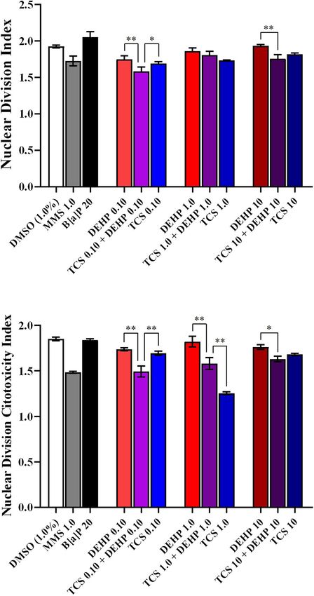
Duarte et al.                                                                                                    Triclosan and DEHP Induced Toxicity

also known that the degradation of TCS may generate                         and detoxification of several toxicants, such as cytochrome
compounds of higher toxicity and greater persistence, such as               P450 isoenzymes (CYP1A1, 1A2, 2B, 2C, 3A, and 2E1)
dioxins and chlorophenols (Pusceddu et al., 2018).                          and sulfotransferases (SULTs), glutathione-S-transferase (GSTs),
    Previous in vitro and in vivo studies showed that TCS presents          UDP-glucuronosyltransferase (UGT), and N-acetyl-transferase
estrogenic, antiestrogenic, and antiandrogenic properties, which            (NAT); moreover, HepG2 cells also have proficient p53 gene
appears to be related to cell lines, tissues, and animal species (for       expression, as well as functional DNA repair systems. Previous
a comprehensive review, see Ishibashi et al., 2004; Kumar et al.,           studies showed that p53-competent cells, with active expression
2009; Jung et al., 2012; Alfhili and Lee, 2019).                            of phases I and II drug metabolizing enzymes, as well as
    One of the most important mechanisms related to TCS-                    functional DNA repair systems may significantly reduce the rate
induced toxicity is the capacity to cause disturbances of the redox         of false positive results comprising genotoxicity assays (for review
status of cells, leading to oxidative damage in lipids, proteins,           see Knasmüller et al., 2004; Mersch-Sundermann et al., 2004;
and DNA (Binelli et al., 2009; Park et al., 2016; Zhang et al.,             Fowler et al., 2012; OECD, 2014).
2018). Previous in vivo studies showed that TCS is able to                      As mentioned above, both chemicals are widely used on daily
induce genotoxicity in several aquatic organisms, such as algae,            life and there is enough data comprising their hazard to aquatic
ciliated protozoa, crustaceans and microcrustaceans, and zebra              organisms; however, it still has a lack of information of their
mussels (Ciniglia et al., 2005; Binelli et al., 2009; Gao et al.,           behavior in mammalian cells. Both compounds are lipophilic,
2015; Silva et al., 2015; Xu et al., 2015). On the other hand,              and it seems to cause damage after phase I drug metabolization
few studies were carried out aiming to assess the impact of TCS             system, resulting in compounds with higher toxicity (for a
exposure on mammalian organisms, either in vitro and in vivo.               comprehensive review, see Caldwell, 2012; Wu et al., 2019);
For example, Li et al. (2018) showed that exposure to TCS                   moreover, previous human biomonitoring studies have detected
(concentrations ranging from 5.0 to 40 µM) was able to induce               the coexposure of TCS and DEHP (or their metabolites), in urine
cell cycle arrest and apoptosis, in HepG2 cells. In weanling rats,          samples of children and adults, in several regions worldwide
Riad et al. (2018) found that TCS caused testicular DNA damage,             (Casas et al., 2011; Larsson et al., 2014; Braun et al., 2017;
increase of malondialdehyde (MDA), and decrease of superoxide               Rocha et al., 2017, 2018; Lim, 2020; Li et al., 2021). Therefore,
dismutase (SOD). In humans, exposure to TCS was associated                  assessing to possible combined effect of TCS and DEHP would
with high levels of urinary 8-hydroxy-20 -deoxyguanosine (8-                provide further information about molecular effects of these
OHdG) in children from China (Lv et al., 2016) and from Brazil              toxicants, as well as their interactions, in the environment and/or
(Rocha et al., 2018).                                                       in organisms (Arambula and Patisaul, 2018). This study aimed to
    As well as TCS, the di-2-ethylhexyl phthalate (DEHP),                   assess the impact of the exposure to TCS and DEHP, as well as
derived from phthalic acid, is classified as endocrine-disrupting           their combination on biomarkers of acute toxicity and of DNA
compounds (EDCs), which comprise a group of toxicants that                  stability, in HepG2 cells.
interacts with natural hormones, interfering with their synthesis,
secretion, binding, transport, and elimination, by changing
their functions (Kabir et al., 2015). EDCs are widely present
in products used in daily life, such as PCP, pesticides, food
                                                                            MATERIALS AND METHODS
packaging, plastics, and flame retardants (Arambula and Patisaul,
2018). Due to its capacity to give malleability in polyvinyl chloride
                                                                            Reagents
(PVCs), DEHP is widely used by several industries sectors,                  TCS (triclosan; 5-chloro-2-(2,4-dichlorophenoxy) phenol; CAS
being found in medical devices, plastic packages, toys, building            3380-34-5), DEHP (bis(2-Ethylhexyl) phthalate; CAS 117-81-7),
materials, among others (Rusyn et al., 2006; Caldwell, 2012).               3,4-benzopyrene (B[a]P; CAS 50-32-8), methyl methanesulfonate
    DEHP is highly lipophilic and easily absorbed by the                    (MMS; CAS 66-27-3), cytochalasin B (Cyt-B; CAS 14930-
gastrointestinal tract, as well as by skin and lungs. Once absorbed,        96-2), dimethyl sulfoxide (DMSO; CAS 67-68-5), Giemsa
DEHP is quickly metabolized mainly in liver, resulting in several           (CAS 51811-82-6) and Dulbecco’s Modified Eagle’s Medium
metabolites, such as the mono(2-ethylhexyl) phthalate (MEHP),               (DMEM, low glucose) were obtained from Sigma-Aldrich
which is associated with most of the toxic effects induced by               (St. Louis, MO, United States), Trypsin-EDTA (0.25%) and
DEHP exposure (Fay et al., 1999; Rusyn et al., 2006; Caldwell,              antibiotic-antimycotic solution (10,000 units/mL of Penicillin,
2012). Previous in vitro and in vivo studies also showed that               10,000 µg/mL of Streptomycin, and 25 µg/mL of Amphotericin
DEHP can induce disturbances of cell homeostasis, leading to                B) came from Gibco (Grand Island, NE, United States) and
DNA instability, dysfunctions of the mitotic spindle, and cell              fetal bovine serum (FBS) was purchased from LGC Biotecnologia
death (Turner et al., 1974; Choi et al., 2010; Caldwell, 2012; Li           (Cotia, Brazil). All other chemicals, reagents, and buffers
et al., 2014; Rowdhwal and Chen, 2018; Silano et al., 2019).                were analytical grade products from Sigma-Aldrich (St. Louis,
    HepG2 cells are widely used for testing the genotoxic                   MO, United States).
properties of several environmental toxicants, mainly by use of
micronucleus (MN) and of comet assays (for review, see Guo                  Cell Culture Conditions and Treatments
et al., 2020). The employment of this cell line is particularly             HepG2 cells were kindly provided by Prof. Dra. Lusânia M. G.
advantageous due its ability to express several phases I and II             Antunes from School of Pharmaceutical Sciences of Ribeirão
drug metabolizing enzymes, which can catalyze the activation                Preto, University of São Paulo, Brazil.

Frontiers in Genetics | www.frontiersin.org                             2                                      April 2021 | Volume 12 | Article 649845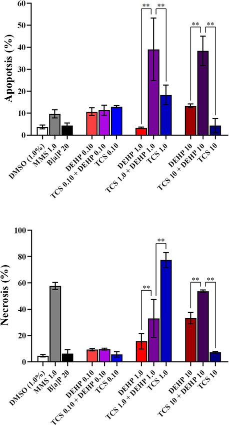
Duarte et al.                                                                                                Triclosan and DEHP Induced Toxicity

   Briefly, the cells were maintained in DMEM low glucose                Overdispersion was tested by chi-square tests. Values of NDI and
with 10% of FBS and 1.0% of antibiotic-antimycotic, in a CO2             NDCI were log-transformed due to their skewed distribution and
incubator with 5% atmosphere at 37◦ C and 96% of relative                analyzed by a Generalized Linear Model with Gaussian deviates.
humidity. All experiments were conducted between the third and           Normality of residuals was tested by Kolmogorov-Smirnov tests
eighth cell passage.                                                     with Lilliefors’ corrected p-values. Homogeneity of variances was
   106 cells were seeded in 25 cm2 cultures flasks for 24 h in the       assessed by Brown-Forsythe tests.
complete medium; after, the medium was removed, cells were                  Values of exposed cells were compared to their controls
washed twice with PBS (pH 7.2) and, then, cells were incubated           by Wald’s chi-square tests with Bonferroni corrected p-values.
with culture medium without FBS for the period of 4 h, with TCS,         Furthermore, cells treated with the combined exposures were
DEHP, as well as their combinations (Figure 1); moreover, vehicle        compared to the group that was treated with its respective
(DMSO 1.0%) and positive controls (B[a]P 20 µM, and MMS                  single exposure (i.e., TCS vs. TCS + DEHP; and DEHP vs.
1.0 µM) were also included in the experiments. Concentrations            TCS + DEHP; at the same doses) using the same procedure.
of positive controls, of TCS, DEHP and their combinations were           All analyses were done by Stata 13.1 (StataCorp, College Station,
chosen based on previous MTT assays (data not shown).                    TX, United States).

Cytokinesis-Block Micronucleus Cytome
(CBMNCyt) Assay                                                          RESULTS
CBMNcyt assay was carried out in three independent                       Table 1 summarizes the impact of treatments of HepG2 cells with
experimental replicates following the protocol published by              TCS, DEHP, as well as their combinations on the parameters of
Fenech (2007). After exposure of the cells to TCS, DEHP, and             cell death, cell viability, and DNA damage.
their combinations, cells were washed twice with PBS (pH 7.2)                It can be seen that the lower and the intermediate doses
and incubated with cytochalasin B (3.0 µg/mL) in complete                of TCS (0.10 and 1.0 µM) were able to increase the number
medium for further 30 h. After, cells were washed, trypsinized,          of cells in apoptosis when compared to the vehicle control
and centrifuged for 5 min at 180 g. Then, the pellets were               group, while only the intermediate concentration of TCS (1.0
resuspended in 5.0 mL of hypotonic solution (sodium citrate              µM) increased significantly the number of necrotic cells; on the
1.0%; 100 µL of formaldehyde (25%) at 4◦ C) for 4 min. The cells         other hand, significant increase of apoptosis and necrosis were
were centrifuged twice and fixed in with methanol and acetic             seen in the cells treated with all doses of DEHP (0.10–10 µM).
acid (3:1 v/v) and then, slides were prepared. Slides were stained       Cotreatments of the cells with the combinations of TCS + DEHP
with 5.0% Giemsa in PBS (pH 7.2) and cells were analyzed by              increased significantly the percentage of both biomarkers related
light microscopy (Carl Zeiss, AxioLab A1, Jena, Germany) at              to cell death, with the exception of the treatment of TCS 1.0
magnification of 630x.                                                   µM + DEHP 10 µM. Concerning the parameters related to cell
   In total, 3,000 binucleated cells (1,000 cells per experimental       division kinetics, TCS presented higher cytostatic effects, when
replicate) were analyzed for each experimental point. Analyses           compared to the groups that were exposed to DEHP, measured
of nuclear anomalies named micronucleus (MN), nuclear buds               by NDI and NDCI. Moreover, only the combination of TCS 1.0
(NBUDs), nucleoplasmatic bridges (NPBs), as well as the number           µM + DEHP 10 µM did not induce significantly cytostatic effects,
of apoptotic and necrotic cells were carried out according               when compared to the respective vehicle control groups.
to the scoring criteria described by Fenech (2007). Pictures                 Concerning the parameters related to DNA instability, TCS,
of each assessed endpoint are depicted in Supplementary                  DEHP, and their combinations were able to increase the DNA
Figure 1. Moreover, nuclear division index (NDI) was scored              damage of cells, assessed by MN and NBUDs endpoints. TCS at
in 1,500 cells (500 cells per experimental replicate) according          all concentrations (0.10, 1.0, and 10 µM) was able to increase
to the formula proposed by Eastmond and Tucker (1989),                   the MN frequencies when compared to the vehicle control
as follow: NDI = (M1 + 2.M2 + 3.M3 + 4.M4)/N, where                      group and only the lowest dose of DEHP (0.10 µM) did not
“M1-M4” represent the number of cells with one, two,                     induce MN formation. When the HepG2 cells were exposed
three, and four nuclei, respectively, and “N” is the number              to the combination of TCS and DEHP, the treatments TCS
of cells scored. Nuclear division cytotoxicity index (NDCI)              0.10 + DEHP 10; TCS 1.0 + DEHP 0.10, and TCS 10 + DEHP
was also scored in 1,500 cells (500 cells per experimental               0.10 did not increase the MN formations, when compared to the
replicate) according to the formula proposed by Fenech (2000):           respective vehicle control group. It is also important to mention
NDCI = (Ap + Nec + M1 + 2.M2 + 3.M3 + 4.M4)/N, where                     that a significant increase of MNs was seen in cells treated
“ap” and “nec” are the number of apoptotic and necrotic cells,           with MMS and B[a]P.
“M1-M4” represent the number of cells with one, two, three, and              Increase of NBUDs formation was seen in the groups treated
four nuclei, respectively, and “N” is the number of cells scored         alone with TCS at the intermediate and highest concentrations,
(both viable and non-viable ones).                                       i.e., at 1.0 and 10 µM, while higher NBUDs frequencies were
                                                                         seen in the cells that were treated with all doses of DEHP (0.10–
Statistical Analyses                                                     10 µM). Concerning the cells that receive the cotreatments with
Nuclear anomalies (MN, NBUDs, and NPBs) and counts of                    to TCS + DEHP, combinations between TCS 0.10 + DEHP 1.0,
necrotic and apoptotic cells were analyzed by Poisson regression.        TCS 0.10 + DEHP 10 and TCS 10 + DEHP 0.10 did not increase

Frontiers in Genetics | www.frontiersin.org                          3                                     April 2021 | Volume 12 | Article 649845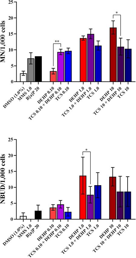
Duarte et al.                                                                                                                        Triclosan and DEHP Induced Toxicity

  FIGURE 1 | Schematic overview of treatments carried out in the study.

TABLE 1 | Impact of exposure to triclosan (TCS), to phtalate DEHP, as well as their combinations on biomarkers of cell death (apoptosis and necrosis), cell viability
(nuclear division index, (NDI); and nuclear division citotoxicity index (NDCI) and DNA instability (micronuclei (MN), nuclear buds (NBUDs) and nucleoplasmatic bridges
(NPBs), in HepG2 cells.

Treatments (µM)                               Cell death                             Cell viability                                  Mutagenic effects

                                 Apoptosis             Necrosis               NDI                     NCDI               MN                NBUDs                 NPBs

DMSOa (1.0%)                      4.0 ± 1.1            5.0 ± 1.1          1.9 ± 0.040            1.8 ± 0.030          2.7 ± 1.1           1.0 ± 1.7                0
MMSb 1.0                          10 ± 2.3**           58 ± 3.2**         1.7 ± 0.11**          1.5 ± 0.020**         7.3 ± 3.1*          1.3 ± 1.1                0
B[a]Pc 20                         4.0 ± 1.5            6.0 ± 3.6          2.0 ± 0.13*            1.8 ± 0.030          8.0 ± 0**           2.7 ± 3.1                0
TCS 0.10                          13 ± 0.8**           6.0 ± 2.5         1.7 ± 0.040**          1.7 ± 0.040*         9.7 ± 1.5**          2.3 ± 2.3           0.33 ± 0.58
TCS 1.0                           18 ± 6.3**           77 ± 7.0**        1.7 ± 0.010**          1.2 ± 0.020**        11 ± 2.5**           11 ± 6.8**          1.67 ± 0.58
TCS 10                            4.0 ± 4.7            7.0 ± 0.82         1.8 ± 0.020           1.7 ± 0.020**        10 ± 5.0**          8.7 ± 8.1**          0.50 ± 0.71
DEHP 0.10                         11 ± 2.5**           9.0 ± 1.1*        1.7 ± 0.090**           1.7 ± 0.030          3.3 ± 1.5           3.7 ± 1.5*               0
DEHP 1.0                         30 ± 0.50**           16 ± 7.2**         1.9 ± 0.080            1.8 ± 0.10          14 ± 1.1**           14 ± 10**            1.0 ± 1.0
DEHP 10                           13 ± 1.2**           33 ± 6.2**         1.9 ± 0.020            1.8 ± 0.050         17 ± 3.6**           13 ± 5.0**           1.7 ± 2.0
TCS 0.10 + DEHP 0.10              11 ± 3.3**           10 ± 0.90*         1.6 ± 0.10**          1.5 ± 0.10**         9.3 ± 1.5**          4.7 ± 2.1*           2.3 ± 1.5
TCS 0.10 + DEHP 1.0               23 ± 8.8**           20 ± 13**         1.6 ± 0.040**          1.5 ± 0.060**         6.3 ± 2.3*          1.7 ± 1.1                0
TCS 0.10 + DEHP 10                30 ± 11**            30 ± 24**         1.7 ± 0.090**          1.6 ± 0.16**          1.0 ± 0.0          0.33 ± 0.58               0
TCS 1.0 + DEHP 0.10               31 ± 23**            28 ± 20**         1.7 ± 0.050**          1.5 ± 0.17**         3.3 ± 0.58           15 ± 2.6*           0.33 ± 0.58
TCS 1.0 + DEHP 1.0                39 ± 20**            33 ± 20**          1.8 ± 0.10*           1.6 ± 0.11**         15 ± 2.6**          7.7 ± 4.5**           1.3 ± 1.5
TCS 1.0 + DEHP 10                2.0 ± 0.90            7.0 ± 1.7         1.8 ± 0.070**           1.8 ± 0.070         16 ± 2.0**           12 ± 8.0**          0.67 ± 0.58
TCS 10 + DEHP 0.10               9.0 ± 0.80*           11 ± 3.3*          1.7 ± 0.10**          1.7 ± 0.090**         6.0 ± 2.6           1.3 ± 0.58          0.33 ± 0.58
TCS 10 + DEHP 1.0                 11 ± 7.3**           34 ± 3.7**        1.8 ± 0.070*            1.7 ± 0.13*         9.3 ± 4.9**          4.3 ± 3.2*           1.0 ± 1.0
TCS 10 + DEHP 10                  38 ± 9.5**           54 ± 1.2**         1.7 ± 0.10**          1.6 ± 0.050**        11 ± 4.6**          8.7 ± 8.3**          0.50 ± 1.71
a Negative  (solvent) control: dimethyl sulfoxide.
b Positive control: methylmethane sulfonate (no required biotransformation/activation).
c Positive control: benzo[a]pyrene (required biotransformation/activation).

*p < 0.050; **p < 0.010 (both in bold) compared to vehicle control group (DMSO).

significantly the formation of NBUDs. On the other hand, TCS,                             at 1.0 µM had lower necrotic events than the cotreatment with
DEHP and their associations did not increase the frequencies of                           TCS + DEHP (both at 1.0 µM); on the other hand, treatment with
NPBs, when compared to the respective vehicle control.                                    TCS 1.0 µM increases the percentage of necrosis, when compared
   Figures 2–4 depict the comparisons between the treatments                              to the group that receives TCS 1.0 + DEHP 1.0 µM. It can be also
with TCS + DEHP and their respective groups that receive TCS                              observed a significant increase of necrotic cells in the treatment
or DEHP alone, on parameters of cell death (apoptosis and                                 with the combination of TCS and DEHP (both at 10 µM) when
necrosis), of cell viability (NDI and NDCI), and DNA damage                               compared to the groups that were exposed to the compounds
(MNs, NBUDs, and NPBs), respectively.                                                     alone (Figure 2).
   There were no significant differences in the percentage of                                 Figure 3 illustrates the NDI and NDCI of cells treated
apoptosis and necrosis among the cells that receive the lowest                            with TCS, DEHP, and their associations. Combined effects were
dose of TCS, DEHP, as well as their combination, while an                                 observed in the groups that receive the lowest doses of TCS and
increasing number of apoptotic cells were seen in the groups that                         DEHP, i.e., cells treated with TCS + DEHP (both at 0.10 µM)
were treated with TCS 1.0 + DEHP 1.0; and TCS 10 + DEHP 10,                               showed a decrease of NDI and NDCI when compared to those
when compared to the cells that were exposed only to TCS and to                           that were exposed only to TCS or DEHP alone. Lower NDI and
DEHP at 1.0 and 10 µM, respectively. Cells treated with DEHP                              NDCI were also seen in the groups treated with TCS + DEHP

Frontiers in Genetics | www.frontiersin.org                                          4                                             April 2021 | Volume 12 | Article 649845
Duarte et al. Triclosan and DEHP Induced Toxicity
FIGURE 2 | Comparisons between the treatments with TCS + DEHP and
their respective groups that receive TCS or DEHP alone, on parameters of cell FIGURE 3 | Comparisons between the treatments with TCS + DEHP and
death: (A) apoptosis and (B) necrosis. All doses are in µM. ∗∗ p < 0.010; their respective groups that receive TCS or DEHP alone, on parameters of cell
Wald’s chi-square tests followed by Bonferroni correction tests. viability: (A) nuclear division index (NDI) and (B) nuclear division cytotoxicity
index (NDCI). All doses are in µM. ∗ p < 0.050; ∗∗ p < 0.010; Wald’s
chi-square tests followed by Bonferroni correction tests.
(both 10 µM) when compared to the cells that receive only DEHP treatments induced NPBs formation, no combined effects can be
10 µM, while no statistical difference was seen between TCS 10 seen for this endpoint (data not shown).
µM and TCS 10 + DEHP 10. No differences of NDI were seen
among groups that receive only TCS 1.0 µM; DEHP 1.0 µM
and their association, while the lowest NDCI was observed in the DISCUSSION
cells treated with TCS 1.0 µM (DEHP 1.0 > TCS 1.0 + DEHP
1.0 > TCS 1.0). Previous studies showed that TCS exposure induces acute
Although the exposure to TCS and DEHP, as well as their toxicity, leading to cell death. For example, Li et al. (2018)
combination, induces DNA instability, no combined effects were showed that TCS at concentrations of 5.0 and 10 µM was able
seen on the biomarkers related to DNA damage (MN and to induce DNA instability, promoting apoptosis mediated by p53
NBUD formations) (Figure 4); moreover, since none of assessed expression, in HepG2 cells. Li et al. (2019) observed that PC12
Frontiers in Genetics | www.frontiersin.org 5 April 2021 | Volume 12 | Article 649845Duarte et al. Triclosan and DEHP Induced Toxicity
with a diet containing 0.08% of TCS for 8 months. It is well
established that the expression of TNFα, TNFβ, and IL-6 are
associated with cell death signaling pathways, mainly by necrosis
(D’Arcy, 2019).
Regarding to DEHP, Fang et al. (2019) assessed the impact of
DEHP exposure at high dose (512 µM for 4 h) on parameters
of cell viability, in differentiated human embryonic stem cells,
and observed that exposure to the compound was able to induce
apoptosis by triggering the PPARγ/PTEN/AKT pathway, which
is related to cell proliferation and survival. Like to TCS, cell
death induced by DEHP also appears to be related to Bax
expression, as well as to an increase of caspases 3 and 8, as seen
previously by and Hannon et al. (2016) and Sun et al. (2018),
using in vitro laboratory models. Ha et al. (2016) observed an
increase of apoptotic events, in hepatocytes of Sprague-Dawley
rats treated with DEHP at 250, 500, and 750 mg/kg for 30 days;
according to the authors, the increase of cell death was mediated
by p53 overexpression caused by DEHP exposure. Our results
demonstrated that DEHP can induce apoptosis even at lower
concentrations, since we observed increases in cell death at doses
of 0.10 and 1.0 µ M.
We also observed that exposure to the both EDCs was able
to impact the NDI of HepG2 cells and previous studies showed
that oxidative damage in DNA is related to cell cycle arrest
(Droge, 2002; Murray and Carr, 2018). Reduced NDI is associated
with more mononucleated cells, when compared to bi-, tri-, and
polynucleated ones, giving evidence of cell cycle arrest (Fenech,
2007, 2020). Interestingly, the most pronounced effects on NDI
were seen when the cells were treated with the combinations of
TCS and DEHP, at all doses. This parameter is a measure of the
proliferative status of viable cells fraction, if the NDI is lower
than control, it can be assumed that more cells with one nucleus
were scored, suggesting cytostatic effects (Fenech et al., 2011).
Therefore, one hypothesis for our observations may be related to
the activation of DNA repair pathways. In the cell cycle, G2/M
checkpoint prevents the entry of mitosis, when DNA damage
was not properly repaired. Delaying entry into mitosis allows
repair mechanisms of DNA lesions before cell division, avoiding
passing the damage to the new cells (Li and Zou, 2005; Gomez
and Hergovich, 2016; Choi and Chung, 2020). In the NDCI,
FIGURE 4 | Comparisons between the treatments with TCS + DEHP and
their respective groups that receive TCS or DEHP alone, on parameters of necrotic and apoptotic cells are included in the number of cells
DNA damage: (A) micronuclei (MN) formation and (B) nuclear buds (NBUDs) scored, since the toxic effects induced by chemical compounds
formation. All doses are in µM. ∗ p < 0.050; ∗∗ p < 0.010; Wald’s chi-square may provide a large proportion of cells becoming non-viable.
tests followed by Bonferroni correction tests. Therefore, an overestimated cytostatic effect can be observed in
NDI if necrotic and apoptotic cells were not included in the
scoring (Fenech, 2000).
cells exposed to TCS at doses of 10 and 50 µM for 12 h was able In our study, clear mutagenic effects were seen in the cells
to activate the expression of p38 mitogen-activated protein kinase exposed to TCS, DEHP and their combinations, by significant
and Bax, which are related to apoptosis signaling. Moreover, increase of MN and NBUD frequencies. An earlier study carried
Park et al. (2016) also showed that TCS-induced apoptosis in rat by Li et al. (2019) suggested exposure to TCS may be related
neural stem cells (NSCs) was mediated by Bax expression and to double strand breaks (DSBs), since the authors observed a
activation of caspase 3, at the dose of 50 µM. Our findings showed significant increase of comet formations in HepG2 cells treated
that the concentrations of TCS at 0.10 and 1.0 µM increased with TCS at 20 and 40 µM, when compared to the negative
the number of apoptotic cells, while the concentration of 1.0 control group; in the same investigation, the authors described
µM enhanced the necrotic cells. Furthermore, Yueh et al. (2014) that DNA-dependent protein kinase (DNA-PKcs) is required
observed an increase of expression of the inflammatory cytokines for the DNA double-strand break repair through the non-
named TNFα, TNFβ, and IL-6 in the liver of male mice fed homologous end-joining (NHEJ) pathway. The formation of MN
Frontiers in Genetics | www.frontiersin.org 6 April 2021 | Volume 12 | Article 649845Duarte et al. Triclosan and DEHP Induced Toxicity
occurs during cell division when the genetic material is exposed environment, studying the interactions between these toxicants
to mutagenic compounds promoting the break of chromosomes is more complex, since they can interact with more than
(by clastogenic agents, which are related to acentric fragments) one compound and be metabolized by bacteria, or suffering
or loss of whole chromosomes (by aneugenic agents, which alterations by UV radiation from sunlight, for example
are related to disturbances of mitotic spindle formation); (Rehberger et al., 2018). For this reason, studies involving
therefore, both (whole chromosomes or their fragments) are not mixtures of toxicants are important to emphasize the risk of
incorporated into the main nucleus during the telophase and these compounds to the environment and human health and
they are surrounded by a nuclear membrane, generating a MN it can contribute with primary data for assessing exposure
(Fenech, 2007; Fenech et al., 2011). Earlier laboratory studies also biomarkers related to DNA damage and further studies with
described an association between DEHP exposure and increased other compounds in other cells models or in in vivo models.
DNA instability. For example, Kim et al. (2019) showed higher Taken together, our results provide further evidence
γH2AX formation in 8505C thyroid gland carcinoma cells treated concerning the mutagenic effects of TCS and DEHP in
with DEHP at concentrations ranging from 5.0 to 50 µM, while mammalian cells. Also, DNA damage induced by exposure to
increased comet formations were seen in the cells treated with the both compounds may be related to delay in cell cycle and to
DEHP at doses from 5.0 to 100 µM. γH2AX is a very sensitive acute toxicity. Interestingly, the most significant damages were
biomarker associated with cellular response to the induction of related to exposure of lower doses of TCS, DEHP, as well as
DNA double-strand breaks (DSBs) (Kopp and Audebert, 2019). their combinations, showing that EDCs are able to induce several
Our results provide further pieces of evidence that TCS and disturbances on cells, even at low concentrations.
DEHP are able to induce chromosome breaks, which resulted
in an increase of MN frequencies, especially when the cells
were exposed simultaneously to both chemicals. In addition, DATA AVAILABILITY STATEMENT
NBUDs have a similar morphology to MN; however, they are
connected to the main nucleus by a stem of nucleoplasmatic The raw data supporting the conclusions of this article will be
material. Most of the NBUDs originate from interstitial or made available by the authors, without undue reservation.
terminal acentric fragments (Fenech, 2007; Fenech et al., 2011).
In this context, MNs may also arise through gene amplification
through fusion bridge breaking (BFB) cycles. In the case of AUTHOR CONTRIBUTIONS
NBUDs, DNA is selectively located at specific locations on the
periphery of the nucleus and can be eliminated through nuclear GB, LL, and ND: conceptualization. EN, FM, LL, and ND:
sprouting, during S phase of the cell cycle; then, as consequence, methodology, investigation. MK: statistics. EN, GB, and ND:
extrusion of amplified DNA via nuclear sprouting may result in writing original draft preparation. GB and ND: writing, review,
MN formations (Fenech and Crott, 2002; Mateuca et al., 2006; and editing. All authors contributed to the article and approved
Fenech, 2020). It is also important to mention that we did not the submitted version.
observed increase of NPBs in cells treated with TCS and DEHP.
According to Bonassi and Fenech (2019), NPBs breaks generate
a pair of abnormal chromosomes lacking telomeres and results FUNDING
in further end-fusion and gene amplification; amplified gene
sequences and unresolved DNA repair complexes are removed This study was supported by grant #2018/02543-5 (São Paulo
by NBUDs formations. Research Foundation, FAPESP, Brazil), by the National Council
Previous data report that toxicant mixtures may increase the for Scientific and Technological Development (CNPq, Brazil) and
adverse effects when compared to one only exposure (Kabir by the Coordination for the Improvement of Higher Education
et al., 2015; Hernández et al., 2017). On the other hand, the Personnel (CAPES, Brazil).
chemical compounds may compete for similar metabolizing
pathways, resulting in high biotransformation rating, which can
increase or decrease the toxic effects of xenobiotics (Lin, 2006). ACKNOWLEDGMENTS
These interactions depend on several factors, including affinity
and concentration of substrates, time of exposure, and CYP450 We are grateful to the Cell Culture facilities from Federal
system inhibition, for example (Deodhar et al., 2020). In this University of São Paulo (Santos, Brazil).
context, it is important to highlight that we assessed the exposure
of TCS and DEHP in HepG2 cells, which although a tumoral cell
line, they express phases 1 and 2 metabolizing enzymes (Mersch- SUPPLEMENTARY MATERIAL
Sundermann et al., 2004), which may influence to toxicity related
to the TCS and DEHP exposure. The Supplementary Material for this article can be found
Moreover, it is well known that laboratory model experiments online at: https://www.frontiersin.org/articles/10.3389/fgene.
are run under restricted controlled conditions; once in the 2021.649845/full#supplementary-material
Frontiers in Genetics | www.frontiersin.org 7 April 2021 | Volume 12 | Article 649845Duarte et al. Triclosan and DEHP Induced Toxicity
REFERENCES Fenech, M., Kirsch-Volders, M., Natarajan, A. T., Surralles, J., Crott, J. W., Parry,
J., et al. (2011). Molecular mechanisms of micronucleus, nucleoplasmic bridge
Agüera, A., Fernández-Alba, A. R., Piedra, L., Mézcua, M., and Gómez, M. J. and nuclear bud formation in mammalian and human cells. Mutagenesis 26,
(2003). Evaluation of triclosan and biphenylol in marine sediments and urban 125–132. doi: 10.1093/mutage/geq052
wastewaters by pressurized liquid extraction and solid phase extraction followed Fowler, P., Smith, R., Smith, K., Young, J., Jeffrey, L., Kirkland, D., et al.
by gas chromatography mass spectrometry and liquid chromatography mass (2012). Reduction of misleading (“false”) positive results in mammalian cell
spectrometry. Anal. Chim. Acta 480, 193–205. doi: 10.1016/S0003-2670(03) genotoxicity assays. II. Importance of accurate toxicity measurement. Mutat.
00040-0 Res. - Genet. Toxicol. Environ. Mutagen. 747, 104–117. doi: 10.1016/j.mrgentox.
Alfhili, M. A., and Lee, M. H. (2019). Triclosan: an update on biochemical and 2012.04.013
molecular mechanisms. Oxid. Med. Cell. Longev. 2019:28. doi: 10.1155/2019/ Gao, L., Yuan, T., Cheng, P., Bai, Q., Zhou, C., Ao, J., et al. (2015). Effects of
1607304 triclosan and triclocarban on the growth inhibition, cell viability, genotoxicity
Arambula, S. E., and Patisaul, H. B. (2018). Endocrine disrupting chemicals and and multixenobiotic resistance responses of Tetrahymena thermophila.
behavior. Encycl. Endocr. Dis. 1, 812–820. Chemosphere 139, 434–440. doi: 10.1016/j.chemosphere.2015.07.059
Binelli, A., Cogni, D., Parolini, M., Riva, C., and Provini, A. (2009). In vivo Gomez, V., and Hergovich, A. (2016). Cell-Cycle Control and DNA-Damage
experiments for the evaluation of genotoxic and cytotoxic effects of Triclosan in Signaling in Mammals, Genome Stability. Amsterdam: Elsevier Inc.
Zebra mussel hemocytes. Aquat. Toxicol. 91, 238–244. doi: 10.1016/j.aquatox. Guo, X., Seo, J. E., Li, X., and Mei, N. (2020). Genetic toxicity assessment using liver
2008.11.008 cell models: past, present, and future. J. Toxicol. Environ. Health B. Crit. Rev. 23,
Bonassi, S., and Fenech, M. (2019). “The micronucleus assay in toxicologyin,” 27–50. doi: 10.1080/10937404.2019.1692744
in Issues in Toxicology, ed. D. Anderson (London: The Royal Society of Ha, M., Wei, L., Guan, X., Li, L., and Liu, C. (2016). P53-dependent apoptosis
Chemistry), 38–52. contributes to di-(2-ethylhexyl) phthalate-induced hepatotoxicity. Environ.
Braun, J. M., Bellinger, D. C., Hauser, R., Wright, R. O., Chen, A., Calafat, A. M., Pollut. 208, 416–425. doi: 10.1016/j.envpol.2015.10.009
et al. (2017). Prenatal phthalate, triclosan, and bisphenol a exposures and child Hannon, P. R., Niermann, S., and Flaws, J. A. (2016). Acute exposure to Di(2-
visual-spatial abilities. Neurotox 58, 75–83. doi: 10.1016/j.neuro.2016.11.009 Ethylhexyl) phthalate in adulthood causes adverse reproductive outcomes later
Caldwell, J. C. (2012). DEHP: genotoxicity and potential carcinogenic in life and accelerates reproductive aging in female mice. Toxicol. Sci. 150,
mechanisms-a review. Mutat. Res. - Rev. Mutat. Res. 751, 82–157. 97–108. doi: 10.1093/toxsci/kfv317
doi: 10.1016/j.mrrev.2012.03.001 Hernández, A. F., Gil, F., and Lacasaña, M. (2017). Toxicological interactions of
Casas, L., Fernández, M. F., Llop, S., Guxens, M., Ballester, F., Olea, N., et al. (2011). pesticide mixtures: an update. Arch. Toxicol. 91, 3211–3223. doi: 10.1007/
Urinary concentrations of phthalates and phenols in a population of Spanish s00204-017-2043-5
pregnant women and children. Environ. Int. 37:5. doi: 10.1016/j.envint.2011.02. Ishibashi, H., Matsumura, N., Hirano, M., Matsuoka, M., Shiratsuchi, H., Ishibashi,
012 Y., et al. (2004). Effects of triclosan on the early life stages and reproduction of
Choi, J. E., and Chung, W. (2020). Functional interplay between the oxidative stress medaka Oryzias latipes and induction of hepatic vitellogenin. Aquat. Toxicol.
response and DNA damage checkpoint signaling for genome maintenance in 67, 167–179. doi: 10.1016/j.aquatox.2003.12.005
aerobic organisms. Arch. Pharm. Res. 38:5. doi: 10.1007/s12275-020-9520-x Jung, E. M., An, B. S., Choi, K. C., and Jeung, E. B. (2012). Potential
Choi, S., Park, S. Y., Jeong, J., Cho, E., Phark, S., Lee, M., et al. (2010). Identification estrogenic activity of triclosan in the uterus of immature rats and rat
of toxicological biomarkers of di(2-ethylhexyl) phthalate in proteins secreted by pituitary GH3 cells. Toxicol. Lett. 208, 142–148. doi: 10.1016/j.toxlet.2011.
HepG2 cells using proteomic analysis. Proteomics 10, 1831–1846. doi: 10.1002/ 10.017
pmic.200900674 Kabir, E. R., Rahman, M. S., and Rahman, I. (2015). A review on endocrine
Ciniglia, C., Cascone, C., Lo Giudice, R., Pinto, G., and Pollio, A. (2005). disruptors and their possible impacts on human health. Environ. Toxicol.
Application of methods for assessing the geno- and cytotoxicity of Triclosan Pharmacol. 40, 241–258. doi: 10.1016/j.etap.2015.06.009
to C. ehrenbergii. J. Hazard. Mater. 122, 227–232. doi: 10.1016/j.jhazmat.2005. Kim, S., Park, G., Jo, Y., Seong, J., Taek, K., Jee, S., et al. (2019). Di-2-
03.002 ethylhexylphthalate promotes thyroid cell proliferation and DNA damage
D’Arcy, M. S. D. (2019). Cell death: a review of the major forms of apoptosis, through activating thyrotropin-receptor-mediated pathways in vitro and
necrosis and autophagy. Cell Biol. Int. 43, 582–592. doi: 10.1002/cbin.11137 in vivo. Food Chem. Toxicol. 124, 265–272. doi: 10.1016/j.fct.2018.
Deodhar, M., Rihani, S. B., Al, Arwood, M. J., Darakjian, L., Dow, P., et al. (2020). 12.010
Mechanisms of CYP450 inhibition: understanding drug-drug interactions due Knasmüller, S., Mersch-sundermann, V., Kevekordes, S., and Darroudi, F. (2004).
to mechanism-based inhibition in clinical practice. Pharmaceutics 12, 1–18. Use of human-derived liver cell lines for the detection of environmental and
Droge, W. (2002). Free radicals in the physiological control of cell. Physiol. Rew. dietary genotoxicants. Curr. State Knowledge 198, 315–328. doi: 10.1016/j.tox.
82, 47–95. doi: 10.1152/physrev.00018.2001 2004.02.008
Eastmond, D. A., and Tucker, J. D. (1989). Identification of aneuploidy-inducing Kopp, B., and Audebert, L. K. M. (2019). Validation of the γH2AX biomarker for
agents using cytokinesis-blocked human lymphocytes and an antikinetochore genotoxicity assessment: a review. Arch. Toxicol. 93, 2103–2114. doi: 10.1007/
antibody. Environ. Mol. Mutagen. 13, 34–43. doi: 10.1002/em.2850130104 s00204-019-02511-9
Fang, H., Fang, W., Cao, H., Luo, S., Cong, J., Liu, S., et al. (2019). Di-(2- Kumar, V., Chakraborty, A., Kural, M. R., and Roy, P. (2009). Alteration of
ethylhexyl)-phthalate induces apoptosis via the PPARγ/PTEN/AKT pathway in testicular steroidogenesis and histopathology of reproductive system in male
differentiated human embryonic stem cells. Food Chem. Toxicol. 131, 1–8. rats treated with triclosan. Reprod. Toxicol. 27, 177–185. doi: 10.1016/j.
Fay, M., Donohue, J. M., and De Rosa, C. (1999). ATSDR evaluation of health reprotox.2008.12.002
effects of chemicals. VI. Di(2-ethylhexyl)phthalate. Toxicol. Ind. Health 15, Larsson, K., Ljung Björklund, K., Palm, B., Wennberg, M., Kaj, L., Lindh, C. H.,
651–732. doi: 10.1191/074823399678847023 et al. (2014). Exposure determinants of phthalates, parabens, bisphenol A and
Fenech, M. (2000). The in vitro micronucleus technique. Mutat. Res. 455, 81–95. triclosan in Swedish mothers and their children. Environ. Int. 73, 323–333.
doi: 10.1016/s0027-5107(00)00065-8 doi: 10.1016/j.envint.2014.08.014
Fenech, M. (2007). Cytokinesis-block micronucleus cytome assay. Nat. Protoc. 2, Li, H., An, J., Zhong, Y., He, H., Wang, L., Yang, Y., et al. (2018). Comparison
1084–1104. doi: 10.1038/nprot.2007.77 of hepatotoxicity and mechanisms induced by triclosan (TCS) and methyl-
Fenech, M. (2020). Cytokinesis-Block micronucleus cytome assay evolution into triclosan (MTCS) in human liver hepatocellular HepG2 cells. Toxicol. Res.
a more comprehensive method to measure chromosomal instability. Genes (Camb) 8, 38–45. doi: 10.1039/c8tx00199e
11:1203. doi: 10.3390/genes11101203 Li, L., and Zou, L. (2005). Sensing, signaling, and responding to DNA damage:
Fenech, M., and Crott, J. W. (2002). Micronuclei, nucleoplasmic bridges and organization of the checkpoint pathways in mammalian cells. J. Cell. Biochem.
nuclear buds induced in folic acid deficient human lymphocytes - Evidence 94, 298–306. doi: 10.1002/jcb.20355
for breakage-fusion-bridge cycles in the cytokinesis-block micronucleus assay. Li, S. J., Chen, P., Peres, T. V., Villahoz, B. F., Zhang, Z., Miah, M. R., et al.
Mutat. Res. 504, 131–136. doi: 10.1016/s0027-5107(02)00086-6 (2019). Triclosan induces PC12 cells injury is accompanied by inhibition of
Frontiers in Genetics | www.frontiersin.org 8 April 2021 | Volume 12 | Article 649845Duarte et al. Triclosan and DEHP Induced Toxicity
AKT/mTOR and activation of p38 pathway. Neurotoxicology 74, 221–229. doi: association with oxidative DNA damage. Sci. Total Environ. 586, 152–162.
10.1016/j.neuro.2019.07.008 doi: 10.1016/j.scitotenv.2017.01.193
Li, X., Fang, E. F., Scheibye-Knudsen, M., Cui, H., Qiu, L., Li, J., et al. (2014). Di- Rocha, B. A., Asimakopoulos, A. G., Honda, M., da Costa, N. L., Barbosa,
(2-ethylhexyl) phthalate inhibits DNA replication leading to hyperPARylation, R. M., Barbosa, F., et al. (2018). Advanced data mining approaches in the
SIRT1 attenuation, and mitochondrial dysfunction in the testis. Sci. Rep. 4:6434. assessment of urinary concentrations of bisphenols, chlorophenols, parabens
Li, X., Zhong, Y., He, W., Huang, S., Li, Q., Guo, C., et al. (2021). Co-exposure and benzophenones in Brazilian children and their association to DNA damage.
and health risks of parabens, bisphenols, triclosan, phthalate metabolites and Environ. Int. 116, 269–277. doi: 10.1016/j.envint.2018.04.023
hydroxyl polycyclic aromatic hydrocarbons based on simultaneous detection Rowdhwal, S. S. S., and Chen, J. (2018). Toxic effects of Di-2-ethylhexyl phthalate:
in urine samples from guangzhou, south China. Environ. Pollut. 272:115990. an overview. Biomed Res. Int. 2018:1750368. doi: 10.1155/2018/1750368
doi: 10.1016/j.envpol.2020.115990 Rusyn, I., Peters, J., and Cunningham, M. (2006). Modes of action and species-
Lim, S. (2020). The associations between personal care products use, and urinary specific effects of di-(2-ethylhexyl)phthalate in the liver. Crit. Rev. Toxicol. 36,
concentrations of phthalates, parabens, and triclosan in various age groups: 459–479. doi: 10.1080/10408440600779065
the korean national environmental health survey cycle 3. 2015-2017. Sci. Total Silano, V., Barat Baviera, J. M., Bolognesi, C., Chesson, A., Cocconcelli, P. S.,
Environ. 742:140640. doi: 10.1016/j.scitotenv.2020.140640 Crebelli, R., et al. (2019). Update of the risk assessment of di-butylphthalate
Lin, J. H. (2006). Expert review CYP induction-mediated drug interactions: in vitro (DBP), butyl-benzyl-phthalate (BBP), bis(2-ethylhexyl)phthalate (DEHP), di-
assessment and clinical implications. Pharm Res. 23, 1089–1116. doi: 10.1007/ isononylphthalate (DINP) and di-isodecylphthalate (DIDP) for use in food
s11095-006-0277-7 contact materials. EFSA J. 17:5838. doi: 10.2903/j.efsa.2019.5838
Lv, Y., Rui, C., Dai, Y., Pang, Q., Li, Y., Fan, R., et al. (2016). Exposure of Silva, A. R. R., Cardoso, D. N., Cruz, A., Lourenço, J., Mendo, S.,
children to BPA through dust and the association of urinary BPA and triclosan Soares, A. M. V. M., et al. (2015). Ecotoxicity and genotoxicity of a
with oxidative stress in Guangzhou, China. Environ. Sci. Process. Impacts 18, binary combination of triclosan and carbendazim to Daphnia magna.
1492–1499. doi: 10.1039/c6em00472e Ecotoxicol. Environ. Saf. 115, 279–290. doi: 10.1016/j.ecoenv.2015.
Mateuca, R., Lombaert, N., Aka, P. V., and Decordier, I. (2006). Chromosomal 02.022
changes: induction„ detection methods and applicability in human Sun, Y., Shen, J., Zeng, L., Yang, D., Shao, S., Wang, J., et al. (2018). Role
biomonitoring detection methods, and applicability in human biomonitoring. of autophagy in di-2-ethylhexyl phthalate (DEHP)-induced apoptosis in
Biochimie 88, 1515–1531. doi: 10.1016/j.biochi.2006.07.004 mouse Leydig cells. Environ. Pollut. 243, 563–572. doi: 10.1016/j.envpol.2018.
McAvoy, D. C., Schatowitz, B., Jacob, M., Hauk, A., and Eckhoff, W. S. (2002). 08.089
Measurement of triclosan in wastewater treatment systems. Environ. Toxicol. Turner, J. H., Petricciani, J. C., Crouch, M. L., and Wenger, S. (1974). An evaluation
Chem. 21:1323. doi: 10.1002/etc.5620210701 of the effects of diethylhexyl phthalate (DEHP) on mitotically capable cells in
Mersch-Sundermann, V., Knasmüller, S., Wu, X. J., Darroudi, F., and Kassie, F. blood packs. Transfusion 14, 560–566. doi: 10.1111/j.1537-2995.1974.tb04577.x
(2004). Use of a human-derived liver cell line for the detection of cytoprotective, Wu, M., Xu, L., Teng, C., Xiao, X., Hu, W., Chen, J., et al. (2019). Involvement
antigenotoxic and cogenotoxic agents. Toxicology 198, 329–340. doi: 10.1016/j. of oxidative stress in di-2-ethylhexyl phthalate (DEHP)-induced apoptosis of
tox.2004.02.009 mouse NE-4C neural stem cells. Neurotoxicology 70, 41–47. doi: 10.1016/j.
Murray, J. M., and Carr, A. M. (2018). Integrating DNA damage repair with the cell neuro.2018.10.013
cycle. Curr. Opin. Cell Biol. 52, 120–125. doi: 10.1016/j.ceb.2018.03.006 Xu, X., Lu, Y., Zhang, D., Wang, Y., and Zhou, X. (2015). Toxic assessment of
OECD (2014). Test No. 487: in Vitro Mammalian Cell Micronucleus Test. Test triclosan and triclocarban on Artemia salina. Bull. Environ. Contam. Toxicol.
No. 487 Vitr. Mamm. Cell Micronucleus Test. Paris: OECD, doi: 10.1787/ 95, 728–733. doi: 10.1007/s00128-015-1641-2
9789264224438-en Yueh, M. F., Taniguchib, K., Chena, S., Evansc, R. M., Hammockd, B. D., Karinb,
Olaniyan, L. W. B., Mkwetshana, N., and Okoh, A. I. (2016). Triclosan in water, M., et al. (2014). The commonly used antimicrobial additive triclosan is a
implications for human and environmental health. Springerplus 5:1639. doi: liver tumor promoter. Proc. Natl. Acad. Sci. U S A. 111, 17200–17205. doi:
10.1186/s40064-016-3287-x 10.1073/pnas.1419119111
Park, B. K., Gonzales, E. L. T., Yang, S. M., Bang, M., Choi, C. S., and Shin, C. Y. Zhang, P., Yang, M., Zeng, L., and Liu, C. (2018). P38/TRHr-
(2016). Effects of triclosan on neural stem cell viability and survival. Biomol. Dependent regulation of TPO in thyroid cells contributes to the
Ther. 24, 99–107. doi: 10.4062/biomolther.2015.164 hypothyroidism of triclosan-treated rats. Cell. Physiol. Biochem. 45, 1303–
Pusceddu, F. H., Choueri, R. B., Pereira, C. D. S., Cortez, F. S., Santos, D. R. A., 1315. doi: 10.1159/000487558
Moreno, B. B., et al. (2018). Environmental risk assessment of triclosan and
ibuprofen in marine sediments using individual and sub-individual endpoints. Conflict of Interest: The authors declare that the research was conducted in the
Environ. Pollut. 232, 274–283. doi: 10.1016/j.envpol.2017.09.046 absence of any commercial or financial relationships that could be construed as a
Rehberger, K., Kropf, C., and Segner, H. (2018). In vitro or not in vitro: a short potential conflict of interest.
journey through a long history. Environ. Sci. Eur. 30:23. doi: 10.1186/s12302-
018-0151-3 Copyright © 2021 Duarte, Lima, Maraslis, Kundi, Nunes and Barcelos. This is an
Riad, M. A., Abd-Rabo, M. M., Abd, El Aziz, S. A., El Behairy, A. M., and open-access article distributed under the terms of the Creative Commons Attribution
Badawy, M. M. (2018). Reproductive toxic impact of subchronic treatment with License (CC BY). The use, distribution or reproduction in other forums is permitted,
combined butylparaben and triclosan in weanling male rats. J. Biochem. Mol. provided the original author(s) and the copyright owner(s) are credited and that the
Toxicol. 32:e22037. doi: 10.1002/jbt.22037 original publication in this journal is cited, in accordance with accepted academic
Rocha, B. A., Asimakopoulos, A. G., Barbosa, F., and Kannan, K. (2017). Urinary practice. No use, distribution or reproduction is permitted which does not comply
concentrations of 25 phthalate metabolites in Brazilian children and their with these terms.
Frontiers in Genetics | www.frontiersin.org 9 April 2021 | Volume 12 | Article 649845You can also read